
Pediatrics - medbox.org · Pediatrics Clinical treatment guidelines 1 Ministry of Health P. O. Box...
176
Pediatrics Ministry of Health P. O. Box 84 Kigali www.moh.gov.rw Republic of Rwanda Pediatrics Clinical Treatment Guidelines September 2012
Transcript of Pediatrics - medbox.org · Pediatrics Clinical treatment guidelines 1 Ministry of Health P. O. Box...

1Clinical treatment guidelinesPediatrics
Ministry of HealthP. O. Box 84 Kigaliwww.moh.gov.rw
Republic of Rwanda
P e d i a t r i c s
Cl in ica l treatment guidel ines
September 2012


3Clinical treatment guidelinesPediatrics
Ministry of HealthP. O. Box 84 Kigaliwww.moh.gov.rw
Republic of Rwanda
P e d i a t r i c s
Clinical treatment guidelines
September 2012


Clinical treatment guidelinesPediatrics iii
Acronyms..................................................................................................viForeword...................................................................................................ix
1. Respiratory Diseases..............................................................................11.1. Rhinitis and Rhinopharyngitis..............................................................11.2. Pneumonia...............................................................................................21.3. Wheezing child/Asthma and Bronchiolitis.........................................6
1.3.1. Acute Bronchiolitis..................................................................61.3.2. Asthma......................................................................................8
1.4. Ear Nose and Throat Conditions........................................................161.4.1. Acute Otitis Media................................................................161.4.2. Chronic Suppurative Otitis Media......................................171.4.3. Tonsillitis................................................................................191.4.4. Acute Mastoiditis..................................................................211.4.5. Epistaxis.................................................................................221.4.6. Sinusitis..................................................................................241.4.7. Laryngitis................................................................................261.4.8. Epiglottitis..............................................................................271.4.9. Pertussis (Whooping cough)...............................................281.4.10. Allergic Rhinitis....................................................................29
2. Gastro-intestinal Disorders..................................................................312.1. Acute Gastroenteritis............................................................................312.2. Persistent diarrhea................................................................................352.3. Bloody diarrhea....................................................................................362.4. Constipation...........................................................................................372.5. Constipation and Encopresis...............................................................392.6. Upper Gastro-Intestinal Tract Bleeding.............................................412.7. Peptic Ulcer disease..............................................................................442.8. Gastroesophagial Reflux.......................................................................472.9. Tropical Splenomegaly (Hyperreactive malarious splenomegaly) (HMS)..........................................................................49
3. Cardiovascular Diseases......................................................................513.1. Heart failure (Congestive Cardiac Failure)........................................51
Table of Contents

Clinical treatment guidelinesPediatricsiv
3.2. Carcinogenic shock...............................................................................533.3. Pulmonary Oedema..............................................................................543.4. Congenital Heart diseases....................................................................55
3.4.1. Non Cyanotic Heart diseases..............................................553.4.2. Cyanotic Heart diseases.......................................................56
3.5. Acquired Heart diseases......................................................................593.5.1. Acute Rheumatic Fever.........................................................593.5.2. Rheumatic Heart diseases...................................................633.5.3. Infective Endocarditis...........................................................64
3.6. Cardiomyopathies..................................................................................673.6.1. dilated Cardiomyopathy......................................................673.6.2. Hypertrophic Cardiomyopathy...........................................683.6.3. Restrictive cardiomyopathy..................................................69
3.7. Pericarditis/Pericardial Effusion.........................................................703.8. Hypertension in children......................................................................713.9. Cardiac Arrhythmias in children........................................................773.10. Bradyarrhythmias.................................................................................81
4. Haematological Conditions...............................................................834.1. Anemia....................................................................................................834.2. Sickle Cell Anemia.................................................................................914.3. Idiopathic Thrombocytopenic Purpura (ITP)..................................94
5. Endocrine System Conditions............................................................975.1. diabetes Mellitus (Type I and Type II)...............................................975.2. diabetic Ketoacidosis..........................................................................1015.3. Hypoglycemia.......................................................................................105
6. Musculoskeletal Conditions.............................................................1076.1. Septic Arthritis....................................................................................1076.2. Juvenile Rheumatoid Arthritis..........................................................109
7. Central Nervous System...................................................................1137.1. Epilepsy.................................................................................................113
7.1.1. Convulsive Status Epilepticus............................................1187.2. Cerebral Palsy......................................................................................122

Clinical treatment guidelinesPediatrics v
8. Dermatology.......................................................................................1258.1. Eczema..................................................................................................1258.2. Bacterial Infections (Impetigo).........................................................1278.3. Fungal Infections.................................................................................129
8.3.1. dematophytes......................................................................129
8.4. Viral Infections....................................................................................1318.4.1. Herpes Zoster Virus (HZV) Infection..............................131
8.5. Parasitic Infections..............................................................................1328.5.1. Scabies..................................................................................132
9. Infectious Diseases...........................................................................1359.1. Malaria...................................................................................................1359.2. Meningitis.............................................................................................1429.3. Tetanus..................................................................................................1449.4. Hepatitis................................................................................................1479.5. Acute Liver Failure..............................................................................1529.6. Septicaemia..........................................................................................1559.7. Salmonella Infections (Typhoid Fever)............................................157
References..............................................................................................161

Clinical treatment guidelinesPediatricsvi
ABC : Airway, Breathing, CirculationABG : Arterial Blood GasesACE : Angiotensin Converting EnzymeACT : Artemisinin Combination Therapy ACTH : Adrenocorticotrophic HormoneADH : Antidiuretic Hormone AHF : Acute Heart Failure AIDS : Acquired Immunodeficiency SyndromeALAT : Alanine TransaminaseALCAPA : Aberrant Left Coronary Artery from the Pulmonary ArteryARA : Angiotensin Receptor AntagonistsARDS : Acute Respiratory distress SyndromeARF : Acute Rheumatic FeverASLO : Anti-Streptolysin OAST : Aspartate AminoTransferaseAVSD : Atrio Ventricular Septal defectAVPU : Alert, Voice, Pain, UnresponsiveBBE : Benzyl Benzoate EmulsionBCG : Bacille Calmette -GuérinBD, BID : Twice per dayBE : Base ExcessBP : Blood PressureBW : Birth WeightCAB : Circulation Airway Breathing CBC : Complete Blood Count CCF : Congestive Cardiac FailureCHD : Congenital Heart diseaseCK, CPK : Creatinine (Phospho) KinaseCKD : Chronic Kidney diseaseCMV : CytoMegalo VirusCNS : Central Nervous System COPD : Chronic Obstructive Pulmonary diseaseCPR : Cardio Pulmonary ResuscitationCRC : Corrected Reticulocyte CountCRP : C – Reactive ProteinCSF : Cephalo Spinal Fluid CT : Computerized TomographyCVD : CardioVascular diseaseCVS : CardioVascular SystemCXR : Chest X-RayDIC : disseminated Intravascular CoagulationDKA : diabetic Keto-Acidosis DM : diabetes MellitusDNA : deoxyribonucleic Acid
Acronyms

Clinical treatment guidelinesPediatrics vii
DVT : deep Venous ThrombosisEBV : Epstein-Barr VirusECG : ElectrocardiogramEEG : Electroencephalography ENT : Ear Nose and ThroatESR : Erythrocyte Sedimentation Rate FBC : Full Blood CountGER : Gasto-Eosophageal RefluxGFR : Glomerular Filtration RateGTCS : Generalized Tonic Clonic Seizures GIT : Gastro-Intestinal TractGORD : Gastro-Oesophageal Reflux diseasesGXM : Group and Cross-Match Hb : HemoglobinHCV : Hepatitis C VirusHHS : Hyperosmolar Hyperglycemic StateHIE : Hypoxic Ischemic EncephalopathyHIV : Human Immunodeficiency Virus HR : Heart RateHSV : Herpes Simplex VirusHT : HematocriteHTN : HypertensionHZV : Herpes Zoster VirusICU : Intensive Care UnitIE : Infective EndocarditisIM : Intra-muscular IR : Intrarectal INH : IsoniazideINR : International Normalized RatioITP : Idiopathic Thrombocytopenic PurpuraIU : International UnitsIV : IntravenousJVP : Jugular Venous PressureKD : Kidney diseaseKOH : Potassium HydroxideLBW : Low Birth WeightLDH : Lactate dehydrogenaseLE : Lupus ErythematosisLGS : Lennox-Gastaut SyndromeLFM : Life Style ModificationLFT : Liver Function TestsLGIB : Lower Gastro-Intestinal Bleeding LMWH : Low Molecular Weight HeparinLP : Lumbar Puncture LV : Left Ventricle MAP : Mean Arterial PressureMCV : Mean Cell Volume
Clinical treatment guidelinesPediatricsviii
MRI : Magnetic Resonance Imaging NHL : Non-Hodgkin’s LymphomaNGT : Naso Gastric TubeNPO : Nil Per Os (Nil By Mouth)NSAID : Non Steroidal Anti Inflammatory drugsNVE : Native Valve EndocarditisOD : Once per dayORS : Oral Rehydration Salts PA : Postero-AnteriorPaO2 : Partial Pressure Oxygen PCP : Pneumo Cystis PneumoniaPDA : Patent ducus ArteroususPE : Pulmonary EmbolusPEF : Peak Expiratory Flow PEEP : Positive End Expiratory PressurePO : Per Os (Take orally)PPI : Proton Pump InhibitorPT : Prothrombin TimePTT : Partial Thromboplastin TimeQID : Four times a dayPUD : Peptic Ulcer disease RBC : Red Blood CellRNA : Ribonucleic AcidRHD : Rheumatic Heart diseasesRSV : Respiratory Syncytial Virus RR : Respiratory RateRV : Right VentricleSBP : Systolic Blood PressureSL : SublingualSLE : Systemic Lupus ErythematosisSSSS : Staphylococcal Scaled skin SyndromeSMEI : Severe Myoclonic Epilepsy of InfancyT4 : ThyroxineTB : TuberculosisTDS, TID : Three times per dayTORCH : Toxoplasmosis Other Rubella Cytomegalovirus HerpesTSH : Thyroid Stimulating HormoneUGIB : Upper Gastro-Intestinal Bleeding ULN : Upper Limit of Normal UTI : Urinary Tract InfectionVLBW : Very Low Birth WeightVSD : Ventricular Septal defectVZV : Varicella-Zona VirusWAS : Wiskott Aldrich SyndromeWBC : White Blood CountWHO : World Health Organization

Clinical treatment guidelinesPediatrics ix
Foreword
The guidelines and protocols presented in this document are designed to provide a useful resource for healthcare professionals involved in clinical case management in Rwanda. They were developed by taking
into consideration services provided at different levels within the health system and the resources available, and are intended to standardize care at both the secondary and tertiary levels of service delivery across different socio-economic levels of our society.
The clinical conditions included in this manual were selected based on facility reports of high volume and high risk conditions treated in each specialty area. The guidelines were developed through extensive consultative work sessions, which included health experts and clinicians from different specialties. The working group brought together current evidence-based knowledge in an effort to provide the highest quality of healthcare to the public. It is my strong hope that the use of these guidelines will greatly contribute to improved the diagnosis, management, and treatment of patients across Rwanda. And it is my sincere expectation that service providers will adhere to these guidelines and protocols.
The Ministry of Health is grateful for the efforts of all those who contributed in various ways to the development, review, and validation of the Clinical Treatment Guidelines. We would like to thank our colleagues from district, Referral, and University Teaching Hospitals, and specialized departments within the Ministry of Health, our development partners, and private health practitioners. We also thank the Rwanda Professional Societies in their relevant areas of specialty for their contributions and for their technical review, which enriched the content of this document, as well as the World Health Organization (WHO) and the Belgium Technical Cooperation (BTC) for their support.
We would like to especially thank the United States Agency for International development (USAId) for both their financial and technical support through the Management Sciences for Health (MSH) Integrated Health System Strengthening Project (IHSSP) and Systems for Improved Access to Pharmaceuticals and Services (SIAPS).
To end with, we wish to express our sincere gratitude to all those who continue to contribute to improving the quality of health care of the Rwanda population.
Dr Agnes Binagwaho Minister of Health


1Clinical treatment guidelinesPediatrics
1. Respiratory Diseases
1.1. Rhinitis and Rhinopharyngitis
Definition: Rhinitis and rhinopharyngitis are very common viral infections of the nasal or pharyngeal mucosa, which occur with seasonal variations in children under 5 years old (more frequent in cold and rainy seasons)
Causes
- Most common virus : Rhinoviruses- Other viruses: Coronaviruses, respiratory syncytial viruses,
human metapneumovirus, influenza viruses, para influenza viruses, adenoviruses, enteroviruses rarely
- Other causes include allergies (in case of recurrence), iron deficiency, passive tobacco smoke
Signs and Symptoms
- Nasal congestion- Sore throat- Sneezing- Productive cough- Fever sometimes - Watery red eyes- Headache
Note: Suspect allergic rhinitis in case of recurrent signs of rhinitis with itching of nose, eyes, ears and palate.
Complications
- Otitis media- Sinusitis (in children over 6 years old)- Tonsillitis - Exacerbation of asthma
Management
- No specific treatment- Nasal irrigation with 0.9% Sodium chloride, 4 to 6 times/ day to
clear the airway.
Respiratory
Diseases
1

2 Clinical treatment guidelinesPediatrics
- In patients with fever give Paracetamol as follows:• 15 mg/kg/dose maximum 4 times a day (maximum dose
60mg/kg/day)- Air humidification using nebulisation with 0.9% Sodium chloride
once a day to clear the airway- Postural drainage- For allergic rhinitis only, give an Antihistamine
(chlorpheniramine) for 3 to 5 days as follows:• From2to5years:1mg,toberepeated4to6timeswithout
exceeding 6 mg/day• From 6 to 12 years: 2 mg, to be repeated 4 to 6 times
without exceeding 12mg/day• Avoiding the allergen
Recommendation
- Antibiotics are not indicated in viral rhinitis and rhinopharyngitis except in cases of evident super-infection
1.2. Pneumonia
Definition: Pneumonia is an inflammation of the parenchyma of the lungs classified according to the infecting organism.
Causes
- Bacterial: Streptococcus pneumonia is the most common at all ages followed by Chlamydia pneumonia and Mycoplasma pneumonia (over 5 year old age), Chlamydia trachomatis (infant) Staphylococcus aureus, Haemophilus influenza (in case of no vaccination), Pseudomonas aeruginosa (In immunocompromised patients), Klebsiella pneumonia
- Viral: Respiratory Synctitial Virus, Adenovirus, Influenzae A and B, Parainfluenzae types 1 and 3, Metapneumovirus
- Fungal: Cryptococcus neoformans, Aspergillus spp- Mycobacterial: Mycobacterium tuberculosis, Mycobacterium
avium, Mycobacterium intracellulare, - Parasites: Pneumocystis jiroveci
Signs and Symptoms
- Fever- Tachypnea- Respiratory distress (inter-costal, sub-costal recession)
Chapiter 1: Respiratory Diseases

3Clinical treatment guidelinesPediatrics
- Nasal flaring- Use of accessory muscles- Cyanosis and respiratory fatigue (in severe case especially for
infant)- Crackles and wheezing in auscultation- bronchial breathing
Findings suggestive of viral and bacterial pneumonia
Findings Viral pneumonia Bacterial pneumonia
Initial signs Upper respiratory tract infection
Upper respiratory tract infection (in case of super-infection)
Fever Low High
Pulmonary sign TachypneaBronchial, crackles
TachypneaCrackles
Clinical signs
WBC <20000Lymphocytes predominance
15000-40000Granulocytes predominance
Inflammatory test (CRP and ESR)
Low High
Chest X-Ray Perihilar changesdiffuse findings on chest exam are commonOften peribronchial thickening
Alveolar pneumoniaBronchopneumonia usually bilateralLobar pneumoniaLung abscess
Note: It is often not possible to distinguish viral pneumonia from disease caused by bacterial pathogens.
Respiratory
Diseases
1Chapiter 1: Respiratory Diseases

4 Clinical treatment guidelinesPediatrics
Clinical staging of pneumonia
Type Signs Symptoms
Very severe pneumonia
CyanosisInability to drink/breastfeedAVPU = V, P or UGrunting
History of cough or difficulty breathingFeverAbdominal/chest pain (sometimes)
Severe pneumonia
Lower chest indrawingNasal flaringgrunting
Non severe Pneumonia
Fast breathingpresence or absence of crackles
Complications
- Empyema- Pleural effusion- Pneumothorax- Sepsis/ Meningitis / Arthritis
Investigations
- FBC- Chest x-ray- Blood culture- HIV test
Management
Factors for admission of children with pneumonia- Age < 6 months- Sickle cell anaemia with acute chest syndrome- Multiple lobe involvement- Immunocompromised state- Toxic appearance- Very severe or severe pneumonia (clinical staging)- Severe respiratory distress
• Supplemental oxygen• dehydration• Vomiting• No response to appropriate oral antibiotic therapy
Chapiter 1: Respiratory Diseases

5Clinical treatment guidelinesPediatrics
Management summary of pneumonia
Type Management Comments
Very severe pneumonia
- Hospitalization- Oxygen- Correct shock, hypoglycaemia and
dehydration - Fluid maintenance- Ampicillin 200mg/kg Q6hr or
Benzyl penicillin 50,000 units/kg IM/IV Q6hr Plus
- Gentamycine IV 7.5mg/kg IV over 3-5 minutes Q24hr
Or- cefotaxime 50mg/kg/dose Q8hr
(second line)
duration 10 daysSwitch to oral treatment with amoxicillin if improvement in clinical symptoms
Severe pneumonia
- Hospitalization- Oxygen- Correct hypoglycaemia and
dehydration - Fluid maintenance- Ampicillin 100mg /kg/day ( 33 mg/
kg/dose Q8h)
duration 7 days
Non severe Pneumonia
- Amoxicillin 50 mg/kg/dose Q12hr duration 5 days
Note: If pneumonia due to staphylococcus is suspected give cloxacillin 100mg/kg/day for 7 days in 3doses and Gentamycine
Use vancomycin as second line therapy if no response.
Recommendations
- Recurrent/persistent pneumonia In case of persistent pneumonia (abnormal X-ray more than
30 days after treatment) the patient should be referred for investigations (CT Scan, bronchoscopy) to exclude:
• Foreign body• Congenital malformation (adenomatosis)• Immotile cilia syndrome
- Likewise, in case of recurrent pneumonia, an underlying cause should be suspected and the child referred for further investigations.
• Pleural effusion
Respiratory
Diseases
1Chapiter 1: Respiratory Diseases

6 Clinical treatment guidelinesPediatrics
- In case of pleural effusion, think of Staphylococcus aureus, streptococcus pneumonia, mycoplasma pneumonia, tuberculosis
• Exclude Tuberculosis• Ultrasound to measure the volume of liquid and aspiration
for culture• drainage of fluid if important and respiratory distress
1.3. Wheezing child/Asthma and Bronchiolitis
Wheezing child
Definition: A wheeze is a musical and continuous sound that originates from oscillations in narrowed airways. Wheezing is heard mostly in expiration as a result of critical airway obstruction.
Causes/ Risk factors
- Bronchiolitis- Asthma- Oesophageal foreign bodies- Aspiration syndrome (gastro-oesophageal reflux diseases)
1.3.1. Acute Bronchiolitis
Definition: Bronchiolitis is an inflammation of the bronchiole tubes due to viral organism resulting in wheezing. In children under 2 years old, it may lead to fatal respiratory distress. Occurs with seasonal variations and has epidemic potential.
Causes
- Acute bronchiolitis is predominantly a viral disease- Respiratory Syncytial Virus is the most common (>50% cases)- Other agents: parainfluenza, adenovirus, Mycoplasma, and
occasionally other viruses especially Human metapneumovirus
Clinical signs
- dyspnea with cough (both day and night)- distension of the thorax- Low-grade fever - Prolonged expiration with diffuse wheeze on pulmonary
auscultation
Chapiter 1: Respiratory Diseases

7Clinical treatment guidelinesPediatrics
- Occasionally fine, diffuse, bilateral late inspiratory crepitations- Signs of serious illness include tachypnea, central cyanosis
(tongue and gingiva), Nasal flaring, Chest indrawing, periods of apnoea, altered level of consciousness, difficulty drinking or breastfeeding, and silence on auscultation (corresponding to an intense bronchospasm)
Complications
- Bacterial secondary infection- Atelectasis- Apnoea especially in neonatal and infant period
Investigations
- FBC- CRP (Less contributory as viral infection)- Chest X-ray: show hyperinflated lungs with patchy atelectasis - Viral testing (usually rapid immunofluorescence, polymerase
chain reaction, or viral culture) is helpful if the diagnosis is uncertain or for epidemiologic purposes
Management
- Treatment is symptomatic • Hospitalize children if signs of serious illness• Administer high humidified oxygen at 8L/min in 30 to 40
% oxygen• Attention to pulmonary toilet including suctioning,
percussion and postural drainage• IV fluid > maintenance• Tube feeding when the child is in improved respiratory
distress state• In case of respiratory failure, use non-invasive naso CPAP
or mechanical ventilation
Recommendations
- Antibiotic treatment only indicated for children with secondary infection according to severity of clinical signs, high fever > 39°C, purulent sputum, aggravation of respiratory symptoms.
- Give oral or parenteral antibiotics for 5 days based on severity and/or condition of the patient as follow:
• Amoxicillin 25mg per dose/kg/day Q12hr PO Or
Respiratory
Diseases
1Chapiter 1: Respiratory Diseases

8 Clinical treatment guidelinesPediatrics
• Ampicillin IM: 100 mg/kg/day in 3 divided doses or injections
- Alternative treatment:• Erythromycin 30 -50 mg per dose/kg/day x3/day/7-10days
Note: Evidence on Treatment of bronchospasms does not support routine use of bronchodilators, steroids or antibiotics. If bronchodilators are to be used, closely monitor effect as it might worsen respiratory distress
1.3.2. Asthma
Definition: Asthma is a chronic inflammatory condition of the lung airways resulting in episodic airflow obstruction.
Causes
- Unknown but the following factors have been identified: • Allergens (e.g., house dust, perfumes, food, animal airs,
mites) • Medicine (e.g., propranolol and aspirin) • Environmental (e.g., change of weather, polluants), • Infections (viral or bacterial) • Emotions • Family history (genetic factors) • Gastro-esophageal reflux
Signs and Symptoms
- Breathlessness- Wheezing/ prolonged expiratory- Cough (chronic nocturnal cough)- Exercise induced cough- Chest tightness - Sputum production
Chapiter 1: Respiratory Diseases

9Clinical treatment guidelinesPediatrics
Seve
rity
of A
sthm
a Ex
acer
batio
nPa
ram
eter
Mild
Mod
erat
eSe
vere
Res
pira
tory
arr
est i
mm
inen
tBr
eath
less
Wal
king
Can
lie
dow
nTa
lkin
gIn
fant
- so
fter,
shor
ter c
ry; d
ifficu
lty
feed
ing
Pref
ers s
ittin
g
At re
stIn
fant
stop
s fee
ding
Hun
ched
forw
ard
Talk
s in
Sent
ence
sPh
rase
sW
ords
Ale
rtne
ssM
ay b
e ag
itate
dU
sual
ly a
gita
ted
Usu
ally
agi
tate
dd
row
sy o
r con
fuse
d
Res
pira
tory
rate
Incr
ease
dIn
crea
sed
Gre
atly
incr
ease
d
Nor
mal
rate
s of b
reat
hing
in a
wak
e chi
ldre
n
< 2
mon
ths
< 60
/min
2-12
mon
ths
< 50
/min
1-5
year
s<
40/m
in
6-8
year
s<
30/m
in
Acc
esso
ry m
uscl
es a
nd
supr
aste
rnal
retr
actio
nsU
sual
ly n
otU
sual
lyU
sual
lyPa
rado
xica
l tho
raco
-abd
omin
al
mov
emen
tW
heez
eM
oder
ate,
often
onl
y an
d ex
pira
tory
Loud
Usu
ally
loud
Abs
ence
of w
heez
e
Respiratory
Diseases
1Chapiter 1: Respiratory Diseases

10 Clinical treatment guidelinesPediatrics
Seve
rity
of A
sthm
a Ex
acer
batio
n (c
ont.)
Para
met
erM
ildM
oder
ate
Seve
reR
espi
rato
ry a
rres
t im
min
ent
Puls
e/m
in.
<100
100
- 200
>120
Brad
ycar
dia
Gui
de to
lim
its o
f nor
mal
pul
se ra
te in
child
ren
Infa
nts
2-12
mon
ths
< 16
0/m
inPr
esch
ool
1-2
year
s<
120/
min
Scho
ol a
ge2-
8 ye
ars
< 11
0/m
in
Puls
us p
arad
oxus
Abs
ent <
10
mm
Hg
May
be
pres
ent 1
0 -
25 m
m H
gO
ften
pres
ent >
25
mm
H
g (a
dult)
20
- 40
mm
Hg
(chi
ldre
n)
Abs
ence
sugg
ests
resp
irato
ry
mus
cle
fatig
ue
PEF
after
initi
al b
ronc
hodi
lato
r %
pre
dict
ed o
r % p
erso
nal b
est
Ove
r 80%
Appr
ox. 6
0-80
%<
60%
pre
dict
ed o
r per
sona
l be
st o
r res
pons
e la
sts <
2 h
rs
PaO
2 (o
n ai
r)†
and/
or p
aCO
2†N
orm
al<
45 m
m H
gTe
st n
ot u
sual
ly
nece
ssar
y!
>60
mm
Hg
< 45
mm
Hg
< 60
mm
Hg
> 45
mm
Hg
Poss
ible
cyan
osis
and
re
spir
ator
y fa
ilure
!
SaO
2% (o
n ai
r)†
>95%
91 -
95%
<90%
Hyp
erca
pnia
(hyp
erve
ntila
tion)
dev
elop
s mor
e re
adily
in y
oung
child
ren
than
in a
dults
and
ado
lesc
ents
*Not
e: Th
e pre
senc
e of s
ever
al p
aram
eter
s, bu
t no
nece
ssar
ily a
ll, in
dica
tes t
he ge
nera
l cla
ssifi
catio
n of
the e
xace
rbat
ion
Chapiter 1: Respiratory Diseases

11Clinical treatment guidelinesPediatrics
Signs and Symptoms
Note: Asthma can often be diagnosed on the basis of a patient’s symptoms and medical history.
Presence of any of these signs and symptoms should increase the suspicion of asthma:
- Wheezing high-pitched whistling sounds when breathing out-especially in children.
- History of any of the following: • Cough, worse particularly at night • Recurrent wheeze • Recurrent difficult breathing• Recurrent chest tightness • Symptoms occur or worsen at night, awakening the patient • Symptoms occur or worsen in a seasonal pattern• The patient also has eczema, hay fever, or a family history
of asthma or atopic diseases
- Symptoms occur or worsen in the presence of: • Animals with fur • Aerosol chemicals • Changes in temperature• domestic dust mites • drugs (aspirin, beta blockers) • Exercise • Pollen • Respiratory (viral) infections • Smoke • Strong emotional expression
- Symptoms respond to anti-asthma therapy - Patients colds “go to the chest” or take more than 10 days to clear
up
Complications
-Uncontrolled/poorly controlled asthma can lead to severe lung damage
- Severe asthma exacerbation can cause respiratory failure and death
Respiratory
Diseases
1Chapiter 1: Respiratory Diseases

12 Clinical treatment guidelinesPediatrics
Investigations
- Lung function to confirm diagnosis and assess severity- Peak expiratory flow rate can help diagnosis and follow up- Additional diagnostic tests
• Allergy testing (where applicable) • Chest X-ray (for differential diagnosis)• FBC for exclusion of super-infection
Management
- Asthma attack requires prompt treatment• Bronchodilators
→Salbutamol: begin with 2-4 puffs/20 min first hour then depending on severity:
■ Mild: 2-4 puffs/3 hours■ Moderate: up to 10 puffs / hour
→Alternatively (especially in severe cases), use nebulization of Salbutamol 2.5mg in 2 ml of normal Saline /20 min first hour
• Glucocorticosteroïds: early if moderate or severe attack → Prednisolone per os 0.5 to 1 mg/kg or equivalent over
a 24 hour period Alternatively, → Hydrocortisone IV, 5 mg / kg (Adult 400 mg), repeat
every 6 hours during 24 hours
• Oxygen: Very efficient bronchodilator to achieve SaO2 ≥ 95 % if hypoxemic patient
• Alternative treatment→ Ipratropium bromide (if available): nebulization
increases effect of salbutamol → Theophylline can be used if salbutamol not available
but causes many side effects→ Adrenaline in case of anaphylaxis but not indicated
for asthma attack (10µg/kg IM then infusion 0.1µg/kg/min)
- Monitor response to treatment• Clinical evolution (signs of respiratory distress)• Peak flow if possible• Oxygen saturation• Arterial blood gas (severe cases)
Chapiter 1: Respiratory Diseases

13Clinical treatment guidelinesPediatrics
- Maintenance treatment: see tables below• Clinical initial check- up• Check risk factors • Patient education: discuss the management plan and the
importance of adherence to treatment• Medication: inhaled corticosteroids
→Example: start with Beclomethasone inhaled 250μg, once to twice a day with inhalation chamber then step up or step down according to the evolution (close follow up after discharge)
• Treatment of co-morbid conditions (Rhinits, sinusitis, gastroesophagial reflux)
Stepwise approach for maintenance treatment
Level of control Treatment actionControlled Maintain and find lowest controlling stepPartially controlled Consider stepping up to gain controlUncontrolled Step up until controlledExacerbation Treat exacerbation
Step 1 Step 2 Step 3 Step 4 Step 5Asthma education environmental control.
(If step-up treatment is being considered for poor symptom control, first check inhaler technique, check adherence, and confirm symptoms are due to asthma).
As needed rapid acting ß2-agonist
As needed rapid acting ß2-agonist
Controller option
Select one Select one To step 3, select one or more
To step 4 Add either
Low dosis ICS (inhaled
costicostreroid)
Low dosis ICS plus long
acting ß2-agonist
Medium or high dosis ICS
plus long acting ß2-agonist
Oral gluco-corticostréroids
(lowest dose)
Leucotriène modifier
Medium or high dosis ICS
Low dosis ICS plus
leukotriene modifier
Anti IgE treatment
Low dosis ICS plus sustained release theophylline
Respiratory
Diseases
1Chapiter 1: Respiratory Diseases

14 Clinical treatment guidelinesPediatrics
Estimated equipotent dose of inhaled Gluco-corticostreroid
Drug Low Dose (μg)
Medium Daily Dose (μg)
High Daily Dose (μg)
Beclomethasone dipropionate - CFC
200 - 500 > 500 – 1000 > 1000 - 2000
Beclomethasone dipropionate - HFA
100 - 250 > 250 – 500 > 500 - 1000
Budesonide 200 - 400 > 400 – 800 > 800 - 1600
Ciclesonide 80 - 160 > 160 – 320 > 320 - 1280
Flunisolide 500 - 1000 > 1000 – 2000 >2000
Fluticasone propionate 100 - 250 > 250 – 500 >500 - 1000
Mometasone furoate 200 >400 >800
Triamcinolone acetonide 400 - 1000 >1000 – 2000 >2000
The most important determinant of appropriate dosing is the clinician’s judgment of the patient’s response to therapy. The clinician must monitor the patient’s response in terms of clinical control and adjust the dose accordingly. Once control of asthma is achieved, the dose of medication should be carefully titrated to the minimum dose required to maintain control, thus reducing the potential for adverse effects.
Chapiter 1: Respiratory Diseases

15Clinical treatment guidelinesPediatrics
INITIAL ASSESSMENT• History• Physical Examination
AuscultationUse of accessory musclesHearth and Respiratory rate
• Peak Flow (PEF)• Oxygen saturation• Blood gas in severe cases
INITIAL TREATMENT• O2 to achieve SaO2 ≥ 95%• Inhaled rapid acting ß2-agonist• Systemic glucocorticosteroids
if severe or if no immediate response
• Sedation is contra-indicated
REASSESS AFTER 1 HOURPhysical Examination, PEF, O2 saturation, other tests as needed (Chest X-ray, lab test, blood gas …)
MODERATE EPISODE (see table)• O2 to achieve SaO2 ≥ 95%• Inhaled rapid acting ß2-agonist,
up to 10 puffs/hour (combine with anti-cholinergic if available)
• Oral glucocorticosteroids• Continue for 1-3 hours, assess
improvement
MILD EPISODE(see table)
Go to next step
GOOD RESPONSE: Criteria for discharge• Sustained 60 min after last treatment• Normal Physical examination : NO dISTRESS• PEF > 70%• SaO2 ≥ 95% at room air
HOME TREATMENT:• Continue inhaled rapid acting ß2-agonist• Consider oral glucocorticosteroids (most cases)• Increase control treatment• Patient education
Takes medication correctlyReview action planClose follow up
REASSESS AFTER 2-3 HOURPhysical Examination, PEF, O2 saturation, other tests as needed (Chest X-ray, lab test, blood gas …)
REASSESS AT INTERVALS
INCOMPLETE OR POOR RESPONSE• Risk factor for near fatal asthma• Admit to Intensive Care Unit• IV Glucocorticosteroids• Consider IV ß2 agonist, IV
theophylline• Possible mechanical ventilation
SEVERE EPISODE (see table) or history of previous severe
episode• O2 to achieve SaO2 ≥ 95%• Inhaled rapid acting ß2-
agonist, up to 10 puffs/hour (combine with anti-cholinergic if available), consider continuous
• Oral glucocorticosteroids• Consider IV Magnesium
ALGORITHM FOR MANAGEMENT OF ASTHMA EXACERBATION
Respiratory
Diseases
1Chapiter 1: Respiratory Diseases

16 Clinical treatment guidelinesPediatrics
1.4. Ear Nose and Throat Conditions
1.4.1. Acute Otitis Media
Definition: It is the inflammation of the middle ear cavities.
Causes
- Viral- Bacterial (Streptococcus pneumoniae, Haemophilus influenzae,
Moraxella catarrhalis etc.)- Predisposing factors include poor living conditions, adenoids,
sinusitis, allergic rhinitis, tonsillitis, asthma etc.
Signs and Symptoms
- Fever- Retroauricular pain - Crying with ear scrubbing - Gastro intestinal signs- Otalgia- Cervical lymphadenopathy- Otorrhea (if tympanic membrane perforated)- Impaired hearing - Redness of eardrum - Sometimes bulging of the eardrum
Complications
- Secretory otitis media (ear glue) - Chronic otitis media with perforation- Acute mastoiditis sometimes with periosteal abscess- Intracranial (meningitis, brain abscess, subdural abscess, etc)- Facial paralysis - Labyrinthitis
Investigations
- Clinical including otoscopy - FBC and CRP if signs of sepsis
Management
- General measures: elimination of risk factors
Chapiter 1: Respiratory Diseases

17Clinical treatment guidelinesPediatrics
Pharmacological - Treatment of first choice
• Amoxicillin, Po 30mg/kg/dose P.O. Q8h for 7-10 days • When associated with rhinitis add Xylometazoline
(Otrivine) 0.5% nose drops or simple argyrol drops 1% , 0.05%
• Paracetamol 10-15mg/kg/dose Q6hr if high fever or pain
- Alternative treatment• Amoxi-clav (Augmentin) 50mg/kg/day P.O, Q8h for 7 -10
days; Or
• cefadroxyl (Oracefal): 25mg/kg/dose Q12h for 7 days• cefuroxime (Zinat): 15mg/kg /dose Q12h for 7 days• Azithromycine 5mg/kg/dose Q24h for 3 days• Erythromycine 20 mg/kg/dose Q8h for 10 days
- Surgical: Myringotomy (if necessary)
Recommendation
- Avoid getting in the inside of the wet ear
1.4.2. Chronic Suppurative Otitis Media
Definition: It is a chronic inflammation of the middle ear with recurrent ear discharges or otorrhoea through a tympanic perforation for more than 2 weeks.
Predisposing risk factors
- Inadequate management of otitis media- Frequent upper respiratory tract infections- Anatomic factor: short eustachian tube - Poor living conditions, poor housing, hygiene and nutrition
analphabetism - Immunosupression (e.g.: HIV infection)
Causes
- Tuberculosis - P. aeruginosa - S.pneumoniae- Staphyllococcus aureus- H. Influenza
Respiratory
Diseases
1Chapiter 1: Respiratory Diseases

18 Clinical treatment guidelinesPediatrics
Signs and Symptoms
- Recurrent pus ear discharge - Large perforation of the eardrum on examination- Progressive hypoacousia with impaired hearing- Buzzing (acouphene)- History of recurrent otitis media - Loss of transparency of tympanic membrane
Complications
- Subperiosteal abscesses- Facial nerve paralysis- Lateral sinus thrombophlebitis- Suppurative labyrinthitis- Brain abscess- Meningitis- Mastoiditis- Extradural and subdural Empyema- Otitic hydrocephalus- Hearing impairment- deafness
Investigations
- Bacterial Cultures - Search for predisposing factors- Audiogram - CT-scan
Management
Non pharmacological management• dry mopping• Aural toilet by medicines’ droppers (with Hydrogen
peroxide or polyvidone iodine saline solutions) • Avoid getting the inside of the ear wet. e.g.: bathing and
swimming
Pharmacological management• Topical quinolones (ciprofloxacin ear drops Q12h for 7
days)• Systemic treatment: ceftazidime IV or IM 50mg/kg/dose
Q8h (max:6gr/day) for 7 days
Chapiter 1: Respiratory Diseases

19Clinical treatment guidelinesPediatrics
Surgical • In case of mastoiditis: Mastoidectomy
Recommendations
- Proper management of acute otitis media- Avoid getting the inside of the ear wet. e.g: bathing and
swimming- Refer to the tertiary health facility for further management
1.4.3. tonsillitis
Definition: It is an inflammation of the tonsils
Causes
- Bacterial infection (Group A β-hemolytic streptococcal, staphylococcal)
- Viral infection (Rhinoviruses, influenza)- Fungal infection
Signs and Symptoms
- difficult and painful swallowing (dysphagia)- Refusal of breastfeeding - Fever, chills- Headache- Vomiting - Sore throat - lasts longer than 48 hours and may be severe - Enlarged and tender submandibular lymph nodes- Swollen red tonsils with white spots
Complications
- Rheumatic heart disease - Acute glomerulonephritis- Middle ear infections- Peritonsillar abscess (quinsy)- Abscess of the pharynx- Sinusitis- Septicaemia- Bronchitis or pneumonia- Airway obstruction
Respiratory
Diseases
1Chapiter 1: Respiratory Diseases

20 Clinical treatment guidelinesPediatrics
Investigations
- Swab for laboratory analysis- Complete blood count if signs of sepsis- Streptococcal screen
Management
Medical treatment • Ensure enough fluids to avoid dehydration • Amoxicillin 15-30 mg/kg/dose Q8h for 10 days
Or • Penicillin V tabs: 15mg/kg/dose Q12h for 10days
Or• Erythromycine 15-20mg/kg/dose Q8h for 10 days Or
Azithromycine 5mg/kg/dose Q24h for 3 days In case of allergy to penicillins use
• If fever or pain, give Ibuprofen: 2-3mg/kg/dose Q8h Or Paracetamol 10-15mg/kg Q6h, max 60mg/kg/day
If no response with the first choice• Amoxi-clav (Augmentin) 15-20mg/kg/dose P.O, Q8h 7 -10
days; Or
• cefuroxime (Zinat): 15mg/kg /dose Q8h for 7 days
Surgical treatment • Tonsillectomy indicated in:
→ Chronic repetitive tonsillitis→ Obstructive tonsils
Recommendations
- Systematically give Antibiotherapy to children > 3 years in order to prevent rheumatic heart disease
- For chronic and obstructive tonsillitis refer to the ENT specialist
Chapiter 1: Respiratory Diseases

21Clinical treatment guidelinesPediatrics
1.4.4. Acute Mastoiditis
Definition: Acute mastoiditis is sudden onset bacterial infections of the mastoid bone
Cause
- Spread of pathogens causing acute otitis media to the mastoid bone
Signs and Symptoms
- Fever- Pain, tenderness, discomfort and swelling behind the ear- In some instances, the ear on the affected side seems pushed out
and quite prominent: this is caused by a high concentration of pus in the mastoid
- Sometimes associated suppurative otitis media- Tympanic membrane is usually perforated with otorrhoea - Occasionally, pus breaks through the mastoid tip and forms an
abscess in the neck (Bezold’s abscess)- Headache- Hearing loss
Complications
- Facial paralysis- Brain abscess- Meningitis- Neck abscess- Extradural abscess - Septicemia- Subdural abscess
Investigations
- X-Ray of the mastoid bone - In selected cases
• CT-scan of the middle ear• Culture of the pus from the mastoid bone• Hemoculture • LP if signs of meningitis
Respiratory
Diseases
1Chapiter 1: Respiratory Diseases

22 Clinical treatment guidelinesPediatrics
Management
Pharmacological• Treatment of first choice
→ cephalosporine 3rd generation:cefotaxime IV 30-50 mg/kg/dose Q8h for 7-10
days Or
ceftriaxone IV 100mg/kg/dose Q24h for 7-10 days
→ If 3rd generation cephalosporine not availableAmpicillin IV 50mg/kg/dose Q6h for 7-10 days
and Gentamycin IV 5mg/kg/dose Q24h 5 days
→ If fever or pain, give Ibuprofen: 2-3mg/kg/dose Q8h or Paracetamol
10-15mg/kg Q6h, max 60mg/kg/day
Surgical• Mastoidectomy • Incision of abscess
→ When anaerobic infection is suspected: Add Metronidazole IV, 15-20 mg/kg/dose Q8h and culture sensitivity where possible
1.4.5. Epistaxis
Definition: Epistaxis is nose bleeding
Causes
- Local : Trauma, inflammation, foreign bodies, tumours of the nose and rhinopharynx, chronic using of nasal steroides, intra nasal growth like polyps
- Systemic : Cardiovascular diseases, blood diseases, liver diseases, kidney diseases, febrile diseases
- Upper respiratory disease : Sinusitis, allergic rhinitis - Juvenile nasopharyngeal angiofibroma if profuse unilateral
epistaxis associated with a nasal mass in adolescent boys- Idiopathic (causes not known)
Chapiter 1: Respiratory Diseases

23Clinical treatment guidelinesPediatrics
Signs and Symptoms
- Blood coming from the nose or the rhinopharynx- History of recurrent nasal bleeding
Complications
- Hypovolemic shock- Anaemia
Investigations (In complicated or recurrent cases)
- Full blood count, clotting time, bleeding time, prothrombin time- CT scan and MRI if juvenile nasopharyngeal angiofibroma- Other investigations should be requested based on general
examination findings
Management
Non pharmaceutical• Sit the patient up to avoid aspiration• Cleaning of blood clots from the nose• direct pressure applied by pinching the soft fleshy part
of the nose applied for at least five minutes and up to 20 minutes
• Application of cold compresses on the nose • Room humidifier• Pack with ribbon gauze impregnated with topical
ointments (Vaseline) and remove it after 12-24 hours
Pharmaceutical• Application of a topical antibiotics ointment to the nasal
mucosa has been shown to be an effective treatment for recurrent epistaxis
• Topical vasoconstrictor: Xylometazoline spray (otrivine) 0.5mg/ml
• Cauterization of the bleeding site with silver nitrate or 20% of solution trichloracetic acid under topical anesthesia
• Electro coagulation• If severe bleeding with shock/or anemia, immediate blood
transfusion is recommended
Respiratory
Diseases
1Chapiter 1: Respiratory Diseases

24 Clinical treatment guidelinesPediatrics
Recommendations
- Investigate for underlying causes- Refer cases of severe and recurrent epistaxis - Refer to ENT specialist for otolaryngologic evaluation if bilateral
bleeding or hemorrhage that did not arise from Kiesselback plexus persists
1.4.6. Sinusitis
Definition: Sinusitis is the inflammation of one or more sinus cavities.
Causes
- Rhinitis (most common cause)- Trauma with open sinuses- Bacterial infections: (Bacteria : S.pneumoniae, H. Influenza,
Moraxella catarrhalis, staphylococcus Aureus, anaerobies)- Viral: Common predisposing factors include: abscess and tooth
extraction,chemical irritants, nasal polyp, deviation of nasal septum, perfumes or paint fumes, and changes in the weather
Signs and Symptoms
- Purulent nasal discharge (unilateral or bilateral)- Fever and cough- Nasal obstruction and congestion- Frontal headache and heaviness of the head exaggerated on
bending the head - Persistant symptoms of upper respiratory tract infection- On clinical examination, pressure on frontal and maxillary
sinuses causes pain- decreased sense of smell- Periorbital oedema- Anterior rhinoscopy shows pus coming through the middle
meatus
Complications
- Local: Osteomylitis, orbital cellulitis, orbital abscess- descending infections: pharyngitis, tonsillitis, bronchitis,
pneumonia - Systemic: septicemia, meningitis, brain abscess,
thrombophlebitis of cavernous sinus, subdural empyema
Chapiter 1: Respiratory Diseases

25Clinical treatment guidelinesPediatrics
Investigations
- Paranasal X-ray (shows opacification with air-fluid level)- CT scan
Management
Medical treatment (consists of nasal decongestants and antibiotics)• Treatment of first choice
→ Amoxicillin, Po 15-20mg/kg/dose Q8h 7-10 days → Paracetamol 10-15mg/kg/dose Q6hr
• Alternative treatment→ Amoxicillin-clavulanate (amoxi-clav, augmentin®) 15-
20 mg/kg/dose PO, Q8h 7 -10 days → Add Xylometazoline (Otrivine) 0.5% drops or simple
argyrol drops 1% , 0.05%Or
→ cefadroxyl (Oracefal): 25mg/kg/dose Q12h for 7 days→ cefuroxime (Zinat): tabs 15mg/kg/dose Q12h for 7
days→ Azithromycine 5mg/kg/dose Q24h for 3 days→ Erythromycine 15-20 mg/kg/dose Q8h for 10 days→ Rovamycine 3MI units: 50000-100000 UI/kg/dose Q8h
for 10 days→ Argyrol-ephedrin nasal drops 2% 3 drop x3/day/7
days
Recommendation
- do not use nasal decongestants taking a monoamine oxidase inhibitor in hypertensive patient
Respiratory
Diseases
1Chapiter 1: Respiratory Diseases

26 Clinical treatment guidelinesPediatrics
1.4.7. Laryngitis
Definition: Laryngitis is inflammation involving the vocal cords and structures inferior to the cords
Cause
- Viral respiratory tract infection (Parainfluenza Virus Type 1 and 2, Rhinoviruses, Syncytial Viruses, adenoviruses)
Signs and Symptoms
- Progressive Laryngeal dyspnea- Sore throat- Hoarseness of voice- Stridor- Barking cough- Fever- Erythema and Edema of larynx
Complications
- Severe respiratory distress- Secondary infection- Airway obstruction
Investigation
- Unless there are signs of secondary infection
Management
Non Pharmacological management• Humidified O2 therapy• Plenty of fluids
Pharmacological treatment • Adrenaline nebulisation 0.5ml/kg [of diluted 1:1000 (1 mg/
ml)] in 3 ml Normal saline. Maximum dose 2.5ml for ≤ 4yrs old and maximum 5ml for > 4yrs old.
• Dexamethasone IM 0.3-0.6mg/kg per dose x 2/day/2days or Prednisolone PO 1-2mg/kg/day divided in 2 doses (Maximum dose 50mg in 24hrs)
Recommendation
- Patients who don’t improve after treatment should be intubated
Chapiter 1: Respiratory Diseases

27Clinical treatment guidelinesPediatrics
1.4.8. Epiglottitis
Definition: Acute epiglottitis is a life-treatening emergency due to respiratory obstruction. It is due to intense swelling of epiglottis and surrounding tissues with septic signs.
Cause
- It is caused by Haemophilus influenza type b. Since systematic vaccination, this condition has become very rare
Presentation
Signs and Symptoms
Croup (laryngitis) Epiglottitis
Onset Over days Over hoursPreceding coryza Yes NoCough Severe, barking Absent or slightAble to drink Yes Nodrooling saliva No YesAppearance Unwell Toxic, very illFever <38,5°C >38,5°CStridor Harsh, rasping Soft, whisperingVoice, cry Hoarse Muffled, reluctant to speak
Management
- Urgent hospital admission and treatment- Move the child only when ready for intubation under anesthaesia- Intubation by senior anesthaesist, paediatrician and ENT in
surgical room- Urgent tracheostomy if intubation impossible- Antibiotic treatment: cefotaxime IV 30-50 mg/kg/dose Q8h for
7-10 daysOr
- ceftriaxone IV 100mg/kg/dose Q24h for 7-10 days
Respiratory
Diseases
1Chapiter 1: Respiratory Diseases

28 Clinical treatment guidelinesPediatrics
1.4.9. Pertussis (Whooping Cough)
Definition: this is a highly infectious form of bronchitis caused by bordetella pertussis. It has become rare since vaccination but it is endemic with epidemics every 3-4 years. Particular attention should be paid to young infants (before complete vaccination), adults (weaning effect of vaccine) and unvaccinated.
Cause
- Bordetella pertussis
Signs and Symptoms
- After one week of coryza (catarrhal phase), the child develops a characteristic paroxysmal cough followed by characteristic inspiratory whoop (paroxysmal phase, 3-6 weeks). It worsens at night with occasional vomiting.
- during paroxysm, the face goes red or blue and mucus flows from nose and mouth. It may cause apnea in young infants. The symptoms gradually decrease and may persists for months (convalescent phase)
Investigations
- Culture if available- FBC: marked lymphocytosis (>15 109/l)
Management
- Admit to hospital if infant (risk of apnoea)- Symptomatic treatment: O2, Naso-Gastro tube feeding - Erythromycin 15-20 mg/kg/dose Q8h for 14 days
• Infants and children aged >6 months→ 10 mg/kg (maximum: 500 mg) on day 1, followed by
5 mg/kg/dose Q24h (maximum: 250 mg) on days 2-5.Or
- Azitrhomycin, Infants aged <6 months: 10 mg/kg/dose Q24h for 5 days.
• Infants and children aged >6 months: 10 mg/kg (maximum: 500 mg) on day 1, followed by 5 mg/kg/dose Q24h (maximum: 250 mg) on days 2-5
Recommendation
- Prophylaxis for close contacts
Chapiter 1: Respiratory Diseases

29Clinical treatment guidelinesPediatrics
1.4.10. Allergic Rhinitis
Definition: It is an inflammation of the mucous lining of the nose due to hypersensitivity to inhaled allergens
Causes
- Allergy with common predisposing factors that include polluting environment, dust, fumes, animals
- Overuse of nasal decongestants (Rhinitis medicamentosa)- Viral infection - Bacterial infection secondary to viral infection
Signs and Symptoms
- Nasopharyngeal discomfort with nasal congestion- dry cough - Headache- Watery eyes- Sneezing and watery running nose- Sensation of nasal obstruction - Asthenia - Thick, sticky mucus (after 3-days)
Complications
- Otitis media- Sinusitis - Pharyngitis - Laryngo-bronchitis
Investigations
- Blood tests for allergens (Serum immunoassays for specific IgE) - Skin testing for specific allergens - Nasal smears for specific allergens
Management
- Avoid allergens- There is no cure for the common cold; treatment is given for
symptom relief- Supportive care includes bed rest and drinking plenty of fluids
Respiratory
Diseases
1Chapiter 1: Respiratory Diseases

30 Clinical treatment guidelinesPediatrics
Treatment of first choice • 2-5 years : chlorpheniramine tabs/syrup :1mg x3/day/1-3
days• 6-11years: chlorpheniramine tabs/syrup: 2mg x3/day/1-3
days• 12 years: chlorpheniramine tabs/syrup: 4mg x3/day/1-3
days• nasal steroids, 1-2 spray/nostril/dose Q12-24h• Avoid local nasal decongestants as they have long term side
effects
Alternative treatment• clarytine tabs/syrup
→ Children 2 to 12 years of age: Body Weight > 30 kg, 10 ml [10 mg], (two 5 ml spoonful)
→ Children : Body Weight 30 kg; 5 ml [5 mg], (one 5 ml spoonful
→ Children 12 years of age and over: One tablet [10 mg] once daily or 2- 5 ml spoonful [10 mg] once daily
Or• cetrizine
→ Children 6 months to <2 years: 2.5 mg (½ teaspoon) once daily
→ Children 2 to 5 Years: 2.5 mg (½ teaspoon) syrup once daily increased to a maximum dose of 5 mg per day given as 1 teaspoon syrup once a day or one ½ teaspoon syrup
→ Children 6 to 11 Years: 5 mg or 10 mg once daily depending on symptom severity
→ Children 12 Years and Older: 5 mg or 10 mg per day
Chapiter 1: Respiratory Diseases

31Clinical treatment guidelinesPediatrics
2. Gastro-intestinal Disorders
2.1. Acute Gastroenteritis
Definition: Gastroenteritis is an inflammation of the stomach and intestines that causes diarrhea, vomiting, nausea and other symptoms of digestive upset.
diarrhea is the passage of three or more loose or watery stools per day. It can be watery, bloody or containing mucus.
Causes
- Viral gastroenteritis: Rotaviruses are the most likely cause of infectious diarrhea in children under age 5
- Bacterial gastroenteritis: Campylobacter, Salmonella or E. coli- Intestinal parasites: Giardia lamblia- Other causes include life threatening conditions that may be
initiated by diarrhea: intussusceptions, appendicitis
Signs and Symptoms
CLINICAL EVALUATION OF DEHYDRATIONMild dehydration : 3 - 5% loss of body weight (Plan A)
- No signs of dehydration
Moderate dehydration : 6-9% loss of body weight (Plan B)
- Able to drink plus 2 or more of the following:
•SunkenEyesand/or•Skinpinch1-2seconds•Restlessness/Irritability
Severe dehydration : 10-15% loss of body weight (Plan C)
- Pulse fine but unable to drink plus:•SunkenEyes•Skinpinch≥2seconds
Gastro-intestinal
Disorders
2

32 Clinical treatment guidelinesPediatrics
Chapiter 2: Gastro-intestinal Disorders
Complications
- Hypovolemic shock: (Tachycardia, cold hands, weak or absent pulse, capillary refill > 2 seconds, not alert)
- Electrolytes imbalance: severe hyponatremia (<130mmol/L), severe hypernatremia (>150mmol/L), severe hypokalemia (<3mmol/L)
- Cerebral œdema: (headache, convulsions, vomiting, nausea, weakness) due to rapid rehydration with hypotonic solutions
- Intracerebral haemorrhage: due to severe dehydration in infants and young children
Investigations
- Stool exam: direct/culture (if blood or pus in stool)- FBC, CRP, Hemoculture if suspicion of bacterial blood stream- Electrolytes (Sodium and Potassium)- Glyceamia, urea/Creatinine if shock
Note: Qualitative evaluation of dehydration (according to natremia)
- Isotonic dehydration: Na 130 to 150 mmol/L- Hypertonic dehydration: Na > 150 mmol/L- Hypotonic dehydration: Na < 130 mmol/L
Management
- Admit the child: Absolute criteria of admission:• Profuse diarrhoea (> 8 stools/24h) with vomiting • Incoercible vomiting • Severe dehydration • Failure of home oral rehydration
- If dehydration and shock without signs of malnutrition, give appropriate treatment as follows:
• Consider CAB• 20ml/kg of normal saline (nS) or Ringers Lactate(RL)
as quickly as possible IV Or PO in 15 minutes (see table below for estimation of required volume for 20ml/kg)
• Repeat the bolus of NS or RL 3-4 times if persistence of signs of shock
• Treat as severe dehydration after correction of shock
33Clinical treatment guidelinesPediatrics
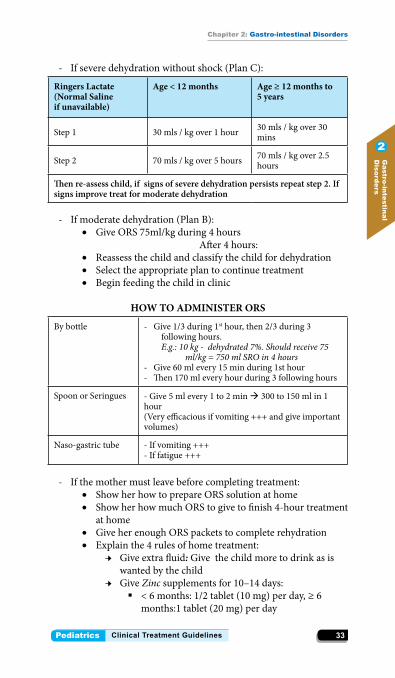
- If severe dehydration without shock (Plan C):
Ringers Lactate(Normal Saline if unavailable)
Age < 12 months Age ≥ 12 months to 5 years
Step 1 30 mls / kg over 1 hour 30 mls / kg over 30 mins
Step 2 70 mls / kg over 5 hours 70 mls / kg over 2.5 hours
Then re-assess child, if signs of severe dehydration persists repeat step 2. If signs improve treat for moderate dehydration
- If moderate dehydration (Plan B):• Give ORS 75ml/kg during 4 hours
After 4 hours:• Reassess the child and classify the child for dehydration• Select the appropriate plan to continue treatment• Begin feeding the child in clinic
HOW TO ADMINISTER ORS
By bottle - Give 1/3 during 1st hour, then 2/3 during 3 following hours.E.g.: 10 kg - dehydrated 7%. Should receive 75
ml/kg = 750 ml SRO in 4 hours- Give 60 ml every 15 min during 1st hour- Then 170 ml every hour during 3 following hours
Spoon or Seringues - Give 5 ml every 1 to 2 min 300 to 150 ml in 1 hour (Very efficacious if vomiting +++ and give important volumes)
Naso-gastric tube - If vomiting +++- If fatigue +++
- If the mother must leave before completing treatment:• Show her how to prepare ORS solution at home• Show her how much ORS to give to finish 4-hour treatment
at home• Give her enough ORS packets to complete rehydration • Explain the 4 rules of home treatment:
→ Give extra fluid: Give the child more to drink as is wanted by the child
→ Give Zinc supplements for 10–14 days: < 6 months: 1/2 tablet (10 mg) per day, ≥ 6
months:1 tablet (20 mg) per day
Chapiter 2: Gastro-intestinal Disorders
Gastro-intestinal
Disorders
2

34 Clinical treatment guidelinesPediatrics
→ Continue feeding: Initial 4-hour rehydration period, breastfed children should continue to breastfeed frequently throughout
→ Return the child to the health facility if :drinking poorly or unable to drink or breastfeedBecomes more sickdevelops feverHas blood in the stool
- If no dehydration (Plan A):• Treat the child as an outpatient; give ORS 10ml/kg after
each watery stool • Counsel the mother on the 4 rules of home treatment:
→Give extra fluid →Give zinc supplements→Continue feeding→Give advice on when to return for review
Particular forms of dehydration
Type Intervention Comment
Hyponatremia(Na < 130mmol/L)
Na Deficit = 0.6 x W in kg x (Na+
d - Na+m) during 4 hours
W= weight d = desired sodium m = measured sodium
do not correct too quickly to avoid CNS lesion
Hypernatremia(Na > 150mmol/L)
Slowly correct dehydration over 48 hours
Risk of convulsions in case of rapid correction
Hypokalemia If Potassium< 2.5 mmol/L give KCl 30-40 mmol/L/24hours
Give KCl
Chapiter 2: Gastro-intestinal Disorders

35Clinical treatment guidelinesPediatrics
2.2. Persistent Diarrhea
Definition: Persistent diarrhea is a diarrhea, with or without blood, which begins acutely and lasts for 14 days or longer.
Causes
AGE ETIOLOGIES
Infancy - Post gastroenteritis mal-absorption syndrome- Cow’s milk/soy protein tolerance- Secondary disaccharidase deficiencies- Cystic fibrosis
Childhood - Secondary disaccharidase defiencies- Giardiasis- Post-gastroenteritis mal-absorption syndrome- Celiac disease- Cystic fibrosis- HIV- Malnutrition
Adolescence - Irritable Bowel Syndrome- HIV- Inflammatory Bowel disease
Complications
- dehydration- Failure to thrive, malnutrition- Immunosuppressant
Investigations (Will vary according to the suspected etiology)
- Stool examination: PH, White blood count, Fat, Ova, osmolarity, Culture
- FBC, CRP, electrolytes, urea and creatinine- Sweat chloride if suspicion of cystic fibrosis- Barium study- Small bowel biopsy- Endoscopy: Sigmoidoscopy or coloscopy with biopsy
Management
- Oral rehydration- Treat the cause (see algorithm)
Chapiter 2: Gastro-intestinal Disorders
Gastro-intestinal
Disorders
2

36 Clinical treatment guidelinesPediatrics
2.3. Bloody Diarrhea
Definition: Frequent (>3/day) passage of blood and/or mucus in the stool
Causes
- Amoebic dysentery is the most common serious cause in children
- Bacterial infections (e.g. Shigella, salmonella)- Parasitic infestations (e.g. amoebic dysentry)- Milk allergy- Chronic inflammatory bowel disease
Signs and Symptoms
- Sudden onset- Abdominal cramps- Peritonism urgency, fever and diarrhea with blood and mucus in
the stool- meningismus and convulsions may occur- Exclude intussusceptions which includes:
• pain or abdominal tenderness• bile-stained vomitus• red currant jelly-like mucus
Complications
- dehydration - Convulsions- Shock - Toxic megacolon- Acidosis - Rectal prolapse- Renal failure - Haemolytic uraemic syndrome
Investigations
- Stool culture to confirm diagnosis of Shigellosis- Stool microscopy reveals many polymorphs and blood- Immediate microscopy of warm stool to diagnose amoebic
dysentery
Chapiter 2: Gastro-intestinal Disorders

37Clinical treatment guidelinesPediatrics
Management
Non-pharmacological• Ensure adequate nutrition and hydration
Pharmacological• Fluid and electrolyte replacement (see Acute diarrhea)• ciprofloxacin, oral, 15 mg/kg/dose 12 hourly for 3 days
Or• ceftriaxone, IV, 20–80 mg/kg as a single daily dose for 5
days (If hospitalised or if unable to take oral antimicrobial
agents)• Metronidazole, oral, 15 mg/kg/dose 8 hourly for 7 – 10 days
(If amoebic dysentery, seen on stool microscopy)
Recommendation
- Refer patient to the specialist, if dysentery with complications, e.g. persistent shock, haemolytic uraemic syndrome and toxic megacolon
2.4. Constipation
Definition: Constipation is an acute or chronic condition in which bowel movement occurs less often than usual or consist of hard, dry stool that are painful or difficult to pass.
Causes
- Lack of exercise - Certain medicines- Metabolic, endocrine, neurogenic and lower bowel abnormalities- Psychogenic disorders - Chronic use of enemas- Not drinking enough water- diet that does not include an adequate amount of fiber-rich
foods - Anal fissure (a tear or crack in the lining of the anus)- Chronic kidney failure- Hischprung disease- Colon or rectal cancer- depression- Hypercalcemia (abnormally high levels of calcium in the blood)
Chapiter 2: Gastro-intestinal Disorders
Gastro-intestinal
Disorders
2

38 Clinical treatment guidelinesPediatrics
- Hypothyroidism (underactive thyroid gland)- Illness requiring complete bed rest- Irritable Bowel Syndrome- Stress
Signs and Symptoms
- Symptomatic bowel impaction- Blood in the stool- Changes in bowel patterns- Abdominal pain, distension
Complications
- Bowel obstruction - Chronic constipation - Hemorrhoids- Hernia - Spastic colitis- Laxative dependency
Investigations
- Abdominal X-ray- Barium meal - reveals blockage inside the intestine in particular
cases- Laboratory analysis of blood and stool samples for internal
bleeding- Sigmoidoscopy (examination of the sigmoid area of the colon
with a flexible tube equipped with a magnifying lens), rarely indicated.
Management
Principles• Treatment involves 3 steps:
→ Initial clearance of stool→ Prevent reaccumulation of hardened retained stool
(diet change with additional natural fibre from fruit, vegetables and bran)
→ Retraining of the gut to achieve regular toilet habitsManagement is long-term, and requires the active involvement of the parents
Chapiter 2: Gastro-intestinal Disorders

39Clinical treatment guidelinesPediatrics
Pharmacological• Enema twice daily for 3 days for faecal clearance if faecal
loading• Lactulose (Duphalac) for 1 week but if 3 stools are passed/
day stop it• Bowel re-training• In refractory cases:
→ Lactulose, oral, twice daily■ < 1 year 2.5 mL■ 1–6 years 5 mL■ > 6 years 10 mL
• determine and treat the underlying cause
Recommendations
- Refer patient to the specialist, if an organic cause e.g. constipation from birth in a breast-fed baby is suspected
- If faecal loading continues, maintenance therapy should be continued for months to years
2.5. Constipation and Encopresis
Definition: Constipation is the delay or difficulty in passage of stool during defaecation that has been present for 2 weeks or longer. Stool is usually hard.
Encopresis also known as faecal soiling is the involuntary leakage of small amounts of soft or watery stool in a child with chronic constipation.
Causes
- Psycho social precipitants- Functional (incorrect diet, lack of exercise, poor fluid intake) - Metabolic or Neurological Abnormalities - Endocrine abnormalities (Hypothyroidism)- Chronic use of laxatives - Obstructive lesions (acquired and congenital defects)
Signs and Symptoms
- Abdominal pain often associated with encopresis- Infrequent defecation - Pain or strain on defecation- Hard stool - Feeling of incomplete evacuation (Tenesmus)
Chapiter 2: Gastro-intestinal Disorders
Gastro-intestinal
Disorders
2

40 Clinical treatment guidelinesPediatrics
Complications
- Anal fissure, ulcers and prolapse- Over flow incontinence (Encopresis)- Stasis syndrome with bacterial overgrowth
Investigations
- Barium Enema- Abdominal x-ray in suspected obstructive lesions- Thyroid function tests when indicated- Stool analysis- Investigate other functional lesions
Management
Non-pharmacological management• Rehydrate to increase fecal bulk and soften stool• Education of patients/parents on diet, exercise, etc. • diet change with additional natural fibre from fruit and
vegetables• Treatment involves 3 steps:
→ Initial clearance of stools → Prevent re-accumulation of hardened retained
stool → Retraining of the gut to achieve regular toilet habits
Pharmacological management• Glycerin Suppositories 1 suppo/dose according to
occurrence of symptoms Or
• Lactulose syrup <1 yr: 5-10ml/24 hr PO Od; 1-6 Yrs 10-20 ml/24 hours PO Od; 7-14 yrs 20-50ml/24 hrs PO Od
Or• Bisacodyl (Dulcolax) 0.3mg/kg/day PO Od maximum dose
30mg/24 hours
Recommendations
- Refer to tertiary health facility in cases of inadequate response to therapy for further investigation
- If continued constipation therapy should be continued for months to years
Chapiter 2: Gastro-intestinal Disorders

41Clinical treatment guidelinesPediatrics
2.6. Upper Gastro-Intestinal Tract Bleeding
Definition: Upper gastrointestinal bleeding (arising proximal to the ligament of Treits in the distal duodenum) commonly manifested by hematemesis and/or melena.
Causes
- Neonates• False bleeding (maternal blood swallowed• Vit K1 deficiency• Stress gastric/ ulcer• Coagulopathy (infection, liver failure, coagulation
disorder)• Hemangioma
- Infants and toddlers• Malory Weiss Syndrome• Non steroid anti-inflammatory drugs• Oesophagitis• Caustic ingestions, iron poisoning• Oesophageal varices bleeding
- Older children and adolescents• Malory Weiss Syndrome• Peptic ulcer/gastritis• Rendu Osler syndrome• Gastric polypes• Oesophagal varices
Clinical manifestations
- Hematemesis- Melena- Other signs according to the causative agent
Assessment
History — The clinical history should include information concerning:
•The time course of the bleeding episode• Estimated blood loss, and any associated symptoms • Gastrointestinal symptoms including dyspepsia,
heartburn, abdominal pain, dysphagia, and weight loss. In infants, these features may be reflected in poor feeding and irritability.
Chapiter 2: Gastro-intestinal Disorders
Gastro-intestinal
Disorders
2

42 Clinical treatment guidelinesPediatrics
• The history should also include information about the following symptoms or signs which may provide clues to an underlying disorder:
→ Recent onset of jaundice, easy bruising or change in stool color, which may suggest underlying liver disease
→ Recent or recurrent epistaxis, to investigate the possibility of a nasopharyngeal source of bleeding
→ History of easy bruising or bleeding, which suggests a disorder of coagulation, platelet dysfunction, or thrombocytopenia
→ Personal or family history or liver, kidney or heart disease, or coagulation disorders
→ A drug history is important to assess potential contribution from medication that may induce ulceration (such as NSAIds and corticosteroids); Tetracyclines, may cause a pill esophagitis
If the patient has been taking drugs or has a cardiac condition that affects homeostatic responses (such as beta-adrenergic antagonists), considering that these may mask tachycardia associated with life-threatening hypovolemia and shock
Physical examination — The physical examination should include the following elements:
• The skin for cutaneous signs of generalized vascular malformations/disorders (cutaneous hemangiomas. mucocutaneous telangiectasia)
• Evidence of portal hypertension, (splenomegaly , prominent abdominal and hemorrhoid vessels)
• Inspection of the nasopharynx • Check for hemodynamic failure (signs of shock? )
Nasogastric tube — in patients presenting with unexplained gastro-intestinal bleeding NGT is sometimes used to confirm the diagnosis and determine if the bleeding is ongoing. The lavage will also remove particulate matter, fresh blood, and clots to facilitate endoscopy and decrease the risk of aspiration.
Ice water lavage (an older practice) does not slow bleeding and may induce iatrogenic hypothermia, particularly in infants and small children, and is not recommended
Chapiter 2: Gastro-intestinal Disorders
43Clinical treatment guidelinesPediatrics
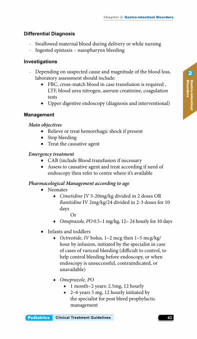
Differential Diagnosis
- Swallowed maternal blood during delivery or while nursing- Ingested epistaxis – nasopharynx bleeding
Investigations
- depending on suspected cause and magnitude of the blood loss, laboratory assessment should include:
• FBC, cross-match blood in case transfusion is required , LTF, blood urea nitrogen, aserum creatinine, coagulation tests
• Upper digestive endoscopy (diagnosis and interventional)
Management
Main objectives• Relieve or treat hemorrhagic shock if present • Stop bleeding• Treat the causative agent
Emergency treatment• CAB (include Blood transfusion if necessary• Assess to causative agent and treat according if need of
endoscopy then refer to centre where it’s available
Pharmacological Management according to age• Neonates
→ cimetidine IV 5-20mg/kg divided in 2 doses OR Ranitidine IV 2mg/kg/24 divided in 2-3 doses for 10 days Or
→ Omeprazole, PO 0.5–1 mg/kg, 12– 24 hourly for 10 days
• Infants and toddlers → Octreotide, IV bolus, 1–2 mcg then 1–5 mcg/kg/
hour by infusion, initiated by the specialist in case of cases of variceal bleeding (difficult to control, to help control bleeding before endoscopy, or when endoscopy is unsuccessful, contraindicated, or unavailable)
→ Omeprazole, PO■ 1 month–2 years: 2.5mg, 12 hourly ■ 2–6 years 5 mg, 12 hourly initiated by
the specialist for post bleed prophylactic management
Chapiter 2: Gastro-intestinal Disorders
Gastro-intestinal
Disorders
2

44 Clinical treatment guidelinesPediatrics
• Older children and adolescents → Omeprazole, PO
■ < 20 kg: 10 mg Qd■ >20 kg: 20 mg Qd
Note: Endoscopy is recommended to be performed within 24 to 48 hours for infants and children presenting with UGI bleeding that is acute and severe, it can be performed for diagnosis and treatment (sclerotherapy in oesophageal variceal)
Alternative treatment• Propranolol oral, 2–8 mg/kg/24 hours in 3 divided doses
(to reduce the pulse rate by 25%)• Surgical over sewing if endoscopy and sclerotherapy or
banding have failed
Recommendations
- Refer all cases to the specialist for appropriate diagnosis and treatment
- Refer all bleeding varices - after commencement of resuscitation and octreotide, if available
2.7. Peptic Ulcer Disease
Definition: This refers to ulceration of gastric or duodenal mucosa that tends to be chronic and/or recurrent.
Cause
- Helicobacter pylori (H. pylori) - in developing nations, the majority of children are infected with H. pylori before the age of 10 and adult prevalence peaks at more than 80 percent before age 50
Signs and Symptoms
- Peptic ulcers may be present with dyspeptic or other gastrointestinal symptoms or may be completely asymptomatic, sometimes until complications such as hemorrhage or perforation occur. The symptoms associated with peptic ulcers are not sensitive or specific and the differential diagnosis is broad.
- Most common: Ulcer-like or acid dyspepsia (burning pain; epigastric hunger-like pain; relief with food, antacids, and/or antisecretory agents).
Chapiter 2: Gastro-intestinal Disorders

45Clinical treatment guidelinesPediatrics
- Food-provoked dyspepsia or indigestion (postprandial epigastric discomfort and fullness, belching, early satiety, nausea, and occasional vomiting): food-stimulated acid secretion persists for three to five hours; thus, classic dU symptoms occur two to five hours after meals.
- Reflux-like dyspepsia
Complications
- The natural history of peptic ulcer ranges from resolution without intervention to development of complications: acute or Chronic blood loss or perforation
- Iron deficiency anaemia
Investigations
- Stool analysis for occult blood- FBC - For HP:
• It is recommended that the initial diagnosis of H. pylori infection be based on positive histopathology plus positive rapid urease test, or positive culture.
• A validated ELISA for detection of H. pylori antigen in stool is a reliable non-invasive test to determine whether H. pylorus has been eradicated.
• Tests based on the detection of antibodies (IgG, IgA) against H. pylori in serum, whole blood, urine and saliva are not reliable for use in the clinical setting.
Note: Specialists recommend: In children with refractory iron deficiency anemia, where other causes have been ruled out, testing for H. pylori infection may be considered (grade of evidence - low)
Management
- Avoid any foods that cause pain to the patient (e.g. acid foods, soda drinks)
- Avoid gastric irritating drugs (NSAIds)- Give magnesium-based antacids or combined magnesium-
aluminium
Chapiter 2: Gastro-intestinal Disorders
Gastro-intestinal
Disorders
2

46 Clinical treatment guidelinesPediatrics
First line H pylori eradication regimens are:• Triple therapy with:
→ PPI + Amoxicillin + Imidazole Or
→ PPI + Amoxicillin + clarithromycin Or
→ Bismuth salts + Amoxicillin + Imidazole Or
• Sequential Therapy Triple therapy for eradication of H. pylori (duration: 10 – 14 days)
→ Omeprazole PO ■ 15-30 kg: 10 mg twice daily ■ >30 kg: 20 mg twice daily
Or → cimetidine 20–40mg/kg/day + clarithromycin :
500mg BId + Amoxicillin 1g twice daily Or
→ cimetidine 20–40mg/kg/day +Clarithromycin : 500mg + Metronidazole 500 mg (15–20mg/kg/day ) twice daily
Note: A reliable non-invasive test for eradication is recommended at least 4 to 8 weeks following completion of therapy
Recommendations
- Refer to a specialist, if there is severe hemorrhage- Stabilize the patient before transfer- Infuse IV fluids/blood to maintain normal volume/pulse- Ensure continuous assessment of further blood loss (Persistent
tachycardia, postural hypotension, continuing haematemesis)- definitive treatment / eradication of H. pylori
Chapiter 2: Gastro-intestinal Disorders

47Clinical treatment guidelinesPediatrics
2.8. Gastroesophageal Reflux
Definition: GER is the passage of gastric contents into the esophagus with or without regurgitation and vomiting. GER is a normal physiologic process occurring several times per day in healthy infants, children, and adults. Most episodes of GER in healthy individuals last <3 minutes, occur in the postprandial period, and cause few or no symptoms. In contrast, Gastroesophagial reflux disease GERd is present when the reflux of gastric contents causes troublesome symptoms and/or complications.
Causes and risk factors
- The cause is still unclear - Anatomical abnormalities such as a hiatal hernia- Long term use of nasal gastric tube - diet that stimulates gastric acid production- Neurologic impairment (NI), obesity, certain genetic syndromes,
esophageal atresia (EA), chronic lung diseases, and those with a history of premature birth
Signs and Symptoms
In infants and toddlers, there is no symptom or symptom complex that is diagnostic of GERd or predicts response to therapy. In older children and adolescents, as in adult patients, history and physical examination may be sufficient to diagnose GERd if the symptoms are typical. The following is suggestive:
- In newborn: Recurrent vomiting, stridor, apnea- In infant: Recurrent vomiting, respiratory manifestations, (dry
cough, recurrent wheeze or cough, chronic obstructive airway disease) recurrent aspiration pneumonia, stridor, apnea
- In children /adolescent: Heartburn, epigastria or chest pain. Respiratory manifestations: (dry cough, recurrent wheeze or cough, chronic obstructive airway disease
Complications
- dysphagia - Odynophagia- Weight loss- Anemia- Esophagitis- Aspiration pneumonia- Barrett’s esophagus - Abnormal posturing or opisthotonus (Sandifer Syndrome)
Chapiter 2: Gastro-intestinal Disorders
Gastro-intestinal
Disorders
2

48 Clinical treatment guidelinesPediatrics
Investigations (when GER is persisting despite basic management)
- 24 hours esophageal PH monitoring- Endoscopy with biopsy to rule out oesophagitis- Barium X-rays for severity of oesophagus stenosis- FBC look for anemia
Management
Non-pharmacological• Postural treatment: Prone and lateral positions are
associated with an increased incidence of sudden infant death syndrome (SIdS). The risk of SIdS outweighs the benefit of prone or lateral sleep position on GER; therefore, in most infants from birth to 12 months of age, supine positioning during sleep is recommended.
• dietary measures such as thickened food if not breastfeeding, frequent small volume of solid foods
Pharmacological• Less Severe or Non Erosive
→ Anti-acids:■ Sodium alginate (Gaviscon Enfant) /antacid
combination, oral, month 1ml after each mealo 1-2 months 1.5 mls after each mealo 2-4 months 2mls after each meal
→ Aluminium and Magnesium hydroxide (Maalox) Syrup 0.5 ml/kg/dose PO QId
→ H2 Antagonists: ■ cimetidine IV/syrup/tab
o Neonates: 5-20mg/kg/24 hr divided in 2 doses
o Infants: 10-20 mg/kg/24hrs divided in 2 doses
o Children: 20-40mg/kg/24hr divided in 2 doses
• Severe or Erosive→ Omeprazole, oral
■ Neonate 0.5–1 mg/kg, 12– 24 hourly■ Children 1- 16 years
o 5 kg to <10 kg: 5 mg once dailyo 10 kg to ≤20 kg: 10 mg once daily
Chapiter 2: Gastro-intestinal Disorders

49Clinical treatment guidelinesPediatrics
o >20 kg: 20 mg once daily Alternate dosing: 1 mg/kg/dose once or
twice daily; higher doses may be necessary in children between 1-6 years
→ Add■ Pro-Kinetics: Domperidone (Motilium) 0.3 – 0.6
mg/kg/24hrs PO divided in 3 doses (TdS). Maximum 30mg/24 hours
→ ANd■ Metoclopromide IV/IM/PO 0.1-0.2mg/kg/dose
TdS. Maximum dose 0.5mg/kg/24 hours
Recommendations
- Refer to tertiary level gastro-oesophageal reflux not responding to treatment
- Educate parents/guardians on patient diet - Eat small, frequent meals
2.9. Tropical Splenomegaly (Hyperreactive malarious splenomegaly) (HMS)
Definition: It is a massive enlargement of the spleen resulting from abnormal immune response to repeated attacks of malaria
Signs and Symptoms
- Chronic abdominal swelling and pain.- Weight loss- Intermittent fever- Some patients present with anaemia, generalized weakness,
cough, dyspnea, epistaxis, headache, increased skin and respiratory infections
Clinical diagnosis
- Splenomegaly of at least 10cms- Regression of the spleen by at least 40% by 6 months on
antimalarial therapy.
Chapiter 2: Gastro-intestinal Disorders
Gastro-intestinal
Disorders
2

50 Clinical treatment guidelinesPediatrics
Complications
- Hypersplenism leading to anemia, leukopenia and thrombocytopenia, bleeding
- Splenic lymphoma- death
Investigations
- Blood smear- Complete blood count (for Hb, Platelets) - Serum levels of IgM (at least 2Sd above normal limit)
Management
Pharmacological treatment• Doxycline tabs /day for 6 months
→ Children >8 years (<45 kg): 5 mg/kg/day Od → Children >8 years (>45 kg): treat as adults
Or • Mefloquine 5mg/kg weekly without exceeding 250mg/week
of adult dose for 6 months
Note: Generally, splenectomy in the management of HMS is not recommended as mortality is high from sepsis and thrombocytosis unless there is a splenic rupture.
Chapiter 2: Gastro-intestinal Disorders

51Clinical treatment guidelinesPediatrics
3. Cardiovascular Diseases
Most cardiac diseases in young children are congenital, while those in older children may be acquired or congenital.
3.1. Heart Failure (Congestive Cardiac Failure)
Definition: It is a clinical syndrome reflecting the inability of the myocardium to meet the oxygen and nutritional metabolic requirements of the body.
Causes
- In normal heart anatomy• Anemia• Infection/sepsis• Volume overload• Arrhythmia • Cardiomyopathies• Hypertension • Renal failure• Acquired valvulopathies• Hypothyroidism• Kawasaki disease
- In Congenital heart disease • Left to Right shunt (Ventricular Septal defect, Patent
ductus Arteriosus)• Aortic coarctation• Aortic valvular stenosis• Supra valvular aortic stenosis• Mitral stenosis, mitral regurgitation • Pulmonary veins stensosis• Single ventricle
Signs and Symptoms
- Tachypnea/dyspnea- Cough- Sweating- Excessive weight gain/oedema- Poor feeding/ failure to thrive
Cardiovascular
Diseases
3

52 Clinical treatment guidelinesPediatrics
- Tachycardia- Gallop rhythm with or without heart murmur- Weak pulses- Hypotension - Palor- Cold extremities - Prolonged capillary refill > 2seconds- Oliguria- Hepatomegaly / increased jugular vein pressure- Crepitations (in older children) / wheezing
Investigations
- FBC, Electrolytes, Urea and Creatinine, Blood Gas if available- Chest X-ray - ECG- Echocardiogram
Management
Non pharmacological• Oxygen therapy • Semi- Sitting position (cardiac bed)• Restrict fluids to 2/3 of maintenance ( aim at urine output
of 2ml/kg/h)• Low sodium diet • Strict bed rest• Ensure adequate nutrition• Recognize and treat the underlying conditions e.g. fluid
overload, hypertension, infection• Monitoring of vital signs: RR, HR, BP, O2 saturation, urine
output
Pharmacological• Frusemide IV 1-4mg/kg divided in 2 doses (to be increased
progressively) • Digoxin per os 0.01mg/kg/day (no loading dose!!)• captopril 1-4mg/kg/day divided in 3 doses if normal
creatinine (to be increased progressively, beware of hypotension)
• carvedilol for stable older children > 30 kg: initiate with 3.125mg BId, increase every 15 days if good tolerance. Maximum dose: 12.5mg BId
Chapiter 3: Cardiovascular Diseases

53Clinical treatment guidelinesPediatrics
Recommendations
- If isolated right sided heart failure: use Furosemide (see dosage above) and Aldactone 2mg/kg/day divided in 2 doses
- Administration of carvedilol and Aldactone should be discussed with the cardiologist
Note: Any patient with Heart Failure due to Heart disease must be referred to the cardiologist
3.2. Carcinogenic Shock
Definition: It is a dramatic syndrome characterized by inadequate circulatory provision of oxygen due to cardiac pump failure secondary to poor myocardial function, so that the metabolic demands of vital organs and tissues are not met.
The patient often has a known case of heart disease with signs of heart failure but may also be a new case with heart failure.
Signs and Symptoms
- Hypotension- Tachycardia- Gallop rhythm- Hepatomegaly- Crackles/wheezes- Weak and fast pulses (or absent)- Cold extremities/ palor- Capillary refill > 2 seconds- Oliguria/anuria
Management
Non pharmacological management• Avoid excessive IV fluids, the patient is fluid overloaded in
this case, give 2/3 of maintenance (aim at urine output of 2ml/kg/h)
• Oxygen therapy: 10-15l/min with mask and reservoir bag• Semi- Sitting position (cardiac bed)• Low sodium diet • Strict bed rest• Ensure adequate nutrition• Correct hypoglycemia with 3-5ml/kg IV of Dextrose 10%
Chapiter 3: Cardiovascular Diseases
Cardiovascular
Diseases
3

54 Clinical treatment guidelinesPediatrics
Pharmaceutical treatment• Dopamine IV 5-10 microgram/kg/min, may increase to 20
microgram/kg/min Or• Dobutamine IV 2 to 20 microgram/kg/min • Furosemide IV 2mg/kg/dose if adequate peripheral
perfusion. Repeat the dose according to estimated fluid overload up to 8mg/kg/day
• Correct arrhythmia if present with Digoxin 0.04mg/kg/day in 3 devided doses ( maintenance: 0.01mg/kg/day)
• Monitor: Heart rate, Respiratory rate, BP, Urine output, Pulse Oxymetry for oxygen saturation
3.3. Pulmonary Oedema
Definition: Pulmonary oedema is the accumulation of fluid in the alveoli due to an increase in pulmonary capillary venous pressure resulting from acute left ventricular failure.
Causes
- Heart not removing fluid from lung circulation properly (cardiogenic pulmonary edema)
- A direct injury to the lung parenchyma
Signs and Symptoms
- Breathlessness/ respiratory distress- Sweating- Cyanosis (decreased oxygen saturation)- Frothy blood-tinged sputum- Ronchi, and crepitations/wheezers
Investigations
- Chest x-ray shows loss of distinct vascular margins, Kerley B lines, diffuse haziness of lung fields, pleural effusion
- Blood Gas if possible- ECG - Echocardiography
Management
- Maintain patient in a semi sitting position- Oxygen by facial mask with reservoir bag if available
Chapiter 3: Cardiovascular Diseases

55Clinical treatment guidelinesPediatrics
- IV Furosemide 2mg/kg/dose, maximum 8mg/kg/day- Inotropic support with Dopamine or Dobutamine if signs of
shock- Transfer to cardiologist for further management
3.4. Congenital Heart Diseases
Definition: Congenital heart disease refers to a problem with the heart’s structure and function due to abnormal heart development before birth. Often divided into two types, non-cyanotic and cyanotic (blue discoloration caused by a relative lack of oxygen).
3.4.1. Non Cyanotic Heart Diseases
Common lesions
- Ventricular Septal defect (VSd) most common congenital heart disease
- Patent ductus arteriosus (PdA)- Atrio-ventricular septal defect (AVSd) or endocardial cushion
defect (common in trisomy 21)- Atrial septal defect (rarely causes heart failure)- Coarctation of aorta
Signs and Symptoms
- Tachypnea, dyspnea- Tachycardia- Sweating- Feeding difficulties / failure to thrive - Recurrent chest symptoms- Hepatomegaly- Increased jugular venous pressure
Complications
- Failure to thrive- Infective Endocarditis- Pulmonary vascular obstructive disease (pulmonary
hypertension) which can lead to Eisenmenger syndrome
Chapiter 3: Cardiovascular Diseases
Cardiovascular
Diseases
3
56 Clinical treatment guidelinesPediatrics
Investigations
- Chest X-Ray- ECG- Echocardiogram- Cardiac catheterization/angioscan in special cases
Management
Treatment depends on the specific condition. Some congenital heart diseases can be treated with medication alone, while others require one or more surgeries.
- Lasix 2mg/kg/day- captopril 1-3mg/kg/day (start with 1mg/kg)- Increase calories in feeding- Iron if Hb less than 10g/dl (preferably reach 15g/dl)- Surgical repair generally before 1 year if possible
3.4.2. Cyanotic Heart Diseases
Definition: Cyanotic heart disease is a heart defect, present at birth (congenital), that results in low blood oxygen levels (< 90 % even with oxygen).
Common lesions
- decreased flow to the lungs (does not cause heart failure)• Tetralogy of fallot • Pulmonary atresia
- Increased flow to the lungs (does cause heart failure and failure to thrive):
• Transposition of great vessels (TGA)• Truncus arteriosus• Single ventricle / Tricuspid atresia
Tetralogy of Fallot
Definition: Tetralogy of Fallot refers to a type of congenital heart defect comprising of:
- Large ventricular septal defect - Narrowing of the pulmonary outflow tract (pulmonary stenosis)- Overriding aorta - Right ventricular hypertrophy
Chapiter 3: Cardiovascular Diseases

57Clinical treatment guidelinesPediatrics
Signs and Symptoms
- Progressive cyanosis with pulmonary systolic murmur- digital clubbing occurs after long time- Hallmark: Paroxysmal hyper cyanotic attacks (blue spells) with
the following manifestations• Hyperpnea and restlessness• Increased cyanosis• Gasping respiration• Syncope or convulsions• Spontaneous squatting position is frequent (in older
children)• Heart murmur disappears
Complications
- delayed development/growth- Polycythemia- Hypercyanotic attack, sometimes associated with seizures and
death- Infective endocarditis- Brain abscess
Investigations
- Chest x-ray- Complete blood count (CBC)- Echocardiogram- Electrocardiogram (ECG)
Management
- Avoid dehydration and stress - Propanolol 0.5-1mg/kg every 6 hours to prevent hypercyanotic
attacks- Iron 5mg/kg /day to prevent microcytosis - Surgical repair, urgent as soon as spells begin- In case of Hypercyanotic attacks
• Squatting position (hold the infant with the legs flexed on the abdomen)
• Oxygen 6l/min with mask• Diazepam 0.3mg/kg IV or 0.5mg PR if convulsing • normal saline 10-20ml/kg/ 30 minutes• Sodium bicarbonate 8.5% 1ml/kg to correct acidosis
Chapiter 3: Cardiovascular Diseases
Cardiovascular
Diseases
3

58 Clinical treatment guidelinesPediatrics
• Morphine 0.1mg/kg IV if persistent attacks (but risk of respiratory depression)
• Propranolol IV 0.1 – 0.2 mg/kg slowly then continue oral maintenance to relax the infundibular spasms
Recommendations
- All children with cyanotic heart diseases who come with diarrhea and vomiting should be admitted for closer observation. Furosemide is contra-indicated.
- All new born babies with suspected cyanotic heart disease should be referred to a cardiologist/tertiary hospital immediately.
- Common causes of heart failure in Neonates:
Clinical manifestations Likely lesions
Very poor pulses - Hypoplastic Left Ventricle Syndrome- Critical aortic stenosis
Poor femoral pulses - Coarctation of aorta
Bounding pulses - Patent ductus arterious (PdA)- Troncus arteriosus- Severe anemia
Chapiter 3: Cardiovascular Diseases

59Clinical treatment guidelinesPediatrics
3.5. Acquired Heart Diseases
3.5.1. Acute Rheumatic fever
Definition: This is an acute, systemic connective tissue disease in children related to an immune reaction to untreated group A Beta haemolytic streptococcus infection of the upper respiratory tract. The initial attack of acute rheumatic fever occurs in most cases between the ages of 3 and 15 years.
Cause
- Auto-immune disease
Signs and Symptoms (Revised Jones Criteria)
Major manifestations Minor manifestations
Group A Strep (GAS) Infection
Carditis Fever GAS on throat swab (culture)
Arthritis Arthralgia Raised Anti-streptolysin O titre
(ASOT)
Sydenham’s Chorea Prolonged P-R interval
on ECG
Raised Anti-deoxyribonuclease B (Anti-dNase B)
Erythema marginatum Raised ESR or CRP
Subcutaneous nodules
Criteria for ARF diagnosis according to WHO- The first episode of ARF can be confirmed if:
• 2 MAJOR, or 1 MAJOR and 2 MINOR manifestations are present plus there is evidence of preceding Group A streptococcal infection.
- Recurrent ARF (with no RHd) can be confirmed if• 2 MAJOR, or 1 MAJOR and 2 MINOR manifestations
are present plus there is evidence of preceding Group A streptococcal infection.
- Recurrent ARF (with existing RHd) can be confirmed if• 2 MINOR manifestations are present plus there is evidence
of preceding Group A streptococcal infection.
Chapiter 3: Cardiovascular Diseases
Cardiovascular
Diseases
3

60 Clinical treatment guidelinesPediatrics
Complication
- Rheumatic heart disease
Investigations
- Throat swab for culture (positive throat culture of group A Streptoccocal infection)
- Raised ASOT/ASLO antibodies titre (Anti-streptolysin-0-titre – ASOT of 1:300)
- Anti dNase B- FBC/ ESR/CRP- Chest x-ray – features of cardiomegaly- ECG - Echocardiogram
Management
- Persons with symptoms of ARF should be hospitalized to ensure accurate diagnosis, and to receive clinical care and education about preventing further episodes of ARF.
- The diagnosis should include an initial echocardiogram used to help identify and measure heart valve damage.
- Long-term preventative management should be organized before discharge.
- All cases of ARF should receive:• A single injection of Benzathine penicillin G (Extencilline):
25,000–50,000 units/kg/dose, maximum 1.2 mega units dose
Or • Oral Penicillin (Pen V) 25–50mg/kg/day in divided 3 doses
for 10 days Or (Erythromycin 30-50mg/kg/day divided in 3 doses if penicillin allergy)
- Symptomatic Treatment • Arthritis and fever
→ Aspirin 75–100mg/kg/day in 4–6 divided doses. Continue treatment until fever and joint inflammation are controlled and then gradually reduced over a 2-week periodAdd an antacid to reduce risk of gastric irritation
→ Prednisolone 1-2mg Od for 2 weeks then taper for 2 weeks with good response begin
→ Aspirin in the 3rd week and continue until 8th week tapering in the final 2 weeks
Chapiter 3: Cardiovascular Diseases

61Clinical treatment guidelinesPediatrics
• Chorea → Most mild-moderate cases do not need medication→ Provide calm and supportive environment (prevent
accidental self-harm)→ For severe cases: carbamazepine per os
<6 years: 10-20mg/kg/day divided in 3 doses 6-12 years: 400-800mg/day divided in 3 doses>12 years: 200mg x 2/day
OR→ Valproic acid 20-30mg/kg/day divided in 2 doses;
duration: 2 weeks
• Carditis → Bed rest if in cardiac failure → Anti-failure medication as above→ Anti-coagulation medication if atrial fibrillation is
present→ Management plan when the acute episode is
controlled administer the first dose of secondary prophylaxis
→ Register the individual with the local health authority or RHd Program
→ Provide disease education for the person with ARF and the familyUnderstanding of ARF and RHd and risks of
ARF recurrenceImportance of regular secondary prophylaxis
and medical reviewRecognising own signs and symptoms of ARF
and RHdRisks associated with future RHd (e.g.
pregnancy, surgery and high level of aftercare) Importance of dental health
→ Include an ARF diagnosis alert on computer systems and/or medical files (if applicable)
→ Refer to local health facility for ongoing management→ Arrange dental review (and provide advice about
endocarditis prevention)
Chapiter 3: Cardiovascular Diseases
Cardiovascular
Diseases
3

62 Clinical treatment guidelinesPediatrics
- Long-term Management• Regular secondary prophylaxis (Refer to 5.5 Table 6
Recommended Secondary Prophylaxis Regimen)• Regular medical review• Regular dental review• Echocardiogram (if available) following each episode of
ARF, and routine echocardiogram:→ Every 2 years for children (sooner if there is evidence
of cardiac symptoms)
- Secondary prophylaxis • Prevents the occurrence of GAS infections which can lead
to recurrent ARF • Reduces the severity of RHd (and can result in cure of
RHd after many years)• Helps prevent death from severe RHd• Secondary prophylaxis is indicated for people who have:
→ ARF confirmed by the Jones Criteria→ RHd confirmed on echocardiogram→ ARF or RHd not confirmed, but highly suspected
• dosage→ Benzathine Penicillin G IM every 4 weeks
1,200,000 units for ALL people ≥30kg 600,000 units for children <30kg
→ Penicillin V if injections not tolerated or contraindicated250mg oral, twice-daily for all children
→ Erythromycin if proven allergy to Penicillin: 250mg oral, twice-daily for ALL people.
- Recommended duration of Secondary ProphylaxisDisease Classification Duration of Secondary Prophylaxis
ARF with No proven carditis
- Minimum of 5 years after last ARF, or Until age 18 years (whichever is longer)
Mild-moderate RHd(or healed carditis)
- Minimum 10 years after last ARF, or- Until age 25 years (whichever is longer)
Severe RHd andfollowing Cardiac Surgery for RHd
- Continue medication for life
Chapiter 3: Cardiovascular Diseases

63Clinical treatment guidelinesPediatrics
3.5.2. Rheumatic Heart Diseases
Definition: It is an inflammatory damage of the heart valves, as a complication of acute rheumatic fever. The mitral valve is the most commonly involved valve, although any valve may be affected.
Types of valvular lesions - Mitral regurgitation/stenosis- Aortic regurgitation/stenosis- Tricuspid regurgitation - Mixed regurgitation and stenosis- Multivalvular heart diseases
Signs and Symptoms
- May be asymptomatic when minor lesions- Heart murmurs over affected valve
Complications
- Congestive cardiac failure with pulmonary oedema- Bacterial endocarditis
Investigations
- Chest x-ray- ECG- Echocardiography
Management
- Treat underlying complication, e.g., heart failure, pulmonary oedema
- Continue prophylaxis against recurrent rheumatic fever- Ensure oral hygiene - Endocarditis prophylaxis if dental procedures, urinary tract
instrumentation, and GIT manipulations• Procedure done above the diaphragm
→ Amoxicillin 50mg/kg (Max 2gr) 1 hour before the procedure
Or→ Erythromycin 50mg/kg (max 1.5gr) – if allergic to
penicillins
Chapiter 3: Cardiovascular Diseases
Cardiovascular
Diseases
3

64 Clinical treatment guidelinesPediatrics
• Below the diaphragm→ Ampicillin 50mg/kg IV or IM (max 2gr) with
Gentamycine, 2mg/kg (max 120mg) 30minutes before the procedure
Then→ Amoxycillin per os 25mg/kg (max1gr) 6 hours after
the procedure
- Ensure good follow up by cardiologist
3.5.3. Infective Endocarditis (IE)
Definition: Infection of the endothelial surface of the heart. Suspect infective endocarditis in all children with persistent fever and underlying heart disease.
Causes/predisposing factors
- Rheumatic valvular disease- Congenital heart disease
Signs and Symptoms
- Persistent low grade fever without an obvious underlying cause- Fatigue, joint pain, new murmurs, clubbing, splenomegaly and
haematuria
Chapiter 3: Cardiovascular Diseases
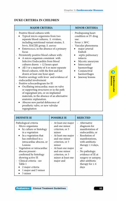
65Clinical treatment guidelinesPediatrics
DUKE CRITERIA IN CHILDREN
MAJOR CRITERIA MINOR CRITERIA
- Positive blood cultures with:• Typical micro-organisms from two
separate blood cultures; S. viridans, including nutritional variant strains, S. bovis, HACEK group, S. aureus
• Enterococci, in the absence of a primary focus
- Persistently positive blood culture with:• A micro-organism consistent with
Infective Endocarditis from blood cultures drawn > 12 hours apart
• All 3 or a majority of 4 or more separate blood cultures, with the first and last drawn at least one hour apart
- Positive serology with fever and evidence of endocardial involvement
- Positive echocardiogram for IE• Oscillating intracardiac mass on valve
or supporting structures or in the path of regurgitant jets, or on implanted materials, in the absence of an alternative anatomic explanation.
• Abscess new partial dehiscence of prosthetic valve, or new valvular regurgitation
- Predisposing heart condition or IV drug use:
- Fever ≥ 38ºC- Vascular phenomena• major arterial
Emboli• septic pulmonary
infarcts• Mycotic aneurysm• Intercranial
haemorrhage• Conjunctival
haemorrhages• Janeway lesions
DEFINITE IE POSSIBLE IE REJECTED
Pathological criteria- Micro-organisms• by culture or histology
in a vegetation• In a vegetation that
has embolised in a intracardiac abscess, or Lesions
- Vegetation or intracardiac abscess present - confirmed by histology showing active IE
- Clinical criteria - see Table 1• 2 major criteria• 1 major and 3 minor • 5 minor
- At least one major and one minor criterion, or 3 minor
- At least one major and one minor criterion, or 3 minor
- At least one major and one minor criterion, or 3 minor at least one major and
- Alternative diagnosis for manifestation of endocarditis, or
- Resolution of manifestations, with antibiotic therapy ≤ 4 days, or
- No pathologic evidence of IE at surgery or autopsy, after antibiotic therapy for ≤ 4 days
Chapiter 3: Cardiovascular Diseases
Cardiovascular
Diseases
3

66 Clinical treatment guidelinesPediatrics
Investigations
- Blood cultures( at least 3 cultures) before antibiotics- FBC /CRP/ESR- Urine test strips – haematuria- Echocardiography
Management
Non-pharmacological• Bed rest/limit physical activity• Ensure adequate nutrition• Maintain haemoglobin > 10 g/dL• Measures to reduce fever
Pharmacological• Paracetamol, oral, 20 mg/kg at once, then 10–15 mg/kg/
dose, every 6 hours as required• Antibiotics regimen: IV antibiotics are always given, based
on culture and sensitivity results→ Native Valve Endocarditis (NVE) due to Streptococci
Benzylpenicillin (Penicillin G), IV, 300 000 units/kg/day divided in 4 doses for 4 weeks Or
ceftriaxone 100mg/kg/day as single dose (maximum 2g) for 4 weeks PLUS
Gentamicin, IV, 3mg/kg/day divided in 3 doses (maximum 240mg/day) for 2 weeks.
→ Patients allergic to penicillin and cephalosporines Vancomycine 40mg/kg/day divided in 3 doses
(max 2g/day) for 4 weeks.
→ NVE due to staphylococcicloxacillin 200mg/kg/day divided in 4 doses 6 for
4 weeks PLUSGentamicin 3mg/kg/day devided in 3 doses
(maximum 240mg/day) for first 5 days OrVancomycine 40mg/kg/day divided in 3 doses
(max 2g/day) for 6 weeks. (In Cloxacillin-resistant strains or allergy to penicillin)
Note: All highly suspected cases of infective endocarditis must be referred to the cardiologist where blood cultures and proper management will be done.
Chapiter 3: Cardiovascular Diseases

67Clinical treatment guidelinesPediatrics
3.6. Cardiomyopathies
Definition: dilated cardiomyopathy refers to a group of conditions of diverse etiology in which both ventricles are dilated with reduced contractility.
Classification
Classification based on the predominant structural and functional abnormalities
- dilated Cardiomyopathy: primarily systolic dysfunction - Hypertrophic Cardiomyopathy: primarily diastolic dysfunction - Restrictive Cardiomyopathy: primarily diastolic but often
combined with systolic dysfunction
3.6.1. Dilated Cardiomyopathy
Causes
- Infections (e.g. Viral+++, Rickettsia, Chagas disease)- Neuromuscular disorders (e.g. duchenne dystrophy, Becker
dystrophy) - Endocrine, metabolic and nutritional (e.g. hyperthyroidism,
Fatty acid oxidation disorders, beriberi, kwashiorkor)- diseases of coronary arteries (e.g. Kawasaki, Aberrant Left
Coronary Artery - ALCAPA)- Autoimmune diseases (e.g. Rheumatic carditis, juvenile
rheumatoid arthritis, systemic lupus erythematosus, dermatomyositis, systemic lupus erythematosus)
- drugs toxicity (e.g. doxorubicin, cyclophosphamide, IPECA)- Hematologic diseases (e.g. anemia, Sickle cell anemia,
hypereosinophilic syndrome Löffler syndrome)
Signs and Symptoms (See signs of congestive heart failure)
Diagnosis- ECG: proeminent P wave, LV or RV hypertrophy, nonspecific
T-wave abnormalities- Chest X-ray: cardiomegaly, pulmonary edema- Echocardiogram: confirm diagnosis and shows LA and LV
dilation, poor contractility- FBC, Urea and creatinine, Electrolytes (Na, K)- Myocardial biopsy, PCR
Chapiter 3: Cardiovascular Diseases
Cardiovascular
Diseases
3

68 Clinical treatment guidelinesPediatrics
Management
- Treatment: (Refer to principles and medication of congestive heart failure)
3.6.2. Hypertrophic Cardiomyopathy
Causes
- Left ventricle obstruction (Coartation of aorta, hypertension, aortic stenosis)
- Secondary (infants of diabetic mothers, corticosteroids in premature infants)
- Metabolic (Glycogen storage disease type II (Pompe disease) - Familiar hypertrophic cardiomyopathy- Syndroms (Beckwith - Wiedman syndrom, Friedereich, ataxia)
Signs and Symptoms
- Weakness- Fatigue- dyspnea on effort- Palpitations- Angina pectoris- dizziness and syncope- Increased risk of sudden death
Diagnosis
- ECG: LV hypertrophy- Chest X-ray: Mild cardiomegaly- Echocardiogram: LV hypertrophy, ventricular outflow tract
gradient- doppler flow studies may demonstrate diastolic dysfunction
before the development of hypertrophy
Management
- Prohibit competitive sports and strenuous physical activities- Propranolol 0.5 -1mg/kg/day devised in 3 doses or Atenolol- Implantable cardioverter-defibrillator if documented arrhythmias
or a history of unexplained syncope- Open heart surgery for septal myotomy: rarely indicated
Chapiter 3: Cardiovascular Diseases

69Clinical treatment guidelinesPediatrics
3.6.3. Restrictive Cardiomyopathy
Definition: Restrictive cardiomyopathy refers to a group of disorders in which the heart chambers are unable to properly fill with blood because of stiffness in the heart muscle. Its prognosis is poor, and clinical deterioration can be rapid.
Causes
- Idiopathic, Systemic disease (scleroderma, amyloidosis, or sarcoidosis)
- Mucopolysaccharidosis- Hypereosinophilic syndrome; malignancies- Radiation therapy - Isolated non compaction of the left ventricular myocardium
Signs and Symptoms
- dyspnea- Edema and ascites- Hepatomegaly with increased venous pressure- Pulmonary congestion
Complications
- Arrhythmias- Mitral regurgitation - Progressive heart failure- Tricuspid regurgitation
Investigations
- ECG: Prominent P waves, ST segment depression, T-wave inversion
- Chest X-ray: mild to moderate cardiomegaly- Echocardiogram: markedly enlarged atria and small to normal-
sized ventricles with often preserved systolic function but highly abnormal diastolic function
Management
- Lasix 2mg/kg divided in 2 doses - Aldactone 1-2mg/kg devised in 2 doses- Antiarrhythmic agents / biventricular pacing are used as required- Aspirin or Warfarin in case of non compaction LV with an
increased risk of mural thrombosis and stroke - Cardiac transplantation where possible and indicated
Chapiter 3: Cardiovascular Diseases
Cardiovascular
Diseases
3

70 Clinical treatment guidelinesPediatrics
3.7. Pericarditis/Pericardial Effusion
Definition: Pericarditis is the inflammation of the pericardium. Pericardial effusion is the abnormal build-up of excess fluid that develops between the pericardium, the lining of the heart, and the heart itself.
Causes
- Infection such as viral, bacterial (tuberculosis) - Inflammatory disorders, such as lupus - Cancer that has spread (metastasized) to the pericardium - Kidney failure with excessive blood levels of nitrogen - Heart surgery (postpericardectomy syndrome).
Signs and Symptoms
- Pericardial tamponade- Chest pressure or pain and signs of congestive heart failure which
can sometimes lead to shock.
Note: Many patients with pericardial effusion have no symptoms. The condition is often discovered on a chest x-ray or echocardiogram that was performed for another reason.
Investigations
- ECG• Small complexes tachycardia• diffuse T wave changes
- Chest X-ray: “water bottle” heart, or triangular heart with smoothed out borders
- Echocardiogram- Tuberculin skin test- diagnostic pericardiocentesis
• In all patients with suspected bacterial or neoplastic pericarditis and patients whom diagnosis is not readily obtained
- Cell count and differential, culture, gram stain, PCR
Chapiter 3: Cardiovascular Diseases

71Clinical treatment guidelinesPediatrics
Management
Non-pharmacological• Semi-sitting position if tamponnade suspected• Pericardiocentesis
→ preferably under ultrasound guidance→ Performed by an experienced person→ Indicated in children with symptomatic pericardial
effusion
Pharmacological• If hypotensive, rapidly administer intravenous fluids 20ml/
kg of Normal saline over 30min to 1 hour• If suspected TB pericarditis: standard anti TB treatment +
steroids• In case of purulent pericarditis: cloxacillin, IV 50 mg/kg/
dose 6 hourly for 3 – 4 weeks + ceftriaxone, IV, 100 mg/kg as a single daily dose, to adapt according to culture results.
• Treat heart Failure (See section on heart failure)
Recommendation
- All patients with pericardial effusion should be referred to a cardiologist
3.8. Hypertension in children
Definition: Hypertension is defined as systolic and/or diastolic blood pressure ≥ the 95th percentile for gender, age and height percentile on at least three consecutive occasions.
A sustained blood pressure of > 115/80 is abnormal in children between 6 weeks and 6 years of age.
Causes
- Severe hypertension suggests renal disease- Coarctation of Aorta- Rarely pheochromocytoma- Long term steroid therapy- Most common causes of secondary hypertension by age
• New born → Renal abnormalities→ Coarctation of the aorta
Chapiter 3: Cardiovascular Diseases
Cardiovascular
Diseases
3
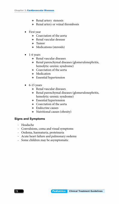
72 Clinical treatment guidelinesPediatrics
→ Renal artery stenosis→ Renal artery or veinal thrombosis
• First year→ Coarctation of the aorta→ Renal vascular desease→ Tumor → Medications (steroids)
• 1-6 years→ Renal vascular diseases→ Renal parenchymal diseases (glomerulonephritis,
hemolytic-uremic syndrome)→ Coarctation of the aorta→ Medication→ Essential hypertension
• 6-15 years→ Renal vascular diseases→ Renal parenchymal diseases (glomerulonephritis,
hemolytic-uremic syndrome)→ Essential hypertension→ Coarctation of the aorta→ Endocrine causes→ Nutritional causes (obesity)
Signs and Symptoms
- Headache- Convulsions, coma and visual symptoms- Oedema, haematuria, proteinuria- Acute heart failure and pulmonary oedema- Some children may be asymptomatic
Chapiter 3: Cardiovascular Diseases

73Clinical treatment guidelinesPediatrics
Blood pressure in children correlates with body size and ageAge of child 95th Percentile of Systolic and Diastolic Blood Pressure
First 12 hours First weeknewborn prem 65/45 mmHg 80/50 mmHgnewborn fullterm 80/50 mmHg 100/70 mmHg
Systolic mmHg diastolic mmHg6 weeks-6 Years 115 808 years 120 829 years 125 8410 years 130 8612 years 135 8814 years 140 90
95th Percentile of systolic and diastolic BP correlated with Height
Height cm Systolic mmHg Diastolic mmHg
100 114 70
110 116 72
120 118 74
130 120 74
140 125 75
150 130 75
160 135 (131) 77
170 140 (133) 80
180 145 (135) 83
Investigations
- Urea, creatinine, electrolytes (Na+, K+) - Fundoscopy- ECG- Echocardiogram- Abdominal ultrasound (focused on kidneys)- Others according to the suspected etiology
Chapiter 3: Cardiovascular Diseases
Cardiovascular
Diseases
3

74 Clinical treatment guidelinesPediatrics
Management
Acute hypertension (hypertension of sudden onset)Non-pharmacological
• Admit patient to paediatric high dependence care unit• Monitor BP every 10 minutes until stable – thereafter every
30 minutes for 24 hours• Insert two peripheral intravenous drips• Rest on cardiac bed• Control fluid intake and output (restriction)• Restrict dietary sodium
Pharmacological• do not combine drugs of the same class• Furosemide, IV, 1–2 mg/kg as a bolus slowly over 5
minutes, Increase up to 8 mg/kg/day • nifedipine 0.25-0.5mg/kg (max: 10mg) sublingual OR
Amlodipine, oral, 0.2 mg/kg/dose. May be repeated 6 hours later, thereafter every 12 hours
• Refer the patient to a specialist when the patient is stable
Recommendations
- For acute or chronic hypertension Blood Pressure needs to be lowered cautiously
- Aim to reduce the SBP slowly over the next 24 - 48 hours- do not decrease BP to < 95th percentile in first 24 hours- Advise a change in lifestyle- Institute and monitor a weight reduction program for obese
individuals- Regular aerobic exercise is recommended in essential
hypertension- dietary advice- Limit salt and saturated fat intake- Increase dietary fiber intake
Chronic Hypertension
Non-pharmacological management• Introduce physical activity, diet management and weight
reduction, if obese• Advise against smoking in teenagers• Follow up to monitor Blood Pressure and educate patient
on hypertension• If Blood Pressure decreases, continue with non-drug
management and follow up
Chapiter 3: Cardiovascular Diseases

75Clinical treatment guidelinesPediatrics
• If BP is increasing progressively, reinvestigate to exclude secondary causes or refer to the specialist
• If BP is stable but persistently > 95th percentile and secondary causes have been excluded, start drug treatment after failed non-drug management for 6 months
• Consider earlier initiation of drug treatment if positive family history for cardiovascular disease, essential hypertension or diabetes mellitus
Pharmacological management
Recommended medication and dosage for patients with chronic hypertension
Drug Dosage Side effect/commentFirst lineHydrochlorothiazide 1-2mg/kg/day once daily
(maximum 25mg/day).Hypokalemia
Second linenifedipine ORAmlodipine
0.3-1mg/kg/day divided in 3 doses0.1mg/kg/day (maximum dose 10mg/day) once daily
Not well studied in children less than 6 years of age
Third linecaptopril
Or
Lisinopril
0.5 – 4mg/kg/day divided in 2 doses
0.07- 0.6mg/kg daily
- Hyperkalaemia - Check renal function and Serum-K periodically, not used in bilateral renal artery stenosis, contraindicated in renal failure- Can cause cough
Fourth lineAtenolol 0.5-1mg/kg/day once daily
(max up to 2mg/kg/day, do not exceed /100mg/day).
Bradycardia
Furosemide (lasix) if associated edema or stage 4 chronic kidney disease.Note: do not associate Furosemide with Hydrochlorothiazide
1-4mg/kg/day in 2 to 4 divided doses
HyponatremiaHypokalemia
Chapiter 3: Cardiovascular Diseases
Cardiovascular
Diseases
3

76 Clinical treatment guidelinesPediatrics
Recommended Hypertension medication for patients with Renal FailureFor CKd 1-3 (GFR>=30, creatinine <2x normal value for ageFirst- line drug LisinoprilSecond -line drug HydrochlorothiazideThird- line drug AmlodipineForth- line drug Atenolol ( use half of normal recommended dose)For CKd 4 or 5 (GFR < 30, creatinine >=2x normal value for ageFirst-line drug FurosemideSecond-line drug AmlodipineThird-line drug Atenolol (use half of normal recommended dose)
Recommendations
- All patients with hypertension and persistent proteinuria should be treated with an ACE inhibitor
- Always exclude bilateral renal artery stenosis before treating with an ACE inhibitor
- Renal function must be monitored when an ACE inhibitor is prescribed because it may cause a decline in GFR resulting in deterioration of renal function and hyperkalaemia
- Patients with hypertension due to a neuro-secretory tumour (phaeochromocytoma or neuroblastoma) should receive an a-blocker either as single drug or in combination with ß-adrenergic blocker
- For patients with persistent hypertension despite the use of first line drugs, a second/third drug should be added
- Specific classes of antihypertensive drugs should be used according to the underlying pathogenesis or illness
- For patients with a predominant fluid overload: use diuretics with/without ß-blocker
Chapiter 3: Cardiovascular Diseases

77Clinical treatment guidelinesPediatrics
3.9. Cardiac Arrhythmias in children
Definition: Heart rate that is abnormally slow or fast for age or irregular.
There are three types of arrhythmias in children- Heart block- Ventricular arrhythmias- Paroxysmal atrial tachycardia
Type of Arrhythmia
Causes Signs and symptom
Heart block: A delay or complete block of the electrical impulse as it travels from the sinus node to the ventricles
- Idiopathic and familial- Electrolyte disturbances
(hyperkalaemia),- digoxin toxicity- Congenital heart
disease, particularly transposition of the great arteries, and especially after surgery
- Myocarditis- Post infective, for
example in endocardial fibroelastosis or rheumatic fever
- Chest pressure or pain- Fainting, also known as
syncopy, or near-syncope- Fatigue- Lightheadedness or
dizziness- Palpitations, which can
be skipping, fluttering or pounding in the chest
- Shortness of breath
Ventricular arrhythmias: A rapid heart rate, usually with a regular rhythm, originating from above the ventricles
- Heart attack- Cardiomyopathy- Heart failure- Heart surgery- Myocarditis- Valvular heart disease
- May be asymptomatic- Chest disconfort (angina)- Fainting (syncope)- Light-headedness or
dizziness- Sensation of feeling the
heart beat (palpitations)- Shortness of breath - Absent pulse- Loss of consciousness- Normal or low blood
pressure- Rapid pulse
Paroxysmal atrial tachycardia: A rapid heart rate, usually with a regular rhythm, originating from above the ventricles.
- Palpitation- lightheadedness- Weakness - Shortness of breath - Chest pressure
Chapiter 3: Cardiovascular Diseases
Cardiovascular
Diseases
3

78 Clinical treatment guidelinesPediatrics
NORMAL HEART RATE/MINUTE FOR AGE
Age Heart rate
Newborn 100–160
< 1 year 110–160
1–2 years 100–150
2–5 years 95–140
5–12 years 80–120
> 12 years 60–100
Signs and Symptoms
Symptoms Signs
Infants Color changes (pale, mottled) Irregular pulse
Irritability Tachycardia
Feeding difficulties Bradycardia
Sweating Signs of cardiac failure
Tachypnoea/apnoeic spells
Children Chest Pain Signs Of Cardiac Failure
dizziness Tachycardia
Palpitations Bradycardia
Fatigue Syncope
Note: All patients with arrhythmias should be referred to a cardiologist
Investigations
- ECG is essential for diagnosis, preferably a 12 lead ECG- Echocardiogram- Other according to the suspected etiology
Chapiter 3: Cardiovascular Diseases

79Clinical treatment guidelinesPediatrics
TACHYARRHYTHMIAS
Sinus tachycardia
ECG CriteriaRate: > upper limit for age P wave: present and normalRhythm: regular QRS: normal
Supraventricular Tachycardia
ECG CriteriaRate: usually > 200 beats per minute P wave: abnormalRhythm: regular QRS: narrowed
Ventricular Tachycardia
ECG CriteriaRate: generally 100–220 beats per minute P wave: mostly not seenRhythm: generally regular QRS: abnormal, large with QRS > 120 millisecond
Chapiter 3: Cardiovascular Diseases
Cardiovascular
Diseases
3

80 Clinical treatment guidelinesPediatrics
Management
Non-pharmacological treatment• Sinus tachycardia usually requires management of the
underlying condition• ABC of resuscitation• Admit to High Care or Intensive Care Unit• Monitor ECG, Oxygen saturation, Blood Pressure,
Haemoglobin, Heart rate, Acid–base status and blood gases, Respiratory rate, maintain adequate nutrition and hydration, treat pyrexia
Pharmacological management• Emergency treatment
→ Stable patient: Attempt vagal stimulation→ Place icebag on face
Infants: immerse face in ice-cold water for a few seconds
Older children: try a valsalva manoeuvre, e.g. ask the patient to blow through a straw
→ Place NGT if other means are not available
Note: Eye-ball pressure and carotid massage is contra-indicated in children
• Adenosine, IV, 0.1 mg/kg initially, increasing in increments
of 0.05 mg/kg to 0.25 mg/kg. Follow with a rapid flush of at least 5 ml normal saline.
• Unstable patient: Heart failure / shocked→ dC synchronised cardioversion in increments of
0.5–1–2 J/kg→ Empty the stomach before cardioversion is attempted→ Amiodarone, IV, 5 mg/kg slowly over 20 minutes
(NEVER as a rapid infusion)
Chapiter 3: Cardiovascular Diseases

81Clinical treatment guidelinesPediatrics
3.10. Bradyarrhythmias
Causes
- Hypoxia- Hypothermia- Head injuries and increased intracranial pressure- Toxins and drug overdose- Post operative- Congenital excessive vagal stimulation- Electrolyte disturbances (Hypo- or hyperkalaemia,
Hypocalcaemia)
Sinus Bradycardia
ECG CriteriaRate: < lower limit for age P wave: present, all look the sameRhythm: regular QRS: normal, 80–120 millisecond
Heart Block (Complete)
ECG CriteriaRate: low, usually < 60 beats per minute P wave: independent P waves QRS’s with no relationship between the two (AV dissociation)
Management
- If syncope and Heart rate - below 50/min • Start i.v. Isuprel (Isoprenaline) 0. 05 – 0. 4 microgram/kg/
min. Or
• Dobutamine (Dobutrex) 2 - 20 microgram/kg/min• Insert pacemaker if ineffective
Chapiter 3: Cardiovascular Diseases
Cardiovascular
Diseases
3


83Clinical treatment guidelinesPediatrics
4.1. Anemia
Definition: Anemia is defined as a reduction of the red blood cell (RBC) volume or hemoglobin concentration below the range of normal values occurring in healthy persons.
Cause
- Anemia is classified according to physiologic process (decreased production, increased destruction or blood loss). In practice, classifying anemia according MCV is a useful approach to assessing the common causes of anemia in children.
Signs and Symptoms
- Pale mucous membranes, palms and nail beds- dizziness, fainting- Headache - Shortness of breath on exertion (exercise intolerance)- Palpitations- Visual disturbances- Poor growth- Confusion, decreased mental activity- Rapid heartbeat or palpitations- dyspnoea, tachypnea- Signs of cardiac failure if severe anemia
4. Haematological Conditions
Haem
atological C
onditions
4
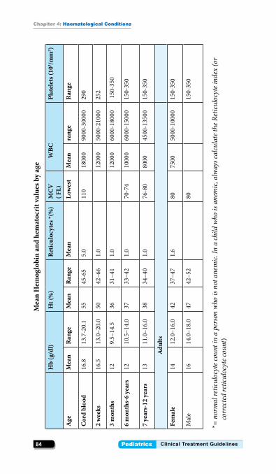
84 Clinical treatment guidelinesPediatrics
Mea
n H
emog
lobi
n an
d he
mat
ocri
t val
ues b
y ag
e
Hb
(g/d
l)H
t (%
)R
etic
uloc
ytes
*(%
)M
CV
( FL)
WBC
Plat
elet
s (10
3 /mm
3 )
Age
Mea
nR
ange
Mea
nR
ange
Mea
nLo
wes
tM
ean
rang
eR
ange
Cor
d bl
ood
16.8
13.7
-20.
155
45-6
55.
011
018
000
9000
-300
0029
0
2 w
eeks
16.5
13.0
–20.
050
42–6
61.
012
000
5000
-210
0025
2
3 m
onth
s12
9.5–
14.5
3631
–41
1.0
1200
060
00-1
8000
150
-350
6 m
onth
s-6
year
s12
10.5
–14.
037
33–4
21.
070
-74
1000
060
00-1
5000
150-
350
7 ye
ars-
12 y
ears
1311
.0–1
6.0
3834
–40
1.0
76-8
080
0045
00-1
3500
150-
350
Adu
lts
Fem
ale
1412
.0–1
6.0
4237
–47
1.6
8075
0050
00-1
0000
150-
350
Mal
e16
14.0
–18.
047
42–5
280
150-
350
*= n
orm
al re
ticul
ocyt
e cou
nt in
a p
erso
n w
ho is
not
ane
mic.
In a
child
who
is a
nem
ic, a
lway
s cal
cula
te th
e Ret
iculo
cyte
inde
x (o
r co
rrec
ted
retic
uloc
yte c
ount
)
Chapiter 4: Haematological Conditions

85Clinical treatment guidelinesPediatrics
Normal values vary by age, sex and ethnicity. Means and ranges for hemoglobin and hematocrit values by age groups of well-nourished children
Reticulocytes
Reticulocytes are circulating immature RBC. Normal range are in the table above. However, if a person has anemia and his bone marrow is able to produce new blood cells, his/her reticulocyte percentage should be higher than “normal” Thus, calculating the corrected reticulocyte count is an important step in understanding whether the reticulocyte count is appropriate or inappropriate to the situation.
- Corrected Reticulocyte Count calculations:
Corrected Reticulocyte Count (CRC): = Reticulocyte % x (Patients’ Hematocrit/Normal hematocrit per age)
- A CRC >1.5 suggests increased Red Blood Cells production as a result of hemolysis and blood loss.
Chapiter 4: Haematological Conditions
Haem
atological C
onditions
4

86 Clinical treatment guidelinesPediatrics
ORDER• CBC with Platelet Count and Automated Differential (including RBC indices and morphology on manual differential)• Reticulocytes, Percent & Number
• Iron Deficiency • thalassemia• Chronic disease• Sideroblastic anemia • Lead toxicity
Suspecthemorrhageand acuteblood loss
Suggests hemolytic process:• Hemoglobinopathies (eg, sickle cell)• RBC membrane defects (eg spherocytosis)• Enzymatic defects (g6PD)• Immune destruction (autoimmune hemolytic anemia, isoimmune hemolytic anemia})• Non-immune destruction
• Aregenative anemias - (Aplastic anemia,
Parvovirus, tEC (transient, erythroblastopenia of childhood), DBA (Diamond Blackfan anemia) (can also be macrocytic)
• Anemia of acute or chronic disease or inflammation
• Mixed fe and vit B12 or folate deficiency
• If other lines involved: leukemia, aplastic anemia
• Folic acid deficiency• Vitamin B 12 deficiency• Hypothyroidism• fanconi anemia• Diamond Blackfan anemia
Corrected Reticulocyte Count (CRC): =Reticulocyte % x (Patients’Hematocrit/Normal hematocrit per age)
Abnormal peripheral smear
ORDER• Iron and Iron Binding Capacity• ferritin
Iron deficiencySuggests:• Inflammation• Chronic disease
Bone marrowbiopsy may be
necessary
B12, folate stud-ies
If no obvious chronicdisease present,consider bone marrowbiopsy
Anemia present on CBC (see normal values for age)
CRC>1.5CRC>1.5
Classify by RBC indices fragmented cells onperipheral smear
© 2006-201 1 ARUP Laboratories, All Rights Reserved, 5/16/2011
INDICAtIONS fOR tEStINgfatigue, weakness, pallor, dizziness, fainting
yesno
Macrocytic(high MCV)
(suggests maturation defects)
Microcytic, hypochromic(low MCV, MCHC)
(suggests maturation defects)
Normocytic, normochromic(normal MCV, MCHC)
(suggests hypoproliferation)
Low/normal tIBCNormal/high ferritin
Low/ normal iron
High tIBCLow iron
Low ferritin
Workup based onsmear
characteristics
yesno
no
Chapiter 4: Haematological Conditions

87Clinical treatment guidelinesPediatrics
Physiologic classification of Anemia- Anemia due to reduced red blood cell/hemoglobin production
• Bone marrow aplasia: → Fanconi’s anemia (congenital aplastic anemia)→ Acquired aplastic anemia, diamond-Blackfan
anemia, Transient Erythroblastopenia of childhood (red blood cell aplasia).
• Bone marrow replacement by tumour cells - leukaemias, secondary metastases.
• Bone marrow replacement by fibrous tissue or granulomas - granulomas can occur in the congenital toxoplasmosis, other, rubella, cytomegalovirus, herpes simplex (TORCH), infections (or in tuberculosis infection. Parvovirus is directly cytotoxic because replicates in the erythroid precursors
• deficiency of iron can be related to :→ Increased requirement for Fe in periods of high
growth rate (ex-premature babies, toddlers, teenagers)
→ Poor Iron intake:The early introduction of whole cow’s milk
(before 9-12 months)Vegetarian dietLate weaning. without iron supplementation
→ Blood loss (reflux esophagitis, menorrhagie)→ Parasitic infection (hookworms)→ Inability to absorb iron (coealiac disease, Crohn’s
disease…)
• deficiency of folic acid: Megaloblastic anemia of infancy can develop due to:
→ Folic acid deficiency during rapid growth. → Malabsorption syndromes such as coeliac disease, in
inflammatory bowel disease and in children taking anticonvulsants.
• deficiency of vitamin B12 can occur in:→ Infants who are breast-fed by a vegetarian mother → Mal-absorption → Worm infestation→ Congenital pernicious anemia where there is inability
to secrete gastric intrinsic factor
Chapiter 4: Haematological Conditions
Haem
atological C
onditions
4

88 Clinical treatment guidelinesPediatrics
• Thalassaemias:→ Normal hemoglobin is under produced due to
mutations in the alpha or beta globin chains.
- Anemia of chronic disease (e.g: chronic pyelonephritis, chronic renal failure, bacterial endocarditis, osteomyelitis ) due to:
• Impaired erythropoietin production. • Anemia can also be associated with hypothyroidism.• Sideroblastic anemia (Extremely rare, heterogeneous
group of diseaeses, either acquired (drugs, toxins, malignancy), or congenital . diagnosis by bone marrow biopsy (presence of sideroblasts) anemia
- Anemia due to increased red blood cell destruction (haemolysis)• Congenital
→ Red cell membrane defects - including hereditary spherocytosis.
→ Red cell enzyme abnormalities - including glucose-6-phosphate dehydrogenase (G-6-Pd) deficiency, pyruvate kinase deficiency.
→ Hemoglobinopathies - including sickle cell disease, thalassaemias.
• Acquired → Autoimmune haemolysis→ Isoimmune haemolysis (haemolytic disease of the
newborn, blood transfusion reactions).→ Infections (including malaria, septicaemia)→ drug- and toxin-induced→ disseminated intravascular coagulation→ Hypersplenism.
- Anemia due to blood loss• Including gastrointestinal blood loss, traumatic, heavy
menstruation in girls.
Complications
- Pulmonary edema- Congestive heart failure- Acute respiratory distress syndrome (ARdS)
Chapiter 4: Haematological Conditions

89Clinical treatment guidelinesPediatrics
Investigations according to clinical situation
- FBC and reticulocyte count and peripheral blood smear examination
- Blood film for malaria parasites- Stool examination for eggs of hookworm / Stool for occult blood,
ova and parasites- Sickling test- Hemoglobin electrophoresis- Analysis for nutritional deficiencies- Bone marrow aspiration to assess the decreased production of
red cells- Coombs direct and indirect (in cases of hemolytic anemia)- Iron studies (Fe, Ferritin, TIBC, transferring % saturation)- Other investigations will be dependent on the clinical evaluation
of the patient
Management
- Obtain a detailed history from the patient or care givers- Examine the anaemic patient carefully and perform the
appropriate investigations with a goal of:• Confirming that the patient is anaemic• Establishing the type of anemia • determining the cause of the anemia• determining whether or not there are complications
arising from the anemia, the cause of the anemia or both• Remove or correct the underlying cause • Always investigate cause of anemia before initiating
treatment• In an emergency, take all blood samples before treatment
Therapeutic objectives• Treat underlying cause of anemia • In sickle cell disease patients restore hemoglobin to steady
state level• In iron deficiency replenish iron stores after correction of
anemia (continue to treat for 2-3 months)
Non-Pharmaceutical• Advise on a balanced diet especially iron-rich foods such
as liver; beef kidneys; molasses; meat; sardines; eggs, fish; fresh green leafy vegetables..
• Malaria prevention
Chapiter 4: Haematological Conditions
Haem
atological C
onditions
4

90 Clinical treatment guidelinesPediatrics
• Encourage exclusive breastfeeding until 6 months, then supplementation with iron rich food. discourage use of cow’s milk before 12 months and excessive intake of cows milk.
Pharmaceutical management• For iron deficiency anemia, prescribe Elemental Iron
4-6 mg/kg/day divided in 3 doses daily until the Hb has reached the normal range. Pay attention to type of iron supplementation prescribed (Ferrous Sulphate has 20% elemental iron, Ferrous Fumarate has 33% elemental iron and Ferrous gluconate has 12% elemental iron). Continue for 2-3 months after normalization of Hb to build up iron stores.
• Sickle cell disease patients should receive iron tablets only if there is evidence of iron deficiency. They should however, receive Folic acid. Similarly, patients whose anemia is possibly due to malaria should receive folic acid
• Folic acid, oral: 5 mg every 2 days for 30 days or for as long as required.
• If anemia is due to hookworms treat appropriately (Albendazole 400 mg po x 3 days or mebendazole 100 mg po x 3 days)
• Vitamin B12 deficiency: (Hydroxycobalamin) injection IM: Initially 100mcg/day X 10-15 days. Maintenance dose 30-50 mcg/month. Lifelong treatment may be required.
• Severe anemia with signs of cardiac failure will need treatment of the heart failure in addition to blood. Transfusion with packed cells. Look for signs of decompensation before deciding to transfuse and look for these signs during transfusion.
Transfuse the patient if Hb < 5 g/dl and decompensation signs are present: Packed cells: 10-15 ml/kg body weight slowly over 4 hours and Whole blood: 2oml/kg body weight
Side effects of iron therapy • diarrhea, abdominal discomfort, constipation, or black
stools
Chapiter 4: Haematological Conditions

91Clinical treatment guidelinesPediatrics
Recommendations
- Refer all patients with anemia related to poor diet to a nutritionist or a health center for nutritional follow-up
- Refer all patients with recurrent anemia or with anemia of unknown cause to a referral hospital
4.2. Sickle Cell Anemia
Definition: Chronic haemolytic anemia characterized by sickle−shaped Red blood cells as a result of mutation in the β chain of Hemoglobin
Cause
- Homozygous inheritance of mutated HBS (amino−acid valine is substituted for glutamic acid in the position 6 of the β−chain)
Signs and Symptoms
- Impaired growth and development - Anemia and mild jaundice - Hepatosplenomegaly (in younger children)- Bone pain (especially long bones in children) - Pain and swelling of the hands and feet (hand - foot syndrome)
in children between 6 months and 3 years old. - Arthralgia with fever- Severe abdominal pain with vomiting - Acute Chest Syndromes (sudden onset of fever, cough, chest
pain, tachypnea leucocytosis and pulmonary infiltrates on x−ray): Must be aggressively treated may be fatal
- Tower shaped (“frontal and parietal bossing”) skull
Complications
- Infections (especially from encapsulated organism such as Streptococcus pneumoaniea:
• Osteomyelitis (Streptotococcus pneumonia and Salmonella)
• Meningitis
- Aplastic crisis (Infection by Parvovirus B19 that infects RBC progenitors resulting in a very rapid drop in Hb).
- Stroke (infarctive) with hemiparesis and convulsions- Gangrene (vaso-occlusive)
Chapiter 4: Haematological Conditions
Haem
atological C
onditions
4

92 Clinical treatment guidelinesPediatrics
- Pulmonary hypertension- Acute chest syndrome (sudden onset of fever, cough, chest pain,
tachypnea leucocytosis and pulmonary infiltrates on x−ray): Must be aggressively treated as may be fatal
- Gall bladder stones +/- cholecystitis- Splenic Sequestration (in 5 first years of life): onset of life
threatening anemia with rapidly enlarging spleen and high reticulocyte counts
- Avascular necrosis of the femoral head is common - Occlusion of major intracranial vessels may lead to hemiplegia- Cranial nerve palsies and other neurological deficits - Priapism
Investigations
- Full blood count - Peripheral blood thick smear - Sickling test (Test d’Emmel) - Hb electrophoresis- X-ray of long bones, cortical thinning - X-ray of skull bone (shows widening of diploic space)
Management
Aims at three types of crisis• Thrombotic (vaso−occlusive, painful or infarctive) • Aplastic (sequestration) • Haemolytic
Non-pharmacological• IV or oral fluids 2L/m2/day• Oxygen if in respiratory distress
Pharmaceutical For complications
• Analgesics (WHO Step wise pain management)→ Paracetamol 10-15mg/kg/dose po every 4-6 hours
associated with Brufen 5-10mg/kg/dose po every 6-8 hours
→ codeine 0.5-1mg/kg/dose every 6 hours → Pethidine 0.5−2mg/kg 4hrly) → Morphine (titrate to effect) PO: 0.2-0.5 mg/kg/dose
every 4-6 hours, IV, IM, SC: 0.1-0.2 mg/kg/dose every 2-4 hours
Chapiter 4: Haematological Conditions

93Clinical treatment guidelinesPediatrics
• If patient has an infection treat according to the bacteria, the site and the severity of the infection
• Aggressively search for cause of infection (hemoculture, urine culture, chest X ray) and start empiric antibiotic treatment if child sick with fever
• Blood Transfusion: A child with sickle cell disease has chronic anemia which is usually well tolerated.
→ Transfusion should be reserved for the following circumstances:Urgently for sudden, severe anemia due to acute
splenic sequestration, Parvovirus B19 infection, or hyperhemolytic crises.
In acute chest syndrome and perioperatively.
→ Acute red cell exchange transfusion is indicated in the following situationsAcute infarctive strokeSevere acute chest syndromeMultiorgan failure syndromesPriapism that does not resolve after adequate
hydration and analgesia
• Additional treatment→ Give supplementary Folic Acid (5 mg oral daily) but
avoid iron (risk of hemochromatosis). → Hydroxyurea should be given to patients with more
than 3 crises per year. Start at a dose of 10 mg/kg PO daily and titrate by 5mg/kg every 8 to 12 weeks to a maximum dose of 25mg/kg/day.
→ Homozygous should be vaccinated for salmonella, Pneumococcal and Haemophilus influenzae
Recommendations
- Education of patient on sickle cell disease and crisis to avoid complications
• Should drink much water daily• Avoid getting cold (dress with warm clothes by cold
weather)
- Sickle cell screening before marriage for suspected carriers and genetic counseling if possible
- Heterozygote carriers should have family members screened for sickle cell disease
Chapiter 4: Haematological Conditions
Haem
atological C
onditions
4

94 Clinical treatment guidelinesPediatrics
4.3. Idiopathic Thrombocytopenic Purpura (ITP)
Definition: Idiopathic Thrombocytopenic Purpura (also called immune thrombocytopenic purpura), is a blood – clotting disorder that can lead to easy or excessive bruising and bleeding. Children often develop ITP after a viral infection and usually recover fully without treatment.
Signs and Symptoms
- History• A previously healthy 1–4 yr old child who has sudden
onset of generalized petechiae and purpura• A history of a preceding viral infection 1–4 wk before the
onset of thrombocytopenia• Acute bleeding from the gums and mucous membranes
- Clinical manifestations• Findings on physical examination are normal, other than
the finding of petechiae and purpura. • Splenomegaly is rare, as is lymphadenopathy or pallor. • Fewer than 1% of patients have intracranial hemorrhage• The severity of bleeding in ITP is based on symptoms and
signs, but not on platelet count→ No symptoms→ Mild symptoms: bruising and petechiae, occasional
minor epistaxis, very little interference with daily living
→ Moderate: more severe skin and mucosal lesions, more troublesome epistaxis and menorrhagia
→ Severe: bleeding episodes—menorrhagia, epistaxis, melena—requiring transfusion or hospitalization, symptoms interfering seriously with the quality of life
Investigations
- Laboratory:• FBC with differential (should not show any anemia
(unless significant bleeding) or anomaly of WBC count) - Profound thrombocytopenia (platelet count <10 × 109/L).
• Peripheral blood film examination (will show large or giant platelets)
• Bone marrow examination only indicated if no response to therapy or before starting steroids
• HIV test
Chapiter 4: Haematological Conditions

95Clinical treatment guidelinesPediatrics
- Additional investigations are done as clinically indicated
Differential diagnosis: (ITP is a diagnosis of exclusion)- Systemic Lupus Erythematosus (SLE),- HIV infection- Wiskott-Aldrich Syndrome (WAS)) must be considered in young
males found to have low platelet counts, particularly if there is a history of eczema and recurrent infection.
Management
- No therapy other than education and counseling of the family and patient for patients with minimal, mild, and moderate symptoms, as defined earlier
- Intravenous Immunoglobulin (IVIG). • IVIG at a dose of 0.8–1.0 g/kg/day for 1–2 days induces a
rapid rise in platelet count (usually>20× 109/L) in 95% of patients within 48 hr.
• IVIG appears to induce a response by down regulating Fc-mediated phagocytosis of antibody-coated platelets.
• IVIG therapy is both expensive and time-consuming to administer
• After infusion, there is a high frequency of headaches and vomiting, suggestive of IVIG-induced aseptic meningitis.
- IV Rh (D) Immune globulin can be used in Rh positive patients at a dose of 50-75 microgram/kg. This causes a rise in platelet count above 20 x 109/L within 48-72h in 90% of patients.Anti-d Therapy can also induce mild hemolytic anemia
- Prednisone. Corticosteroid therapy has been used for many years to treat acute and chronic ITP in adults and children. doses of prednisone of 2 mg/kg/24 hr for up to 3 days appear to induce a more rapid rise in platelet count than in untreated patients with ITP. Corticosteroid therapy can be continued up to 2–3 wk or until a rise in platelet count to>20× 109/L has been achieved, with a rapid taper to avoid the long-term side effects of corticosteroid therapy, especially growth failure, diabetes mellitus, and osteoporosis
- The role of splenectomy in ITP should be reserved for 1 of 2 circumstances
• The older child (> 4 yr) with severe ITP that has lasted >1 yr (chronic ITP) and whose symptoms are not easily controlled with therapy is a candidate for splenectomy
.
Chapiter 4: Haematological Conditions
Haem
atological C
onditions
4

96 Clinical treatment guidelinesPediatrics
• Splenectomy must also be considered when life-threatening hemorrhage (intracranial hemorrhage) complicates acute ITP, if the platelet count cannot be corrected rapidly with transfusion of platelets and administration of IVIG and corticosteroids.
Note: Guidelines for transfusion: See national protocol GEnERAL TRAnSFUSIOn POLIcY MAnAGEMEnT: (for
newborns, see national neonatology protocols)
Chapiter 4: Haematological Conditions

97Clinical treatment guidelinesPediatrics
5. Endocrine System Conditions
5.1. Diabetes Mellitus (Type I and Type II)
Definition: diabetes mellitus is a disorder of absolute or relative insulin deficiency that results in increased blood glucose and disruption of energy storage and metabolism. diabetes Mellitus is generally divided into two classifications: diabetes Mellitus I and diabetes Mellitus Type II.
- diabetes Mellitus Type I: This results from the destruction of the pancreatic beta cells that leads to absolute insulin deficiency. Type IA is secondary to the autoimmune destruction of the beta cells. Type IB is secondary to non-autoimmune destruction of the beta cells. Type I diabetes accounts for approximately 2/3 of the new diagnosis of diabetes in patients < 19 years old. There is a component of genetic susceptibility and close relatives of patients with type I dM are at higher risk of developing the disease.
- diabetes Mellitus Type II: This is secondary to varying degrees of insulin resistance and insulin deficiency and is related to both genetic and environmental influences including predisposing medication such as steroids and some ARVs. It is the most common type of diabetes mellitus in adults.
- Neonatal diabetes: This is defined as persistent hyperglycemia occurring in the first months of life that lasts more than 2 weeks and requires insulin therapy for management. It is a rare cause of hyperglycemia in the neonate and has an estimated incidence of 1/500,000 births. The majority of affected infants are small for gestational age and present with weight loss, volume depletions, hyperglycemia and glucosuria with or without ketonuria and ketoacidosis.
Signs and Symptoms
- Polyuria: This occurs when the serum glucose concentration rises above 180 mg/dL exceeding the renal threshold for glucose and leads to increased urinary glucose excretion and a subsequent osmotic diuresis. This may present as nocturia, bedwetting, or daytime incontinence in a previously toilet trained child, or heavy diapers.
Endocrine S
ystem
Conditions
5

98 Clinical treatment guidelinesPediatrics
- Polydipsia: This is secondary to increased thirst from increased serum osmolality and dehydration.
- Polyphagia: This is due to an increased appetite that’s initially secondary to loss of calories from glycosuria. These symptoms are not always present.
- Weight loss: This is due to hypovolemia and increased catabolism.
- Weakness/Lethargy with ultimate progression to coma: This is secondary to hypovolemia and electrolyte disturbances including progressive acidosis.
Visual disturbances: This is secondary to osmotic changes in the lens.
Complications
- Short-term complications: • diabetic ketoacidosis (dKA): Occurs more frequently in
type I diabetes mellitus, but may also occur in some forms of type I diabetes mellitus.
• Hyperosmolar hyperglycaemic state (HHS): Occurs in type II diabetes mellitus.
• Insulin resistance secondary to hyperglycemia: This occurs in both type I and type II diabetes mellitus.
• Infections due to immunosuppression commonly include oral and vaginal candidiasis and Urinary Tract Infections.
• death: Patients presenting with dKA or HHS have a high mortality rate.
- Long Term complications:• Vascular complications including both micro-angiopathy
and macro-angioapthy:→Nephropathy→Retinopathy→Neuropathy→Cardiovascular disease →Hypertension
• dyslipidemia• Growth retardation or obesity depending on the insulin
therapy. Patients may also have delayed puberty secondary to poor growth.
• Psychiatric disorders including depression related to their chronic disease.
Chapiter 5: Endocrine System Conditions

99Clinical treatment guidelinesPediatrics
Investigations
- Blood sugar: The diagnosis is made based on abnormalities of the blood glucose. See diagnostic criteria below.
- Additional studies to evaluate severity and complications of the disease:
• Blood gas if concern for diabetic ketoacidosis.• Electrolytes • Renal function tests (urea and creatinine) to evaluate for
diabetic nephropathy and dehydration.• Urine analysis to check for glycosuria, ketones, and protein• HbA1c: This can be used for diagnosis (see below) or to
assess severity of disease and to assess response to therapy.• Lipid profile• Fundoscopy: This is to evaluate for diabetic retinopathy.• Foot examination: This is to evaluate for diabetic
neuropathy and assess for wounds that may already be present.
• Further history and physical examination to exclude other co-existing autoimmune diseases such as hypothyroidism, vitiligo, rheumatoid arthritis, etc., and to further ask about family history of endocrinopathies or autoimmune diseases.
• Thyroid-stimulating hormone (TSH): This should be performed in type I diabetics as autoimmune diseases may occur together.
Diagnosis criteria for diabetes mellitus
DIABETES MELLITUS (DM)Symptoms of dM plus random plasma glucose ≥200 mg/dL (11.1 mmol/L)OrFasting plasma glucose ≥126 mg/dL (7.0 mmol/L). Fasting is defined as no oral intake of foods for at least 8 hours.OrTwo-hour plasma glucose ≥200 mg/dL during an oral glucose tolerance test (OGTT) as described by the WHO.OrHgA1C > 6.5% This test should be performed in a certified laboratory with an assay standardized to the diabetes control and complications trial (dCCT).
Chapiter 5: Endocrine System Conditions
Endocrine S
ystem
Conditions
5

100 Clinical treatment guidelinesPediatrics
Management
General Objectives• Maintain normal glycemia with insulin therapy or oral
medications (in type II diabetes mellitus) to prevent both the signs and symptoms of uncontrolled hyperglycemia and the complications mentioned above.
Non-Pharmaceutical Management• Assess A-B-C-d (Airway, Breathing, Circulation, drug) • If patient has signs or symptoms of diabetic ketoacidosis
(dKA) or hyperosmolar hyperglycaemic state, this is an emergency and treatment must be initiated immediately
• The patient and the family should be counselled on the cause and treatment of diabetes and its management. The patient and the family should be taught how to monitor blood glucose, record the test results, administer and adjust insulin doses based on blood glucose values and food intake.
• They family should be counselled on the complications of diabetes mellitus and how to manage them. In particular, they should know the signs and symptoms of acute hypoglycemia and its management. They should also understand the importance of maintaining normoglycemia to avoid long-term complications. They should be instructed on how to manage acute illnesses in the context of diabetes mellitus, for example how to manage an insulin dose if the the patient is unable to tolerate oral intake.
• diet modification is important in both type I and type II diabetes mellitus. A nutritionist should be involved in providing individualized recommendations.
Pharmaceutical management• The majority of children with diabetes mellitus have type
I diabetes and may present with diabetic ketoacidosis (dKA). The management of dKA is detailed below.
→ diabetes Mellitus Type I: Children with diabetes Mellitus Type I require insulin therapy. The patient is insulin dependent and while the insulin therapy may be adjusted based on the clinical condition and blood glucose results; the insulin therapy should NEVER be stopped completely as this could result in the development of dKA and death.
Chapiter 5: Endocrine System Conditions

101Clinical treatment guidelinesPediatrics
5.2. Diabetic Ketoacidosis
Definitions: It is defined as an increase in the serum concentration of ketones greater than 5 mEq/L, a blood glucose level greater than 250 mg/dL (although it is usually much higher), and a blood (usually arterial) pH less than 7.3. Ketonemia and ketonuria are characteristic, as is a serum bicarbonate level of 18 mEq/L or less (< 5 mEq/L is indicative of severe dKA).
Mainly occurs in patients with type I diabetes, however it is not uncommon in type II diabetes
Causes
- Previously undiagnosed diabetes - Interruption of insulin therapy - Underlying infection and intercurrent illness- Poor Management of dM type I- Stress- Medication like corticosteroids, clozapine etc.
Signs and Symptoms
Symptoms Signs
Polyuria dehydration with dry skin, reduced skin turgor or sunken eyes
polydypsia deep and fast breathing (Kussmal respiration) with acetone (ketotic) breath odor
Nausea, vomiting Low Blood Pressure
Abdominal pain Fast and weak pulse
Relatives may report alteration in sensorium or collapse
Confusion, stupor or unconsciousness
Investigations
- Blood glucose - Urine glucose - Urine ketones - Blood urea and electrolytes - Blood film for malaria parasites ( Unconscious in highly endemic
area)
Chapiter 5: Endocrine System Conditions
Endocrine S
ystem
Conditions
5

102 Clinical treatment guidelinesPediatrics
- Full blood count - Blood and urine culture- Electrocardiography
Management
Principles• Manage A,B, C• Admission in ICU if possible• Correction of fluid loss with intravenous fluids• Correction of hyperglycemia with insulin• Correction of electrolyte disturbances, particularly
potassium loss• Correction of acid-base balance• Treatment of concurrent infection, if present
Rehydration
AGE 1st hour Next 7 hours Next 16 hours
< 1 yr 20 ml/kg 15 ml/kg 7 ml/kg
1 - 7 yrs 8 – 14 yrs> 15 yrs
20 ml/kg 20 ml/kg 20 ml/kg
10 ml/kg 9 ml/kg8 ml/kg
5 ml/kg 5 ml/kg4 ml/kg
• Correction of hydro-electrolytic disorder: Initial correction of fluid loss is either by ISotonic Sodium chloride solution or by lactated ringer solution
• If blood glucose falls to < 14mmol/l (250mg/dl) before dKA has resolved (PH < 7.3) add 5% glucose and continue with insulin
Emergency Insulin Therapy• delay insulin until serum K+ is known to be > 3,5 mmol/l • Insulin should only be started after ½ - 1 hour of fluid
therapy, provided shock has been treated.
Doses and route• Low dose hourly regimen
→ Regular (neutral, soluble) Insulin (Actrapid or Humulin R), give 0.1 unit/kg per hour i.vGiving hourly bolus doses ensure regular
medical and nursing supervision of the patient
Chapiter 5: Endocrine System Conditions

103Clinical treatment guidelinesPediatrics
If glucose fall inadequate, i.e. a fall of < 4 mmol/l/hr - double the dose
If glucose fall is excessive, i.e. a fall of > 5,5 mmol/l/hr - half the dose
Continue with hourly insulin until blood glucose and ketoacidosis are controlled. If blood glucose stable and urine ketones negative, then start standard insulin regimen
→ Potassium (K+)■ If hyperkalaemia (serum K+ or ECG) withhold
potassium supplementation■ If serum K+ is normal or low and patient
is passing urine: Start K+ supplementation immediately
■ K+ replacement will be necessary in all cases (even with initial hyperkalaemia)
DOSES
SERUM POTASSIUM
POTASSIUM SUPPLEMENT (as KCl add to each liter of iv fluid)
<3,0 mmol/l 40 mmol
3,0 - 4,0 mmol/l 30 mmol
4,1 - 5,0 mmol/l 20 mmol
5,1 - 6,0 mmol/l 10 mmol
6,0 mmol/l None
Transitional insulin therapy (- Sliding Scale)
Monitor Blood Glucose 4−hourly and give the corresponding amount of Soluble/Regular insulin subcutaneously
Blood Glucose Result Amount of Soluble/Regular Insulin to be given
Less than 6 mmol/L No Insulin
6.1 − 9.0 mmol/L 0.06 units/kg body weight
9.1 − 12.0 mmol/L 0.09 units/kg body weight
12.1−15.0 mmol/L 0.12 units/kg body weight
15.1−18.0 mmol/L 0.15 units/kg body weight
Chapiter 5: Endocrine System Conditions
Endocrine S
ystem
Conditions
5

104 Clinical treatment guidelinesPediatrics
• For transitional therapy consider the following in a patient:→ No coma (maybe still some clouding of
consciousness), No acidosis→ Continue the sliding scale, making appropriate
adjustments to the doses of insulin, until the patient is eating normally and the urine is free of ketones. This may take on average between 12 − 24 hours
Maintenance insulin therapy• determine dose on normal requirement: 1 units/kg/day
→ 2 injections regimen■ Administer subcutaneously in the form of 50%
intermediate−acting insulin (NPH or Lente) and 50% rapid insulin. Total dose divided in 2 doses:
o 2/3 before breakfast (1/2 Rapid insulin and 1/2 Intermediate acting insulin)
o Remaining 1/3 before the evening meal( 1/2 Rapid insulin and 1/2 Intermediate acting insulin)
Or→ 4 injections regimen (Prandial regimen)
■ Total dose divided in 4 doses:o 50% of intermediate−acting insulin at bed
timeo 50% of Rapid acting insulin dived in 3
doses – 20% before breakfast, 10% before lunch and 20% before dinner
→ Treatment of intercurrent infection ■ Start empiric antibiotics on suspicion of
infection until culture results are availableo cefotaxime 100mg/kg/day/7days
Recommendations
- Regular follow-up of all individuals with diabetes is important to assess their metabolic control
- dietary education- Physical activity- diabetes education- Keep urine free of ketones
Chapiter 5: Endocrine System Conditions

105Clinical treatment guidelinesPediatrics
5.3. Hypoglycemia
Definition: Blood glucose levels below the lower limit of the normal range (blood glucose < 2.2 mmol/L, for malnourished children <3 mmmol/L).
Causes/Risk factors
- Individuals with diabetes- Excessive dose of medication anti−diabetic medication- Omitted or inadequate amount of food - Unaccustomed physical over activity - Alcohol intake
Signs and Symptoms
- dizziness- Blurred vision- Headaches- Palpitation- irritability and abnormal behavior- Sweating- Tremors- Tachycardia- Confusion- Unconsciousness- Convulsions
Investigation
- Blood glucose
Management
- 10% Glucose, IV, 2−4 ml/kg body weight 1 to 3 minutes through a large vein followed by 5−10% Glucose, IV, according to total daily fluid requirement until the patient is able to eat normally
Alternatively• Glucagon, IV, IM or subcutaneous, • Age over 8 years (or body weight over 25 kg);
→ Give 1 mg stat IM if available • Age less than 8 years (or body weight less than 25 kg);
→ Give 500 microgram stat IM if available
Recommendation
- Control blood glucose 30 minutes after 10% bolus of Glucose
Chapiter 5: Endocrine System Conditions
Endocrine S
ystem
Conditions
5


107Clinical treatment guidelinesPediatrics
6. Musculoskeletal Conditions
6.1. Septic Arthritis
Definition: Septic arthritis is defined as an acute articular suppurative infection caused by pyogenic micro-organisms
Causes
Neonates S.aureus, Group B. Streptococci, E. coli, fungi
Infants/children S.aureus, H. influenzae, Group A StreptococciS. pneumonia
Children - sexually active N. gonorrhoea
Chronic septic arthritis Brucella, tuberculosis, atypical mycobacteria, fungi and other uncommon organisms
Risk factors
- Trauma- Rheumatoid arthritis or osteoarthritis- Sickle cell disease- Skin infections- Sexual activity- Immune deficiency (HIV, etc.)
signs and Symptoms
- In neonates and infants • Signs and symptoms may be subtle (not well remarked)• diminished movement of the extremity• digestive disturbance • Poor progression of weight • Fever• Septicemia• Swollen, warm and painful joints
- Older infants and children• Acute onset of pain, warm, and swollen joints • Usually monarticular and affecting large weight-bearing
joints (knee, shoulder or hip)
Musculoskeletal
Conditions
6

108 Clinical treatment guidelinesPediatrics
Complications
- Sepsis- Osteomyelitis- destruction of articular cartilage, permanently damaging the
joint - Secondary infectious site (bacterial endocarditis, brain abscess,
etc.)
Investigations
- Joint ultrasonography- Arthrocentesis with synovial fluid examination - FBC and CRP- X-ray- Blood culture and sensitivity before starting antibiotic treatment- Scintigraphy - MRI
Management
Non-pharmacological management• Emergency surgical drainage of pus from infected joints
Pharmacological management• Antibiotics: minimum duration of therapy is 4–6 weeks
→ Neonates cloxacillin IV
o1st -2nd week of life: 50 mg/kg/dose every 12 hours
o3rd – 4th week of life: 50mg/kg/dose every 8 hours
o> 4 weeks of life 50mg/kg/dose 6 hourly + Cefotaxime, IV, 50 mg/kg/dose ( preterm 12 hourly, 1st week of life 8 hourly and > 2 weeks every 6 hours)
→ Infants and children cloxacillin IV 50mg/kg/dose, every 6 hours +
Cefotaxime IV 25–50mg/kg/dose, every 6 hours do arthrocentesis and culture to treat
appropriately to sensitivities Alternative: Vancomycine 50mg/kg/day divided
in 3 doses. Maximum dose is 1g/dose
Chapiter 6: Musculoskeletal Conditions

109Clinical treatment guidelinesPediatrics
Antipyretics and anti-inflammatories• Ibuprofen, oral, 5–10 mg/kg/dose, every 6 hours
Recommendations
- Penicillin antibiotic given for up to 6 weeks, with the first 2 weeks administered intravenously followed by a switch to oral treatment if an oral option exists and clinical signs, symptoms, and inflammatory markers are settling
- IV antibiotics regimen is adjusted based on the results of culture and sensitivity testing
6.2. Juvenile Rheumatoid Arthritis
Definition: Juvenile Rheumatoid Arthritis is a chronic non-suppurative inflammatory condition of the synovium.
OCCURS IN dIFFERENT FORMS- Systemic onset arthritis (Still’s disease), occurs at any age (mostly
between 2−4 years old)- Polyarticular onset arthritis, typically involves five or more
joints, usually small joints Pauci − Articular onset Arthritis, most common type of juvenile rheumatoid arthritis (50 %), less than five joints affected
SYSTEMIC ONSET ARTHRITIS
Signs and Symptoms
- Swinging/spiking fever - Rash − maculo−papular, especially on the torso - Lymphadenopathy - Hepato−splenomegaly - Arthralgia - Arthritis, multiple joints - Serositis, i.e. pericarditis and pleuritis
POLYARTICULAR ONSET ARTHRITIS
Signs and Symptoms
- Affects ≥ 5 joints in the first 6 months- Involves large and small joints- Rheumatoid factor either positive or negative
Chapiter 6: Musculoskeletal Conditions
Musculoskeletal
Conditions
6

110 Clinical treatment guidelinesPediatrics
- Aggressive form of diseases with chronic course persisting into adulthood
PAUCI − ARTICULAR ONSET ARTHRITIS
Signs and Symptoms
- Involves the large joints (wrists, knees, ankles or elbows)- Often asymmetrical distribution- ≤ 4 joints are involved- Associated with an increased risk of iridocyclitis/uveitis
Complications
- Leg length discrepancy- Scoliosis - Contractures- Iridocyclitis/uveitis
Investigations
- FBC, differential, ESR - Rheumatoid factor - X−ray of affected joints - Anti Nuclear Antibodies (ANA)
Management
Non-pharmaceutical management• Occupational and physiotherapy are essential• Education of the patient and their families
Pharmaceutical management• First Choice: Brufen 5-10 mg/kg/dose x 3/day• Alternative: Prednisone PO 2 mg/kg as a single daily dose
for 1–2 weeks, continue with 0.3–0.5 mg/kg/day as single dose for 3 months
If Arthritis not controlled• Give Methotrexate PO, 0.3 mg/kg/week as a single dose on
an empty stomach, increase at monthly intervals up to 1 mg/kg/week until there is satisfactory response, maximum dose is 25 mg/week + folic acid 5mg daily for methotrexate treatment.
Chapiter 6: Musculoskeletal Conditions

111Clinical treatment guidelinesPediatrics
Recommendation
- Refer patient for rheumatology specialist consultation and adequate management (methotrexate treatment).
Chapiter 6: Musculoskeletal Conditions
Musculoskeletal
Conditions
6


113Clinical treatment guidelinesPediatrics
7.1. Epilepsy
Definition: Epilepsy is a condition characterized by recurrent seizures associated with abnormal paroxysmal neuronal discharges. When seizures are recurrent, persistent or associated with a syndrome, then the child may be diagnosed with epilepsy.
Causes
- Idiopathic (70-80%)- Secondary causes:
• Cerebral dysgenesis or malformation• Cerebral vascular occlusion• Cerebral damage like Hypoxic Ischemic Encephalopathy
(HIE), intraventicular hemorrhage or ischemia, head injury, infections
• Cerebral tumors • Neuro-degenerative disorders
Signs and Symptoms
Type Clinical Signs/Symptoms
Infantile spasms (West’s Syndrome)
- Onset is during the child’s first year- Epileptic spasms (flexion and extension)
associated with hypsarrhythmia on the EEG
- developmental regression- Child appears to stare, with a sudden
flexion of the trunk and head, limbs either flung in or out but held in a tonic spasm for a few seconds
- Red appearance in the face and may cry out
Severe Myoclonic Epilepsy of Infancy (SMEI)
- Occurs in children under 1 year of age- Recurrent clusters of febrile convulsions,
severe neuro-regression and other non-febrile seizures by 2 - 3 years of age
7. Central Nervous System
Central N
ervous S
ystem
7

114 Clinical treatment guidelinesPediatrics
Type Clinical Signs/Symptoms
Lennox-Gastaut syndrome (LGS)
- Onset between at 2 - 3 years of age- Combination of Generalized Tonic
Clonic Seizures (GTCS), atypical absences, myoclonic seizures, atonic drop attacks
- Occasionally complex partial seizures- Behavioral problems and neuro-
regression
Benign rolandic epilepsy with centrotemporal spikes (BRECTS)
- Onset at ± 6–10 years (can occur before or after 6 years up to 10 years of age)
- Sleep related events of hemi-facial clonic spasm
- Inability to speak with retained awareness
- Usually resolves by late adolescence
Primary generalized absence seizure of childhood (petit mal)
- Onset 4 - 6 years of age- Short spells of motor arrest of maximum
15 seconds duration with little or no associated movements and no post-ictal effect
Generalized epilepsy with febrile seizures
- Febrile convulsions which persist beyond 6 years of age
- Often family history of febrile convulsions
- Occasionally associated with afebrile convulsions
Note: Infantile spasms, Severe Myoclonic Epilepsy of Infancy and Lennox-Gastaut syndrome are regarded as malignant forms of epilepsy and are associated with neuro-regression and behavioral problems.
Complications
- Status Epilepticus - Trauma secondary to loss of consciousness during seizures- Mental retardation
Investigations
- EEG- MRI of the brain- CT scan of the brain
Chapiter 7: Central Nervous System

115Clinical treatment guidelinesPediatrics
Management
Non Pharmaceutical • Acute management
→ Manage Airway-Breathing-Circulation-disability and continue to monitor throughout seizures
→ Place patient on side at 20 – 30° head up to prevent aspiration
→ Monitor heart rate, respiratory rate, blood pressure, oxygen saturation (SaO2), neurological status, fluid balance
→ Monitor laboratory values including blood glucose, electrolytes, blood gases, toxicology screen and if indicated anticonvulsant blood levels
→ Control fever with tepid sponging → Administer oxygen to maintain SaO2 of ≥ 95%→ If unable to protect airway or poor ventilation,
consider use of an oral airway, bag-mask ventilation and/or intubation
→ Admit to pediatric ward or to Intensive Care Unit if indicated
• Long-term management→ Minimize the impact of the epilepsy by obtaining
complete seizure control to maximize child’s full potential
→ Educate the patient and parent or caregiver about epilepsy and associated complications (i.e. learning difficulties)
Pharmacological treatment • Children <1 month of age
→ Refer to neonatology protocols for management of convulsions
• Children >1 month of age→ Monotherapy is preferred but combination therapy
may be necessary. Combination therapy should be initiated by or in close consultation with a pediatric specialist or neurologist. drug levels are rarely indicated unless there is concern about toxicity or compliance
Chapiter 7: Central Nervous System
Central N
ervous S
ystem
7

116 Clinical treatment guidelinesPediatrics
→ For acute generalized tonic clonic seizures ■ Diazepam rectal 0.5 mg/kg once OR IV 0.2-
0.3mg/kg once May be repeated every 5 minutes for a total of
3 doses, monitor airway and breathing closely with repeat dosing
■ Alternative Medication (in the absence of diazepam)
o Lorazepam IV 0.05- 0.1 mg/kg once, may be repeated in 5 minutes for a total of 3 doses
Oro clonazepam IV 0.1 -0.15 mg/kg loading
dose by slow IV injection
→ For refractory status epilepticus ■ Midazolam IV 0.1-0.3 mg/kg bolus followed by a
continuous infusion starting at 1 ug/kg/minute. The infusion can be titrated upwards every 5 minutes as needed.
→ If persistent seizure activity after benzodiazepines■ Phenobarbital 15 mg/kg IV or by NG tube
loading dose over 15minutes, may use a dextrose containing solution. If no response after 30 minutes, may repeat a 7.5 -10 mg/kg IV loading dose.
Or■ Phenytoin 15-20 mg/kg IV infused over 30
minutes in a dextrose-free solution
→ If seizures persist after loading dose of either Phenobarbital or Phenytoin
■ Please consult a specialist physician regarding combination therapy and referral for specialized care. Phenytoin and Phenobarbital may be used together but vital signs must be monitored closely and patient should be referred as soon as possible.
■ Monitor for bradycardia, arrhythmias, and hypotension and pause the infusion if these signs occur and restart at 2/3 of the initial loading dose.
Chapiter 7: Central Nervous System

117Clinical treatment guidelinesPediatrics
MA
INTE
NA
NC
E D
RUG
TR
EATM
ENT
CH
OIC
ES F
OR
DIF
FER
ENT
TY
PES
OF
EPIL
EPTI
C S
EIZU
RES
:
Trea
tmen
tSe
izur
e Ty
pe
Gen
eral
ized
toni
c and
/or c
loni
c Pa
rtia
l sei
zure
s w
ith/w
ithou
t ge
nera
lizat
ion
Infa
ntile
spas
ms
Abs
ence
Ju
veni
le M
yocl
onic
Ep
ileps
y
1st li
ne
- Le
vetir
acet
am
-
Valp
roic
Aci
d (*
do
not u
se v
alpr
oic a
cid
if <2
yea
rs;
if no
oth
er fi
rst l
ine
med
icat
ion
is av
aila
ble,
use
Phen
obar
bita
l in
thes
e in
fant
s)-
Lam
otrig
ine
- Le
vetir
acet
am,
- O
xcar
bam
azep
ine
- Re
fer t
o a
neur
olog
ist.
-
Med
icat
ion
optio
ns
depe
nd o
n th
e ty
pe o
f in
fant
ile sp
asm
s and
in
clud
e A
CTH
and
V
igab
itrin
as fi
rst l
ine
agen
ts.
- Et
hosu
xom
ide
-
Valp
roic
Aci
d-R
efer
all
susp
ecte
d ca
ses t
o a
neur
olog
ist
for e
valu
atio
n.
- Lev
etira
ceta
m,
- Lam
otrig
ine,
- V
alpr
oic A
cid.
2nd li
ne-
Topi
ram
ate,
-
Oxc
arba
maz
epin
e -
Phen
ytoi
n
- Va
lpro
ic A
cid
- La
mot
rigin
e-
Pred
niso
ne
- Va
lpro
ic a
cid
- To
pirim
ate
-
Zoni
sam
ide
- Be
nzod
iaze
pine
s
Refe
r to
a ne
urol
ogist
. M
edic
atio
n op
tions
incl
ude:
- Va
lpro
ic A
cid
- La
mot
rigin
e3rd
Lin
eRe
fer t
o a
neur
olog
ist.
Med
icat
ion
optio
ns in
clud
e:
-
Phen
obar
bita
l
-
Zoni
sam
ide
- Pr
imid
one
Refe
r to
a ne
urol
ogist
. M
edic
atio
n op
tions
in
clud
e:
-
Lac
osam
ide
-
Topi
ram
ate
- Zo
nisa
mid
e
-
Phen
ytoi
n
Chapiter 7: Central Nervous System
Central N
ervous S
ystem
7

118 Clinical treatment guidelinesPediatrics
Recommendations
The following conditions require referral for specialized services:- All cases of suspected infantile spasms or myoclonic seizures.- If there is concern for a secondary cause of epilepsy requiring
further evaluation (examples include brain tumors, tuberous sclerosis, brain abscess, cysticercosis, etc.). This is particularly true in partial seizures where there may be a focal neurological problem.
- Seizures that are not controlled with first-line medication within 1 month.
- Seizures associated with neuro-regression.- Mixed seizure types within one patient.
7.1.1. Convulsive Status Epilepticus
Definition: Status epilepticus is a convulsion that persists for > 30 minutes or is repeated frequently enough to prevent recovery of consciousness and return to baseline between attacks.
Causes
- Epilepsy syndromes may be present first as status epilepticus or status epilepticus may occur with inadequate anti-epileptic drug levels
- CNS infection- Hypoxic ischemic insult- Traumatic brain injury- Cerebrovascular accidents- Metabolic disease including severe hypoglycemia and inborn
errors of metabolism- Electrolyte imbalance - Intoxication - Cancer including primary brain tumors and metastatic disease
Signs and Symptoms
- Seizure lasting > 30 minutes or repetitive seizure activity without return to baseline consciousness.
Complications
- death - Neurologic morbidity including persistent seizures or
encephalopathy
Chapiter 7: Central Nervous System

119Clinical treatment guidelinesPediatrics
- Respiratory depression or failure due to neurologic status or aspiration
- Blood Pressure disturbances including severe hypotension or severe hypertension
- Hyperthermia- Metabolic derangement including hypoglycemia, alterations in
sodium, and acidosis- Rhabdomyolysis - Renal failure
Investigations
- Laboratory evaluation for underlying cause may include blood glucose, electrolytes, NFS, arterial blood gas, toxicology screen, and anticonvulsant drug levels if indicated.
- If there is no contraindication, a lumbar puncture should be performed to exclude infectious etiology.
- EEG- CT scan of the brain - MRI of the brain
Management
Non-pharmaceutical Acute Management• Manage Airway-Breathing-Circulation-disability and
continue to monitor throughout seizures• Place patient on side at 20 – 30° head up to prevent
aspiration• Monitor heart rate, respiratory rate, Blood Pressure, oxygen
saturation (SaO2), neurological status, fluid balance • Monitor laboratory values including blood glucose,
electrolytes, blood gases, toxicology screen and if indicated anticonvulsant blood levels
• Control fever with tepid sponging • Administer oxygen to maintain SaO2 of ≥ 95%• If unable to protect airway or poor ventilation, consider use
of an oral airway, bag-mask ventilation and/or intubation • Admission to Intensive Care Unit if possible
Chapiter 7: Central Nervous System
Central N
ervous S
ystem
7

120 Clinical treatment guidelinesPediatrics
Pharmacological
A flowchart showing medical management of Status Epilepticus:
Fourth AED:- Midazolam 0.1-0.3 mg/kg bolus followed by infusion of 1 meg/kg/minute.- Phenobarbital 3-15 mg/kg bolus followed by continuous infusion of 1-5
mg/kg/hour Alternatives include general anesthetics such as thiopental or propofol.* This will require intubation and intensive care unit management.
I f seizure ≥ minutes
I f no response after 10 minutes
I f no response after infusion:
I f no response after infusion :
I f no response after infusion:
Manage the ABCs (Airway, Breathing, Circulation).Administer oxygen.Check blood glucose.
First AED:If no IV: Diazepam 0.5 mg/kg/dose PR (maximum mg/dose)If IV: Lorazepam 0.5 -1 mg /kg IV (maximum 5 mg IV over 1-4 minutes) May repeat benzodiazepine dosing every 5 minutes x2 if persistent seizure activity.
Second AED:- Phenytoin 15-20 mg/kg IV infused over 30 minutes in a dextrose free solution.- If phenytoin unavailable, give: Phenobarbital 20 mg/kg IV over 15 minutes.Monitor for arrhythmias including bradycardia and hypotension. If they occur, stop infusion, stabilize patient, then re-start at 2/3 the initial rate.
Repeat dose of the second AED:- Phenytoin 5-10 mg//kg IV over 30 minutes in dextrose free solution- Phenobarbital 15-20 mg/kg IV infused over 15 minutes.
Third AED:- If Phenobarbital not yet given: Phenobarbital 20 mg/kg IV over 15 minutes- If previously given Phenobarbital, start: Levetiracetam or Valproic Acid. If
not available, pass to next step.
Chapiter 7: Central Nervous System
121Clinical treatment guidelinesPediatrics
While following medication flow chart above, it is important to continue to address and manage the following:
→ ABCs→ Hypoxia: Administer oxygen, oral airway, bag-mask
ventilation or intubation.→ Hemodynamic: Assess for shock or hypertension and
manage accordingly.→ Hyperthermia: Treat with Paracetamol 10-15 mg/kg
orally or rectally every 4-6 hours as required.→ Hypoglycemia: Treat with IV dextrose solution.→ Hyponatremia: Assess etiology and manage
accordingly.→ If cerebral edema and normal renal function,
consider Mannitol IV 0.5-1 gram/kg administered over 30–60 minutes.
→ If there is a known space-occupying lesion, consider Dexamethasone IV 1-2 mg/kg IV as a single dose then 1-1.5 mg/kg/day divided into 4 doses.
Recommendations
- Once status epilepticus is resolved, consider maintenance therapy with an appropriate anti-epileptic drug depending on the etiology of seizure.
- Referral to a specialist is always appropriate in the case of status epilepticus. If possible, control seizures and stabilize the patient before referral. If status epilepticus has resolved, further work-up by a neurologist may be indicated.
Chapiter 7: Central Nervous System
Central N
ervous S
ystem
7

122 Clinical treatment guidelinesPediatrics
7.2. Cerebral Palsy
Definition: Cerebral palsy is a group of non-progressive clinical syndromes due to brain abnormalities from a variety of causes that is characterized by motor and postural dysfunction of varying severity. Though it is not progressive, the appearance of the brain lesions and the clinical manifestations may change over time as the brain matures.
Causes
- The etiology of the disorder is unknown in 70% of cases- Congenital infections (TORCH) - Obstetric complications (toxemia, placenta previa, abruptio
placentae, etc.)- Teratogenic substances- Congenital abnormalities including brain malformations and
hereditary disorders- Prematurity - Intracranial hemorrhage - Asphyxia: Please note that though this is often suspected as the
cause, in reality perinatal asphyxia accounts for only a small percentage of cases
- Cerebral trauma- Infections (Bacterial sepsis, meningitis, herpes) - Metabolic disturbances (kernicterus, severe prolonged
hypoglycemia, Reye’s syndrome) - Intoxication (i.e. lead)
Signs and Symptoms
Findings are consistent with a specific CNS lesion and commonly include:
- Spastic syndromes : diplegia, hemiplegia, or quadriplegia- dyskinetic syndromes : athetosis, chorea or dystonia- Ataxic syndromes- Atonic syndromes- Abnormal persistence or absence of infantile reflexes
Complications
- Intellectual disability- Psychiatric disorders : Behavioral, emotional or psychiatric
disorders- Epilepsy: This occurs in 45% of patients with CP and the onset is
generally in the first 2 years of life
Chapiter 7: Central Nervous System

123Clinical treatment guidelinesPediatrics
- Speech, swallowing, vision and hearing problems- Growth failure: This is generally due to poor nutrition- Pulmonary disease: This is usually due to chronic aspiration and
chronic pulmonary disease is a leading cause of death in patients with CP
- Orthopedic disease: This includes hip and foot deformities and spinal curvatures. Patients may have chronic back, neck, and joint pain
- Osteopenia: This is multifactorial related to poor nutrition, lack of motility and chronic medication use
- Urinary disorders including enuresis, urgency, frequency and stress incontinence
Investigations
- Neuro-imaging including brain ultrasound, CT or MRI - Lumbar puncture if indicated- Basic lab-work to exclude other abnormalities (liver and renal
function tests)- Genetic screening depending on clinical and family history- Metabolic screening depending on clinical and family history as
well as basic lab work- EEG- Audiogram and visual evaluation to exclude correctable hearing
or vision loss- X-rays if indicated
Management
- Perinatal asphyxia may be managed by passive or active hypothermia as per neonatology protocols.
- Pharmacologic management of spasticity:• Botulinum toxin injections: Must be done by trained
provider.• Dantrolene oral 0.5 mg/kg/dose once daily for 7 days, then
increase to 1.5 mg/kg divided 3 times/day for 7 days, then increase to 3 mg/kg/day divided 3 times/day for 7 days, then increase to 6 mg/kg/day divided 3 times/day. do not exceed 400 mg/day.
• Benzodiazepines: dose varies based on medication. Diazepam may be used: If 5 years: <8.5 kg: 0.5-1 mg at bedtime; 8.5-15 kg: 1-2 mg at bedtime; >5 years: 1.25 mg given 3 times per day up to 5 mg given 4 times per day.
• Baclofen oral: <2 years: 10-20 mg divided every 3 times per
Chapiter 7: Central Nervous System
Central N
ervous S
ystem
7

124 Clinical treatment guidelinesPediatrics
day, titrate dose every 3 days in increments of 5-15 mg/day to a maximum of 40 mg daily; 2-7 years: 20-30 mg/day divided 3 times per day, titrate dose every 3 days in increments of 5-15 mg/day to a maximum of 60 mg/day, >8 years: 30-40 mg/day divided every 8 hours, titrate dose every 3 days in increments of 5-15 mg/day to a maximum of 120 mg/day.
• Intrathecal Baclofen: Requires neurosurgical intervention to place pump to deliver medication. The benefits and complications should be discussed in detail with the neurosurgeon.
- Multidisciplinary services to address and promote social and emotional development, communication, education, nutrition, mobility and maximal independence and normal appearance.
• Physical, occupational, and speech language therapy as necessary
• Social services provided in a variety of contexts to aid in the coordination of care.
• Nutritional assessment and support for those with dysphagia and/or poor growth
• Mobility aids including crutches, walkers, or wheelchairs as needed
• Surgical procedures to correct spasticity, contractures, scoliosis, or hip disorders
Chapiter 7: Central Nervous System

125Clinical treatment guidelinesPediatrics
8. Dermatology
8.1. Eczema
Definition: Eczema, also known as dermatitis, is a syndrome characterized by superficial inflammation of the epidermis and itching.
types
- Atopic dermatitis: Chronic disease that affects the skin and often occurs together with asthma, dermatitis, rhinitis and conjunctivitis.
- Contact dermatitis: Acute or chronic inflammation caused by allergens or irritants
- Napkin (Or diaper area) dermatitis
Signs and Symptoms
- Pruritus (constant symptom)- And any of the following:
• Blisters• Exudates and Erosions• Crusting/Excoriations• Xerosis • Erythroderma
Complications
- Secondary infection (bacterial, viral, fungal, etc) - Post inflammatory Hypo or Hyper pigmentation- Lichenification
Investigations
- Identification of allergens (Prick Skin Test or Patch test)- Full blood count (Increase of Eosinophiles)
Derm
atology
8

126 Clinical treatment guidelinesPediatrics
Management
If Atopic dermatitisNon-pharmacological management
• Patient education • Recommend Emollient to restore cutaneous barrier • Aqueous cream: Apply > 2 times/day• Emulsifying Ointment: apply > 2 times/day
Pharmacological management• Local Treatment
→ Antiseptic – Exudative lesions, Potassium permanganate diluted at 1/10,000 (500mg Tablet in 5 liters)
→ Antibiotics – Impetiginized lesions, Fucidine 2% 1 application/day/5 days
→ Topical steroids■ According to topography and thickness of the
lesion■ Short course of topical steroid treatment is
recommended to avoid local side effects and gradual loss of efficiency
→ First choice■ Clobetasol propionate (Dermovate) cream 2
applications/day for 3-4 days, then 1 application/day for 3 days then 1 application every 2 days/week for 2 weeks
Or■ Betamethasone dipropionate (Diprosone,
Diprolene) cream/ointment 2 applications/day for 3-4 days, then 1 application/day for 3 days then 1 application every 2 days/week for 2 weeks
→ Alternatives: According to the severity of the lesions and location
■ Betamethasone valerate (Betneval) cream/ointment 2 applications/day for 3-4 days, then 1 application/day for 3 days then 1 application every 2 days/week for 2 weeks
Or■ Methylprednisolone (Advantan) cream/
ointment 1 application/day/3-4days then every 2 days/week for 1 week
Chapiter 8: Dermatology

127Clinical treatment guidelinesPediatrics
Or ■ Hydrocortisone cream/ointment 2 applications/
day for 3-4 days, then 1 application/day for 3 days then 1 application every 2 days/week for 2 weeks
Note: Side effects of topical steroids:- Skin atrophy- Skin bleaching
• Systemic treatment→ Sedative antihistaminics: Promethazine Syrup: >
2 yrs of age 7.5-12.5ml at bed time until relief of scratching.
→ Combined Phototherapy UVAB in Erythrodermic atopic dermatitis
Recommendations
- Short duration of topical steroids whenever possible (stop topical steroids as soon as skin lesions disappear)
- Encourage use of emollient - Avoid medicated soap- Other eczema, consider topical steroids as indicated in atopic
dermatitis above
8.2. Bacterial Infections (Impetigo)
Definition: A contagious intra-epidermal infection caused by streptococcus or staphylococcus and presenting as bullous lesions which rupture and crust. It comprises of two types namely:
- non Bullous Impetigo: more common form and is a superficial infection of the skin that appears first as a discrete papullovesicular lesion surrounded by a localized area of redness. The vesicle becomes rapidly purulent and covered with crust. The lesions may occur anywhere but is more common on the face and extremities. There is usually neither fever nor systemic signs. Also occurs in traumatized skin that forms vesicles or pustules initially and rapidly develops crust.
- Bullous Impetigo: less common and occurs most often in neonates and young infants on a previously healthy skin. It is characterized by transparent bullae usually < 3cm diameter. The distribution involves the face buttocks trunk and perineum. Staphylococcus aureus usually responsible.
Derm
atology
8
Chapiter 8: Dermatology

128 Clinical treatment guidelinesPediatrics
Signs and Symptoms
- Non Bullous Impetigo - Honey colored crusters - Adenopathies- Bullous Impetigo - Flaccid and purulent bullous
Complications
- Ulcerations- Septicemia- Staphylococcal scaled skin syndrome (SSSS)
Investigation
- Swab for bacterial culture and sensitivity test
Management
- Local Treatment: • Antibiotics Fucidic acid ointment (Fucidine 2%) 2
applications/day/7 days • disinfectant with antiseptic solution:
→ Potassium Permanganate diluted at 1/10,000 (500mg in 5 liters)
Or→ chlohexidine solution (dermobacter) 2 applications/
day/7-10 days
- Systemic treatment-diffuse lesions• cloxacilline Syrup/Tabs 50mg/kg/day divided in 3 doses for
7 days Or
• Erythromycine Syrup/Tab 50mg/kg/day divided in 3 doses for 7days
Recommendation
- Follow-up is important to ensure complete clearing of lesions
Chapiter 8: Dermatology

129Clinical treatment guidelinesPediatrics
8.3. Fungal Infections
8.3.1. Dematophytes
Definition: Fungal infection often seen as Tinea or Ringworm with clinical entities/forms depending on the anatomic site and etiologic agents involved. It is of two types:
- Tinea capitis: Fungal infection of the scalp or head and often found in children
- Tinea corporis: Fungal infection of the glabrous skin (hairless part of the body)
Signs and Symptoms
TYPE CLINICAL FORMS (Causative Agent)
SIGNS AND SYMPTOMS
Tinea Capitis Microsporic Tinea (Microsporum spp)
- Large patches/ plaques- Hair fracture at few millimetres
above surface of scalp (no alopecia)
Tricophytic Tinea (Tricophyton Spp)
- Multiple small patches- Hair fracture at the scalp giving
black dots aspect
Inflammatory Tinea/kerion(Microsporum spp and Tricophyton Spp)
- Severe inflammatory reaction with deep abscess causing hair loss with permanent alopecia after healing
- Yellow cup shaped crusts known as scotula
- Hair is eliminated leading to permanent alopecia
Favus (Tricophyton schonleini)
- Raised borders with central normal skin, ring itself is red with dryness and scaling (circinate lesions)
Tinea Corporis
All spp - Itching- Skin rash - Small area of red, raised spots and
pimples- Rash which slowly becomes ring-
shaped, with a red-colored, raised border and a clearer center
- The border of rash may look scaly- Rash may occur on the arms,
legs, face, or other exposed body areas-
Derm
atology
8
Chapiter 8: Dermatology

130 Clinical treatment guidelinesPediatrics
Investigations
- Looking at a skin scraping of the rash under the microscope using a potassium hydroxide (KOH)test
- Skin biopsy for histological exams
Management
Types Therapeutic optionsTinea capitis - Topical treatment (always combined with systemic
treatment)- Ketoconazol (Nizoral) shampooing, 3times/week
apply to moist hair after shower / bath, and then wash off after 15 minutes
Or
Systemic treatment First choice
- Whitefield ointment , apply BId - Griseofulvin (tabs 125mg,250mg, 500mg): 20 mg/
kg/ day, 6 to 8 weeks taken once daily with fatty meal
Alternatives- Fluconazol (Flucazol susp 50mg/ml) 6 mg/kg/day, 6
to 8weeks once a day- If inflammatory Tinea: Add systemic antibiotics to
antifungal as mentioned aboveTinea Corporis Local treatment
- Miconazole nitrate 2% cream, 2 applications/day for 15 days
Or - Clotrimazol cream, 2 applications/ day for 10 days
Or- Ketoconazole cream, 2 applications/ day for 10 days
Systemic treatment(≥3 lesions) First choice
- Griseofulvin 20 mg/kg/ day, 3-4 weeks taken with fatty meals
Alternative- Fluconazol (Flucazole suspension, 50mg/ml) 6 mg/
kg/day, 6 to 8weeks once a day.
Recommendation
- Avoid sharing combs and towels to prevent Tinea capitis
Chapiter 8: Dermatology

131Clinical treatment guidelinesPediatrics
8.4. Viral Infections
8.4.1. Herpes Zoster Virus (HZV) Infection
Definition: It is a highly contagious systemic disease that normally results in lifelong immunity.
Causes/Predisposing factors
- Herpes zoster virus - People with no prior immunologic exposure to varicella
virus, most commonly children, develop the clinical syndrome of varicella, while those with circulating varicella antibodies develop a localized recrudescence zoster (Zona)
Signs and Symptoms
- Small red macules that progress rapidly over 12 to 14 hours to papules
- “dewdrops on a rose petal” Vesicles - pustules, and finally – crusts
- Pruritus usually associated with skin lesions- Prolonged fever
Complications
- Bacterial super infection with subsequent scarring - Extra-cutaneous complication (CNS involvement, rare) with
neurological manifestation- Hemorrhagic complications in immunocompromised children
Management
Immunocompetent children • Symptomatic therapy for non severe cases
→ calamine (ZnO + Fe2O3) lotion 4-5 application /day→ Promethazine sp 5mg/5ml, 7.5mg at bed time > 2 -5
yr ; 12.5mg at bed time >6 yr (oral antihistaminic)• In severe cases (disseminated or mucosal involvement):
→ Acyclovir 20mg/kg a day for 5 days
Immunocompetent ≥ 12 years• Symptomatic therapy in less severe disease
→ calamine (ZnO + Fe2O3) lotion 4-5 application a day
Derm
atology
8
Chapiter 8: Dermatology

132 Clinical treatment guidelinesPediatrics
→ Oral antihistaminic: Promethazine 25mg at bed time associated with oral acyclovir 800 mg 5 times/day for 7 days
Immunocompromised / Immunosuppressed children• Symptomatic therapy
→ calamine (ZnO + Fe2O3) lotion 4-5 application a day→ Oral antihistaminic: Promethazine 25mg at bed time → Oral Acyclovir 800 mg 5 times/day for 7 days
In life threatening conditions• Give IV Acyclovir: 10 mg/kg, infused at a constant rate over
1 h, every 8 hours for 7 days
8.5. Parasitic Infections
8.5.1. Scabies
Definition: Human scabies is a pruritic and contagious skin condition caused by the S. scabies mite var, hominis. It is transmitted via direct and prolonged contact with an infected individual.
Sign and Symptoms
- Nocturnal intense pruritus - Lesion distribution
• Interdigital web spaces• Around the nipples • Genital region
- Lesion characteristics • Papules, pustules or excoriations. • The pathognomonic sign: intradermal tunnel called
scabietic “burrow”
Complications
- Secondary skin infection - Sepsis
Investigation
- Microscopic identification of skin scrapings
Chapiter 8: Dermatology

133Clinical treatment guidelinesPediatrics
Management
- Benzyl Benzoate Emulsion (BBE) 25% (12.5% in children <5 yr, diluted in water 1:1, and 7.5% in infant, diluted in water 1:3), applied for 24 hours for three to five successive days. Apply from chin to toes and under fingernails and toenails. Repeat the same treatment ten days after.
Or - Permethrin 5% cream as follows:
• Apply from chin to toes and under fingernails and toenails• Rinse off in shower / bath 12 hours later; repeat in 1 wk • Pediatric: >2 months old: Apply on head and neck, repeat
in 1 wk + Promethazine sp5mg/5ml, 7.5mg/nocte > 2 -5 yr, 12.5mg nocte > 6 yr for 5 days
Recommendations
- All family members and close contacts must be evaluated and treated for scabies, even if they do not have symptoms
- Instruct patients to launder clothing, bed linens, and towels used within the last week in hot water the day after treatment is initiated and again in 1 week
- Items that cannot be washed may be professionally dry cleaned or sealed in plastic bags for 1 week
Derm
atology
8
Chapiter 8: Dermatology


135Clinical treatment guidelinesPediatrics
9. Infectious Diseases
9.1. Malaria
Definition: Malaria is a febrile hematozoid parasitic illness due to Plasmodium parasites. It can be present in simple or severe form. In Rwanda, the main species is Falciparum (98%). The Malaria Rwanda Program distinguishes 3 forms of severe exposure cases namely:
Simple Malaria
- Auxiliary temperature 37.5 °C (hot body) or history of fever in the last 24 hours with or without the following signs: headache, weakness, chills, loss of appetite, stiffness, and muscular pains.
- Laboratory confirmation using either a blood smear or a rapid test is compulsory in all cases without exception.
Simple Malaria with minor digestive symptoms
- Characterized by signs of Simple Malaria with vomiting that prevents oral medication with or without associated moderate diarrhea.
- The parasitological confirmation of Plasmodium by either blood smear or rapid test is compulsory without any exception.
Severe malaria
- Form marked by the presence of signs of vital distress. This form of malaria is an extreme emergency and requires hospitalization in a district or Referral Hospital.
- It is characterized by positive parasitaemia due to Plasmodium falciparum, accompanied by one or more of the following signs of severity or danger:
• Inability to drink or suckle• Vomiting (leaving nothing in stomach)• Convulsions (≥ 2 convulsions in 24 hours)• Lethargy and unconsciousness
- Severe malaria is a medical emergency. delay in diagnosis and inappropriate treatment, especially in infants and children, leads to rapid worsening of the situation.
- The keys to effective management are early recognition, assessment and appropriate anti-malarial and supportive therapy.
Infectious D
iseases
9

136 Clinical treatment guidelinesPediatrics
Management
Simple Malaria• First line treatment:
→ Artemisinin combination therapy (ACT): Artemether 20 mg and Lumefantrine 120 mg (COARTEM®), taken preferably during meals twice a day for 3 days
Schematic diagram of Coartem dosing according to the body weight of the patient
Category of body weight of the patient in kg
Type of blister administered
Number of tablets of COARTEM per doseDay 1 Day 2 Day 3First dose
8 hours after first dose
24 hours after first dose
36 hours after first dose
48 hours after first dose
60 hours after first dose
5 kg ≤ weight < 14 kg
6*1 (5-15 kg) 1 1 1 1 1 1
15 kg ≤ weight < 24 kg
6*2 (15-25kg) 2 2 2 2 2 2
25 kg ≤ weight < 34 kg
6*3 (25-35 kg) 3 3 3 3 3 3
≥ 35 kg 6*4 (> 35 kg) 4 4 4 4 4 4
Important instructions to follow:• Respect the dose prescribed by the health provider • Artemether-lumefantrine is contraindicated in:
→ Children weighing less than 5 kg→ during first trimester pregnancy→ In cases of allergy to one of the two drugs in
combination → In severe liver or renal disease
• In such cases, oral Quinine sulphate is indicated, 10 mg per kg body 3 times for 7 days:
• If there is no improvement after 48 hours of treatment Artemether-Lumefantrine, verify if the patient swallowed the drugs correctly, re-examine the patient carefully and do another peripheral blood smear, and if the test is positive, change the treatment to oral Quinine sulphate at 10 mg per kg body weight per dose, taken three times a day over seven consecutive days.
Chapiter 9: Infectious Diseases

137Clinical treatment guidelinesPediatrics
• If the peripheral blood smear is negative, exclude and treat other causes of illness and/or refer the patient to a specialist.
If there is no improvement after 48 hours of treatment with Quinine probably due to associated pathologies other than malaria, refer the patient to a specialist.
Note: Monotherapy using artemisinine derivatives is not allowed for the
management of simple malaria in Rwanda.
Simple Malaria with minor digestive symptom• Artesunate IV: 2.4 mg/kg body weight as a single dose on
admission (time= 0) then at 12 hour, then daily thereafter. • If the patient’s condition improves, change to oral
Artemether-lumefantrine twice a day for three consecutive days.
• If the patient’s condition does not improve within 24 hours of treatment, refer the patient to a specialist.
Note: Preparation: Artesunate will be diluted in 1 ml 5% sodium bicarbonate (provided in the package), and then further diluted with 5% dextrose or 0.9% normal saline to a total volume of 6 ml, giving a final concentration of 10 mg/ml.
• In case of contra indications of Arthemether derivatives give
→ Quinine dihydrochloride (salt) intra-rectal: 15mg/kg body weight diluted in 4 ml of distilled water or physiological saline and administered rectally with a 5 ml syringe every eight hours. The drug is administered slowly through the anus, and the buttocks are held together for 5 minutes to prevent a premature reflex ejection of the drug
→ If the patient’s condition improves, change to oral cOARTEMR, 2 times a day for 3 consecutive days, or in the case of contraindications to COARTEMR, administer oral quinine
→ If no improvement after 24 hours of treatment, refer to the hospital
Chapiter 9: Infectious Diseases
Infectious D
iseases
9

138 Clinical treatment guidelinesPediatrics
Recommendations
- If the drug is ejected during the first 10 minutes following its administration, administer another half dose
- diarrhea and anal lesions contraindicates utilisation of intra-rectal route, then give Quinine dihydrochloride (salt) intravenous: 10 mg /kg body weight per dose, diluted in 5 to 10 ml of 5% or 10% glucose, every 8 hours
- Rapid administration of Quinine is unsafe - If the patient’s condition does not improve within 24 hours of
treatment, refer the patient to the hospital - Quinine IM is contraindicated- Supportive treatment in case of diarrhea and/or vomiting
→ Evaluate and monitor the hydration status of the patient
→ Rehydrate the child with ORS or other available liquids, encourage breastfeeding and other modes of feeding and if necessary use a naso-gastric tube
→ Anti-emetics should be avoided as necessary → In case of fever, give oral Paracetamol 15 mg/ kg per
dose
Severe Malaria
- Treatment must be initiated based on malaria positive blood smear or rapid diagnostic test results
- Meanwhile, other investigations to determine severity and prognosis should be undertaken
- The management of Severe Malaria must be done in either district Hospital or the National Referral Hospital (private or public)
Pre-transfer treatment at the Health Centre • It is indicated to administer antimalarial treatment only
after obtaining a positive blood smear or positive rapid diagnostic test
• While preparing for the transfer of the patient, urgently administer IV Artesunate or Quinine IR or IV (IV infusion) if there is a contraindication to artemesinine derivates and depending on the general condition of the patient (weak pulse or not, dehydration or none), the health centre staff will administer, either:
• Give Arthesunate 3.2 mg /kg IV as a single dose before transferring the patient
Chapiter 9: Infectious Diseases

139Clinical treatment guidelinesPediatrics
Or• Quinine by intrarectal route in children, 20mg per kg body
weight diluted in 4ml of distilled water of physiological saline, administered with a 5 ml syringe without a needle
Or • Give Quinine IV, preferably by intravenous infusion as a
loading dose of 20 mg /kg body weight to run in 4 hours (not exceeding a total dose of 1200 mg for the loading dose);
Recommendations
- Give parenteral antimalarials in the treatment of Severe Malaria for a minimum of 24 hours, once started (irrespective of the patient’s ability to tolerate oral medication earlier), and, thereafter, complete treatment by giving a complete course of artemether plus lumefantrine per os three days dose.
- For Cerebral Malaria, administer the first dose of antibiotics; Ampicillin 50 mg/kg body weight per dose, four times a day accompanied by - chloramphenicol 25 mg/ kg body weight per dose, four times a day.
- In case of hypovolaemia (severe anemia, rapid breathing, coma or systolic BP < 80 mm Hg), start with normal saline or Ringer’s lactate infusion in a dose of 20 ml/kg to run for 15 minutes to move the patient out of shock.
- For malnourished children (kwashiorkor or marasmas), give the loading dose of quinine in IV perfusion without fluid replenishment (as it is difficult to asses, hypovolaemia and dehydration increases the risk of circulatory overload).
- The administration of quinine in intravenous infusion is preferable in cases where there are signs of vital distress (repeated convulsions, coma, respiratory distress, cardio-vascular shock)
- In the case where it has been impossible to establish an intravenous line to administer quinine intravenously, use intramuscular artemether or intra-rectal quinine.
Note: The intramuscular use of Quinine is prohibited in all health facilities in Rwanda.
Supportive treatment • If the temperature is higher or equal to 38°C:
→ do tepid sponging→ Give Paracetamol 15 mg /kg body weight by oral
route or suppository and injectable forms
Chapiter 9: Infectious Diseases
Infectious D
iseases
9

140 Clinical treatment guidelinesPediatrics
• To prevent hypoglycemia (characterized by lack of consciousness, severe weakness):
→ Give 3-5ml/kg body weight of 10% Glucose bolus or if not available 1 ml/kg of 50% glucose
Or → Administer water with 10% sugar orally or with
nasogastric tube, at a rate of 5 ml/kg Preparation of 10% sugar/water: take 100
ml of boiled clean water and add 10 g of sugar or 2 teaspoons
• In case of convulsions→ Administer Diazepam 0.5 mg/kg body weight Intra-
rectal; and → if convulsions persist, give Phenobarbital 10-15 mg/
kg IM
Treatment of the Severe Malaria in the hospital • Artesunate 2.4 mg/kg IV or IM given on admission (time
= 0), then at 12 hours and 24 hours, then once a day is the recommended treatment; Quinine is an acceptable alternative if parenteral artesunate is not available or not possible for the patient.
If Quinine is indicated: • Loading dose of 20 mg/kg body weight of Quinine
dihydrochloride (do not exceed 1200 mg) diluted in an isotonic solution or 5 or 10% glucose on the basis of 5 to 10 ml/kg body weight to run for 4 hours in IV perfusion.
• Then run IV Glucose 5 or 10% for 4 hours as maintenance drip. Thereafter, a maintenance dose of 10 mg/kg body weight of quinine dihydrochloride, to run for 4 hours repeated every 8 hours until the patient can swallow, within 48 hours.
• After 48 hours, if the patient’s state does not permit him / her to take Quinine orally, continue the drip of quinine by reducing the doses to 7 mg/kg every 8 hours to run for 4 hours.
• Give parenteral antimalarials in the treatment of Severe Malaria for a minimum of 24 hours, once started (irrespective of the patient’s ability to tolerate oral medication earlier), and, thereafter, complete treatment by giving a complete course of oral Artemether 20 mg and
Chapiter 9: Infectious Diseases

141Clinical treatment guidelinesPediatrics
Lumefantrine 120 mg, as recommended for the treatment of Simple Malaria.
• Change to oral quinine 10 mg/kg of quinine sulphate every 8 hours as soon as the patient can swallow; to complete the 7 days of treatment in case of contraindication in artemesinin derivates.
Recommendations
- For patients with over 60kg bodyweight give the loading dose, and decrease the dose from 1200mg to 800mg after dividing it in two doses which should not exceed 2000mg per day
- The loading dose of quinine is not administered if the patient received quinine the past 12 hours or Mefloquine in the past 7 days
- Never exceed 2 g of daily dose of quinine - For the cerebral form of Severe Malaria, concurrent IV
antibiotherapy is recommended; Ampicillin 50 mg/kg /dose 4 times a day plus Chloramphenicol 25 mg/kg/dose 4 times a day OR Cefotaxime 50mg/kg/dose 4 times until meningitis and sepsis have been excluded
- For the anaemic form of Severe Malaria antibiotherapy is not indicated.
- Syrup Quinine is not recommended
SUMMARY OF ORAL QUININE DOSING SCHEME
Body weight of patient in kg Number of tablets of quinine 300 mg per dose
Weight ≤10 kg ¼ tablet
10 kg < weight ≤ 15 kg ½ tablet
15 kg < weight ≤ 21 kg ¾ tablet
21 kg < weight ≤ 31 kg 1 tablet
31 kg < weight ≤ 36 kg 1+ ¼ tablet
36 kg < weight ≤ 47 kg 1+ ½ tablet
Weight > 48 kg 2 tablets
Chapiter 9: Infectious Diseases
Infectious D
iseases
9

142 Clinical treatment guidelinesPediatrics
9.2. Meningitis
Definition: meningitis is the inflammation of the meninges usually due to infection
Causes
- Bacteria (H.influenzae, streptococcus pneumoniae, meningococcus)
- Viruses (Herpes group)- Fungi (Cryptococcus Neoformans) - Protozoa (Toxoplasma gondii)
Note: Hemophilus Influenza, Streptococci are common causes in infants while neisseria meningitides is responsible for epidemics in older children
Signs and Symptoms
- In younger infants• No specific features e.g. vomiting, restlessness, and poor
feeding • Convulsions and bulging fontanel are more reliable signs
in this age group
- In older children • Headaches• Fever • Convulsions • Stiffness of the neck
Complications
- Convulsions - Brain oedema - Coma- Brain abscess- Cranial nerve palsies - Psycho-motor retardation - Hydrocephalus- Epilepsy
Chapiter 9: Infectious Diseases

143Clinical treatment guidelinesPediatrics
Investigations
- Lumber puncture and laboratory analysis of cerebral spinal fluid- CBC, serum glucose, electrolytes (Na and K)- Blood culture
Interpretation of the CSf results
- Either Bedside examination:• Looks cloudy in bottle (turbid) and not a blood stained tap• And / or laboratory examination with one or more of (if
possible):→ White cell count more than 10x106/l→Gram positive diplocoque or gram negative coco
bacilli ■ If one is positive: definitive meningitis ■ If all labs are negative but one of the following
(coma, stiff neck, bulging fontanel, + LP looks clear: probable meningitis
■ If all of the clinical signs mentioned above and CSF not done: possible meningitis
Management
- General supporting measures• Admit in high dependence unit• Follow ABC guidelines for unconscious patient• Correct hypoglycemia if present• Give maintenance fluids IV • Stop convulsions with Diazepam 0.5mg/kg intra rectal or
Phenobarbital 10- 15mg/kg IV and Dexamethasone 0,5mg• Feeding by NGT with milk, soup and porridge, if stabilized
(then, stop IV fluids)
- Antibiotics • definitive meningitis: 3rd generation cephalosporins
(cefotaxime 50 mg/kg IV every 6 hours for 10 to14 days) or ceftriaxone 50mg/kg every 12 hours for10 to14 days
→ If not available Ampicillin 50 mg/kg IV 6 hourly + chloramphenicol 25mg/Kg IV every 6 hour for 10 to 14 days
• Probable meningitis: chloramphenicol and Penicillin – double dose if age >1m minimum 10 days of treatment IV
• Possible meningitis: IV chloramphenicol and Penicillin – senior review
Chapiter 9: Infectious Diseases
Infectious D
iseases
9

144 Clinical treatment guidelinesPediatrics
- Monitor• Vital signs (temperature, RR, HR, level of consciousness,
diuresis)• Fluid input and output
- If suspected viral Meningo-encephalitis• Add Acyclovir IV 20mg/kg every 8 hours for 3 weeks• If tuberculous meningitis, fungal and protozoal meningitis
treatment refer to the respective treatment services
- Contraindications to performing LP• Focal neurological signs (strabismus, focal convulsions,
unequal pupils)• Papilledema• Glasgow coma scale less than 8/15 or Blantyre scale <3
9.3. Tetanus
Definition: Tetanus is toxi infection of the nervous system with the potentially deadly bacteria clostridium tetani (C. tetani). It occurs in several clinical forms including generalized, neonatal and localized disease.
Cause
- Clostridia tetani
Signs and Symptoms
- Trismus (lock jaw)- Opisthotonos (Rigid arching of back mucles)- dysphagia- Laryngospasm- Autonomic nervous system instability with hypertension,
tachycardia and dysarhythmias
Complications
- Asphyxia- Heart failure - Pneumonia- Fractures - Brain damage due to lack of oxygen during spasms
Chapiter 9: Infectious Diseases

145Clinical treatment guidelinesPediatrics
Investigations
- No specific lab test is available to determine the diagnosis of tetanus
- Other tests done to rule out meningitis, rabies, strychnine poisoning etc.
Management
Non-Drug Treatment• Admit to High or Intensive Care Unit (if available)• Oxygen to prevent hypoxia and ventilatory support if
needed• Monitor:
→ Temperature → Respiration → Heart rate → Blood gases → Sao2→ Blood Pressure→ Blood glucose → Electrolytes→ Acid–base status
• Protect the patient from all unnecessary sensory and other stimuli
• Ensure adequate hydration and nutrition• Wound care and debridement/umbilical cord care• Educate parents/caregivers regarding prevention of tetanus
by vaccination
Pharmacological• Tetanus immunoglobulin, IM, 500–2 000 IU as a single dose • Eliminate toxin production
→ Benzylpenicillin (Penicillin G), IV, 50000IU/kg/day (Neonate every 12hours and in older children every 6 hours)
→ Metronidazole 40mg/kg/day IV in three divided doses for 7-10 days
Chapiter 9: Infectious Diseases
Infectious D
iseases
9

146 Clinical treatment guidelinesPediatrics
Weight DosageNeonates less than 7 days old <1.2 kg 7.5mg/kg/ i.v Every 48 hours
1.2-2 kg 7.5kg/kg i.v Every 24 hrs > 2kg 15kg/kg/day Every 12 hours
Neonates 7 days and older <1.2kg 7.5kg/kg Every 48 hours1.2-2 kg 15mg/kg/day Every 12 hours>2kg 30mg/kg/day Every 12 hourls
Infants and children 30mg/kg/24 i.v every 6 hourls
→ Diazepam, IV, 0.1–0.2 mg/kg/dose 4–6 hourly, titrated according to response. do not exceed dose of 10 mg/dose. Alternating with chlorpromazine 0.5 mg/kg every 6 hours PO (NGT)
After recovery from tetanus, patients should be actively immunized as the disease does not confer immunity
Note: Don’t remove the nGT from the child until at least one week seizure free
Prevention of tetanus- Minor Wounds
• Children with clean minor wounds do not require tetanus immunoglobulin or antibiotics
• Tetanus vaccine should be given, except in fully immunised patients who have received a booster within the past 5 years
- For more severe wounds• If child with penetrating wound not completely immunised
→ Tetanus immunoglobulin, IM< 5 years 75 IU5–10 years 125 IU> 10 years 250 IU
→ Tetanus Toxoid vaccine (TT), IM, 0.5 ml→ Phenoxymethyl penicillin, oral, 12.5 mg/kg/dose every
6 hours for 7 days Or→ Erythromycin, oral, 6.25–12.5 mg/kg/dose, every 6
hours for 7 days (f allergic to penicillins)
Recommendation
- Refer all severe cases of tetanus to Intensive Care Unit
Chapiter 9: Infectious Diseases

147Clinical treatment guidelinesPediatrics
9.4. Hepatitis
Definition: It is an acute inflammation of the liver with varying degrees of hepatocellular necrosis. The most commonly known are hepatitis A, B and less commonly C, d and E viruses.
HEPATITIS A
Causes
- Hepatis A RNA (virus) - Vaccination does exist but provided in developed countries - HAV is spread via the fecal-oral route
Signs and Symptoms
- Abrupt onset with nonspecific symptoms, such as fever, malaise, anorexia, vomiting, nausea, abdominal pain or discomfort, and diarrhea
- Jaundice usually occurs one week after onset of symptoms, along with coluria (bilirubin in the urine) and mild hepatomegaly
- Several young children are asymptomatic. Symptomatic 30% of infected children who are younger than six years of age, jaundice usually lasts for less than two weeks. Conjugated bilirubin and aminotransferases returns to normal within two to three months
- In contrast, older children and adults with HAV infection are usually symptomatic for several weeks. Approximately 70% are jaundiced, and 80% have hepatomegaly. Symptoms lasting for a longer time
- The most common extrahepatic manifestations include an evanescent rash (11%) and arthralgias (14%). And less common extrahepatic manifestations include vasculitis, arthritis, optic neuritis, transverse myelitis, encephalitis, and bone marrow suppression
Complications
- Acute liver failure is rare in developed countries , but account for 60% of liver failure in Latin America
- death
Investigations
- Liver Function tests - Anti-HAV IgM in a patient with the typical clinical presentation - Serological tests for Hepatitis A
Chapiter 9: Infectious Diseases
Infectious D
iseases
9

148 Clinical treatment guidelinesPediatrics
Management
- Improved sanitary conditions, adherence to sanitary practices, hand washing +++ (virus may survive for up to four hours on the fingertips)
- (Chlorination and certain disinfecting solutions are sufficient to inactivate the virus)
- No specific treatment for Hepatitis A- Bed rest may be recommended- Active vaccine is recommended for all children 12- 24 months - Human immunoglobulin prophylaxis for those who had contact
Patients rarely require hospitalization except for those who develop fulminant hepatic failure. The following criteria were proposed by the Pediatric Acute Liver Failure Study Group:
• Absence of known chronic liver disease• Evidence of hepatic injury• PT>15 and/or INR>1.5 with encephalopathy• PT>20 and/or INR>2.0 with or without encephalopathy
These criteria should be fulfilled within eight weeks from the onset of illness, and the above-described coagulopathy (prolonged prothrombin time and/or INR) should be unresponsive to vitamin K therapy. If suspicion refer to a specialist.
HEPATITIS B
Causes
- Hepatitis B dNA virus (HBV)- Perinatal transmission remains the most important cause of
chronic infection because of high rates of disease in pregnant women
- Infants born to women with HBV infection (HBeAg positive or negative) shall be tested for hepatitis B at 9 – 18 months even if vaccinated (at least 5% develop chronic HBV)
- Hepatitis B vaccination is part of national immunization program
- All pregnant women should be screened for HBV infection
Signs and Symptoms
Infection with HBV is associated with characteristic changes in the serum levels of hepatitis B antigens and antibodies. These markers are used to define different clinical states
Chapiter 9: Infectious Diseases

149Clinical treatment guidelinesPediatrics
Acute hepatitis•Acute HBV infection in children has a variable course
ranging from asymptomatic infection to fulminant hepatitis.
→ Constitutional symptoms, anorexia, nausea, jaundice and right-upper-quadrant discomfort
→ The symptoms and jaundice generally disappear after one to three months, but some patients have prolonged fatigue even after normalization of serum aminotransferase concentrations. Older children and adolescents have mild constitutional symptoms during acute HBV infection
Chronic hepatitis •Most children with chronic HBV infection are
asymptomatic and grow and develop normally. Some children note vague right upper quadrant discomfort and fatigue, loss of appetite, occasional boots of mild jaundice. Chronic HBV infection is occasionally associated with extrahepatic manifestations including polyarteritis nodosa and glomerulonephropaty. The diagnosis of chronic HBV infection is based on persistence of HBsAg for more than six months; IgG anti-HBc is positive, while IgM anti-HBc is negative.
Note: Some carriers have large numbers of HVB in their serum and liver without symptoms or signs and without antibodies in their serum.
Chapiter 9: Infectious Diseases
Infectious D
iseases
9

150 Clinical treatment guidelinesPediatrics
Investigations
- Serologic responses to HBV infection:
ALT ALTAcuteInfection
ChronicInfection
HBeAg Anti-HBeHBeAg Anti-HBe
HBsAg Anti-HBsHBsAg
IgM anti-HBC IgG anti-HBcIgG anti-HBc
IgM anti-HBc
HBV DNA
HBV exposureMonths Years
HBV DNA
- Left panel: Acute infection: HBeAg (hepatitis B e antigen), HBsAg (hepatitis B surface antigen), and HBV dNA beginning in the preclinical phase. IgM anti-HBc (hepatitis B core antigen) appears early in the clinical phase; the combination of this antibody and HBs Ag makes the diagnosis of acute infection.
- Recovery: normalization of the serum ALT, the disappearance of HBV dNA, HBeAg to anti-HBe seroconversion, and subsequently HBsAg to anti-HBs seroconversion and switch from IgM to IgG anti-HBc. Then previous HBV infection is characterized by anti-HBs and IgG anti-HBc.
- Right panel: Chronic infection Persistence of HBsAg for more than six months after acute infection is considered indicative of chronic infection: persistence of HBeAg (for a variable period), HBsAg, and HBV dNA in the circulation; anti-HBs is not seen
- Other tests • Liver Function tests (Prothrombin time, Bleeding time)• Glycemia if severe • HBV tests (refer to figure)• Blood ammonia • Urea and electrolytes in cases of liver failure• CBC to determine severity of anaemia
Complications
- Chronic Liver disease: In children born from infected mothers approximately 76% of children remained HBeAg positive at 10 years of age. Rates of spontaneous seroconversion are less than 2% per year in children younger than three years of age, and 4 to 5% after age three. The frequency of spontaneous seroconversion increases during puberty (Cirrhosis)
- Liver failure (hepatic encephalopathy)- Portal hypertension (GIT bleeding, hematemesis and melena
stools)
Chapiter 9: Infectious Diseases

151Clinical treatment guidelinesPediatrics
- Glomerulonephritis /Renal failure- Liver cancer
Management
- General measures • Counseling of the patient including alcohol use in
adolescents and family, surveillance for disease progression and development of complications
• Patients who are in the immune tolerant phase of HBV infection (ie, HBsAg positive, HBeAg positive, serum HBV dNA>20,000 copies/mL) should undergo monitoring of liver biochemical tests every 6 to 12 months
• Patients who are in the inactive carrier phase of hepatitis B infection (ie, HBsAg positive, HBeAg negative, anti HBe positive, persistently normal ALT/AST levels, serum HBV dNA <10(5) copies/mL) should undergo monitoring of liver biochemical tests every 6 to 12 months
- Selection of patients for treatment: • Treatment is generally considered in patients with HBV
dNA positive chronic hepatitis who are in the immune active phase (usually defined as ALT/AST >2 times Upper Limits of Normal and HBV dNA >20,000 IU/mL or 10(5) copies/mL, for at least six months)
• Children with ALT values greater than 10 times the upper limit of normal but with concomitant low HBV dNA levels may be in the process of spontaneous seroconversion, and may not require treatment. These patients should be observed for several months with serial serologic testing
• If there is evidence of hepatic decompensating, such as jaundice or coagulopathy, treatment should be initiated earlier
• Several other considerations may be relevant to treatment decisions (co-infected with HCV, HIV or HdV)
- Choice of treatment• Lamivudine and interferon (IFn), are licensed for use in
children • Adefovir approved for use in children over 12 years of age• Licensed in children with HIV and is a first choice for HBV
in adult • Start using Lamivudine and TDF
Chapiter 9: Infectious Diseases
Infectious D
iseases
9

152 Clinical treatment guidelinesPediatrics
• Use IFN alfa as the first-line treatment (but expensive) for patients with serum ALT more than twice the upper limit of normal, have positive HBeAg, who are committed to adhering to the treatment, and have no comorbid diseases that might be exacerbated by an immunostimulatory agent
• If the patient does not respond to IFN alfa (defined by detectable HBV dNA and elevated serum ALT six months after completion of the course of IFN alfa), a nucleoside/nucleotide analog such as Lamivudine or adefovir can be used – this shall be considered as primary treatment if IFN alpha is not available
9.5. Acute Liver Failure
Definition: Acute liver failure (ALF) is an uncommon condition in which the rapid deterioration of liver function results in coagulopathy and alteration in the mental status of a previously healthy individual. Acute liver failure often affects young people and carries a very high mortality.
Causes
- Massive necrosis of liver cells - Hepatic encephalopathy- Hepatotoxicity due to acetaminophen and idiosyncratic drug
reactions- Viral (Hepatitis, Cymegalovirus, Hemorrhagic fever viruses,
Herpes simplex virus, Paramyxovirus, Epstein-Barr virus)- Autoimmune hepatitis
Signs and Symptoms
- Malaise - Vomiting- Anorexia - Stupor- Encephalopatthy- Foetor hepaticus- Bleeding tendency- Ascites- Jaundice
Chapiter 9: Infectious Diseases
153Clinical treatment guidelinesPediatrics
Investigations
- Raised or low liver enzymes, low serum albumin, raised bilirubin, raised blood ammonia
- Hypoglycaemia- Prolonged prothrombin time- Low fibrinogen- FBC- Urea-creatinine and electrolytes
Management
Non-pharmacological• Admit to High Care or Intensive Care Unit• Monitor Blood Pressure, urine output, heart rate,
neurological state, respiration, gastrointestinal bleeding, haematocrit, blood glucose (every 3 hours if comatose), acid–base status, liver and renal functions, coagulation, competence (INR), electrolytes: sodium, potassium, calcium and phosphate
• Maintain hydration• Aim to reduce ammonia production by the gut and
optimise renal excretion for patients with encephalopathy• Withdraw protein completely initially followed by
restricted intake if level of consciousness improves, i.e. 0.5–1 g/kg/24 hours
• Stop medium chain triglyceride supplements but maintain an adequate energy intake
• Stop sedatives, diuretics and hepatotoxic drugs, if possible
Pharmacological• Lactulose, oral, 1 g/kg/dose every 4–8 hours via nasogastric
tube, then adjust dose to produce frequent soft stools daily (to reduce intestinal protein absorption) Or
• Polyethylene glycol solution with sodium sulphate and electrolytes, oral/via nasogastric tube, 10–25 mL/kg/hour over 6 hours. Follow with lactulose.
• neomycin, or Gentamicin oral, 12.5 mg/kg/dose every 6 hours for 5 days
• Mannitol, IV, 250 mg/kg administered over 30–60 minutes (if cerebral Oedema with serum osmolality < 320)
• Fresh frozen plasma, IV, 20 mL/kg over 2 hours (pre-operative)
Chapiter 9: Infectious Diseases
Infectious D
iseases
9

154 Clinical treatment guidelinesPediatrics
• Vitamin K1, IV/oral, 2.5–10 mg daily never gives IM→ Monitor response to vitamin K1 with INR and PTT
• Platelet transfusion (if platelet count < 10 x 109/L or if < 50 and with active bleeding
• Ranitidine, IV/oral 3–4 mg/kg/day every 8 hours Or
• Omeprazole, oral initiated by the specialist:→ Neonate 1–2 mg/kg, every 12– 24 hours→ 1 month–2 years 5 mg, every 12 hours→ 2–6 years 10 mg, every 12 hours→ 7–12 years 20 mg, every 12 hours
And/Or
• Sucralfate, oral, 250–500 mg every 6 hours• Dextrose 10%, IV bolus 2 mL/kg (for patient with
hypoglycaemia) • Ringers lactate with dextrose 5%, IV, 60–80mL/kg/day,
ensure a minimum of 3–6 mmol/kg/day of potassium (for electrolyte imbalance, maintenance volumes)
• Avoid diuretics• Packed red cells, 10 mL/kg over 3 hours if haemoglobin < 7
g/dL for anaemia• For sedation, if essential
→ Midazolam, IV, 0.1 mg/kg Amelioration of liver injury, especially in idiopathic/toxin cases
• Ampicillin, IV, 25 mg/kg/dose, 6 hourly + cefotaxime, IV, 25–50 mg/kg/dose, every 6–8 hours + nystatin 100 000 units/mL, oral, 0.5 mL after each feed. Keep nystatin in contact with affected area for as long as possible
Recommendation
- Refer all cases to specialized services for determination of the underlying cause to initiate appropriate treatment.
Chapiter 9: Infectious Diseases

155Clinical treatment guidelinesPediatrics
9.6. Septicaemia
Definition: Is a suspected or proven infection plus a systemic inflammatory response syndrome (e.g., fever, tachycardia, tachypnea, and leukocytosis).
Causes
- Bacteremia: (Streptococcus pneumoniae, Haemophilus influenzae type b, Neisseria meningitidis, group A streptococcus, S. aureus, Salmonella)
- Viral infection: (influenza, enteroviruses, hemorrhagic fever group, HSV, RSV, CMV, EBV)
- Encephalitis: (arboviruses, enteroviruses, HSV)- Vaccine reaction (pertussis, influenza, measles)- Toxin-mediated reaction (toxic shock, staphylococcal scalded
skin syndrome)
Clinical evaluation
- Assess Air way, Breathing (RR, signs of respiratory distress and pulse oximetry), Circulation (HR, BP, skin for signs of dehydration, JVP)
- Identify SIRS (on the basis of ≥2 of the following): • Increased heart rate (>90/min)• Increased respiratory rate (>20/min) or PaCO2 <32 mm
Hg• Increased temperature (>38°C) or decreased
temperature(<36°C)• Increased WBC (>12,000/mm3) or decreased (<4000/
mm3)
- Identify source of infection e.g pneumonia, abdominal abscess, meningitis etc.
- Assess organ function e.g. CNS (LOC, focal signs), renal function for urinary output
Complications
- Convulsions- Confusion or coma- dehydration- Shock- Cardiac failure- disseminated intravascular coagulation (with bleeding episodes)
Chapiter 9: Infectious Diseases
Infectious D
iseases
9

156 Clinical treatment guidelinesPediatrics
- Pneumonia- Septicaemic shock is an important cause of death
Investigations
- Identify SIRS; CBC and White-cell differential- Identify source of infection; blood and urine culture and
sensitivity, sputum, CSF analysis, chest radiography and ultrasonography
- Assess organ function;• Renal function: electrolytes, BUN, creatinine• Hepatic function: Bilirubin, AST, alkaline phosphatase• Coagulation: INR, PTT, platelets
Management
- Assess for Air way, Breathing, Circulation, dehydration and manage accordingly
- Control the source of sepsis e.g abscess, peritonitis - First choice treatment:
• I.V cefotaxime 80mg/kg/dose every 8 hours for 7 days Alternative
• I.V infusion ceftriaxone 75-100mg/kg/day once over 30-60 minutes for 7 days
MonitoringThe child should be checked by nurses at least every 3 hours and by a doctor at least twice a day. Check for the presence of complications such as shock, reduced urine output, signs of bleeding (petechiae, purpura, bleeding from venepuncture sites), or skin ulceration.
Recommendation
- Immunization with the conjugate H. influenzae type b and S. pneumoniae vaccines is for all infants
Note: Use of corticosteroids in patients with sepsis has adverse effects like hyperglycemia and immunosuppression thus leading to nosocomial infection and impaired wound healing. Studies reveal that early use of short-course, high-dose corticosteroids does not improve survival in severe sepsis.
Chapiter 9: Infectious Diseases

157Clinical treatment guidelinesPediatrics
9.7. Salmonella Infections (Typhoid Fever)
Definition: is a systemic infection with the bacterium Salmonella enterica serotype typhi.
Cause
- Bacteria (Salmonella typhi)
Signs and Symptoms
- Fever and malaise- dull frontal headache- Poorly localized abdominal discomfort- Anorexia, nausea and diarrhea or constipation - A coated tongue, tender abdomen, hepatomegaly, and
splenomegaly are common- Febrile convulsions - Jaundice may occur
Note: There is no thyphoid fever without fever or hypothermia in infants!!!
Diagnosis
- On examination, key diagnostic features of typhoid are:• Fever with no obvious focus of infection• No stiff neck or other specific signs of meningitis, or a
lumbar puncture for meningitis is negative (note: children with typhoid can occasionally have a stiff neck)
• Signs of systemic upset, e.g. inability to drink or breastfeed, convulsions, lethargy, disorientation/confusion, or vomiting
• Rose spots on the abdominal wall (in light-skinned children)
• Hepatosplenomegaly, tense and distended abdomen
Typhoid fever can atypically be present in young infants as an acute febrile illness with shock and hypothermia. In areas where typhus is common, it may be very difficult to distinguish between typhoid fever and typhus by clinical examination alone. The differential diagnosis is broad and includes malaria, amebiasis, dengue fever, leishmaniasis, and other causes of bacterial gastroenteritis.
Chapiter 9: Infectious Diseases
Infectious D
iseases
9

158 Clinical treatment guidelinesPediatrics
Complications
- GIT: gastrointestinal bleeding, intestinal perforation, abdominal mass due to abscess formation
- CVS: Asymptomatic electrocardiographic changes, myocarditis, shock
- CNS: Encephalopathy, delirium, psychotic behaviour, meningitis, impairment of coordination
- Hematologic: Anemia, disseminated intravascular coagulation- Respiratory: Bronchitis, pneumonia (salmonella enterica
serotype typhi, streptococcus pneumoniae)- Cardiovascular (myocardite)- Others: Focal abscess, pharyngitis, relapse and chronic carriage- Chronic carriers frequently have high serum antibody titers
against the Vi antigen, which is a clinically useful test for rapid identification of such patients
Investigations
- FBC (may show leucocytosis more common in children or leucopenia, thrombocytopenia, severe anaemia follows intestinal bleeding)
- Blood culture (Gold standard) will isolate the bacteria during the first 2 weeks of illness
- Stool culture will isolate the bacteria during the later period of illness
- Plain x-rays of abdomen in erect position will show gas under the diaphragm if there is gut perforation
Note: Bone marrow cultures may be positive in as many as 50% of patients after as many as five days of antibiotics.
Serology — Serologic tests such as the Widal test are of limited clinical utility in endemic areas because positive results may represent previous infection. Positive serology only, shall never be a base for treatment of typhoid fever (several clinicians in Rwanda are still using this )
Chapiter 9: Infectious Diseases

159Clinical treatment guidelinesPediatrics
Management
Pharmacological• Paracetamol to reduce fever• Rectal Diazepam if there are convulsions and blood
transfusion in case of severe bleeding• ciprofloxacin 10mg/kg (max400mg) every 12 hours
ciprofloxacin 15mg/kg (max500mg) orally every 12 hours for7-10 days
• ceftriaxone 50 mg/kg every 12 hours IV for 7-14 days Or
• cefotaxime 50 mg/kg IV every 6 hours for 7-14days
Follow up review: check for the following• Efficacy of treatment: fever • Perforation (abdominal pain, tenderness, transit)• Myocarditis (cardiac frequency, cardiac auscultation)
Chapiter 9: Infectious Diseases
Infectious D
iseases
9


161Clinical treatment guidelinesPediatrics
References
1. Hadjiloizou and Bourgeois: (2007) Antiepileptic drug treatment in children. Expert Rev neurotherapeutics, Updated to 2011.
2. Loddenkemper, T., & Goodkin, H. (2011). Treatment of Pediatric Status Epilepticus. In H. S. Singer (Ed.), Pediatric neurology. In current Treatment Options in neurology. Springer Science + Business Media. dOI 10.1007/s11940-011-0148-3
3. Miller, G. (2009) clinical Features of cerebral Palsy. In: UpTodate., Patterson, MC (Ed), UpTodate, Waltham, MA.
4. Miller, G. Epidemiology and Etiology of Cerebral Palsy. In UpTodate., Patterson, MC (Ed), UpTodate, Waltham, MA.
5. Miller, G., Management and Prognosis of cerebral Palsy. In UpTodate., Patterson, MC (Ed), UpTodate, Waltham, MA.
6. World Health Organization (2005). Pocket Book of Hospital care for children. Geneva, Switzerland: WHO Press.
7. Wilfong, A., Management of status epilepticus in children. In UpTodate., Nordii, d (Ed), UpTodate, Waltham, MA.
8. Wilfong, A. Treatment of seizures and epileptic syndromes in children. In UpTodate., Nordii, d (Ed), UpTodate, Waltham, MA.
9. American diabetes association. (2007) clinical practice recommendations:. Diabetes care. 2007 Updated 2010
10. http://emedicine.medscape.com/article/801117-overview
11. Hume, Petz LD et al: (1996 ) clinical Practice of Transfusion Medicine (eds.) 3rd edition. Published by new York, churchhill Livingstone 1996: 705 – 732.
12. European Society of CardiologyL 2004) Guidelines on Prevention, diagnosis and Treatment of Infective Endocarditis Executive Summary, European Heart Journal (2004) 25, 267–276
13. Gene Buhkman. (2011): The PIH guide to chronic care Integration for Endemic communicable Diseases. Rwanda Edition
14. GREGORY B. LUMA et al. (2006): Hypertension in children and Adolescents. American Family Physician. Volume 73, Number 9

162 Clinical treatment guidelinesPediatrics
15. Brian W. McCrindle. (2010) Assessment and Management of Hypertension in Children and Adolescent.
16. American Heart Association. Stroke, and cardiovascular Surgery and Anesthesia,. 2005; 111: e394-e434


