pediatrics manual - uomustansiriyah.edu.iq03_49_09_PM.d… · Web viewUnconjugated bilirubin is...
30
PEDIATRICS MANUAL
Transcript of pediatrics manual - uomustansiriyah.edu.iq03_49_09_PM.d… · Web viewUnconjugated bilirubin is...

pediatrics manual

1
Contents
Page
Pediatrics1. Neonatal hyperbilirubinemia 22. Gastroenteritis 83. Febrile convulsion 154. Respiratory distress syndrome 18

Hyperbilirubinemia in the Newbornb (Neonatal Jaundice)Background 1-Bilirubin is derived primarily from the breakdown of heme in the reticuloendothelial system. Unconjugated bilirubin is released into the circulation and is rapidly bound reversibly to albumin and transported to the liver. Nonpolar and water-insoluble unconjugated bilirubin is conjugated inside liver cells to form Water-soluble conjugated bilirubin (1).
2-Most conjugated bilirubin is excreted through the bile into the small intestine and eliminated in the stool. Some bilirubin may undergo hydrolysis back to the unconjugated fraction by intestinal glucuronidase, and may be reabsorbed (enterohepatic recirculation) (2).
3-The yellow discoloration of the skin caused by accumulation of bilirubin is called jaundice or icterus. Nearly all newborns develop transient hyperbilirubinemia (serum bilirubin 2 mg/dL) and nearly 65% (two third) are clinically jaundiced (serum bilirubin5 mg/dL) (1).
Causes of Neonatal jaundice Causes may be classified by age onset as shown in table 1. Onset of jaundice in the first 24 hours of life is always pathological (3).
Table 1: Causes of neonatal jaundice (3).Onset Cause
Less than 24 hours old
Excess haemolysis:• Immune-mediated—rhesus or ABO incompatibility• Intrinsic RBC defects—G6PD, pyruvate kinase deficiency, or hereditary spherocytosisCongenital infections
Between 24 hours and 2 weeks old
Physiological jaundiceBreast milk jaundiceInfection, e.g. UTIExcess haemolysis, bruising, or polycythaemia
Persistent jaundice after 2
weeks old
Unconjugated:• Breast milk jaundice• Infections, e.g. UTI• Excess haemolysis, e.g. ABO incompatibility, G6PD deficiency• Hypothyroidism (screened for in newborn)• GalactosaemiaConjugated :• Biliary atresia• Neonatal hepatitis
2

Unconjugated hyperbilirubinemia1-Nonpathologic unconjugated hyperbilirubinemiaA-Physiologic Jaundice1-Physiologic jaundice is an unconjugated hyperbilirubinemia that occurs after the first postnatal day and can last up to 1 week. Total serum bilirubin (TSB) concentrations peak in the first 3 to 5 postnatal days and decline to adult values over the next several weeks (4).
2-The underlying mechanisms for physiologic jaundice in newborn are related to:(a) Increased bilirubin production because of elevated hematocrit and red blood cell volume per body weight and a shorter and shorter life span (70 to 90 days compared with the 120-day erythrocyte life span in adults) (2, 4).(b) Infants have immature hepatic glucuronosyl transferase, a key enzyme involved in the conjugation of bilirubin that facilitates excretion from the body (4). (c) Increased enterohepatic circulation in newborn (2).
B-Breast milk jaundice 1-It occurs in some breast-fed infants because breast milk may contain an inhibitor of bilirubin conjugation or may increase the enterohepatic recirculation of bilirubin because of breast milk glucuronidase (2).
2-Jaundice appears in the seventh day and it gradually increased in severity till it reaches its peak during third week (5). It may persists for several weeks (5). 3-Interruption of breast feeding and use of formula feeding for 1–3 days causes a prompt decline in bilirubin(1) (which do not increase significantly after breastfeeding resumes) (2) but is only recommended for infants with serum bilirubin concentrations that put them at risk for kernicterus (1).
C-Breast feeding jaundice
1-Jaundice can occur when a breastfeeding baby is not getting enough breast milk because of difficulty with breastfeeding or because mother's milk is not in yet (the problem is not with breast milk itself but the baby not getting enough to drink) (6).(Breastfeeding jaundice is due to insufficient milk intake, which leads to infrequent bowel movements and increased enterohepatic circulation of bilirubin. It occurs during the first week of life) (7).
2-Infants need to be fed at least 8 to 12 times in the first few days after birth to help improve the mother’s milk supply. The best way to judge successful breastfeedingis to monitor infant urine output, stool output, and weight. Newborns should have 4-6 wet diapers and 3-4 yellow, seedy stools per day by the fourth day after birth. Breastfed infants should lose no more than 10% of their body weight by the third or fourth postnatal day.
3-Water and dextrose solutions should not be used to supplement breastfeeding because they do not prevent hyperbilirubinemia and may lead to hyponatremia (4).
3
D-Prematurity. 1-Although preterm infants develop hyperbilirubinemia by the same mechanisms as term infants, it is more common and more severe in preterm infants and lasts longer (due to the relative immaturity of the red blood cells, hepatic cells, and gastrointestinal tract) (4).
2-Despite the prevalence of hyperbilirubinemia in preterm newborns, kernicterus is extremely uncommon. However, kernicterus does occur at lower TSB concentrations, even without acute neurologic signs (4).
3-It is unclear, however, at what value of bilirubin central nervous system injury occurs. TSB values as low as 10 to 14 mg/dL have resulted in milder forms of bilirubin-induced neurologic dysfunction (BIND) in preterm infants (4).
2-Pathologic Unconjugated Hyperbilirubinemia.
A-Acute hemolysis:
In this condition, jaundice appears at birth or during the first day and it is commonly severe. Serum bilirubin level may rise rapidly to reach serious levels where kernicterus may occur. Anemia is evident clinically and hemoglobin level may reach below 6 gm/dl. The general condition is commonly affected especially with serious bilirubin levels. Kernicterus is a real risk and it may occur when serum bilirubin exceeds the critical level, which depends on the birth weight and the condition of the baby. The critical level is lower in those with low birth weight and in sick neonates (5).
The cause of hemolysis can be identified by clinical and laboratory evaluation.
1-Rh incompatibility: It is the commonest cause of hemolysis. It occurs in some Rh positive babies born
to Rh negative mothers. Hemolysis occurs due to placental passage of maternal antibodies active against the fetal red cells. The first baby is usually not affected as maternal sensitization usually occurs during delivery of the first baby. Jaundice and anemia are usually severe . Kernicterus is a common complication, which occurs when serum bilirubin exceeds the critical level (5).
Diagnosis is confirmed by the presence of positive Coombs test, anemia (hemoglobin level below 10 gm/dl), and unconjugated hyperbilirubinemia. Exchange transfusion is always necessary to keep serum bilirubin below the critical level (5).
Rh incompatibility can be prevented by injection of Rh immune globulin to the mother within 72 hours after delivery which prevents her from forming antibodies which might affect subsequent babies (5).
2-ABO incompatibility:
4

ABO incompatibility may occur if the mother’s blood type is O and the infant’s blood type is A or B (4). It is less common than Rh disease. The first baby may be affected. The disease is milder than Rh disease. Jaundice and anemia are not severe. kernicterus is rare. Coombs test may be negative (5).
B-Neonatal septicemia: 1-Jaundice in septicemia, if present, usually appears between the fourth and seventh day or later and is usually moderate in severity (5).
2-The most important clinical signs are the markedly affected general condition. The baby is not doing well with lethargy, poor suckling, vomiting, fever or hypothermia. In severe cases, serious complications may occur as septic shock, renal failure and disseminated intravascular coagulation (DIC). Immediate hospitalization and combined parenteral antibiotic therapy are important (5).
C-Other rare causes :1-Hemolysis Present
-Red blood cell enzyme defects: glucose-6-phosphate dehydrogenase, pyruvate kinase-Red blood cell membrane disorders: spherocytosis, ovalocytosis-Hemoglobinopathy: thalassemia (2).
2- Hemolysis Absent-Mutations of glucuronyl transferase enzyme (Crigler-Najjar syndrome, Gilbert disease), hypothyroidism (2).
Conjugated Hyperbilirubinemia
1-Conjugated (Direct-reacting ) hyperbilirubinemia is never physiologic and should always be evaluated thoroughly (2).
2- Conjugated hyperbilirubinemia is defined by a conjugated bilirubin concentration greater than 1 mg/dL (4) (greater than 2mg/dl) (2) when the TSB concentration is 5 mg/dL or less (4). If the TSB concentration is greater than 5 mg/dL , conjugated hyperbilirubinemia is defined when the value is 20% or greater of the TSB concentration (4).
3-Direct-reacting bilirubin (composed mostly of conjugated bilirubin) is not neurotoxic to the infant, but signifies a serious underlying disorder involving cholestasis , hepatocellular injury (2) or biliary atresia (4). (atresia is an unusual closing orabsence of a tube in the body).
4- In unconjugated jaundjee, the skin color is bright yellow or orange and the stool is of normal color while in conjugated jaundice the skin is greenish yellow or muddy yellow and the stool is commonly pale or clay colored (5).
5

Kernicterus (Bilirubin Encephalopathy)
1-Lipid-soluble, unconjugated, indirect bilirubin fraction is toxic to the developing CNS, especially when indirect bilirubin concentrations are high and exceed the binding capacity of albumin. Kernicterus results when indirect bilirubin is deposited in brain cells and disrupts neuronal metabolism and function (2).
2-Kernicterus usually is noted when the biliribin level is excessively high for gestational age. Kernicterus usually does not develop in term infants when bilirubin levels are less than 20 to 25 mg/dL. The incidence of kernicterus increases as serum bilirubin levels increase to greater than 25 mg/dL (2).
3-Kernicterus may be noted at bilirubin levels less than 20 mg/dL in the presence of some conditions like sepsis, meningitis, bilirubin-displacing drugs (sulfa drugs), and prematurity (the less mature the infant, the greater the susceptibility to kernicterus) (2).
Therapy of Indirect (unconjugated) Hyperbilirubinemia
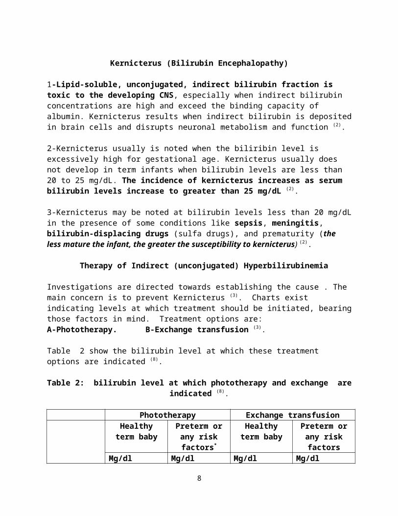
Investigations are directed towards establishing the cause . The main concern is to prevent Kernicterus (3). Charts exist indicating levels at which treatment should be initiated, bearing those factors in mind. Treatment options are:A-Phototherapy. B-Exchange transfusion (3).
Table 2 show the bilirubin level at which these treatment options are indicated (8).
Table 2: bilirubin level at which phototherapy and exchange are indicated (8).
Phototherapy Exchange transfusionHealthy term
babyPreterm or any
risk factors*Healthy term
babyPreterm or any
risk factorsMg/dl μmol/l Mg/dl μmol/l Mg/dl μmol/l Mg/dl μmol/l
Day 1 Any visible jaundice** 15 260 13 220Day 2 15 260 13 220 25 425 15 260Day 3 18 310 16 270 30 510 20 340
Day 4 and after
20 340 17 290 30 510 20 340
* Risk factors include small size ( less than 2.5 kg or born before 37 weeks gestation), haemolysis, and sepsis.** Visible jaundice anywhere on body on day 1.
6

A-Phototherapy
1-Blue light (not ultraviolet) of wavelength 450 nm converts the bilirubin in the skin and superficial capillaries into harmless water-soluble metabolites, which are excreted in urine and through the bowel (3).
2-The eyes are covered to prevent discomfort and additional fluids are given to counteract increased losses from skin (3).
B-Exchange transfusion
1-This is required if the bilirubin rises to levels considered dangerous despite phototherapy. It rapidly reduces the level of circulating bilirubin (3).
2-Techniques vary, but conventionally the exchange is done via umbilical artery and vein catheters. Aliquots of baby's blood (10-20 mL) are withdrawn, alternating with infusions of donor blood of the same volumes. Twice the infant's blood volume (i.e. 2 x 80 mL/kg) is exchanged over about 2 hours (3) ( or 2 x 85 mL/kg) (2).
3-The procedure is carried out through umbilical vein catheter. Phototherapy is usually needed after the first exchange to keep serum bilirubin below critical level. Howecer, a second or even third exchange may be needed in severe cases with rapidly rising bilirubin level (9).
Management of conjugated hyperbilirubinemiaManagement depend on the treatment of the causative diseases (if treatable e.g. surgical correction of biliary atresia) (5).
References1-Thomas P. Green. Pediatrics Just the Facts. Copyright © 2005 by The McGraw-Hill Companies, Inc.2-Robert M. Kliegman. Nelson essentials of pediatrics. 5th edition.3- Shyam Bhakthavasala. Crash course pediatrics.3rd edition 2008.4- Bryon J. Lauer and Nancy D. Spector . Hyperbilirubinemia in the Newborn. Pediatrics in Review 2011;32;3415- Mohammed El-naggar. Practical pediatric diagnosis. 5th edition. 2005.6- Babeta A garwal. Neonatal jaundice a review. International Journal of Biomedical and Advance Research. 2011.7- Brooke T. Davey. Deja Review Pediatrics. Copyright © 2008 by The McGraw-Hill Companies, Inc.8-Pediatrics pocket book.
7
9- Mohammed El-naggar. Practical pediatric therapy. 5th edition.2005Gastroenteritis
It is an infection of the small intestine usually viral, which present with a combination of diarrhea and vomiting (1) , but sometimes present without vomiting (2).
Incidence and etiology
In developed countries it is usually mild and self limiting but in developing countries 5 million children under 5 years old die from GE each year (1).
Rota virus is the most common pathogen (in children under 2 years (3), other causes include:
1. Acute bacterial infections (shigellae, Salmonellae, Campylobacter spp and Yersinia) (3).2. Enterotoxigenic E coli and Vibirio cholera which secrete enterotoxins (3).3. Parasites like E. histolytica, Giardia lambilia, and Cryptosporodium spp. (1).
Note: If a child was given antibiotic in response to infection like otitis media which may precede GE, then the diarrhea may be attributed to the antibiotic rather than to rotaviruse (2)
Clinical Features
Rotaviruse cause low sodium content watery diarrhea (1)
Acute bacterial infection cause invasion of GIT, so there is fever, small volume bloody stool and culture is indicated (3)
Enterotoxigenic bacteria cause high volume diarrhea with high sodium content > 90 mEq/L of stool (3)
Viral infection cause a prodromal illness (1) ( respiratory illness occur in about half of patients (2)) followed by vomiting and diarrhea, the vomiting precede the diarrhea and usually not stained (1).
Ingested toxins ( food poisoning ) usually cause more rapid onset diarrhea, vomiting without prodrome (3) For differential diagnosis.
Abdominal pain and blood or mucus in stool suggest invasive bacterial pathogen (1)
On examination, the most important physical sign is the presence and severity of dehydration (1).
8

Severity of dehydration
Severe dehydration sign and symptoms
Deeply sunken eyes, parched mucous membranes, significantly prolonged capillary refill (this can be assessed by applying enough pressure on the nail bed to cause blanching; refill time of approximately 1 second is normal); (4) skin pinch goes back very slowly 2 seconds (5) cool, mottled extremities; crying without tears; oliguria or anuria; weak or thready pulses; lethargy; poor oral intake; (4) unable to drink water or drink poorly (5) deep respirations; (4) Also weight loss 10% (1,4) , tachycardia and low BP (1).
Mild – Moderate ( Some) dehydration:
Sunken eyes, drinks eagrly, thirsty , skin pinch goes slowly < 2 seconds (5)
Weight loss 3 – 9% (4)
Incubation period and infectivity
Infant may be very infectious to other infants in ward or nursery (2)
Incubation period is 24 – 48 hour, so if 2 or more infants in the ward or nursery have diarrhea , cross infection should be presumed and admission to the ward may have to be stopped (2).
Complication of Gastroenteritis
Dehydration, metabolic disturbances and even death (4)
Infants and children are at high risk for morbidity and mortality secondary to diarrhea for several reasons. Since dehydration can occur easily as acute net intestinal fluid losses are relatively much greater in young children than in adults. This may result from inefficient transport systems in the developing intestine. In addition, the percent of total body water in children is higher than in adults; thus, they are more susceptible to body fluid shifts Finally, the renal capacity to compensate for fluid and electrolyte imbalances in the infant is limited compared with that of adults (4).
Treatment
Uncomplicated viral GE requires no specific treatment except attention to fluid and electrolyte replacement (3) Most of these episodes are self limited (4).
9

The frequency of stool is reduced by dietary treatment, but the abnormal consistency persist up to a week (2).
There is no role for anti emetic or antidiarrheal in GE. (1)
Vomiting associated with gastroenteritis usually resolves in 24 to 48 hours (4). As dehydration is corrected the frequency of vomiting will decrease (4).
Routine use of antiemetics for acute vomiting in children is not recommended because masking of symptoms may delay diagnosis of a treatable illness. In addition, the safety and efficacy of the antiemetics, including metoclopramide, promethazine, trimethobenzamide, and dimenhydrinate, have not been demonstrated (4).
While ondansetron, demonstrated good safety and efficacy when it was tested in children
aged 6 months to 10 years who presented to a pediatric emergency department
with gastroenteritis and dehydration (6) .
Treatment with anti-diarrheal preparations is generally not indicated especially in young infants (3).
Adsorbents, such as kaolin-pectin or attapulgite, adsorb bacterial toxins and water and lessen the symptoms of diarrhea by producing more formed stools, but there is no evidence of effectiveness and they are not recommended. (4) Kaolin should not be prescribed as it deflect the mother’s attention from the main treatment (2).
Drugs such as loperamide that alter GI motility should be avoided, especially in children with high fever, toxemia, or bloody mucoid stools, because they may worsen the clinical course of the bacterial infection. Bismuth subsalicylate preparations, which possess
antisecretory and antimicrobial effects, have not been shown to provide clinical benefit and are not recommended (4).
Antibiotics are rarely indicated except for specific bacterial infections such as invasive salmonellosis or sever campylobacter infection, and amebiasis or giardiasis (1) and also in case of cholera (3).
The key management of GE is rehydration with correction of fluid and electrolyte imbalance, the strategy depend on the severity of dehydration; Unless the child has persistent vomiting Oral fluid is the best means for rehydration, smaller more frequent sips may be better tolerated and should be encouraged (1).
10

Mild Dehydration: ORS are used, these contain dextrose to stimulate sodium and water reabsorption across the bowel wall (1)
Moderate dehydration: Oral rehydration is still indicated if tolerated. IV fluid should be reserved for those with vomiting or sever dehydration (1).
If there are signs of circulatory failure, immediate rescuitation is achieved by administration of IV fluids (1).
Calculation of the amount of fluid that is needed for correcting dehydration (1)
Volume required in 24 hour = Deficit + maintenance + ongoing loss
Deficit = % of dehydration / Body weight in Kg
Maintatinace = 100ml/24 hour for each Kg for the 1st 10 kg
50ml/day for each Kg for the 2nd 10 Kg ( 10 – 20)
And 20ml/day for each Kg thereafter
Ongoing loss: by estimating the volume of vomit and diarrhea.
Give 10% of the total daily amount within 30 min, then the remaining amount given through the day.
Treatment of dehydration (Severe cases)
A. Children with severe dehydration should be given rapid IV rehydration followed by oral rehydration therapy (5)
➤Start IV fluids immediately. While the drip is being set up, give ORS solution if the child can drink. (5)
Note: The best IV fluid solution is Ringer's lactate Solution (also called Hartmann’s Solution for Injection). If Ringer's lactate is not available, normal saline solution (0.9% NaCl) can be used. 5% glucose (dextrose) solution on its own is not effective and should not be used. ( 5)
For those < 12 month (infants) give 30ml/kg in 1 hour then give 70ml/kg within 5 hours
11

For those > 12 months ( Children) give 30ml/kg within 30min and give 70ml/kg within 2.5hour ( pocket book)
Reassess the child every 15–30 minutes. If hydration status is not improving, give the IV drip more rapidly.
➤ Also give ORS (about 5 ml/kg/hour) as soon as the child can drink: usually after 3–4 hours (infants) or 1–2 hours (children). ORS provide additional base and potassium which may not be adequately supplied by IV fluids (5).
ORS solution can be given by nasogastric tube or by mouth when IV therapy is not possible, in this case Start rehydration by tube (or mouth) with ORS solution: give 20 ml/kg/hour for 6 hours (total of 120 ml/kg) (5).
For assessment of hydrations status, if hydration status improved, there will be a strong radial pulse, improved ability to drink, also we need to check skin pinch which should be improved, while sunken eyes recovered more slowly (5).
B. In case of mild – moderate ( some dehydration)
In the first 4 hours, give the child the following approximate amounts of ORS solution, according to the child’s weight ( 75 ml for each Kg of body weight) but if the child want more to drink give more.
Give a child teaspoonful every 1 - 2 minutes if the child under 2 years, frequent sips from a cup for older child. (5)
If the child vomits, wait 10 minutes; then, resume giving ORS solution more slowly (e.g. a spoonful every 2–3 minutes). If the child’s eyelids become puffy, stop ORS solution and give plain water or breast milk. (5)
Advise breastfeeding mothers to continue to breastfeed whenever the child wants (5).
If the child still has some dehydration, repeat treatment for another 4 hours with ORS solution, as above, and start to offer food, milk or juice and breastfeed frequently (5). Fresh fruit juice or banana are preferred because they are a source of potassium (5).
After that use Zinc supplement for 10 – 14 days
12

Infants up to 6 months 1omg /day while for 6 months and more 20 mg/day (5).
C. In case of no dehydration
Give ORS, for those up to 2 years 50 – 100 ml after each loose stool and 100 – 200ml after each loose stool for those 2 years and more (5)
Zinc supplements can be used here also for 10 -14 days ( pocket book)
Notes about ORS
1. ORS should only be abandoned in children with intractable vomiting, loss of consciousness, or bowel obstruction, or if the child is in shock (4).
2. A glucose-containing ORS. Glucose provides calories and enhances salt and water absorption in the small intestine through mechanisms that usually are unimpaired in many toxin-induced diarrheas. however, glucose concentrations greater than 3% can impair sodium absorption because the glucose-coupled sodium transport system becomes saturated at this concentration and any additional glucose acts as an osmotically active solute in the bowel lumen (4). Carbonated beverages and fruit juices do not contain sufficient sodium to replace diarrheal losses (4).
3. Sodium content of ORS: The old WHO formula for ORS containing sodium (90 mEq/L), potassium (20 mEq/L), bicarbonate 30 mEq/L), chloride (80 mEq/L), and 2% glucose for the widespread management of acute diarrhea in third-world countries. This formula, which had a 90% successful rehydration rate for the management of diarrhea, contained a high concentration of sodium because secretory diarrhea (e.g., cholera) is associated with substantial loss of sodium. Malabsorptive diarrheas, such as those associated with rotavirus infections, are associated with much lower loss of sodium (<40 mEq/L). ORS contain less sodium than the WHO formulation, these preparations were equally effective as the WHO formula, even when used to treat the high sodium losses associated with cholera. Furthermore, these lower sodium-containing formulations were associated with less vomiting, lower stool output, and reduced need for IV infusions in non–cholera-associated gastroenteritis. As a result, the WHO, in 2002, promoted a new formulation that consists of 75 mEq/L sodium (4).
General notes in treating gastroenteritis
Continuation of oral feeding, despite diarrheal episodes, minimizes the development of protein and energy deficits; facilitates the maintenance and repair of intestinal mucosa; promotes recovery of brush border membrane disaccharidases; decreases the duration
of illness; and improves nutritional status (4).
13
Zinc supplementation (10–20 mg for 10–14 days) has been recommended by the WHO for the treatment and prevention of diarrheal disease in children in developing countries (4).
Probiotics, live microbial products containing species of Lactobacillus, Bifidobacterium, Saccharomyces, and Streptococcus, can improve the balance of intestinal flora and diminish the effect of enteric pathogens. Probiotics are most useful in infectious gastroenteritis when used early in the course of disease (4).
Admission or medical attention is needed in the following cases: (2,4)
1. If vomit persist 2. Red or black vomit or stool3. Any fever + GE in neonate4. If there is a sign of ear infection with abdominal pain or distention
Reference
1. Crash course2. ABC of the 1st year3. Current essential4. Applied therapeutics 20135. Pocket book of hospital care for children, WHO , 20056. ADVANCES IN Pediatrics . VOLUME 54. 2007 . By Michael S. Kappy, Lewis Barness,
Leslie Barton, Enid-Gilbert Barness, and Moritz Ziegler. Elsiever
14

Febrile convulsion
A febrile convulsion is a fit occurring in a child aged from six months to five years, precipitated by fever ( temp > 38 C) arising from infection outside the nervous system in a child who is otherwise neurologically normal (1,2) and in case of absence of acute electrolyte imbalance (3). Febrile convulsions are usually of the tonic clonic type (2).
Etiology, incidence and pathogenesis
The etiology and pathogenesis are unknown, and there is no evidence that the rate of temp. is important. Genetic predisposition appear to be a risk factor for later development of epilepsy or developing complex or recurrent febrile seizure (1,2).
Incidence: it occur in 2 – 4% of children aged 6 month – 5 years (1). Only 1 – 2.4% of children with febrile seizure have subsequent epilepsy (4).
Typically febrile seizure occurs within the 1st 24 hour of a febrile episodes (1) and most commonly due to acute viral respiratory infections (4)
Types of febrile seizure and clinical features
In simple febrile convulsions the fit lasts less than 20 minutes (while for reference 1 it is 15 minutes), there are no focal features, and the child is aged between six months and five years and has been developing normally (2).
While complex febrile seizures have a longer durations, occur in series, and are associated with focal changes (1) and signify a greater risk of later epilepsy (3)
Diagnosis: Diagnosis is made by exclusion of other causes of symptomatic seizures like meningitis or metabolic abnormalities. Whenever there is a clinical suspicion of meningitis or encephalitis, a lumbar puncture (LP) should be performed (3).
Treatment
The objective of emergency treatment is the prevention of a prolonged fit (lasting over 20 minutes), which may be followed by permanent brain damage, epilepsy, and developmental delay (2).
Antibiotics are not needed in most cases of children with febrile convulsion unless the child has otitis media or meningitis ( there is purpuric rash rather than neck stiffness which is absent sign in those < 1 year ) (2).
1. Control fever (4)
Antipyretic treatment for future illnesses provides comfort and prevent dehydration of the child but does not appreciably reduce the risk of febrile seizures (3).
15

Acetaminophen is the most common antipyretic agent used in children. The usual dose, oral or rectal, is 10 to 15 mg/kg/dose 100 to 150 mg) administered every 4 to 6 hours as needed to maximum of 90 mg/kg/day. Ibuprofen is administered as to 10 mg/kg/dose (50 to 100 mg) orally every 6 to 8 hours as needed to a maximum of 40 mg/kg/day. Ibuprofen is as effective as acetaminophen as an antipyretic and is associated with a low incidence of adverse effects. the risk of renal impairment is small with short-term use and not greater than that with acetaminophen (1). Recent data suggest that ibuprofen is more effective than paracetamol and should be used first (1).
Failure to respond to paracetamol is an indication to add ibuprofen to the paracetamol or to give ibuprofen alone (2) Some data suggest that combination therapy with acetaminophen every 4 to 6 hours and ibuprofen every 6 to 8 hours may be more effective in reducing time with fever in the first 24 hours (1).
2. Stop ongoing seizure. Drugs rarely needed as seizures are usually brief (4)
However rectal diazepam can be used ( 0.2 – 0.5mg/kg) (3), Rectal diazepam should be used as soon as possible after the onset of the convulsion (and used for the duration of febrile illness(4)). The parents should be advised not to give it if the convulsion has stopped (2). Rectal diazepam (0.5 mg/kg) produces an effective blood concentration of anticonvulsant within 10 minutes. The most convenient preparation resembles a toothpaste tube (Stesolid) (2).
The buccal midazolam treatment was superior than diazepam for stopping seizures and was not associated with an increased risk of respiratory depression. The doses given were approximately 0.5 mg/kg to children of all ages included in the study. A similar study found that intranasal lorazepam (0.1 mg/kg) was effective at stopping seizures for children 2 months of age or more presenting to an emergency department [5].
3. Anticonvulsant prophylaxis can reduce recurrent febrile seizures and may be appropriate after the second febrile seizure (4).
The indications for long term anticouvulsant prophylaxis have changed and the sole indication – frequent recurrences, which should be treated with phenobarbitone – is rare. There is no evidence that in the minority of children who later develop epilepsy the prophylactic use of anticonvulsant drugs would have prevented it (2). Both phenobarbital and valproate reduced the risk of a recurrent febrile seizure if regularly administered. However, the vast majority of children with febrile seizures have no long-term consequences and treatment does not alter the risk of developing epilepsy. Adverse drug effects occur in as many as 40% of infants and children treated with phenobarbital, and valproate carries a risk of idiosyncratic fatal hepatotoxicity and pancreatitis (3) Phenytoin and carbamazepines are ineffective (4).
16
Reference:
1. Applied therapeutics 20132. Bernard Valman, ABC of first year, 5th edition, 2002.3. Current Pedaitric therapy, 18th edition, 2006. 4. Judith M. Sondheimer, Current essentials of Pediatrics, 20085. ADVANCES IN Pediatrics . VOLUME 54. 2007 . By Michael S. Kappy, Lewis Barness,
Leslie Barton, Enid-Gilbert Barness, and Moritz Ziegler. Elsiever
17

Acute Respiratory distress syndrome ( ARDS )
Definition: Hyaline membrane disease (HMD) is another name for ARDS1. The ARDS described as the most severe manifestation of acute lung injury 2.This clinical diagnosis is warranted in a preterm newborn with respiratory difficulty, including tachypnea (>60 breaths/min), chest retractions, and cyanosis in room air that persists or progresses over the first 48-96 hours of life, and a characteristic chest x-ray appearance.1
Incidence : ARDS occurs in ~50% of infants with birth weight between 501 and 1500g 1 , mostly in infants less than 28 week of gestational age 3 The incidence is inversely proportional to the gestational age and birth weight , the incidence and severity of ARDS are expected to decrease after the increase in use of antenatal steroids in recent years 1.
Pathophysiology:
1- Lack of surfactant : These surface-active agents are released into the alveoli, where they reduce surface tension and help maintain alveolar stability by preventing the collapse of small air spaces at end-expiration. 3 In the absence of surfactant, the small air spaces collapse1 .
2- Presence of an overly compliant chest wall : In the presence of a chest wall with weak structural support secondary to prematurity, the large negative pressures generated to open the collapsed airways cause retraction and deformation of the chest wall instead of inflation of the poorly compliant lungs1.
3- Decreased intrathoracic pressure : Especially for infant who is <30 weeks' gestational age 1.
4- Shunting : The presence or absence of a cardiovascular shunt may change the presentation or course of the disease process1.
Risk Factors : An increased frequency is associated with infants of diabetic mothers, delivery before 37 week gestation, multifetal pregnancies, cesarean section delivery, and a history of previously affected infants 3.
Diagnosis :1- Chest x-ray2- Laboratory studies : Blood gas sampling , complete blood cell count , Serum glucose
levels , Serum electrolyte levels including calcium (Hypocalcemia can contribute to more respiratory symptoms3 ) 1
3- Echocardiography
Clinical presentationSigns of ARDS usually appear within minutes of birth, although they may not be recognized for several hours in larger premature infants 3.
1- History: The infant is often preterm, either by dates or by gestational examination, or has a history of asphyxia in the perinatal period.
18
2- Physical examination : The infant with ARDS exhibits tachypnea, grunting, nasal flaring, and retractions of the chest wall. The infant may have cyanosis in room air 2 that is often relatively unresponsive to oxygen administration 3.
If the condition is inadequately treated, blood pressure may fall; fatigue, cyanosis, and pallor increase, and grunting decreases or disappears as the condition worsens,Respiratory failure may occur in infants with rapid progression of the disease. In most cases, the symptoms and signs reach a peak within 3 days, after which improvement is gradual 3
Management
Because most cases of ARDS are self-limited, the goal of treatment is to minimize abnormal physiologic variations. Treatment of these infants is best carried out in a specially staffed and equipped hospital unit, the neonatal intensive care unit (NICU)3.
A- Prevention 1- Strategies to prevent preterm birth include, bed rest, treatment of infections, and
administration of tocolytic medications 4 , most important is prevention of prematurity, including avoidance of unnecessary or poorly timed cesarean section.3
2- Antenatal corticosteroids: Use of antenatal betamethasone to enhance fetal pulmonary maturity is now established and generally considered to be standard of care , the recommended glucocorticoid regimen consists of the administration to the mother of two 12-mg doses of betamethasone given intramuscularly 24 h apart1
between 24 and 34 wk of gestation 3 . Dexamethasone is no longer recommended because of increased risk for cystic periventricular leukomalacia among very premature infants exposed to the drug prenatally 1, 3
B- Surfactant replacement: is now considered a standard of care in the treatment of intubated infants with ARDS , surfactant, whether used prophylactically in the delivery room or in the treatment of the established disease, leads to a significant decrease in the risk of death 1 .
C- Respiratory support: Endotracheal intubation and mechanical ventilation are the mainstays of therapy for infants with ARDS in whom apnea or hypoxemia with respiratory acidosis develops. Warm humidified oxygen (>90% saturation) to maintain normal tissue oxygenation while minimizing the risk of oxygen toxicity.1
D- Fluid and nutritional support: For the 1st 24 hour, 10% glucose and water should be infused through a peripheral vein3.
E- Antibiotic therapy: Antibiotics that cover the most common neonatal infections are usually begun initially. Aminoglycoside dosing intervals are increased for the premature infant 1.
19

F- Sedation : is commonly used to control ventilation in these sick infants. Phenobarbital is often used to decrease the infant's activity level 1 .
Prognosis : Although the survival of infants with ARDS has improved greatly, the prognosis for survival with or without respiratory and neurologic sequelae are highly dependent on birth weight and gestational age. Major morbidity and poor postnatal growth remain high for the smallest infants. 1
References
1- NEONATOLOGY: MANAGEMENT, PROCEDURES, ON-CALL PROBLEMS, DISEASES, AND DRUGS - 5th Ed. (2004)
2- The Respiratory Tract in Pediatric Critical Illness and Injury 20093- Nelson text book of Pediatric 19 ED4- Nelson essential of pediatrics
20

![ANNEX I SUMMARY OF PRODUCT …€¢ Liver function ( alanine aminotransferase [ALAT], aspartate aminotransferase [ASAT], albumin, bilirubin) ... • The short needle should be then](https://static.fdocuments.net/doc/165x107/5d3edcab88c993715a8c0898/annex-i-summary-of-product-liver-function-alanine-aminotransferase-alat.jpg)


![· Cl Ureum a C) Total Protein a Albumin Bilirubin Direct C] Bilirubin Total 12 Amylase D ALP a GOT - AST CPT - ALT GCT CK Cholesterol Cl Triglycerides ... 115 125 133 123 108 160](https://static.fdocuments.net/doc/165x107/5c8567f409d3f2fe508c0f26/-cl-ureum-a-c-total-protein-a-albumin-bilirubin-direct-c-bilirubin-total-12.jpg)