Pediatric Upper-Extremity Fractures - Healio: Medical … · ity of pediatric upper-extremity...
9
196 | Healio.com/Pediatrics PEDIATRIC ANNALS 43:5 | MAY 2014 FEATURE Abstract Upper-extremity fractures account for more than half of childhood bony injuries. The frequency of injury increases with increasing mobility. The most common mechanism is a fall on an outstretched hand while playing. Optimal management requires knowledge of the normal anatomy and variants unique to pediatric bones. The physician needs to maintain a high level of suspicion for growth plate injuries because if unrecognized, these may result in growth arrest. Although the vast major- ity of pediatric upper-extremity fractures will heal rapidly with minimal intervention, physicians should be aware of the compli- cations that can arise from these injuries. Pediatric Upper-Extremity Fractures Rajan Arora, MD; Utkarsh Fichadia, MD; Earl Hartwig, MD; and Nirupama Kannikeswaran, MD Rajan Arora, MD, is a Pediatric Emergency Medicine Fellow, Carman and Ann Adam Depart- ment of Pediatrics, Children’s Hospital of Michi- gan. Utkarsh Fichadia, MD, is a Pediatric Emer- gency Medicine Fellow, Carman and Ann Adam Department of Pediatrics, Children’s Hospital of Michigan. Earl Hartwig, MD, is Associate Profes- sor of Pediatrics and Emergency Medicine, Car- man and Ann Adam Department of Pediatrics, Children’s Hospital of Michigan. Nirupama Kan- nikeswaran, MD, is Associate Professor of Pediat- rics and Emergency Medicine, Carman and Ann Adam Department of Pediatrics, Children’s Hos- pital of Michigan. Address correspondence to: Rajan Arora, MD, Carman and Ann Adam Department of Pediat- rics, Division of Emergency Medicine, Children’s Hospital of Michigan, 3901 Beaubien Boulevard, Detroit, MI 48201; email: [email protected]. Disclosure: The authors have no relevant fi- nancial relationships to disclose. doi: 10.3928/00904481-20140417-12 © Shutterstock
-
Upload
nguyenlien -
Category
Documents
-
view
218 -
download
0
Transcript of Pediatric Upper-Extremity Fractures - Healio: Medical … · ity of pediatric upper-extremity...

196 | Healio.com/Pediatrics PEDIATRIC ANNALS 43:5 | MAY 2014
FEATURE
AbstractUpper-extremity fractures account for
more than half of childhood bony injuries.
The frequency of injury increases with
increasing mobility. The most common
mechanism is a fall on an outstretched
hand while playing. Optimal management
requires knowledge of the normal anatomy
and variants unique to pediatric bones.
The physician needs to maintain a high
level of suspicion for growth plate injuries
because if unrecognized, these may result
in growth arrest. Although the vast major-
ity of pediatric upper-extremity fractures
will heal rapidly with minimal intervention,
physicians should be aware of the compli-
cations that can arise from these injuries.
Pediatric Upper-Extremity FracturesRajan Arora, MD; Utkarsh Fichadia, MD; Earl Hartwig, MD; and Nirupama Kannikeswaran, MD
Rajan Arora, MD, is a Pediatric Emergency
Medicine Fellow, Carman and Ann Adam Depart-
ment of Pediatrics, Children’s Hospital of Michi-
gan. Utkarsh Fichadia, MD, is a Pediatric Emer-
gency Medicine Fellow, Carman and Ann Adam
Department of Pediatrics, Children’s Hospital of
Michigan. Earl Hartwig, MD, is Associate Profes-
sor of Pediatrics and Emergency Medicine, Car-
man and Ann Adam Department of Pediatrics,
Children’s Hospital of Michigan. Nirupama Kan-
nikeswaran, MD, is Associate Professor of Pediat-
rics and Emergency Medicine, Carman and Ann
Adam Department of Pediatrics, Children’s Hos-
pital of Michigan.Address correspondence to: Rajan Arora, MD,
Carman and Ann Adam Department of Pediat-rics, Division of Emergency Medicine, Children’s Hospital of Michigan, 3901 Beaubien Boulevard, Detroit, MI 48201; email: [email protected].
Disclosure: The authors have no relevant fi-nancial relationships to disclose.
doi: 10.3928/00904481-20140417-12 © S
hutte
rsto
ck
PEDIATRIC ANNALS 43:5 | MAY 2014 Healio.com/Pediatrics | 197
FEATURE
Childhood injuries account for more than 10 million annual visits and are the second lead-
ing cause for visits to primary care of-fices and the emergency department.1,2 Fractures make up around 10% to 25% of these musculoskeletal injuries. Almost two-thirds of all boys and nearly half of all girls will have had sustained a fracture by 15 years of age, with a peak incidence for fractures of age 14 years for boys and 11 years for girls.3,4 In children, fractures tend to occur more frequently in the up-per extremity in comparison to the lower extremity.5 The distal radius is the most frequently fractured bone. This, along with fracture of metacarpals and phalan-ges, accounts for 50% of all fractures.3,6
With children and adolescents’ ever-increasing participation in sports, it is likely that primary care physicians will increasingly encounter patients with upper-extremity injuries. This article discusses the commonly encountered upper-extremity fractures in children. Common mechanisms of injury, fracture type and classification, possible compli-cations, and current principles of their management are reviewed.
KEY ASPECTS OF PEDIATRIC BONE The skeletal anatomy of a child varies
considerably from that of an adult in sev-eral important ways. The pediatric bone is less dense, more porous, and has a lower mineral content. Being more elas-tic and pliable, it can undergo a greater degree of deformation before breaking. Hence, both greenstick and torus frac-tures are seen almost exclusively in the pediatric population. Increased porosity prevents fracture propagation, thereby resulting in a lower incidence of com-minuted fractures in children. However, since the tensile strength of pediatric bone is lower than that of the ligaments, fractures are more common than sprains.
The two key attributes of imma-ture skeleton are presence of growth plate and thick periosteum. The growth
plates are located at the ends of the long bones and are responsible for con-tinued longitudinal growth. In younger children, fractures closer to a growth plate have great remodeling potential, as the physis of a bent bone will grow eccentrically to help restore alignment over time. Depending upon age, re-modeling can correct varying degrees of displacement and angulation but not axial malrotation. The periosteum in children is much thicker, stronger, and has a greater osteogenic potential. It provides stability, acts as a restraint to displacement, preserves the vascular supply, maintains reduction, and allows for faster fracture healing.
CLASSIFICATION OF PEDIATRIC FRACTURES
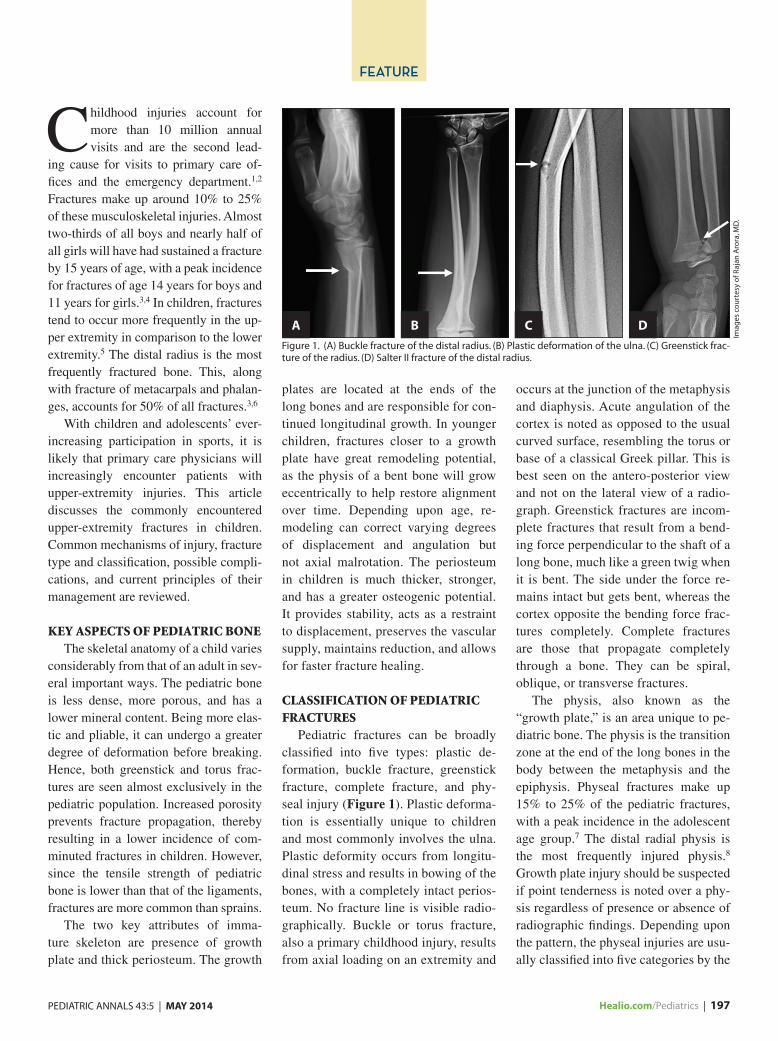
Pediatric fractures can be broadly classified into five types: plastic de-formation, buckle fracture, greenstick fracture, complete fracture, and phy-seal injury (Figure 1). Plastic deforma-tion is essentially unique to children and most commonly involves the ulna. Plastic deformity occurs from longitu-dinal stress and results in bowing of the bones, with a completely intact perios-teum. No fracture line is visible radio-graphically. Buckle or torus fracture, also a primary childhood injury, results from axial loading on an extremity and
occurs at the junction of the metaphysis and diaphysis. Acute angulation of the cortex is noted as opposed to the usual curved surface, resembling the torus or base of a classical Greek pillar. This is best seen on the antero-posterior view and not on the lateral view of a radio-graph. Greenstick fractures are incom-plete fractures that result from a bend-ing force perpendicular to the shaft of a long bone, much like a green twig when it is bent. The side under the force re-mains intact but gets bent, whereas the cortex opposite the bending force frac-tures completely. Complete fractures are those that propagate completely through a bone. They can be spiral, oblique, or transverse fractures.
The physis, also known as the “growth plate,” is an area unique to pe-diatric bone. The physis is the transition zone at the end of the long bones in the body between the metaphysis and the epiphysis. Physeal fractures make up 15% to 25% of the pediatric fractures, with a peak incidence in the adolescent age group.7 The distal radial physis is the most frequently injured physis.8 Growth plate injury should be suspected if point tenderness is noted over a phy-sis regardless of presence or absence of radiographic findings. Depending upon the pattern, the physeal injuries are usu-ally classified into five categories by the
Figure 1. (A) Buckle fracture of the distal radius. (B) Plastic deformation of the ulna. (C) Greenstick frac-ture of the radius. (D) Salter II fracture of the distal radius.
Imag
es c
ou
rtes
y o
f Raj
an A
rora
, MD
.
A B C D

198 | Healio.com/Pediatrics PEDIATRIC ANNALS 43:5 | MAY 2014
FEATURE
Salter and Harris classification system.9 Type I injuries extend through the phy-sis. Type II fractures extend through the physis and exit through the metaphysis. They are the most common and repre-sent approximately 50% of all growth-plate fractures in children.10 Type III begins in the physis and exits through the epiphysis intra-articularly. Type IV injury traverses through the physis, me-taphysis, and epiphysis. Type V involves crush injury to the physis and carries the worst prognosis. Salter-Harris III to V fractures have a higher incidence of growth disturbance, as the likelihood of growth arrest is directly related to the severity of physeal injury. Usually the growth plate repairs well and rapidly
but it is important to recognize physeal fractures as any damage to the growth plate can result in progressive angular deformity, limb-length discrepancy, or joint incongruity.
RISK FACTORSIn otherwise healthy children, skel-
etal fragility is often attributed to low peak bone mass. Several independent risk factors like genetic constitution, birth weight, poor nutrition, and low socio-economic status may influence fracture risk in children. Whiting11 and Goulding et al.12 showed that obesity in childhood and adolescence reduces bone mineral density with an increased propensity for fractures. Furthermore,
obese and overweight children tend to fall more during daily activities as a result of difficulty with balance. Envi-ronmental modifications have not been shown to lower the risk of fractures in obese children, and the only reduction in fracture risk was achieved by attaining a healthy body weight.13
The role of vitamin D in maintaining patients’ bone health, as well as fracture healing and prevention of future frac-tures, is well known. In a recent study by James et al.,14 hypovitaminosis D was common among children with upper-extremity fractures. In their study cohort of 181 patients, 64% had low vitamin D levels, with African-American children being more likely to have an insufficient or deficient level.
Repetitive stress may result in frac-tures due to overuse and fatigue of the surrounding musculature. Though less prevalent than lower-extremity stress fractures, upper-extremity stress frac-tures are now being more frequently recognized. Common examples include “Little Leaguer shoulder” and “gym-nast wrist,” which are a chronic Salter-Harris type I injury to the physis of the proximal humerus and distal radius, respectively. Pediatricians should offer nutritional counseling and discuss the importance of appropriate training and conditioning prior to and during sports participation to avoid these fractures.
INITIAL ASSESSMENTAccurate diagnosis of musculoskel-
etal injuries in children warrants a sys-tematic approach. A detailed history, comprehensive physical examination, and a good understanding of sport bio-mechanics are vital adjuncts in mak-ing the correct diagnosis and planning appropriate management. A pertinent history should include mechanism of injury, direction and magnitude of the force, prior injuries, or any associated symptoms. All splints and bandages must be removed to ensure a thorough
TABLE 1.
Sensorimotor Assessment of Common Upper-Limb Nerves
Nerve Sensory Motor
Radial Dorsum of first web space Thumbs-up sign
Ulnar Volar aspect of little finger Make a star
(Spread fingers wide)
Median Volar aspect of index finger Make a fist with thumb flexion
Anterior Interosseus No sensory function “OK” sign (Making a circle with the
thumb and index finger)
TABLE 2.
Common Upper-Extremity Splints and Their Indications
Splint Indications
Volar/Dorsal Soft tissue injuries to hand and wrist, carpal bone fractures (excluding
scaphoid/trapezium), and distal radius buckle fracture.
Thumb Spica Scaphoid, non-displaced first metacarpal, and stable thumb fracture.
Radial Gutter Non-displaced/non-rotated fractures of second and third metacarpal or
corresponding proximal/middle phalangeal shaft fractures.
Ulnar Gutter Non-displaced/non-rotated fractures of fourth and fifth metacarpal or
corresponding proximal/middle phalangeal shaft fractures.
Sugar Tong Acute distal radial and ulnar fractures.
Long Arm
Posterior
Distal humeral, proximal/midshaft forearm fractures, and non-buckle
wrist fractures.
Aluminum
U-Shaped Splint
Distal phalangeal fractures.
Buddy Taping Stable, non-displaced, non-angulated shaft fractures of the proximal or
middle phalanx.

PEDIATRIC ANNALS 43:5 | MAY 2014 Healio.com/Pediatrics | 199
FEATURE
examination of the injured extremity. Ensure appropriate analgesia, as it will make the child comfortable and comply with the examination. It is imperative to examine the entire limb as well as assess the joint above and below the injury for range of motion, stability, or concomi-tant injuries.
While performing an upper-extrem-ity examination, attention should be paid to the clavicle, scapula, and shoul-der. One should assess for any swelling, deformity, open wound, area of maxi-mal tenderness, and active and passive range of motion. Finally, it is important to assess the distal neurovascular status of the limb. In younger children, motor and sensory assessment can be done by asking them to copy simple movements and touching areas reliably supplied by each nerve (Table 1). Plain radiographs are the mainstay of the diagnosis. Frac-tures should be described in terms of location, displacement, separation, shortening, and presence of angular or rotational deformities. Further im-aging, such as computed tomography (CT) scans, may be necessary to plan operative intervention for intra-articu-lar displacements. A bone scan or mag-netic resonance imaging (MRI) may be needed occasionally to confirm the di-agnosis of a stress fracture.
BASIC PRINCIPLES OF FRACTURE MANAGEMENT IN CHILDREN
Bone healing in children is usually rapid and inversely related to the age of the patient. Further, greater remodeling potential and abundant callus formation lessens the likelihood of long-term com-plications in childhood fractures. There-fore, conservative treatment including pain control, immobilization in a splint or cast, and return of activity in accor-dance with the patients’ symptoms suf-fices in the majority of common upper-extremity fractures.
Splinting may be accomplished us-ing plaster, pre-padded casting material,
or prefabricated and over-the-counter splints. In general, six to 10 sheets of plaster material over appropriate pad-ding — especially over bony promi-nences — are recommended for upper-extremity injuries. Splints should be non-circumferential, somewhat loose, and applied in a position of function. Commonly used upper-extremity splints and their indications are listed in Table 2. Although some angulation is accept-able depending upon patient’s age, the fracture should not be malaligned or malrotated. In most cases, 15 to 20 de-grees of volar/dorsal angulation in the sagittal plane in a skeletally immature child is deemed acceptable.15,16 Marked bowing should usually be corrected by completing the fracture and restor-ing alignment. For displaced fractures, closed reduction and immobilization in a cast can be achieved under procedural sedation. Open fracture warrants broad-spectrum antibiotics and tetanus vacci-nation. Immediate orthopedic evaluation is indicated for fractures that are unsta-ble, open, or associated with neurovas-cular compromise.
CLAVICLE FRACTURESClavicle fractures are the most com-
mon of all pediatric fractures. They may be seen in newborns as a result of birth trauma, or in children and adolescents following a fall on an outstretched hand (FOOSH) or shoulder, or a direct blow to the bone. An overwhelming majority (80% to 85%) occur in the middle one-third of the bone (Figure 2). In younger children, these fractures are typically
greenstick and may go unnoticed until a large callus forms.17 In older children, the fracture is usually displaced and as-sociated with lowering of the affected shoulder, point tenderness, swelling, obvious deformity, and/or ecchymosis at the fracture site. Shoulder movements across different planes usually exac-erbate the pain. X-rays with dedicated clavicle views can accurately diagnose most clavicle fractures. However, one should be aware that clavicle views are different from routine shoulder films.
The majority of clavicle fractures will heal uneventfully and are managed using a simple sling for 2 to 3 weeks, primar-ily for pain control. When compared to a figure-of-eight splint, a sling is more comfortable, less cumbersome to put on, causes fewer skin problems, and has similar outcomes. Indications for opera-tive intervention include open fractures, complicated comminuted fractures, and fractures with neurovascular compro-mise or greater than 100% displacement (width of the clavicle) where skin is tent-ed or its integrity is threatened.18 Parents should be informed that a callus bump at the fracture site is part of the natural heal-ing process and will become less distinct as the bone remodels over the next 6 to 12 months. Complications, though rare, may include brachial plexus injury, pneumo-thorax, and airway compromise. Healing usually happens over 6 to 18 weeks, with average return to non-contact sports in 4 to 6 weeks followed by return to contact sports when there is no pain over the frac-ture site, full pain-free range of move-ments, and normal strength.
Figure 2. (A) Midshaft clavicle fracture with overriding. (B) Healing clavicle fracture with surrounding callous.
A B

200 | Healio.com/Pediatrics PEDIATRIC ANNALS 43:5 | MAY 2014
FEATURE
HUMERUS FRACTURESProximal humerus fractures are a
common injury in children that peaks during adolescence secondary to in-creased sports participation. It compris-es of both physeal and metaphyseal frac-tures (Figure 3A). The most common mechanism of injury is a FOOSH or a hit directly at the proximal humerus. Al-though less common than clavicle frac-tures, this type of fracture may be seen in neonates secondary to birth trauma. The patient usually presents with pain local-ized to the proximal humerus and/or anterior shoulder or, in newborn cases, pseudoparalysis. Physical findings could
be subtle or absent and may include swelling, bruising, and/or an obvious deformity in more severe injuries. Injury to the axillary nerve should be excluded by carefully assessing deltoid function and sensation over the lateral aspect of the proximal humerus. Standard shoul-der radiographs are diagnostic. These fractures carry an excellent prognosis as a result of the abundant remodeling potential of the proximal humerus. Most can be managed by immobilization with a simple sling, or a coaptation splint for the more severely displaced fractures, for approximately 3 to 4 weeks. Surgi-cal intervention is required for associ-
ated neurovascular injuries, open, and/or intra-articular fractures, or severely angulated fractures in older children and adolescents. Complications are quiet rare and include axillary nerve/brachial plexus injury (typically neuropraxia), avascular necrosis, and malunion.
Humeral metaphysial/shaft fractures in the absence of significant trauma (eg, motor vehicle accident, fall from height) should raise the suspicion of abuse, al-though neither age nor fracture pattern is pathognomonic of abuse (Figure 3B). Physicians should also consider patho-logic fracture, as the humerus is a com-mon site for bone cysts and other benign lesions (Figure 3C).19 These fractures also have an enormous remodeling po-tential; hence, the majority are treated with immobilization in a coaptation splint for 3 to 4 weeks and rarely require surgical intervention. Complications are rare, with radial nerve injury being the most common.
ELBOW FRACTURESElbow fractures in children are quite
common and represent approximately 10% to 12% of all pediatric fractures.20,21 Unlike fractures of the clavicle or proxi-mal humerus, elbow fractures are more likely to require precise, often surgical, reduction.
Diagnosis of pediatric elbow frac-tures can be challenging, as this requires distinguishing normal ossification cen-ters from fractures in a radiograph. Ap-plying the mnemonic CRITOE, which refers to the sequence of appearance of six secondary ossification centers at the elbow (Table 3), can help pediatric providers with this.22,23 However, one should be aware that these ages are ap-proximations, and these injuries often occur somewhat earlier in girls.
Supracondylar FracturesSupracondylar fractures account for
60% to 80% of all pediatric elbow frac-tures.5 They occur primarily during the
Figure 3. (A) Displaced fracture through the neck of humerus. (B) Humerus shaft fracture with overrid-ing. (C) Pathologic proximal humerus fracture through a bone cyst.
TABLE 3.
Ossification Centers of the Pediatric Elbow
Ossification Center
Age of Appearance
(years)
C = Capitellum 1
R = Radial Head 3
I = Internal (Medial)
Epicondyle
5
T = Trochlea 7
O = Olecranon 9
E = External (Lateral)
Epicondyle
11
Adapted from Beatty and Kasser22 and Green23
TABLE 4.
Gartland’s Classification of Supracondylar Fractures
Type I Non-displaced.
Type II Partially displaced with
intact posterior cortex.
Type III Completely displaced with
no contact between frac-
tured segments.
Adapted from Gartland24
A B C

PEDIATRIC ANNALS 43:5 | MAY 2014 Healio.com/Pediatrics | 201
FEATURE
first decade of life, with a peak incidence at around 5 to 7 years of age. The most common mechanism is a FOOSH injury with hyperextension of the elbow (eg, a fall off the monkey bars). The child usually presents with a swollen painful elbow and limited range of motion and may have an obvious deformity. Signifi-cant ecchymosis is considered a risk fac-tor for forearm compartment syndrome. Due to its potential to injure the brachial artery, as well as the radial, median, and/or ulnar nerve, a thorough neurovascu-lar assessment should be performed in all patients with a suspected or known elbow fracture. Standard elbow radio-graphs, including an anteroposterior (AP) view in extension and a lateral view at 90-degree flexion, are sufficient in making the diagnosis. On a lateral radiograph, the classic figure-of-eight appearance of the distal humerus and the anterior humeral line should always be assessed. In a normal elbow, a line drawn along the anterior cortex of the humerus should bisect the middle third of the capitellum, whereas in the case of a supracondylar fracture, the capitellum is generally displaced posterior to this line. In absence of a clear fracture line, subtle signs like presence of a posterior fat pad or a large, triangular sail-shaped anterior fat pad are indicative of an intra-articular joint effusion with an associat-ed fracture. Supracondylar fractures are classified on the basis of the degree of displacement of the fractured distal frag-ment (Figure 4, Table 4).24
Type I non-displaced supracondylar fractures can be treated in a long-arm cast for approximately 3 to 4 weeks. Type II and Type III fractures are mostly managed surgically with closed reduc-tion and percutaneous pin fixation. In case of suspected vascular compromise, the patient should be taken immediately to the operating room for fracture reduc-tion. Supracondylar fracture carries one of the highest complication rates of any pediatric fracture.25 Complications in-
clude neurovascular injuries, compart-ment syndrome, Volkmann’s ischemic contractures (contracture deformities of the fingers, hand, and wrists), and cubi-tus varus (angular gunstock deformity).
OTHER ELBOW FRACTURES Lateral condyle
The lateral condyle is the second most common elbow fracture and results from a FOOSH mechanism. Examina-tion findings reveal a swollen elbow, possible localized lateral tenderness, and decreased range of movement. The frac-ture may not always be appreciated on initial radiographs, as it occurs through an area that may be only partially ossi-fied. Radiographic findings may include the presence of a posteriorly displaced metaphysial fragment or disruption of the radial head alignment with the capi-
tellum (Figure 5A). Treatment is usu-ally surgical. The articular nature of the fracture and often delayed diagnosis re-sult in a high incidence of malunion and nonunion with this injury.
Medial EpicondyleMedial epicondyle is the third most
common pediatric elbow fracture. It is usually seen in children between the ages of 9 and 14 years and frequently encountered in baseball pitchers. The mechanism of injury is a valgus stress, and about half of cases are associated with dislocation of elbow. Examina-tion findings include a swelling and tenderness over the medial epicondyle and weakness in the flexor-pronator muscle group. Imaging for epicondylar fractures includes AP and lateral radio-graphs (Figure 5B). Non-displaced and
Figure 4. (A) Type I supracondylar fracture showing anterior fat pad (sail sign) and posterior fad pad sign with normal anatomic alignment of anterior humeral line (black line). (B) Type II posteriorly displaced supracondylar fracture with large effusion. (C) Type 3 supracondylar fracture with severe and complete displacement of the distal segment.
Figure 5. (A) Transverse fracture through lateral condyle and epicondyle with displaced capitellum. (B) Fracture of the medial epicondyle. (C) Fracture of the radial head. (D) Avulsion fracture of the olecranon.
A B C
A B C D

202 | Healio.com/Pediatrics PEDIATRIC ANNALS 43:5 | MAY 2014
FEATURE
stress fractures are treated conservative-ly in a long arm posterior splint or cast for 3 weeks. Displacement of greater than 3 mm to 5 mm or presence of intra-articular loose bodies require surgical fixation.26 The most common compli-cations are stiffness, ulnar nerve injury, and symptomatic non-union.
RADIAL HEAD/NECK FRACTURESChildren usually sustain radial neck
fractures, whereas fractures of the ra-dial head occur primarily in adults. Most fractures result from a FOOSH mecha-nism with the elbow in extension and valgus. Upon examination, there may be local tenderness over the radial head with pain accentuation on pronation/supination of the forearm. At times, pa-tients may present with referred pain at the wrist. Most cases are Salter-Harris type II fractures and are revealed on standard elbow radiographs (Figure 6C). At times, radiographic findings can
be subtle and only demonstrate the pres-ence of a posterior fat pad sign. Undis-placed fractures, or those with less than 30-degree angulation, are managed in a long arm posterior splint or cast. Dis-placed or more angulated fractures are treated surgically. Complications in-clude decreased range of motion, avas-cular necrosis of the radial head, and posterior interosseous nerve injury.
OLECRANON FRACTURESOlecranon fractures occur rarely in
children. If present, they are commonly associated with other elbow injuries. The common mechanism is a fall onto the elbow or a direct blow to the olec-ranon process. Examination may reveal swelling and tenderness over the olecra-non process, and the patient will resist extension at the elbow. If nondisplaced, these fractures can be treated in a long arm cast; however, operative interven-tion is required for displaced fractures.
FOREARM FRACTURESChildhood forearm fractures repre-
sent 40% to 50% of all fractures in chil-dren.27,28 They may occur at the proxi-mal end or in the middle of the forearm, but the majority (75%) are located near the wrist at the distal end of the bone (Figure 6A).29 Distal injuries vary great-ly in severity and complexity, with age determining the pattern and the location of the fracture. In children younger than
10 years of age, buckle and greenstick fractures occur most frequently, whereas growth plate and complete fractures are more likely in patients older than 10 years of age and adolescents, respec-tively. Fractures of both bones of the forearm are mostly distal in location, as well (Figure 6B). Imaging may re-veal plastic deformation, greenstick, or complete injuries with varying degrees of displacement. Fracture dislocations of the forearm can also happen, in which there is a fracture with shortening of one of the two bones with dislocation of the other bone. Monteggia fracture compris-es radial head dislocation plus proximal ulna fracture or plastic deformation of the ulna without obvious fracture (Fig-ure 6C). Galeazzi fracture is a relatively rare injury characterized by fracture of the distal radial shaft with disruption of the distal radioulnar joint (Figure 6D).
Forearm fractures typically result from a FOOSH injury or, occasionally, due to direct blow, as in the case of a both-bone forearm fracture. Physical exam may reveal swelling, tenderness, and decreased motion, and gross defor-mity may or may not be present. While evaluating forearm fractures, if only one bone appears to be fractured, the clini-cian should check the proximal and distal joints for injury. Although rare, compartment syndrome should be ruled out in both-bone forearm fractures. Plain radiographs of the forearm can provide the diagnosis.
Treatment depends on the type of fracture and the degree of displacement. Most pediatric forearm fractures can be treated without surgery. Buckle fractures may simply need the support of a splint or cast for 2 to 3 weeks until they heal. Closed reduction and immobilization for 6 to 12 weeks in a long arm cast is needed for greenstick fracture with more than 10 degrees of angulation, both-bone fracture in children younger than age 10 years, fracture dislocations, distal radius fractures, and Salter-Harris type I
Figure 6. (A) Transverse fracture of the radial metadiaphysis. (B) Fracture of both bones of the forearm. (C) Monteggia fracture characterized by proximal ulnar fracture with radial head dislocation. (D) Gale-azzi fracture depicted by distal radial fracture with disrupted distal radio-ulnar joint.
Figure 7. (A) Fracture through waist of the scaph-oid bone. (B) Fracture through shaft of the 5th metacarpal (“boxer’s fracture”).
A B C D
A B
PEDIATRIC ANNALS 43:5 | MAY 2014 Healio.com/Pediatrics | 203
FEATURE
and II injuries. There is great potential for remodeling, and outcomes are quite good.30 Surgical indications include open fractures, Salter-Harris type III and IV fractures of the distal radial physis, failed or unstable reductions, associated vascular injuries, and fractures in skel-etally mature individuals.
WRIST AND HAND FRACTURESCarpal bones are predominately car-
tilaginous until late childhood. There-fore, mechanisms that would produce bony wrist injuries in adults would pro-duce fracture of the forearm in young children. Acute injuries to the wrist usually result from a FOOSH or blunt trauma. The scaphoid bone is the most common carpal bone to be fractured (Figure 7A). Clinically, there is radial-sided wrist pain and swelling, with ten-derness that is often localized to the an-atomic snuffbox. Scaphoid fractures can be difficult to diagnose on plain films, with reported sensitivities between 70% and 86%.31 Hence in an acute setting, any suspected scaphoid injury should be managed with a splint. CT or MRI may be needed to make the diagnosis of subtle or stress fractures. Treatment and prognosis is dependent on the location of the fracture. Distal and middle one-third scaphoid fractures are often non-displaced and treated with a short arm thumb spica cast for 4 to 8 weeks. Since they have a good vascular supply, dis-tal and middle third scaphoid fractures heal well without complications. Proxi-mal or displaced scaphoid fractures of-ten require surgery as they have a more precarious blood supply and, hence, a higher incidence of nonunion.
Hand injuries in children are common but seldom complicated. Metacarpal fractures are the second most common type of upper extremity fracture. They can result from a direct blow or trauma to a clenched fist, such as punching a wall. The most common site of injury is neck of the fifth metacarpal, known as
a “boxer’s” fracture (Figure 7B). The phalanges too are frequently fractured, with distal phalanx being the most com-mon. The usual mechanism of injury to the distal phalanx is a crush or axial load injury.32 Often there is associated finger-tip or nail bed injury. The assessment for hand injury should include finger alignment, as any rotational deformity is unacceptable. The radiographs should be thoroughly examined for rotation, shortening, and angulation.33 Pediatric hand injuries are mostly managed non-operatively. Non-displaced metacarpal fractures can be immobilized in a radial or ulnar gutter splint. Most phalangeal fractures can be treated with splinting or buddy taping for 3 to 4 weeks. Anti-biotic therapy should be prescribed for distal phalangeal fractures with associ-ated nail trauma, as they are technically open fractures. Fractures associated with open injury, rotational deformity, unac-ceptable angulation, and intraarticular displacement require surgical manage-ment.
CONCLUSIONUpper-extremity fractures occur fre-
quently in the pediatric and adolescent patient population. It is important for the primary care physician to be cogni-zant of the key skeletal differences and unique fracture patterns in children, as they can directly impact patient man-agement and outcome. A thorough, sys-tematic approach will enable providers to accurately identify these injuries, institute the initial treatment, and offer appropriate anticipatory guidance for a positive outcome.
REFERENCES 1. Hambridge SJ, Davidson AJ, Gonzales R,
Steiner JF. Epidemiology of pediatric injury-related primary care office visits in the United States. Pediatrics. 2002;109:559-565.
2. Freedman KB, Bernstein J. The ad-equacy of medical school education in musculoskeletal medicine. J Bone Joint Surg.1998;80(10):1421-1427.
3. Cooper C, Dennison EM, Leufkens HG,
et al. Epidemiology of childhood fractures in Britain: a study using the General Prac-tice Research Database. J Bone Miner Res. 2004;19(12):1976-1981.
4. Lyons RA, Delahunty AM, Kraus D, et al. Children’s fractures: a population based study. Inj Prev. 1999;5(2):129-132.
5. Beaty JH, Kasser JR. The elbow region: general concepts in the pediatric patient. In: Rockwood CA, Wilkins B, eds. Fractures in Children. 5th ed. Philadelphia, PA: Lippincott Williams & Wilkins; 2001.
6. Rennie L, Court-Brown CM, Mok JY, Beattie TF. The epidemiology of fractures in children. Injury. 2007;38(8):913-922.
7. Peterson HA, Madhok R, Benson JT, el al. Physeal fractures: Part 1. Epidemiology in Olmsted County, Minnesota, 1979-1988. J Pediatr Orthop. 1994;14(4):423-430.
8. Neer II CS, Horwitz BZ. Fractures of the epiphyseal plate. Clin Orthop Relat Res. 1965;41:24-31.
9. Salter RB, Harris WR: Injuries involv-ing the epiphyseal plate. J Bone Joint Surg. 1963;45:587-622.
10. Peterson HA. Physeal and apophyseal inju-ries. In: Rockwood CA, Wilkins KE, Beaty JH, eds. Fractures in Children. 4th ed. Philadelphia, PA: Lippincott Williams & Wilkins; 1996.
11. Whiting SJ. Obesity is not protective for bones in child-hood and adolescence. Nutr Rev. 2002;60(1):27-30.
12. Goulding A, Taylor RW, Jones IE, et al. Over-weight and obese children have low bone mass and area for their weight. Int J Obes Relat Metab Disord. 2000;24:627-632.
13. Davidson PL, Goulding A, Chalmers DJ. Biomechanical analysis of arm fracture in obese boys. J Paediatr Child Health. 2003;39(9):657-664.
14. James JR, Massey PA, Hollister AM, Greber EM. Prevalence of hypovitaminosis D among children with upper extremity fractures. J Pe-diatr Orthop. 2013;33:159-162.
15. Bachman D, Santora S. Orthopedic trauma. In: Fleisher GL, Henretig FM, Ruddy RM, Silverman BK, eds. Textbook of Pediatric Emergency Medicine Philadelphia. 4th ed. Philadelphia, PA: Lippincott Williams & Wilkins; 2000.
16. Al Ansari K, Howard A, Seeto B, et al. Mini-mally angulated pediatric wrist fractures: is casting without manipulation enough? CJEM. 2007;9:9-15.
17. Kubiak R, Slongo T. Operative treatment of clavicle fractures in children: a review of 21 years. J Pediatr Orthop. 2002;22(6):736-739.
18. Housner JA. Clavicle fractures: individu-alising treatment for fracture types. Phys Sportsmed. 2003;31:30-36.
19. Shaw BA, Murphy KM, Shaw A, et al. Humerus shaft fractures in young chil-dren: accident or abuse? J Pediatr Orthop. 1997;17(3):293-297.

204 | Healio.com/Pediatrics PEDIATRIC ANNALS 43:5 | MAY 2014
FEATURE
20. Herring JA. Upper extremity fractures. In: Tachdijan’s Pediatric Orthopaedics. 3rd ed. Philadelphia, PA: W.B. Saunders Com-pany; 2002.
21. Tamai J, Lou J, Nagda S, et al. Pediatric el-bow fractures: Pearls and pitfalls. The Uni-versity of Pennsylvania Orthopaedic Journal. 2002;15:43-51.
22. Beaty JH, Kasser JR. The elbow region: general concepts in the pediatric patients. In Beaty JH, Kasser JR, eds. Rockwood and Wilkins’ Fractures in Children. Philadel-phia, PA: Lippincott Williams & Wilkins; 2001:563-570.
23. Green NE. Fracture and dislocations about the elbow. In: Green NE, Swiontkowski MF, eds. Skeletal Trauma in Children. Philadelphia, PA: W.B. Saunders Company; 1994.
24. Gartland JJ. Management of supracondylar
fractures of the humerus in children. Surg Gy-necol Obstet. 1959;109(2):145-154.
25. Rab GT, Grottkau BG. Operative treatment of children’s fractures and injuries of the physes. In: Chapman MW, ed. Orthopaedic Surgery. 3rd ed. Philadelphia, PA: Lippincott Williams & Wilkins; 2001.
26. Benjamin HJ, Hang BT. Common acute upper extremity injuries in sports. Clin Ped Emerg Med. 2007;8:15-30.
27. Price CT, Flynn JM. Management of frac-tures. In: Morrissy RT, Weinstein SL, eds. Lovell and Winter’s Pediatric Orthopaedics. 6th ed. Philadelphia, PA: Lippincott Williams & Wilkins; 2006.
28. Rodríguez-Merchán EC. Pediatric frac-tures of the forearm. Clin Orthop Relat Res. 2005;432:65-72.
29. Pizzutillo PD. Pediatric orthopaedics. In:
Griffin YL, ed. Essentials of Musculoskeletal Care. 3rd ed. Rosemont, PA: American Acad-emy of Orthopaedic Surgeons; 2005.
30. Jones K, Weiner DS. The management of fore-arm fractures in children: a plea for conserva-tism. J Pediatr Orthop. 1999;19(6):811-815.
31. Tiel-van Buul MM, van Beek EJ, Borm JJ, et al. The value of radiographs and bone scintigraphy in suspected scaphoid frac-ture. A statistical analysis. J Hand Surg Br. 1993;18:403-406.
32. De Jonge JJ, Kingma J, van der Lei B, et al. Phalangeal fractures of the hand. An analysis of gender and age-related incidence and aeti-ology. J Hand Surg Br. 1994;19:168-170.
33. Peterson JJ, Bancroft LW. Injuries of the fingers and thumb in the athlete. Clin Sports Med. 2006;25(3):527-542,vii-viii.
'Yuma Regional Medical Center Pediatric Hospitalists' seeks BC/BE Pediatricians for our dynamic Peds Hospitalist program, in place for the past 2 years and extremely supported by our community physicians.
*� is excellent opportunity includes a strong salary, full bene� ts package, a generous relocation allowance and a � exible schedule*
Yuma Regional Medical Center (YRMC), a 406 bed top-in-technology facility, includes a 22-bed pediatric unit and a 15-bed level IIEQ Neonatal ICU to help care for the pediatric patients within our community of 200,000 residents.
� is position will provide limited coverage for healthy newborns and will require no delivery attendance. Sub-specialty support is available through a combination of local physicians, as well as through a� liated tertiary care centers.
Further, YRMC has recently adopted EPIC as an integrated electronic health record system.
Yuma, Arizona is located just over 2 ½ hours drive- either direction- from both San Diego, CA and Phoenix, AZ and o� ers family-friendly, year around recreational opportunities in one of the sunniest cities across the US.
I welcome all inquiries into this exciting position!
Pam Orendor� , Physician Relations SupervisorYuma Regional Medical Center • 928- 336-3032 • porendorff @yumaregional.org
Be part of a wonderful Pediatric Hospitalist program in our Southwest community of Yuma, Arizona!!
Classified Marketplace