Pediatric Spinal Anomalies - UCSDspinwarp.ucsd.edu/NeuroWeb/PPT/sp-congenital.pdf · Pediatric...
45
Pediatric Spinal Anomalies John R. Hesselink, M.D. Department of Radiology University of California San Diego
Transcript of Pediatric Spinal Anomalies - UCSDspinwarp.ucsd.edu/NeuroWeb/PPT/sp-congenital.pdf · Pediatric...

Pediatric Spinal
Anomalies
John R. Hesselink, M.D.
Department of Radiology
University of California
San Diego

Spine Embryogenesis
Barkovich, Pediatric Neuroimaging
Raven Press, New York, 1995
1. Primitive streak
2. Proliferation of cells
at primitive pit
(Hensen's node)
3. Cells enter pit & migrate
cephalad to form
notochordal process
4. Notochord induces
overlying neural plate

Formation of the Spinal Cord
Neurulation
Canalization & retrogressive
differentiation

Formation of the
Vertebral Column
Membrane development
Chondrification
Ossification

Normal Pediatric Spine
Stage I: Birth to 1 Month
Cartilage
Ossified
vertebra
Disk
Vertebra
Disk
T1-weighted image T2-weighted image

Normal Pediatric Spine
Stage II: 1 Month to 6 Months
Disk
Vertebra
Vertebra
Disk
T1-weighted image T2-weighted image

Spine Anomalies
Embryogenesis
Neural crest
Cutaneous ectoderm
Neural ectoderm
Notochord
Neurulation
Dysjunction
Neural plate
Premature
Dysjunction
Non-Dysjunction
Split Notochord
from endoderm-
ectoderm adhesion
Formation of
neural folds
& groove

Anomalies of Notochord Formation
Diastematomyelia
Split notochord syndrome
Dorsal enteric anomalies
(fistula, sinus, cyst)
Adhesion between endoderm &
ectoderm splits notochord

Dorsal Enteric Cyst

Lipomyelo- Lipomyelocele Intradural
meningocele lipoma
Premature Dysjunction
Spine Development

Anomalies of Premature Dysjunction
Intradural Lipoma Lipomyelocele

Myelocele
Myelomeningocele
Dorsal dermal sinus
Hemimyelocele
Chiari II
Non-Dysjunction
Spine Development
Myelocele
Myelomeningocele

History: Newborn male
with a mass on
the lower back
624

Dx: Myelomeningocele
{Page 2}

Myelomeningocele 1
3 2

Postop hematoma
Postop infection
Residual or recurrent tumor
Cord ischemia/infarction
Myelomalacia
Arachnoid cyst
Diastematomyelia
Syringohydromyelia
Cord retethering
Postoperative myelomeningocele
with clinical deterioration

Dorsal Dermal Sinus
A disorder of nondysjunction
Dorsal opening in a hyperpigmented
patch or a hairy nevus
Terminates in spinal canal
(extradural or intradural)
Associated with epidermoid
or dermoid in 50%
May have spina bifida
History of meningitis

Dorsal dermal sinus
with dermoid/epidermoid tumor
2
1
3

History: 9 y/o boy with a lumbar
nevus & brisk leg reflexes
307

Lumbar nevus
{Page 2}

Dx: Dorsal dermal sinus
with an intradural epidermoid
{Page 3}

Hemimyelocele
2 1

{Page 2}

History: One day old boy born
with an occipital mass
39

Dx: Chiari III
- occipital meningoencephalocele
- small posterior fossa
- low tonsils, culpocephaly
- scalloped clivus, beaked tectum
- stenogyria
{Page 2}

Lower Spine Development
Canalization
Caudal cell mass forms
Microcysts coalesce to form neural tube
Retrogressive Differentiation
Neural tube decreases in size
Neural tube forms caudal conus
medullaris & filum terminale

Spine Development
Fibrolipomas of filum terminale
Tight filum terminale syndrome
Caudal regression
Anterior sacral meningoceles
Sacrococcygeal teratomas
Caudal Cell Mass Anomalies

Fibrolipoma of the
Filum Terminale

Congenital scoliosis secondary
to tethered cord

Spinal Dysraphism
Position of conus
Location of neural placode
Fibromuscular tethering bands
Ventral & dorsal nerve roots
Hydrosyringomyelia
Critical Neural Features

Tethered Spinal Cord
Myelomeningocele
Lipomyeloschisis
Diastematomyelia
Dorsal dermal sinus
Caudal regression
Associated Anomalies

Caudal Regression Syndrome

Anterior
Sacral
Meningocele

Sacrococcygeal Teratoma
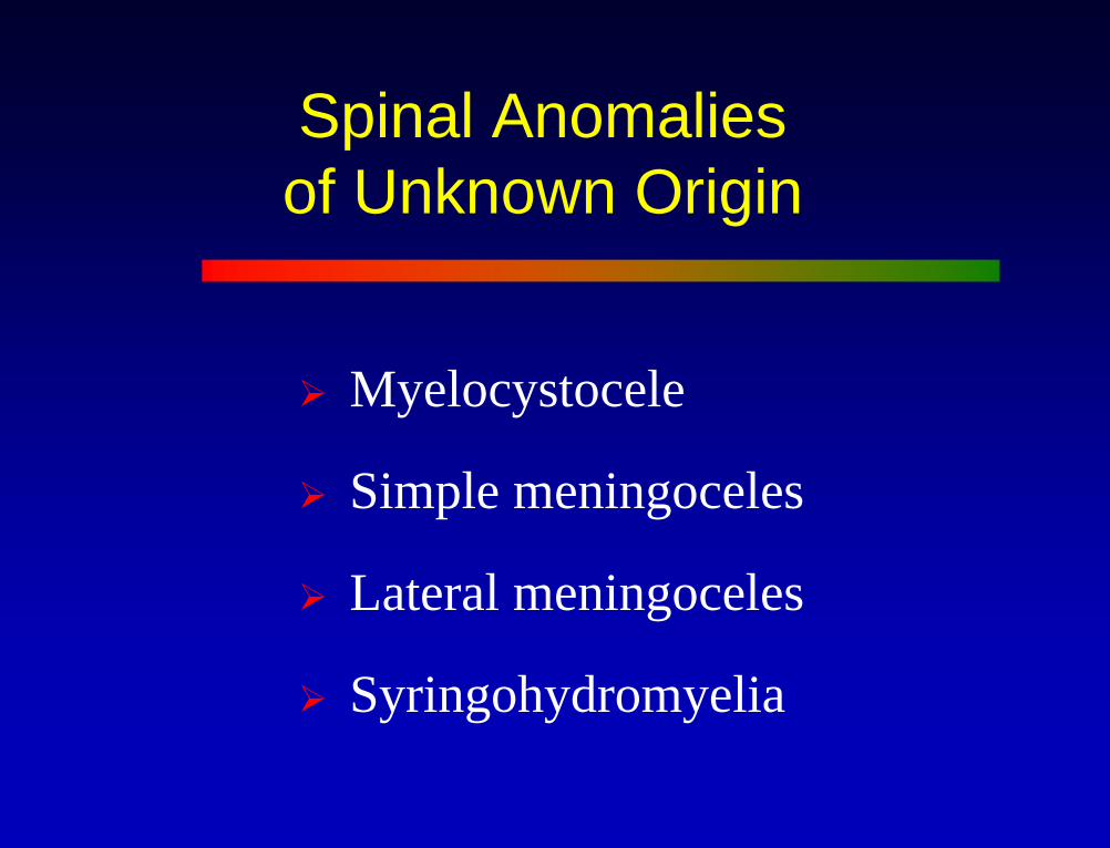
Spinal Anomalies
of Unknown Origin
Myelocystocele
Simple meningoceles
Lateral meningoceles
Syringohydromyelia

Myelocystocele

History: 67 y/o male with NF1
408

Dx: Meningocele
{Page 2}

Postoperative
Myelocystocele

Syringohydromyelia
Etiology
Chiari I
Post-Traumatic
Neoplastic
Idiopathic
42%
15%
15%
28%

Chiari I with Syringohydromyelia

Dx: Chiari I malformation
with syringohydromyelia
339
History: 22 y/o male with
brain stem dysfunction

History: 45 y.o. male
with a progressive
myelopathy
101
C1
C6
C5

Myelopathy {Page 2}
T5
T9
T6

Spinal Cord Cysts
Fluid isointense to CSF
Smooth, well-defined internal margins
Thinned adjacent parenchyma
Cord atrophy
No contrast enhancement
Presence of Chiari malformation
Evidence for Benign Syrinx


![[PPT]Pediatric Airway Managementssu.ac.ir/.../rah_hawaei/pediatric_airway_management.ppt · Web viewCauses of Difficult Airway Congenital Anomalies Tumors Infection Musculoskeletal](https://static.fdocuments.net/doc/165x107/5b95a15d09d3f2214e8d079d/pptpediatric-airway-web-viewcauses-of-difficult-airway-congenital-anomalies.jpg)