Pediatric Lower Extremity Alignment Issues in Primary … · Pediatric Lower Extremity Alignment...
91
Pediatric Lower Extremity Alignment Issues in Primary Care Tom Bush DNP, FNP-BC, FAANP Clinical Associate Professor University of North Carolina at Chapel Hill Schools of Nursing and Medicine 1
Transcript of Pediatric Lower Extremity Alignment Issues in Primary … · Pediatric Lower Extremity Alignment...
Pediatric Lower Extremity Alignment Issues in Primary CareTom Bush DNP, FNP-BC, FAANP
Clinical Associate Professor
University of North Carolina at Chapel Hill
Schools of Nursing and Medicine
1

UNC School of Nursing
Objectives
• Identify physiologic variation in pediatric lower extremity alignment
• Explain developmental changes that influence lower extremity alignment
• Demonstrate techniques for evaluation of lower extremity alignment
• Recognize pathologic lower extremity alignment
• I have no conflicts to disclose
2

UNC School of Nursing
Alignment issues- Etiology
• Extrinsic causes • Infants are otherwise healthy• Associated with fetal crowding
• Oligohydramnios• Breech position• Multiple fetuses• Very large fetus • Compression of uterus
• Small maternal pelvis• Size of maternal organs
• Uterine abnormality• Bicornuate• Septae
3

UNC School of Nursing
Alignment Issues- Etiology
• Intrinsic causes• Fetal abnormalities
• Renal disease• Decreased production of amniotic fluid
• Central nervous system disorders with associated decreased fetal movement
• Positional deformities may be less common in preterm infants
• Assessment must detect abnormality that may indicate intrinsic cause
4

UNC School of Nursing
Genu Varus & Valgus
Rosenfeld, 2016 5

UNC School of Nursing
Overview- Age & Gender
• Some deformities are physiologic for age• Genu varum (bowlegs) normal at birth and progresses to genu
valgus (knock knee) by age 3
• Proximal thigh pain and limp• Ages 4-8 most likely Legg-calve-Perthes.
• Adolescent more likely slipped capital femoral epiphysis (SCFE) or overuse syndrome.
• Hip dysplasia & scoliosis more often in females
• Perthes and SCFE more common in males
6
UNC School of Nursing
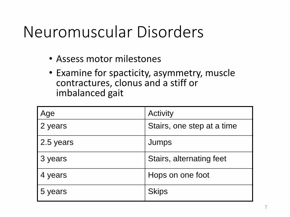
Neuromuscular Disorders
7
• Assess motor milestones
• Examine for spacticity, asymmetry, muscle contractures, clonus and a stiff or imbalanced gait
Age Activity
2 years Stairs, one step at a time
2.5 years Jumps
3 years Stairs, alternating feet
4 years Hops on one foot
5 years Skips

UNC School of Nursing
Intoeing and out toeing
• Foot typically turns out 10-20º by age 2
• Intoeing may be due to:• Foot deformity (metatarsus adductus)
• Inward rotation of femur (femoral anteversion)
• Inward rotation of the (internal tibial torsion)
• Out toeing is less common concern• Femoral retroversion
• External rotation hip contracture
• May be combination of rotation at 2 or more sites
8Rosenfeld, 2016
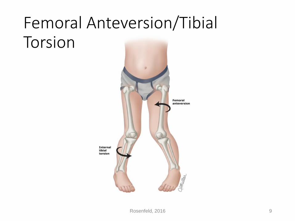
UNC School of NursingFemoral Anteversion/Tibial
Torsion
Rosenfeld, 2016 9

UNC School of Nursing
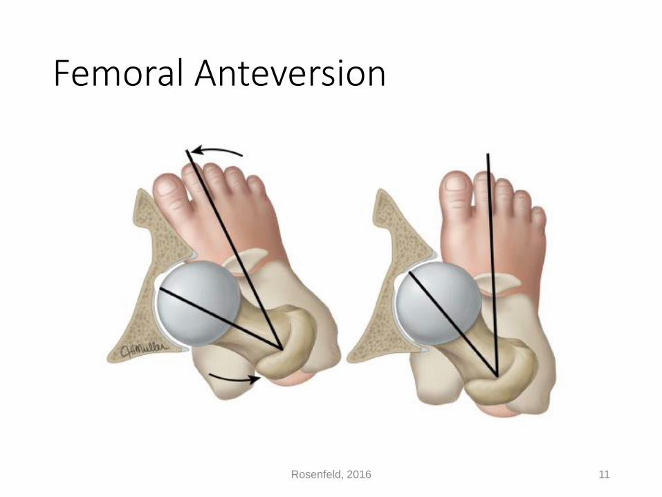
Femoral Anteversion
• Greatest at birth- up to 40º.
• Gradually declines to 10-15º by 8 years• Some are slower to regress
• Usually needs no treatment
• Indirectly measure by assessing hip motion• Hip in extension, child prone, knee flexed.
• Internally rotate legs simultaneously and compare
• Externally rotate while stabilizing pelvis
• Internal rotation > external rotation = anteversion
• External rotation > internal rotation = retroversion
10
UNC School of Nursing
Femoral Anteversion
11Rosenfeld, 2016

UNC School of Nursing
Tibial Torsion
• Normal variant • Intrauterine position
• Most common cause of in-toeing in toddlers
• May be associated • Metatarsus adductus
• Genu varum
• Bilateral in 2/3 of cases• Left side more frequently affected in unilateral cases
12

UNC School of Nursing
Tibial Torsion
• Measure thigh-foot angle• Neutral or internal thigh-foot angle indicates internal tibial torsion
• Most will resolve by 2-4 years of age
Rosenfeld, 2016 13
UNC School of Nursing
14Rosenfeld, 2016
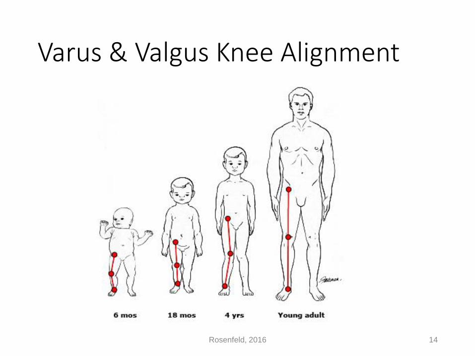
Varus & Valgus Knee Alignment

UNC School of Nursing
Normal Physiologic Alignment
• Varus alignment at Birth
• Varus may increase at onset of gait• May relate to early ambulation
• Neutral alignment at 18-24 months
• Progress to valgus after 24 mohths
• Maximum valgus at age 3-4 years
• Near neutral alignment at age 7
• Most variations are physiologic and can be monitored in primary care
15

UNC School of Nursing
Physiologic Varus• Birth to 2-3 years
• Bilateral • Relatively symmetric
• Normal stature• Document height
• No lateral thrust• Increased risk of
progression
• No pain or limping
• No functional limitations
16Rosenfeld, 2016

UNC School of Nursing
Approach to Alignment Issues
• Exam most important
• Document in history• Parental concern and perception• Birth history• Motor milestones• Onset of alignment issue• Progressive or static• Associated complaints• Family history• Diet and vitamin intake• History of infection, trauma, neoplasm
Armstrong & Hubbard, 2016 17
UNC School of Nursing
Pathologic Varus
• Severe bowing• Greater than 6cm between femoral condyles with
patella forward and malleoli together
• Progressive bowing
• Bowing persists beyond 3 years
• Unilateral or asymmetric bowing
• Lateral thrust on ambulation
• Short stature
• History of metabolic disease, fracture, infection or tumor
18

UNC School of Nursing
Physical Exam- Varus
• Height• <10th-25th%ile
• Look closely
• Family history
• <3rd%ile• Rickets
• Dysplasia
• Weight• Obesity associated with
Blount’s disease
19

UNC School of Nursing
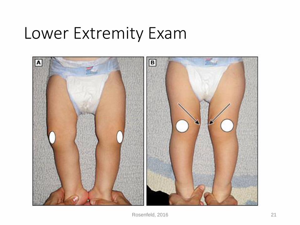
Physical Exam- Varus
• Rotate hips so patella face forward
• Consider distance between knees• Ankles together • Varus with >6 cm considered abnormal at any age
• Symmetry and leg length
• Localize site of angulation
• Assess ROM and palpate hips, knees and ankles
• Observe gait• Note foot/patella progression angle• Look for lateral thrust
20
UNC School of Nursing
Lower Extremity Exam
Rosenfeld, 2016 21
UNC School of Nursing
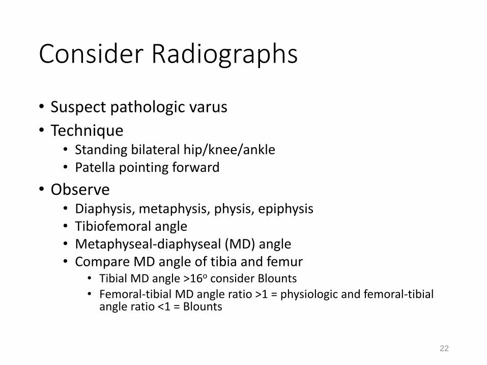
Consider Radiographs
• Suspect pathologic varus
• Technique• Standing bilateral hip/knee/ankle • Patella pointing forward
• Observe • Diaphysis, metaphysis, physis, epiphysis• Tibiofemoral angle• Metaphyseal-diaphyseal (MD) angle• Compare MD angle of tibia and femur
• Tibial MD angle >16o consider Blounts• Femoral-tibial MD angle ratio >1 = physiologic and femoral-tibial
angle ratio <1 = Blounts
22
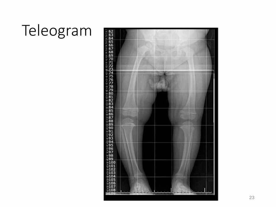
UNC School of Nursing
Teleogram
23

UNC School of NursingGenu Varum
• The metaphyseal-diaphyseal angle
• intersection of transverse plane of the proximal tibialmetaphysis with a line parallel to the long axis of the tibia
• Should be <11-16° (as measured from 90°)
24
0° 15°
UNC School of Nursing
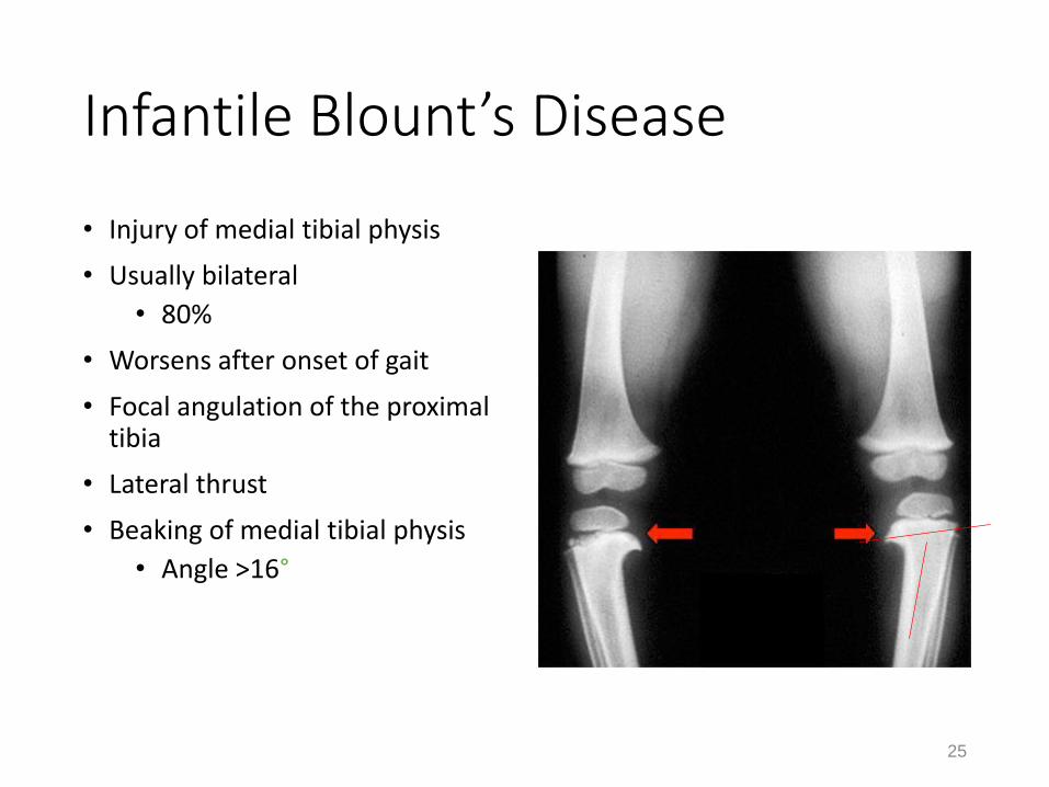
Infantile Blount’s Disease
• Injury of medial tibial physis
• Usually bilateral
• 80%
• Worsens after onset of gait
• Focal angulation of the proximal tibia
• Lateral thrust
• Beaking of medial tibial physis
• Angle >16°
25

UNC School of Nursing
Adolescent Blount’s Disease
• Appears later in childhood
• Unilateral or bilateral
• Risk factors• Obesity
• African American
26

UNC School of Nursing
Genu Varum
• Treatment• Usually requires confident reassurance for parents
• Infantile Blount’s disease• Some consider bracing if younger than 3 years
• Unloads medial tibial physis
• Hemiepiphysiodesis
• Tibial osteotomy
• Adolescent Blount’s disease• Bracing ineffective
• Surgical hemiepiphysiodesis or osteotomy
27

UNC School of Nursing
Genu Varum and Rickets
• Deficient bone mineralization• Reduced calcification and ossification
• Varus if before 18-24 months
• Valgus with later onset
• Nutritional rickets does occur in U.S.• Vitamin and calcium supplements
• Hypophosphatemic rickets• X-linked-dominant inherited condition• Bilateral symmetric bowing of legs• Short stature (<10%ile)
• Treat with calcitriol and phosphate
• May need surgical correction if bowing is severe• May improve with proper medical treatment
28

UNC School of Nursing
29
Blount’s DzRickets
Tibial torsion
Rosenfeld, 2016

UNC School of Nursing
Genu Valgum
• “Knock knees” max at 3-4 years
• Reach adult alignment early adolescence
• Measure height and plot on nomogram.
• Small stature (<25%) should prompt workup
30
UNC School of Nursing
Genu Valgum
• Onset (ie, before or after walking?)
• Progression • physiologic valgus progresses 2-4 and improves between age 4-7
• pathologic valgus worsens after age four years
• Associated complaints
• History of infection, trauma, or fracture
• History of joint swelling/warmth
• Family history
31

UNC School of Nursing
Pathologic Genu Valgum
32

UNC School of Nursing
Physiologic Genu Valgum
• Treatment• Observation and parent
reassurance for otherwise normal 3-4 year old.
• May follow every 4-6 months till age 10
• Braces, shoe modification are not indicated or helpful
• Surgical correction if symptomatic with significant valgus
Bush Stock Photo 33
UNC School of Nursing
34Bush Stock Photo

UNC School of Nursing
Metatarsus Adductus
• Most common congenital foot deformity• May be as many as 13% full term infants
• Often resolves spontaneously but may persist
• Striking lateral convexity of forefoot.
• Heel bisector line crosses forefoot lateral to second web space
• Deep medial crease
35

UNC School of Nursing
36Rosenfeld, 2016
UNC School of Nursing
Metatarsus Adductus
• Assess flexibility of forefoot• Cast if rigid beyond 3 months of age• May observe if flexible
• Photocopy foot to follow progression or resolution
• Consider serial casting persists beyond 3-6 months of age
• High degree of success if started at age 6-9 months• May consider stretching if remains supple
• Important to rule out clubfoot• Hind foot is in valgus or neutral, never varus• Ankle dorsiflexion is normal
37

UNC School of Nursing
Club foot
• Three classifications• Positional
• Intrauterine crowding• Not true club foot
• Congenital• Most common • Idiopathic, isolated
anomaly
• Syndromic• Connective tissue• Genetic• Neuromuscular
• Spina bifida
Richard Henderson Stock Photo 38
UNC School of Nursing
Club foot
• Risk factors- generally not modifiable• Family history of club foot
• Condition that restricts fetal movement• Crowding in multiple gestation
• Oligohydramnious
• Uterine abnormality
• Fetal neuromuscular disorder
39

UNC School of Nursing
Congenital Clubfoot
• Four components (talipes equinovarus)• Ankle plantar flexion (equinus)
• Ankle adduction (varus)
• High arch (cavus)
• Forefoot adduction
• 2:1 male/female ratio
• Bilateral in 30-60%
Armstrong & Hubbard, 2016 40

UNC School of Nursing
Congenital Clubfoot
• Exam• Assess muscle function and sensation
• Absence of either suggest spinal cord disorder
• Passive motion of the foot and ankle• Not fully correctable with true clubfoot
• Correctable if positional
• Radiographs usually not necessary to confirm diagnosis or begin treatment
• Consider if treatment failure or diagnosis unclear
41

UNC School of Nursing
Congenital Clubfoot
• Treatment• Manipulation and casting upon diagnosis
• Stiffer and more resistant to treatment over time
• Two to four months of serial casting often needed
• Recurrence most common in first 2-3 years
• Surgery required if casting fails• Heel cord lengthening vs reconstruction
• Casting after surgery
• Goal is foot in good position without pain
• Affected foot and calf may be smaller
42

UNC School of Nursing
Positional Calcaneovalgus
Bush Stock Photo 43
UNC School of Nursing
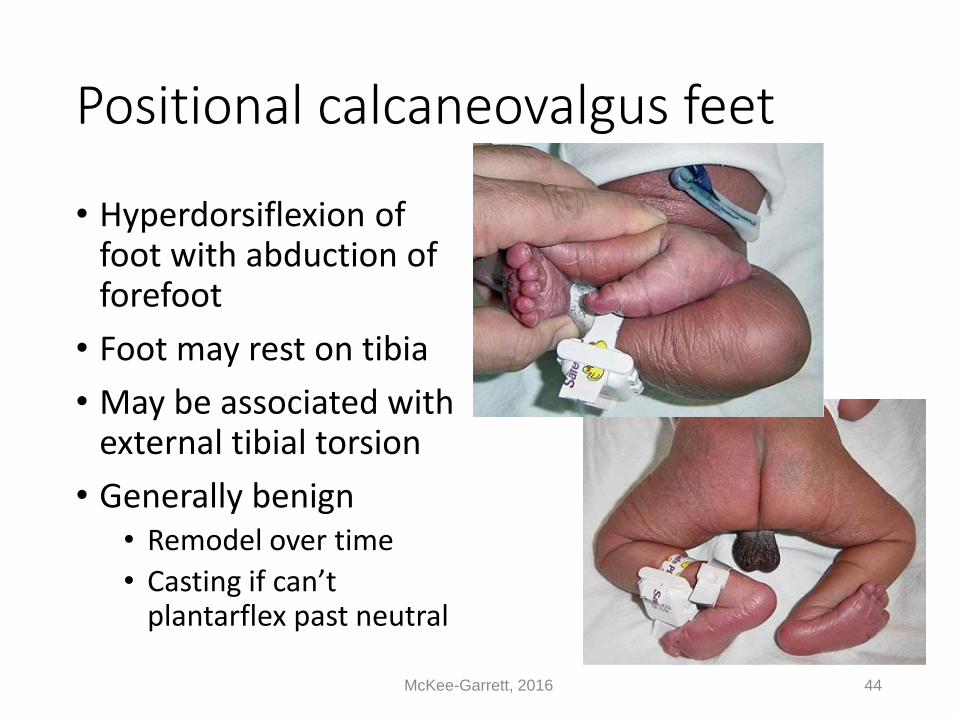
Positional calcaneovalgus feet
• Hyperdorsiflexion of foot with abduction of forefoot
• Foot may rest on tibia
• May be associated with external tibial torsion
• Generally benign• Remodel over time
• Casting if can’t plantarflex past neutral
McKee-Garrett, 2016 44

UNC School of Nursing
Out-toeing
• Persistent external rotation hip contracture from infancy
• Right leg more common, boys and blacks most often affected.• Typically brought in at age 6-12 months.
• Starting to pull up, stand or walk.• Sixty to 70º of external rotation and only 20 to 30º of internal
rotation.
• Resolves spontaneously • May take up to 6 months after begin to walk
• Femoral retroversion
• Increased external rotation at the hip• uncommon
45Rosenfeld, 2016

UNC School of Nursing
Intoeing & Out toeing Treatment
• Shoe modification, braces and exercises do not alter development in femoral or tibial torsion.
• Rotational osteotomy if not improved by age 8-9 years.• More likely with neuromuscular disorder.
• Casting occasionally required for metatarsus adductus if persists beyond 6 months of age.
• May observe if remains supple.
• Some may try putting shoes on wrong foot.
• May consider straight last shoe.
Armstrong & Hubbard, 2016 46
UNC School of Nursing
Flexible Pes Planus “flat feet”
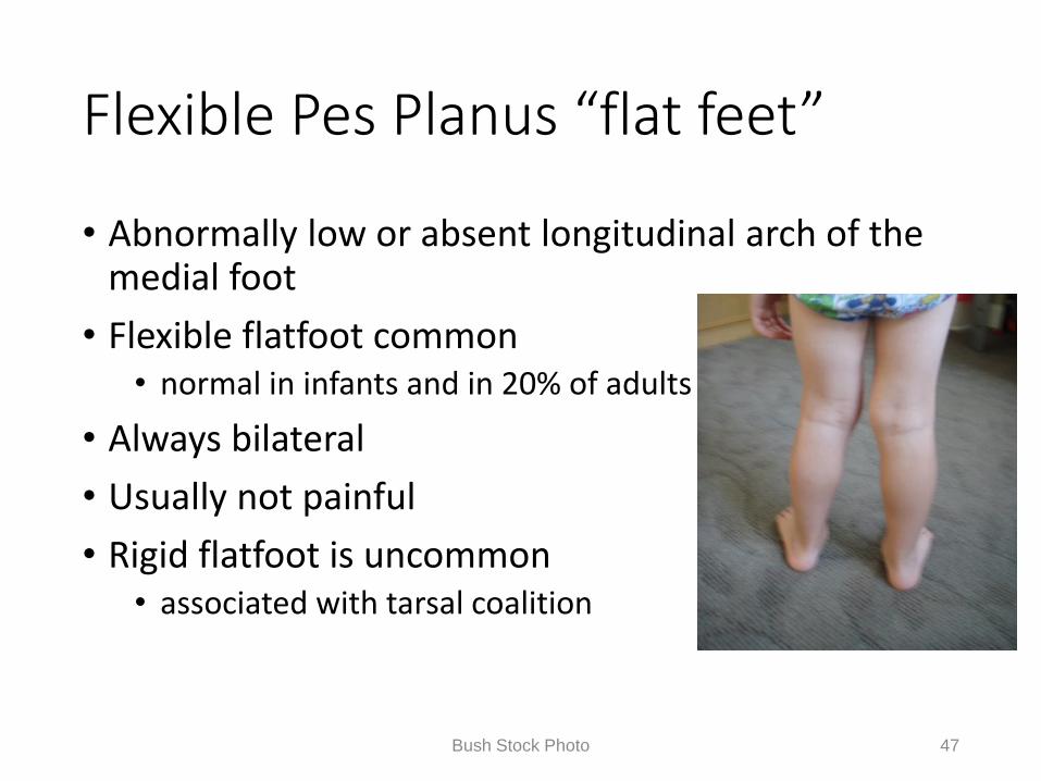
• Abnormally low or absent longitudinal arch of the medial foot
• Flexible flatfoot common • normal in infants and in 20% of adults
• Always bilateral
• Usually not painful
• Rigid flatfoot is uncommon • associated with tarsal coalition
47Bush Stock Photo

UNC School of Nursing
Flexible Pes Planus
• Physical exam• Medial malleolus more prominent
• Heel in valgus
• “Too many toes sign”
Bush Stock Photo 48

UNC School of Nursing
Flexible Pes Planus
• Physical exam• Can recreate arch with “jack toe” test
• Heel moves into varus when standing on toes
Bush Stock Photo 49
UNC School of Nursing
Flexible Pes Planus
• Treatment• Young children usually asymptomatic.
• Reassure parents• Orthotics have not proved helpful in influencing development
of arch
• Arch will likely develop by age 6-8 years.
• Consider gastroc/soleus stretching in older children
• OTC orthotics will help most with symptoms
Armstrong & Hubbard, 2016 50
UNC School of Nursing
Toe walking
• Can be normal variation of gait • Idiopathic• Typically bilateral with onset of gait• Asymptomatic • No functional limitations• May or may not have tight tendoachilles• Can walk heel-toe when reminded• Family history• Unilateral toe walking is red flag
• Cerebral Palsy • Intraspinal abnormality• Leg length inequality
51

UNC School of Nursing
Toe walking
• Idiopathic toe walking is Dx of exclusion• Cerebral Palsy
• Detailed birth history• Prematurity, anoxia/hypoxia, perinatal infection
• May have delayed developmental milestones
• Muscular dystrophy• Family history• Proximal muscle weakness• Begin with heel-toe gait and toe walk at later age
• Intraspinal abnormality• Cavus feet, unilateral or asymmetric involvement
52

UNC School of Nursing
Toe walking
• Treatment• Observe in toddler who is just learning to walk
• May resolve in 3-6 months
• Tendoachilles stretching and ankle ROM• Young child with mild contractures
• Serial casting if persists beyond 3-5 years• Assuming persistent equinus contracture
• Short leg casts changed at 2-3 week intervals
• PT may prevent recurrence
• Tendoachilles lengthening
53

UNC School of Nursing
Shoes for Children
• Properly fit shoes• Do not influence foot development • Do not influence how soon child will walk.
• Shoes should be • Flexible• Flat• Foot shaped• Fitted generously• Friction like skin
• Shoe inserts • Do not alter intoeing, out toeing or flat feet • May treat symptoms by redistributing load
54Armstrong & Hubbard, 2016

UNC School of Nursing
Legg-Calve-Perthes
• Idiopathic osteonecrosis of the femoral head• Bone dies and looses structural integrity.
• Collapse, deformity and arthritis
• Onset usually between ages 4 and 8 years
• Unilateral in 90% of cases
• Four times more common in males
• Uncommon in blacks
55

UNC School of Nursing
Legg-Calve-Perthes
• Complain of hip or thigh pain• Often complain of knee pain
• May present with painless limp
• Limp usually at the end of the day and following vigorous activity.
• Reduced hip range of motion• Abduction limited to 20-30°
• Should be 60-70°
56
UNC School of Nursing
Legg-Calve-Perthes
• AP and frog leg lateral radiographs.
• Increased density of femoral head is early sign• Plain radiographs may be normal
• MRI may show osteonecrosis
57

UNC School of Nursing
Legg-Calve-Perthes
58

UNC School of Nursing
Legg-Calve-Perthes
• Treatment• Observation for children under 6 years who maintain 40-
45° of abduction.
• Observe older children with no involvement of lateral portion of femoral head.
• Abduction brace to maintain femoral head in acetabulum.
• Less commonly used
• Osteotomy for older children and those with reduced abduction.
Armstrong & Hubbard, 2016 59
UNC School of Nursing
Slipped Capital Femoral Epiphysis
60
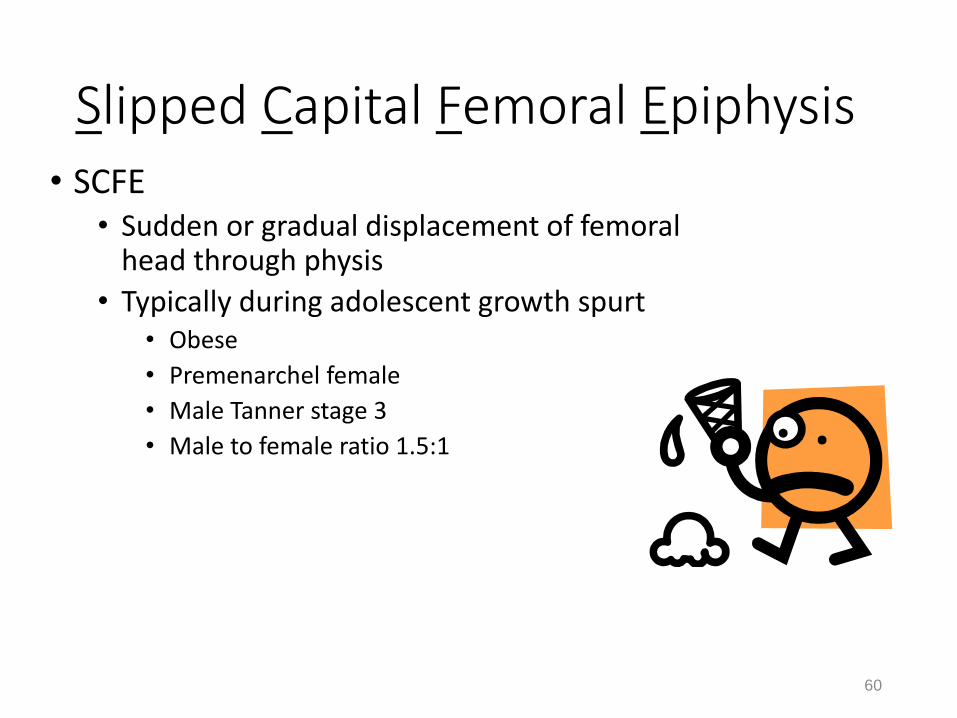
• SCFE• Sudden or gradual displacement of femoral
head through physis
• Typically during adolescent growth spurt• Obese
• Premenarchel female
• Male Tanner stage 3
• Male to female ratio 1.5:1

UNC School of Nursing
Slipped Capital Femoral Epiphysis
• Predisposing factors• Obesity
• 60% > 90th %ile weight for age & gender
• Male gender
• Sports
• Femoral retroversion
• Hypothyroidism and growth hormone deficiency
61

UNC School of Nursing
Slipped Capital Femoral Epiphysis
• Mean age at presentation• 12 years for girls (range:10-14 years)
• 13 years for boys (range: 11-16 years)
• Onset before or after typical range is associated with endocrinopathy.
• Contralateral hip slips in 30 to 60%• 20-40% at presentation
• Not always affected simultaneously • up to 100 percent of patients with underlying endocrine disorders
• May be acute or chronic
• Early detection and treatment imperative
Armstrong & Hubbard, 2016 62

UNC School of Nursing
Slipped Capital Femoral Epiphysis
• Symptoms• Pain worse with activity
• Localized to anterior thigh or knee
• Altered gait
• May be unable to bear weight
• Exam• Loss of hip internal rotation
• Further reduction of internal rotation with hip flexion
• Loss of abduction and extension
• Affected extremity usually shorter by 1-3 cm.
63
UNC School of Nursing
Slipped Capital Femoral Epiphysis
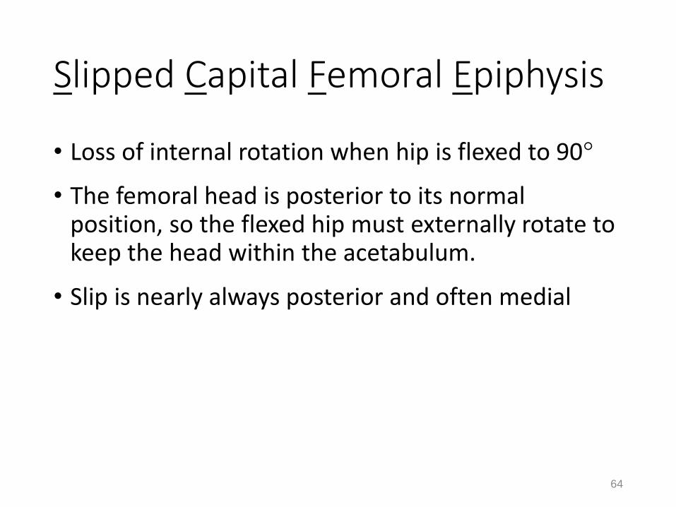
• Loss of internal rotation when hip is flexed to 90°
• The femoral head is posterior to its normal position, so the flexed hip must externally rotate to keep the head within the acetabulum.
• Slip is nearly always posterior and often medial
64

UNC School of Nursing
Slipped Capital Femoral Epiphysis
65

UNC School of Nursing
Slipped Capital Femoral Epiphysis
66

UNC School of Nursing
Slipped Capital Femoral Epiphysis
• Diagnostic tests• AP and frog-leg lateral radiographs of pelvis
• AP view may appear normal
• Severity important in treatment and prognosis
• Severity is estimated by the percentage of femoral neck left exposed:
• Mild- less than 25%
• Moderate- 25-50% is moderate
• Severe- more than 50%
67

UNC School of Nursing
Slipped Capital Femoral Epiphysis
• Treatment • NWB, refer to pediatric orthopedist
• In situ stabilization• Pin in current position to prevent progression.
• If unstable may require urgent ORIF
• Severe deformity may require realignment osteotomy.• Chronic painful limp despite treatment
68
UNC School of Nursing
Evaluation of Limping Child
• Challenging to establish correct diagnosis.
• Possibilities are extensive
• May involve numerous anatomic sites• Spine to foot
• History is often vague.
• Complaints are often nonfocal or referred
• Exam may be unremarkable
69

UNC School of Nursing
Evaluation of Limping Child
• Age of onset• Younger than age 3 years
• DDH
• Leg length inequality
• Ages 4-10 years• Transient synovitis
• Legg-Calve-Perthes
• Adolescence• SCFE
• Overuse syndromes
70

UNC School of Nursing
Evaluation of Limping Child
• Acute symptoms• Trauma
• Fracture apparent on radiograph
• Child abuse suspected if story does not fit injury
• Overuse syndromes
• Infection• Transient synovitis
• Osteomyelitis
• Diskitis
71

UNC School of Nursing
Evaluation of Limping Child
• Chronic symptoms• Neuromuscular disorder
• Legg-Calve-Perthes
• SCFE
• DDH
• Rheumatic disease
• Psychogenic
72
UNC School of Nursing
Evaluation of Limping Child
• Pain in afternoon and following activity?• DDH
• SCFE
• Legg-Calve-Perthes
• Morning pain?• JRA
• Transient synovitis
• Night symptoms suggest leukemia or other neoplasm
73
UNC School of Nursing
Evaluation of Limping Child
• Systemic symptoms?• Malaise
• Fever
• Fatigue
• Pain
74

UNC School of Nursing
Evaluation of Limping Child
• Physical exam• Growth and vital signs (especially temp)
• Palpate spine and extremities
• Inspect skin
• Assess muscle bulk, strength and tone
• Observe gait.
• Asses joint range of motion
• Neuro exam
75

UNC School of Nursing
Evaluation of Limping Child
• Diagnostic studies• CBC
• ESR
• CRP
• Blood cultures/arthrocentesis
• Radiographs above and below affected joint
• Bone scan
• MRI or CT scan
76

UNC School of Nursing
References
• Armstrong & Hubbard. (Eds): Essentials of Musculoskeletal Care, 5th edition. Rosemont, IL, American Academy of Orthopaedic Surgeons, 2016.
• McKee-Garrett, T. M. Lower extremity positional defromations. In: UpToDate, Weisman, L. E., Phillips, W., Kim, M. S. (Eds), UpToDate, Waltham, MA, 2016
• Rosenfeld, S. B. Approach to child with bow legs. In: UpToDate, Phillips, W., Drutz, J., Trochia, M.M.(Eds), UpToDate, Waltham, MA, 2016
• Rosenfeld, S. B. Approach to with in-toeing. In: UpToDate, Phillips, W., Drutz, J., Trochia, M.M.(Eds), UpToDate, Waltham, MA, 2016
• Rosenfeld, S. B. Approach to with knock-knees. In: UpToDate, Phillips, W., Drutz, J., Trochia, M.M.(Eds), UpToDate, Waltham, MA, 2016
77
UNC School of Nursing
Resources
• Saywer, J.R. & Kapoor, M. (2009). The limping child: A systematic approach to diagnosis. American Family Physician, 79(3).
• Henderson, RC. Tibia vara: a complication of adolescent obesity. J Pediatrics 1992; 121:482.
• Loder, RT, Schaffer, JJ, Bardenstein, MB. Late-onset tibia vara. J Pediatr Orthop 1991; 11:162.
• Bradway, JK, Klassen, RA, Peterson, HA. Blount disease: a review of the English literature. J Pediatr Orthop 1987; 7:472.
78

UNC School of Nursing
Case Study #1
• REASON FOR VISIT: Evaluate toe walking in 2 year old child.
• HISTORY OF PRESENT ILLNESS: Parents report child toe walking since she began to walk at age 1 year. No trauma history or complaints of pain. No functional limitations. Parents report pain score today of 0/10.
• PAST MEDICAL HISTORY: A 32-week gestation with spontaneous vaginal delivery. Birth weight 3 pounds 13 ounces. She spent approximately 2 weeks in the NICU primarily for weight gain. She has met developmental milestones on target including walking independently at 12 months. Hospitalization for RSV at age 2 months.
• MEDICATIONS: None.
• SOCIAL HISTORY: Lives with both parents and 2 siblings in Rockingham, North Carolina.
79

UNC School of Nursing
Case Study #1
• REVIEW OF SYSTMS: No recent fever. No vomiting or diarrhea. No seizure history. Child is left hand dominate.
• FAMILY HISTORY: No reports of familial toe walking. No familial history of lower extremity malalignment.
• PHYSICAL EXAMINATION: Well-developed 2 year-old white female in no acute distress. The patient is able walk heel to toe when reminded in clinic. Toe walking is primarily on the right. Ankle flexion to neutral on the right with knee flexed. Unable to bring the ankle to neutral with knee extended. Left ankle flexion past neutral with knee extended. No unusual muscle tone or spasticity of the lower extremities. Examination of her upper extremities reveals symmetric muscle tone. She will perform simple tasks with the right hand but is less fluid than with the left hand. She is appropriate size and weight for age. No skin changes or dimpling at the base of the spine. Child appears developmentally appropriate in clinic today.
80
UNC School of Nursing
Case Study #1
• ASSESSMENT: A 2 year-old female with mild hemiplegic CP and tight heel cord on the right
• PLAN: Greater than 20 minutes of face-to-face consultation with the patient and her parents regarding this diagnosis. Parents are advised that she has a very mild form of CP and most likely will not cause her any significant trouble, but is likely to limit her physical abilities in terms of athletics. She appears to be cognitively on track. I explained to them that this is not progressive disorder and that no intervention is necessary at this time other than observation. I did recommend stretching and parent can demonstrate proper technique in the clinic today. She will follow up with Dr. Richard Henderson for further evaluation of tight heel cords and consideration of bracing or serial casting if she remains symptomatic.
• UPDATE 9/25/07- Casted for persistent symptoms. RTC 10/16.
81

UNC School of Nursing
Case Study #2
• REASON FOR VISIT: Three year old child with reports of limping and right lower extremity pain.
• HISTORY OF PRESENT ILLNESS: Parents report child limps after 5-10 minutes of ambulation. They noticed this shortly after she began to walk at age 16 months. Occasional night pain. No early am pain or stiffness. No trauma history. No functional limitations.
• PAST MEDICAL HISTORY: Induced delivery at 40 weeks with subsequent C-section. Details of the delivery are unclear. No reports of difficulty in the early newborn period and child discharged home on DOL #3. She has met developmental milestones on target. She receives regular well child checks and immunizations are up to date.
82

UNC School of Nursing
Case Study #2
• SOCIAL HISTORY: Child was born in Mexico. She now lives in Franklinton, NC with both parents and an older sibling. Parents do not speak English well and visit today facilitated by UNC hospital interpreter.
• ROS: No recent respiratory or GI symptoms. No other joint or extremity complaints. No history of rash or fevers
• PHYSICAL EXAM: Well developed 3 year old Latina female in NAD. She appears developmentally appropriate in the clinic today. She walks with an antalgic gait. Hip aBduction 40 on the right compared to 70 on the left. Hip motion elicits pain and mechanical symptoms. Skin is intact. No lower extremity LAD. Bilateral muscle tone is robust and symmetric. No spasticity or ankle clonus.
83

UNC School of Nursing
Case Study #2• Radiographs today show dysplastic right
hip with posterior dislocation
• Update 9/14/07- doing well 3 wks status post open reduction and femoral osteotomy
84

UNC School of Nursing
Case Study #3
• REASON FOR VISIT: Evaluate bow legs
• HISTORY OF PRESENT ILLNESS: Parents report intoeing on the right at about one year of age. No trauma or functional limitations. No reports of pain or limping. Mother is concerned that she is “late bloomer”
• PAST MEDICAL HISTORY: Full term gestation with spontaneous vaginal delivery. Discharged home on DOL #3. Has met developmental milestones on target including walking at 15 months. Reported diet high in calcium. Developmental evaluation reported language and fine motor skills WNL.
• FAMILY HISTORY: No familial lower extremity malalignment or short stature.
85

UNC School of Nursing
Case Study #3
• REVIEW OF SYSTEMS: No recent fever or respiratory illness. No fracture history. No joint complaints.
• SOCIAL HISTORY: Lives with both parents in western NC.
• PHYSCIAL EXAM: Two plus 4 year old female appears developmentally appropriate in clinic today. Walks heel to toe without difficulty. Right thigh foot angle 20 degrees, ten degrees on the left. Neutral alignment at the knee. Bilateral lower extremity muscle mass robust and symmetric. No increased tone or spasticity. Bilateral ankle flexion past neutral with knee extended.
86

UNC School of Nursing
Case Study #3
• ASSESSMENT: Internal tibial torsion
• PLAN: Parents reassured that alignment should improve as child continues to mature. They are advised on expected course over the next 6 to 12 months and are aware of the wide variation of normal alignment. They are invited to return to the clinic as needed.
87

UNC School of Nursing
Case Study #4
• REASON FOR VISIT: 7 year old female with left knee pain and limp x2 months.
• HISTORY OF PRESNET ILLNESS: Parents note limp mostly when child is running. No trauma history. Occasional reports of knee pain.
• PAST MEDICAL HISTORY: Spontaneous vaginal delivery at 38 weeks. NICU stay for 2 days due to respiratory effort. She has met developmental milestones on target including walking at 11 months.
• REVIEW OF SYSTEMS: No recent fever or sweats. No nausea, vomiting or diarrhea. No fracture history or coagulopathy.
88
UNC School of Nursing
Case Study #4
• PHYSCIAL EXAM: Well developed 7 y.o. female. Walks without a limp in the clinic today. Indicates left knee as site of pain. Knee motion is full and without complaints. Left hip flexion to 110 and aBduction to 45 degrees with associated knee pain. Left lower extremity is neurovascularly intact. Skin intact throughout. No local tenderness to the hip, knee and ankle. Slight valgus alignment of the knees.
• AP and lateral views of bilateral hips show…..
89

UNC School of Nursing
Case Study #4
90
..collapse of the lateral surface of the femoral epiphysis. Femoral
head is well covered by the acetabulum. No hip dysplasia.
ASSESSMENT: Legg-Calve Perthes Dz

UNC School of Nursing
Case Study #4
• PLAN: Fitted with hip aBduction brace and advised on night time use. Advised on activity modification to avoid high impact activity and ibuprofen or acetaminophen for symptomatic care. She will follow up with Dr. Ed Campion in 4 months with repeat AP pelvis and frog leg lateral films at that time. They are advised to monitor for new symptoms and encouraged to return to clinic sooner if new signs or symptoms develop.
91