Pediatric Infectious Disease Concerns in Primary Care · Pediatric Infectious Disease Concerns in...
54
Pediatric Infectious Disease Concerns in Primary Care Sean P. Elliott, MD Professor of Pediatrics Associate Chair of Education, Department of Pediatrics University of Arizona College of Medicine Tucson, AZ
Transcript of Pediatric Infectious Disease Concerns in Primary Care · Pediatric Infectious Disease Concerns in...

Pediatric Infectious Disease Concerns
in Primary Care
Sean P. Elliott, MDProfessor of Pediatrics
Associate Chair of Education,Department of Pediatrics
University of Arizona Collegeof Medicine
Tucson, AZ

Objectives
• Review challenging pediatric infectious diseases which may be seen in a primary care setting.
• Identify initial steps helpful in initiating an infectious diseases work-up.
• Recognize warning signs that should alert a primary care provider to a possibly fatal, pediatric infectious disease.

Conflict of Interest Disclosure
I have no relevant financial relationships with the manufacturer(s) of any
commercial product(s) and/or provider of commercial services discussed
in this presentation.
I do not intend to discuss an unapproved or investigative use of a commercial product or device in my presentation.

Cervical Adenopathy

Cervical Adenopathy
• Acute (55%):– Reactive– Bacterial– Viral
• Chronic (45%):– Neoplastic disease– Non-tuberculous Mycobacteria– Cat-scratch lymphadenitis– EBV– HIV

Cervical Adenopathy - Bacterial
• Presentation:– Fever– Acute swelling with pain– Erythema & induration
• Diagnosis:– CBC w/ diff– Blood Culture
• Management:– Cephalexin vs Clindamycin
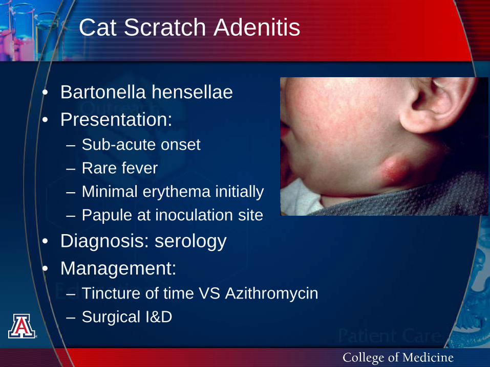
Cat Scratch Adenitis
• Bartonella hensellae• Presentation:
– Sub-acute onset– Rare fever– Minimal erythema initially– Papule at inoculation site
• Diagnosis: serology• Management:
– Tincture of time VS Azithromycin– Surgical I&D

Cervical Adenopathy - Mycobacterial
• Non-tuberculous Mycobacteria (MAI) VS M. tuberculosis
• Presentation:– Cervical vs supra-clavicular nodes– Afebrile– Minimally tender– Minimal erythema– PICA
• Diagnosis: History + PPD

Cervical Adenopathy - Mycobacterial
• Treatment – NTM:– Azithromycin vs Clarithromycin– Ciprofloxacin OR– Rifampin/Rifabutin
• Surgery:– Excisional Biopsy– Fistula formation– Location and surgical risk– Spontaneous resolution?

Prolonged feverRash with abxProminent LADPharyngitis w/ exudateFATIGUE! (> 10 y.o.)Atypical < 10 y.o.
Epstein-Barr Virus

Epstein-Barr diagnosis

HIV Perinatal Exposure Protocol
• HIV DNA PCR at 2 & 4 weeks
• AZT 2 mg/kg/dose Q6 until 5-6 weeks old
• F/U Peds ID at 4 weeks
• Presumptive negative at 4 weeks
• Definite negative at 4 months
Pediatrics 2009; 123 (1), 175

Perinatal HIV – today…
• 66% reduction in risk of perinatal transmission
• 5-10% transmission• On-going issues:
– Breastfeeding– C-section benefit?– Identifying HIV-infected mothers

New trends in HIV/AIDS
• Few perinatal HIV cases• International refugees/adoptees• Emerging teen-young adult HIV:
– Immortality beliefs– HIV/AIDS ≠ Death Sentence – High risk exposures:
• Intercourse• IVDU• Substance abuse and High risk
associates

Adolescent presentation• Generalized lymphadenopathy• FUO• Weight loss (unintentional)• Organomegaly• Acute antiretroviral syndrome• “heterosexual”• “no risks”

Pediatric HIV/AIDS
• Clinical therapy:– HAART– Opportunistic infections– Nutrition, lipodystrophy
• Socioeconomic:– Orphan– Medication adherence– Depression and psychosis– Social isolation
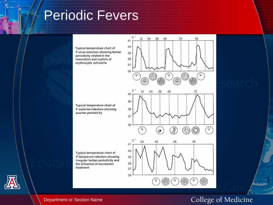
Periodic Fevers
Department or Section Name

Periodic Fevers
• Common• Truly periodic?• 12 febrile illnesses/year• Viruses• AOM• Pharyngitis • UTI• Periodic Fever Syndromes

Periodic Fever Syndromes
• FMF• TRAPS• Cyclic Neutropenia• Hyperimmunoglobulin D Syndrome• PFAPA

FMF • Familial Mediterranean Fever
• Presentation:– Fever X 3 days– Serositis: peritonitis,
pleuritis, arthritis– Exploratory laparotomy
• Diagnosis:– Clinical criteria published– Mutation analysis MEFV
gene• Treatment: Colchicine

TRAPS
• TNF Receptor Associated Periodic Syndrome• Presentation (Q 5 – 6 weeks):
– Fever X 5 – 14 days– 80%: myalgias, rash, conjunctivitis, periorbital
edema• Diagnosis:
– Exclude alternate Dx– ↑ ESR and CRP– Mutation analysis TNFR1 gene
• Treatment: steroids > NSAIDs

Cyclic Neutropenia
• Cyclical retention of Neutrophils• Periodic peripheral neutropenia• ANC = % pmn X total WBC• Presentation: Fevers and mucositis• Diagnosis:
– CBC w/ diff– Timing!!!
• Management:– Heme/Onc referral– GCSF

Hyperimmunoglobulin D Syndrome
• IgD and Mevalonate Kinase Deficiency• Partially predictable events• Presentation (3 – 7 days):
– Fever + rigors– GI symptoms (N/V/D)– Lymphadenopathy– (rash, arthritis, splenomegaly)
• Diagnosis: ↑ IgD and IgA• Treatment: NSAIDs > steroids

PFAPA
• Periodic Fever, Aphthous stomatitis, Pharyngitis, and Adenitis
• Presentation (3 -4 days):– Strikingly predictable fever episodes– Associated cervical LAD, aphthous ulcers– Sore throat vs abdominal pain
• Diagnosis: – Exclude alternate Dx– ESR and CRP, +leukocytosis
• Treatment: Prednisone > T&A

Routine illnesses gone bad…
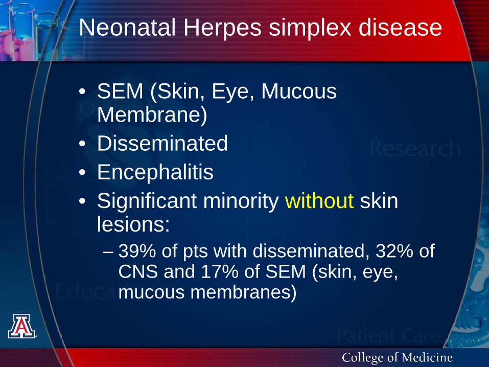
Neonatal Herpes simplex disease
• SEM (Skin, Eye, Mucous Membrane)
• Disseminated• Encephalitis • Significant minority without skin
lesions:– 39% of pts with disseminated, 32% of
CNS and 17% of SEM (skin, eye, mucous membranes)
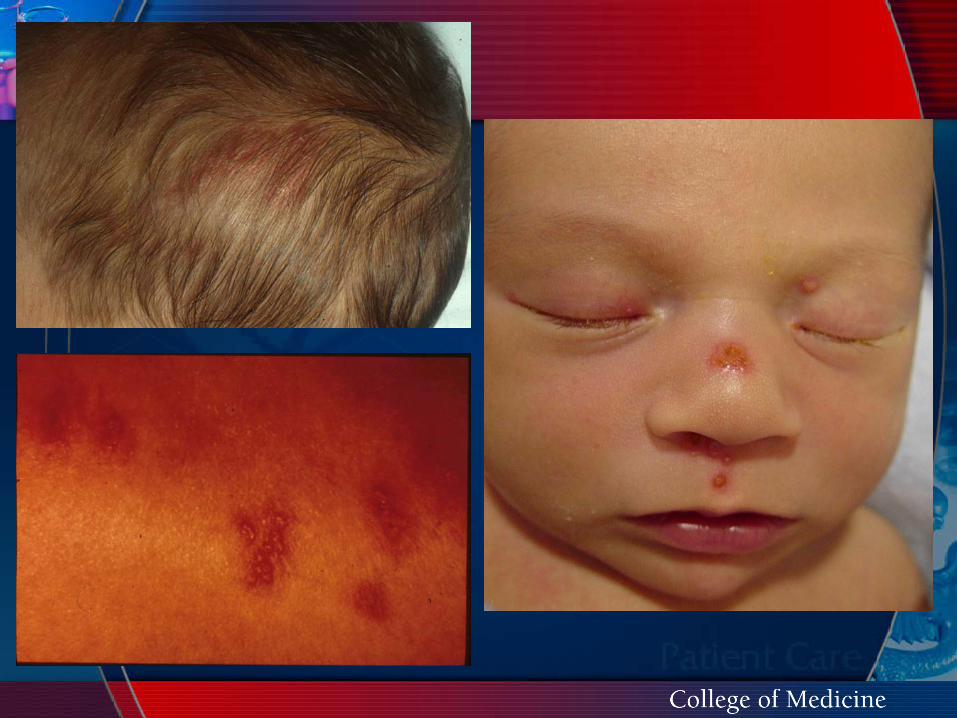
Herpes

Neonatal Herpes simplex disease
• Risk of transmission:– Primary disease = 50%– Reactivated disease = 5%
• Transmission: >70% cervical lesions
• Maternal (paternal) history unreliable!– Kaiser & Washington studies
• Maternal suppressive Acyclovir?

Herpes simplex
• Tests: Viral culture and FA staining of lesions. Throat, CSF, brain, urine cultures, CSF PCR
• Treatment: Acyclovir• Prevention: Deliver
by C-section if active lesions

Neonatal HSV recurrence
• Unclear risk of recurrence• >2 episodes recurrence = cognitive risk• Role of suppressive Acylovir?
– Kimberlin et al.– 74 neonates (45 CNS + 29 SEM)– Acyclovir 300 mg/m2/dose TID X 6
mo.– Improved neurodevelopmental
outcomes
NEJM 2011;365:1284


Croup…

Croup
• Acute laryngotracheobronchitis• Spasmodic croup• Varying degrees of laryngeal obstruction
(subglottic edema)• Age:
– 6 months - 6 years– Incidence = 3/100 children < 6 y.o.
• Significance:– 1.3% hospitalized– Annual cost for hospitalization = $56 million

Croup
• Parainfluenza 1• Peak incidence in fall• Others:
– Parainfluenza 3– RSV– Influenza A & B– Adenoviruses

Croup
• Symptoms:– Nasal stuffiness, sore throat– Fever within 24 hours– Harsh, seal-like barking cough– Inspiratory stridor, retractions, dyspnea
• Degree of obstruction wax and wane• Acute symptoms last 3-4 days• Hypoxemia and CO2 retention may occur
from progressive obstruction

Complications: Pneumonia
Croup-Differential Diagnosis
• Infectious– Epiglottitis– Bacterial tracheitis– Diphtheria– Retropharyngeal
abscess– Peritonsillar
abscess
• Noninfectious– Foreign body– Trauma– Allergic/angio-
neurotic edema– Upper airway
burns– Vascular ring

Croup-Diagnosis
• Clinical diagnosis• Presence of
– Sudden onset– High fever– Increased WBC(left shift - differential)– Muffled/hot-potato voice– Drooling– =BAD

RMSF

Rocky Mountain Spotted Fever
• Rickettsia rickettsii• 16 patients:
– 13 (81%) children (<12 years)– 15 (94%) hospitalized– 2 deaths
• Tick-infested dogs• 4 patients + tick bites; all + contact with
dogs• Rhipicephalus sanguineus
NEJM 2005, 353:587-594

Data and slide courtesy of Dr. J. McQuiston, Rickettsial Branch-CDC
Reported RMSF, 2008-2013*2013 data reporting not complete

Community Risk Factors
Slide courtesy of Dr. J. McQuiston, Rickettsial Branch-CDC

Rocky Mountain Spotted Fever
• Rash:– Characteristic– “drives” the diagnosis– Absent 60%!
• New RMSF criteria:– Fever > 48 hours– No alternative explanation– Exposure to endemic area

Rocky Mountain Spotted Fever
• Diagnosis:– Immunohistochemical stains
(skin/tissue biopsy)– Seroconversion with >4-fold rise– PCR amplification of R. rickettsii DNA
• Treatment:– Doxycycline– Adults: 100 mg po BID– Children < 45 kg: 2.2 mg/kg/dose BID– 10 days

Fever in Infants (1 – 90 days)

Fever in Infants (1 – 90 days)
• 20% ED visits• 8% - 10% SBI• Risk stratification:
– Low-risk: 1.4% SBI– High-risk: 21% SBI
• UTI = most common SBI (80%)• E. coli most common pathogen• Incidence SBI in viral infection = low

Fever in Infants (1 – 90 days)
• Low-risk vs. High-risk:– Clinically ill?– Yale Toxicity Score– Modified Rochester criteria– CBC/diff & UA are key
• Presence of virus:– Upper respiratory viruses year-round– HSV I/II?
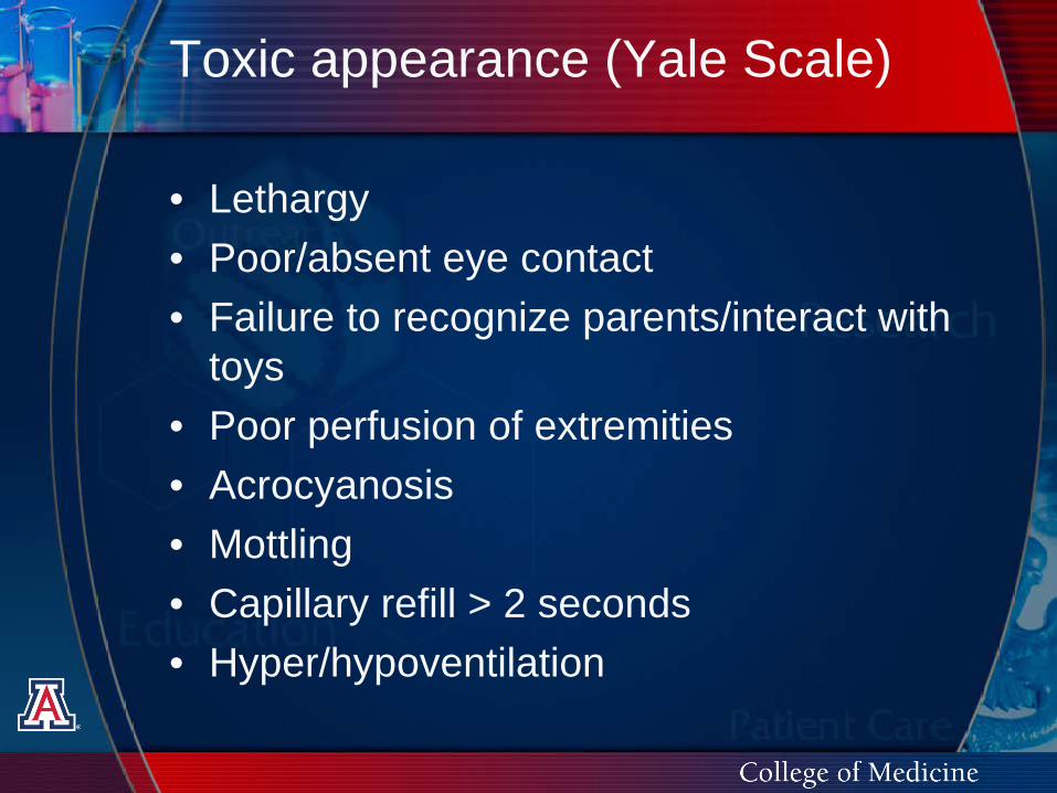
Toxic appearance (Yale Scale)
• Lethargy• Poor/absent eye contact• Failure to recognize parents/interact with
toys• Poor perfusion of extremities• Acrocyanosis• Mottling• Capillary refill > 2 seconds• Hyper/hypoventilation

Decision making tools - WBC
• SBI risk:– < 15K: 21%– 15K: 29%– >20K: 49%
• WBC and/or Band count: sensitivity 11-55%
• PPV WBC > 15K = 6%• Unnecessary Rx: 85-95% cases

Infant sepsis Evaluation
• Procalcitonin assay:– Highly reliable– NPV 96%– Ideal marker of SBI, use in ED setting– Not readily available – Urgent Care clinics
• C-reactive protein:– CRP < 5 mg/dL: 1.9 - 9.7% risk SBI– CRP > 10 mg/dL: 86.5% risk SBI– NPV = 90%– Limitations: Time-specific (< 12 hrs.)
Fever in Infants (1 – 90 days)
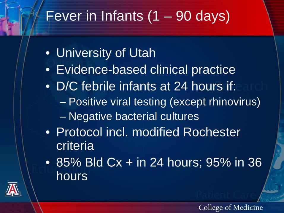
• University of Utah• Evidence-based clinical practice• D/C febrile infants at 24 hours if:
– Positive viral testing (except rhinovirus)– Negative bacterial cultures
• Protocol incl. modified Rochester criteria
• 85% Bld Cx + in 24 hours; 95% in 36 hours

Fever in Infants (1 – 90 days)
• High-risk:– Age < 28 days– WBC <5,000 or >15,000– Absolute band count > 1,500– UA > 10 WBC/hpf– Prematurity (<37 wks) AND underlying
medical condition• Labs: CBC/diff, Bld Cx, U/A, U Cx,
AST/ALT (infants < 42 days), viral studies

Fever in Infants (1 – 90 days)
• Impact:– Reduced hospital stay– Improved cost savings ($3000/admit)– Improved parent satisfaction (98%)– Reduced antibiotic use– Improved use of risk-screening labs– ↑ capture meningitis/bacteremia (91%
to 99%)– No missed SBI (2-year follow-up)

Fever in Infants (1 – 90 days)
• Application to your facility?– ARUP multiplex PCR for respiratory
viruses– Microbiology lab practices– Epidemiologic mix
• Study/apply locally• Stay tuned…

Questions?