Pediatric HEAD Imaging Guidelines 2015 - eviCore · PEDIATRIC HEAD IMAGING GUIDELINES ... o...
49
© 2015 MedSolutions, Inc. Pediatric Head Imaging Guidelines PEDIATRIC HEAD IMAGING GUIDELINES Version 17.0; Effective 02-16-2015 MedSolutions, Inc. Clinical Decision Support Tool for Advanced Diagnostic Imaging Common symptoms and symptom complexes are addressed by this tool. Imaging requests for patients with atypical symptoms or clinical presentations that are not specifically addressed will require physician review. Consultation with the referring physician may provide additional insight. This version incorporates MSI accepted revisions prior to 12/31/14 CPT ® (Current Procedural Terminology) is a registered trademark of the American Medical Association (AMA). CPT ® five digit codes, nomenclature and other data are copyright 2015 American Medical Association. All Rights Reserved. No fee schedules, basic units, relative values or related listings are included in the CPT ® book. AMA does not directly or indirectly practice medicine or dispense medical services. AMA assumes no liability for the data contained herein or not contained herein. MedSolutions, Inc. This tool addresses common symptoms and symptom complexes. Imaging requests for patients with atypical Clinical Decision Support Tool symptoms or clinical presentations that are not specifically addressed will require physician review. Diagnostic Strategies Consultation with the referring physician, specialist and/or patient’s Primary Care Physician (PCP) may provide additional insight.
Transcript of Pediatric HEAD Imaging Guidelines 2015 - eviCore · PEDIATRIC HEAD IMAGING GUIDELINES ... o...
©2015 MedSolutions, Inc. Pediatric Head Imaging Guidelines
PEDIATRIC HEAD IMAGING GUIDELINES Version 17.0; Effective 02-16-2015
MedSolutions, Inc. Clinical Decision Support Tool for Advanced Diagnostic Imaging
Common symptoms and symptom complexes are addressed by this tool. Imaging requests for patients with atypical symptoms or clinical presentations that are not specifically addressed will require physician review. Consultation with the
referring physician may provide additional insight.
This version incorporates MSI accepted revisions prior to 12/31/14
CPT® (Current Procedural Terminology) is a registered trademark of the American Medical Association (AMA). CPT® five digit codes, nomenclature and other data are copyright 2015 American Medical Association. All Rights Reserved. No fee schedules, basic units, relative values or related listings are included in the CPT® book. AMA does not directly or indirectly practice medicine or dispense medical services. AMA assumes no liability for the data contained herein or not contained herein.
MedSolutions, Inc. This tool addresses common symptoms and symptom complexes. Imaging requests for patients with atypicalClinical Decision Support Tool symptoms or clinical presentations that are not specifically addressed will require physician review. Diagnostic Strategies Consultation with the referring physician, specialist and/or patient’s Primary Care Physician (PCP) may provide additional insight.
Version 17.0; Effective 02-16-2015 Pediatric Head RETURN 2 of 49
PEDIATRIC HEAD IMAGING GUIDELINES
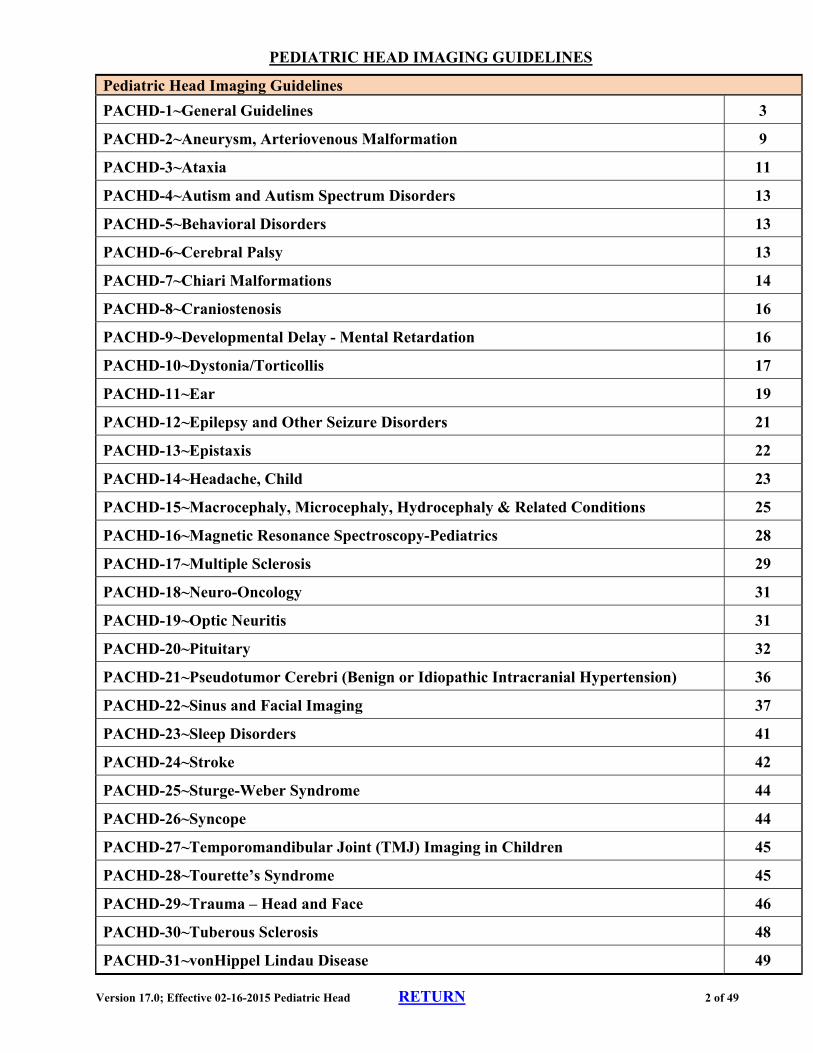
Pediatric Head Imaging Guidelines
PACHD-1~General Guidelines 3
PACHD-2~Aneurysm, Arteriovenous Malformation 9
PACHD-3~Ataxia 11
PACHD-4~Autism and Autism Spectrum Disorders 13
PACHD-5~Behavioral Disorders 13
PACHD-6~Cerebral Palsy 13
PACHD-7~Chiari Malformations 14
PACHD-8~Craniostenosis 16
PACHD-9~Developmental Delay - Mental Retardation 16
PACHD-10~Dystonia/Torticollis 17
PACHD-11~Ear 19
PACHD-12~Epilepsy and Other Seizure Disorders 21
PACHD-13~Epistaxis 22
PACHD-14~Headache, Child 23
PACHD-15~Macrocephaly, Microcephaly, Hydrocephaly & Related Conditions 25
PACHD-16~Magnetic Resonance Spectroscopy-Pediatrics 28
PACHD-17~Multiple Sclerosis 29
PACHD-18~Neuro-Oncology 31
PACHD-19~Optic Neuritis 31
PACHD-20~Pituitary 32
PACHD-21~Pseudotumor Cerebri (Benign or Idiopathic Intracranial Hypertension) 36
PACHD-22~Sinus and Facial Imaging 37
PACHD-23~Sleep Disorders 41
PACHD-24~Stroke 42
PACHD-25~Sturge-Weber Syndrome 44
PACHD-26~Syncope 44
PACHD-27~Temporomandibular Joint (TMJ) Imaging in Children 45
PACHD-28~Tourette’s Syndrome 45
PACHD-29~Trauma – Head and Face 46
PACHD-30~Tuberous Sclerosis 48
PACHD-31~vonHippel Lindau Disease 49
Version 17.0; Effective 02-16-2015 Pediatric Head RETURN 3 of 49
PEDIATRIC HEAD IMAGING GUIDELINES
PACHD-1~GENERAL GUIDELINES
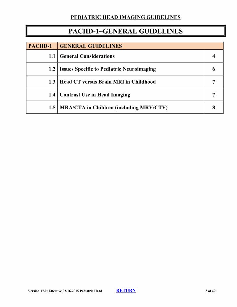
PACHD-1 GENERAL GUIDELINES
1.1 General Considerations 4
1.2 Issues Specific to Pediatric Neuroimaging 6
1.3 Head CT versus Brain MRI in Childhood 7
1.4 Contrast Use in Head Imaging 7
1.5 MRA/CTA in Children (including MRV/CTV) 8
Version 17.0; Effective 02-16-2015 Pediatric Head RETURN 4 of 49
PEDIATRIC HEAD IMAGING GUIDELINES
PACHD-1~GENERAL GUIDELINES
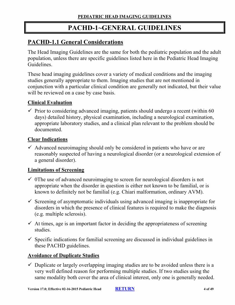
PACHD-1.1 General Considerations
The Head Imaging Guidelines are the same for both the pediatric population and the adult population, unless there are specific guidelines listed here in the Pediatric Head Imaging Guidelines.
These head imaging guidelines cover a variety of medical conditions and the imaging studies generally appropriate to them. Imaging studies that are not mentioned in conjunction with a particular clinical condition are generally not indicated, but their value will be reviewed on a case by case basis.
Clinical Evaluation
Prior to considering advanced imaging, patients should undergo a recent (within 60 days) detailed history, physical examination, including a neurological examination, appropriate laboratory studies, and a clinical plan relevant to the problem should be documented.
Clear Indications
Advanced neuroimaging should only be considered in patients who have or are reasonably suspected of having a neurological disorder (or a neurological extension of a general disorder).
Limitations of Screening
0The use of advanced neuroimaging to screen for neurological disorders is not appropriate when the disorder in question is either not known to be familial, or is known to definitely not be familial (e.g. Chiari malformation, ordinary AVM).
Screening of asymptomatic individuals using advanced imaging is inappropriate for disorders in which the presence of clinical features is required to make the diagnosis (e.g. multiple sclerosis).
At times, age is an important factor in deciding the appropriateness of screening studies.
Specific indications for familial screening are discussed in individual guidelines in these PACHD guidelines.
Avoidance of Duplicate Studies
Duplicate or largely overlapping imaging studies are to be avoided unless there is a very well defined reason for performing multiple studies. If two studies using the same modality both cover the area of clinical interest, only one is generally needed.
Version 17.0; Effective 02-16-2015 Pediatric Head RETURN 5 of 49
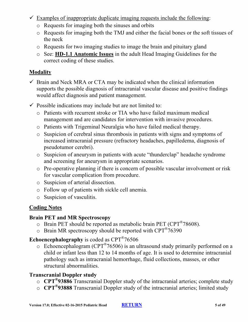
Examples of inappropriate duplicate imaging requests include the following: o Requests for imaging both the sinuses and orbits o Requests for imaging both the TMJ and either the facial bones or the soft tissues of
the neck o Requests for two imaging studies to image the brain and pituitary gland o See: HD-1.1 Anatomic Issues in the adult Head Imaging Guidelines for the
correct coding of these studies.
Modality
Brain and Neck MRA or CTA may be indicated when the clinical information supports the possible diagnosis of intracranial vascular disease and positive findings would affect diagnosis and patient management.
Possible indications may include but are not limited to: o Patients with recurrent stroke or TIA who have failed maximum medical
management and are candidates for intervention with invasive procedures. o Patients with Trigeminal Neuralgia who have failed medical therapy. o Suspicion of cerebral sinus thrombosis in patients with signs and symptoms of
increased intracranial pressure (refractory headaches, papilledema, diagnosis of pseudotumor cerebri).
o Suspicion of aneurysm in patients with acute “thunderclap” headache syndrome and screening for aneurysm in appropriate scenarios.
o Pre-operative planning if there is concern of possible vascular involvement or risk for vascular complication from procedure.
o Suspicion of arterial dissection. o Follow up of patients with sickle cell anemia. o Suspicion of vasculitis.
Coding Notes
Brain PET and MR Spectroscopy o Brain PET should be reported as metabolic brain PET (CPT®78608). o Brain MR spectroscopy should be reported with CPT®76390
Echoencephalography is coded as CPT®76506 o Echoencephalogram (CPT®76506) is an ultrasound study primarily performed on a
child or infant less than 12 to 14 months of age. It is used to determine intracranial pathology such as intracranial hemorrhage, fluid collections, masses, or other structural abnormalities.
Transcranial Doppler study o CPT®93886 Transcranial Doppler study of the intracranial arteries; complete study o CPT®93888 Transcranial Doppler study of the intracranial arteries; limited study
Version 17.0; Effective 02-16-2015 Pediatric Head RETURN 6 of 49
o CPT®93890 Transcranial Doppler study of the intracranial arteries; vasoreactive study
o CPT®93892 Transcranial Doppler study of the intracranial arteries; emboli detection without intravenous microbubble injection
o CPT®93893 Transcranial Doppler study of the intracranial arteries; emboli detection with intravenous microbubble injection
PACHD-1.2 Issues Specific to Pediatric Neuroimaging
Many differences characterize the optimal approach to neuroimaging in the pediatric population versus the optimal approach in adults. Some of these differences arise due to the probability of various illnesses in children versus adults.
The differential diagnoses and relative frequency of disorders often differ considerably between children and adults, and between infancy, early childhood, “middle childhood”, and adolescence.
Congenital anomalies: this is an especially prominent disease category in infancy and childhood. Certain anomalies tend to cluster, and those who have one congenital anomaly are more likely to have certain others, as well.
Craniofacial syndromes sometimes require multiple surgeries and reimaging for pre op planning. Facial imaging may be approved at specialist’s request.
Radiation: children are more sensitive to the harmful effects of ionizing radiation, and can expect longer life spans in which to experience those effects. In general, this favors the use of MRI/MRA over CT/CTA if either modality would otherwise be satisfactory (although see Need for sedation bullet point below). Exceptions will be mentioned in specific guidelines as necessary.
Need for sedation: the scanning times involved for MRI/MRA generally require that infants and young children be sedated or anesthetized for these procedures. o CT can usually be performed successfully without sedation (short scanning times). o Therefore, CT may be performed in children under age 6 rather than MRI if CT
will reasonably provide the imaging information needed. o If MRI is felt to be necessary in these very young children, contrast and coding
approvals should be geared toward avoiding a second imaging session.
Berry Aneurysm: Significant-sized or symptomatic berry aneurysms are rare below age 10 and very uncommon below age 20. o Advanced imaging for occult berry aneurysms will rarely be appropriate prior to
age 20. o Also see: PACHD-2.3 Aneurysm
Version 17.0; Effective 02-16-2015 Pediatric Head RETURN 7 of 49
o Neuroimaging is sometimes needed with infants and young children due to limitations in gathering an accurate history and symptoms, as well as difficulties in interpreting the neurological exam.
PACHD-1.3 Head CT versus Brain MRI in Childhood
Head CT
Head CT is normally performed without contrast (CPT®70450). o With modern CT scanners, the addition of contrast adds little useful diagnostic
information in most settings, doubles the radiation dose, and adds the risk of a contrast reaction.
Head CT is used for the following: o Urgent/emergent settings due to availability and speed of CT o Trauma o Evaluation for recent hemorrhage, whether traumatic or spontaneous o Evaluation for calcifications o Evaluation of the bony structures of the head o Evaluation and follow-up of hydrocephalus o In patients dependent on life support o Uncooperative patients (including very young children—see Need for sedation
bullet in PACHD-1.2) o Prior to lumbar puncture to rule out mass or bleeding
If requested, brain MRI without contrast (CPT®70551) can be substituted for head CT
Brain MRI
Brain MRI is superior to head CT in evaluating known tumors, the pituitary region, epilepsy, white matter, and inflammation within the brain, and is the appropriate imaging study to use in these conditions, even in very young children.
PACHD-1.4 Contrast Use in Head Imaging
Head CT
Head CT is normally performed without contrast (CPT®70450) o Exception: a child with a known or strongly suspected intracranial abscess and an
absolute inability to have an MRI can undergo head CT without and with contrast (CPT®70470).
Brain MRI
In children, brain MRI is generally performed without contrast (CPT®70551)
Brain MRI without and with contrast (CPT®70553) is indicated for the following (especially in young children who require sedation):
Version 17.0; Effective 02-16-2015 Pediatric Head RETURN 8 of 49
o Well-grounded concern regarding the pituitary region, or o Known brain tumor, or o Ataxia, or o Presence of focal neurological signs (including stroke)
PACHD-1.5 MRA/CTA in Children (including MRV/CTV)
Indications for CT and MR angiography and venography are well defined, and it is inappropriate to request these studies as simply an “add-on” to head CT or brain MRI without clinical information that supports performing the angiography or venography studies.
Head MRA, CTA, MRV, CTV should not be used as the only initial neuroimaging study(ies) in children prior to review of a recent head CT or brain MRI.
Coding Notes
Head MRA is normally performed without contrast (CPT®70544) o There are no generally recognized indications for head MRA without and with
contrast (CPT®70546)
MRA of the neck vessels is usually done with contrast (CPT®70548) o For stroke or reasonable suspicion of carotid or vertebral artery dissection, neck
MRA without and with contrast (CPT®70549) can be performed o Some specialists use noncontrast MRA of the neck vessels (CPT®70547) and this
is acceptable when specifically requested.
Head MRV should be reported as report as CPT®70544
MRA vs. CTA
MRA is usually preferred in children in order to avoid radiation exposure, but CTA may be needed in specific conditions (e.g. vasculitis).
Pediatric stroke: (head and neck MRA [CPT®70544 and CPT®70548] or head and neck CTA CPT®70496 and CPT®70498] along with brain MRI without and with contrast (CPT®70553) or noncontrast head CT (CPT®70450) if not already done
References
1. Yousem DM,Grossman RI. Neuroradiology, the requisites. 3rd Ed. Philadelphia, Mosby, 2010. 2. Latchaw RE, Kucharczyk J, Moseley ME. Imaging of the Nervous System. Philadelphia, Elsevier,
2005. 3. Rowland LP (Ed.). Merritt’s Neurology. 12th Ed. Philadelphia, Lippincott, 2010. 4. Menkes JH, Sarnat HB, Maria BL. Child Neurology. 7th Ed. Philadelphia, Lippincott, 2006. 5. Diagnostic Imaging: Pediatric Neuroradiology by A. James Barkovich, Anna Illner, Kevin R.
Moore, Ellen Grant, Blaise V. Jones.
Version 17.0; Effective 02-16-2015 Pediatric Head RETURN 9 of 49
PEDIATRIC HEAD IMAGING GUIDELINES
PACHD-2~Aneurysm, Arteriovenous Malformation
PACHD-2.1 Thunderclap Headache or Headache of Explosive Onset
These types of headaches are much less common in children than in adults. Often, it is very difficult to obtain a clear history describing these headaches.
Within the first 12 hours, noncontrast head CT (CPT®70450) is appropriate when a headache is documented as thunderclap or explosive onset.
After the first 12 hours, noncontrast brain MRI (CPT®70551) can be performed rather than head CT, if head imaging has not already been performed.
PACHD-2.2 Arteriovenous Malformation (AVM)
Also see HD-14.3 Arteriovenous Malformations in the adult Head Imaging Guidelines Hemorrhage from an AVM is the most common source of non-traumatic subarachnoid hemorrhage in the first decade of life. In teenagers, it is somewhat less common than aneurysmal hemorrhage. In newborns, AVM’s may present with high output heart failure due to the large volume of blood traversing the shunt.
Brain MRI without and with contrast (CPT®70553) can be performed for evaluation of AVM.
Brain MRI, contrast as requested plus brain MRV/CTV can be performed.
Head imaging for AVM screening is not indicated except for a family history of familial cavernoma. o AVM’s are not familial lesions except in conditions such as hereditary
hemorrhagic telangectasia syndrome (OWR).
PACHD-2.3 Aneurysm
Most berry aneurysms in children under age 10 are found by age 2 and involve the junction of the anterior cerebral and internal carotid arteries. Many seem to be associated with anterior midline cerebral anomalies. Berry aneurysms are uncommon in the second decade of life.
Emergency noncontrast head CT (CPT®70450) or noncontrast brain MRI (CPT®70551) plus head CTA (CPT®70496) can be performed to evaluate suspected ruptured aneurysm.
Head MRA to screen for berry aneurysms is rarely indicated before age 20. Exceptions may apply to a teenager with a family history of aneurysm either in both parents or in an identical twin.
Version 17.0; Effective 02-16-2015 Pediatric Head RETURN 10 of 49
References
1. Neuroimaging Clinics of North Am 2007;17(2):153-163 2. Pediatr Neurosurg 2008;44:296-301. 3. Menkes JH, Sarnat HB, Maria BL. Child Neurology. 7th Ed. Philadelphia, Lippincott, 2006, pp. 837-
839 4. Nelson PK, et al. Neuroradiology in the imaging and therapeutic management of brain
arteriovenous malformations (Ch 38). In Latchaw RE, Kucharczyk J, Moseley ME Imaging of the Nervous System. Philadelphia, Elsevier, 2005
Version 17.0; Effective 02-16-2015 Pediatric Head RETURN 11 of 49
PEDIATRIC HEAD IMAGING GUIDELINES
PACHD-3~ATAXIA
PACHD-3.1 Ataxia
Ataxia refers to an abnormally ill-coordinated or unsteady gait for age. “Limb ataxia” refers to impaired coordination (for age) of limbs, especially arms. Developmental failure to acquire the ability to walk is a form of developmental retardation, not ataxia. (See: PACHD-9~Developmental Delay - Mental Retardation)
Detailed neurological history and recent (within the past 60 days) clinical examination, including a neurological examination, are indicated prior to considering advanced imaging in the evaluation of ataxia.
Brain MRI without and with contrast (CPT®70553) can be performed to evaluate ataxia, hereditary ataxia, and slowly progressive ataxia. o Cervical spine MRI, contrast as requested, can be performed if brain MRI is non-
diagnostic. o In younger children, brain MRI and cervical spine MRI can be performed together
as initial studies.
PACHD-3.2 Ataxia Telangiectasia
After tumor, ataxia telangiectasia is the most common cause of ataxia in children under age 10. Usually transmitted via autosomal recessive mechanisms, and is a systemic disorder with prominent neurological manifestations. Immune (IgA) deficiencies are usual, and frequent sinopulmonary infections are prominent. These children should be considered immunocompromised.
Sinus CT, chest x-ray, and sometimes chest CT may be needed.
Brain MRI without and with contrast (CPT®70553) can be performed if there is uncertainty about the diagnosis.
PACHD-3.3 Friedreich’s Ataxia
In the United States, Friedreich’s ataxia is the most common cause of progressive ataxia in adolescents. Onset is usually in the teens or early 20’s. A GAA expansion is usually identified on DNA testing. The gene is typically an autosomal recessive. Type I diabetes and cardiac disturbances are often present.
Brain MRI, usually without contrast (CPT®70551), is appropriate.
Practice Notes
The differential diagnosis of ataxia in childhood includes:
Version 17.0; Effective 02-16-2015 Pediatric Head RETURN 12 of 49
o In children less than 2 years old, a normal variation of development o Neuraxis tumor o Ataxia telangiectasia o Friedreich’s ataxia o Juvenile lipofuscinosis, Refsum’s disease, Abetalipoproteinemia, and other
uncommon genetic progressive ataxias. o Sex-linked adrenoleukodystrophy may present atypically, as progressive ataxia in
affected males, usually during adolescence o Benign ataxia of childhood (postviral)—this has become infrequent since the
widespread use of Varicella vaccine.
Reference
1. ACR Appropriateness Criteria, Ataxia, 2009
Version 17.0; Effective 02-16-2015 Pediatric Head RETURN 13 of 49
PEDIATRIC HEAD IMAGING GUIDELINES
PACHD-4~Autism and Autism Spectrum Disorders
The members of this group, including Asperger syndrome, are classified as pervasive development disorder (PDD). This is a clinical diagnosis. No lab or imaging study can confirm the diagnosis of autism. Comprehensive evaluation for autism might include history, physical exam, audiology evaluation, speech, language, and communication assessment, cognitive and behavioral assessments, and academic assessment.
There is insufficient evidence-based data to support the use of PET in patients with autism.
PACHD-5~Behavioral Disorders
PACHD-5.1 Imaging
Behavioral disorders of childhood or adolescence generally require no advanced imaging for diagnosis or management.
PACHD-6~Cerebral Palsy
MRI or CT can identify a treatable problem in about 5% of those cases in which the cause was not determined in the newborn period (usually by ultrasound). In addition, it can prove the timing of the insult in most of the remaining cases.
PACHD-6.1 Imaging
Brain MRI is superior, but generally requires sedation or anesthesia.
Brain MRI without and with contrast (CPT®70553) or head CT without contrast (CPT®70450) is appropriate for cases of cerebral palsy of undetermined origin or if a fixed deficit worsens.
Reference
1. Neurology 2004;62:851-863
Version 17.0; Effective 02-16-2015 Pediatric Head RETURN 14 of 49
PEDIATRIC HEAD IMAGING GUIDELINES
PACHD-7~Chiari Malformations
PACHD-7.1 Initial Imaging
Chiari I Brain MRI without contrast (CPT®70551) and MRI of entire spine without contrast
(CPT®72141, CPT®72146, CPT®72148)
Chiari II and III Brain MRI and MRI of the entire spine, contrast as requested.
Head CT without contrast CT (CPT®70450) to visualize the skull, and/or CT of involved spinal regions may also be necessary.
PACHD-7.2 Follow-up Imaging
Chiari I If initial spine imaging was normal, repeat spine studies should not be necessary
unless signs or symptoms appear that suggest a spinal cord disorder.
Follow-up spine and head imaging in individuals with syringomyelia/hydromyelia is individualized and should be at the discretion of the specialist following the patient.
Patients who have had an MRI confirming Chiari I should not need repeat brain imaging unless there are new clinical abnormalities or a surgical procedure is actively being considered.
Chiari II and III Follow-up imaging is individualized and should be at the discretion of the
specialist following the patient.
PACHD-7.3 Familial Screening
Familial screening is not indicated.
Practice Notes
Classification
o Chiari malformations involve abnormally hindward positioning (“herniation”) of the cerebellum so that the tips of the cerebellar tonsils lie at least 5 mm below the foramen magnum.
o There may be associated abnormalities of the skull base, the cranio-cervical junction, the upper cervical vertebrae, or the spinal cord.
Chiari I o The most common type; involves the cerebellar tonsils and is often associated with
Version 17.0; Effective 02-16-2015 Pediatric Head RETURN 15 of 49
syringomyelia. o Usually, does not include overt hydrocephalus. o Usually, not symptomatic during childhood, but can present later in the teens. o Most cases are discovered accidentally on a head scan performed for another
indication. o Symptoms are usually nonspecific, but can include lower cranial nerve palsies or
sleep apnea.
Chiari II o Incidence is one in 5000 live births; presents in infancy with profound neurological
abnormalities and hydrocephalus. o Includes positioning of the cerebellar vermis, fourth ventricle, and brain stem
below the foramen magnum. o Hydrocephalus occurs. o Tethered cord and lumbar meningomyelocele are also generally present, and
midline cerebral anomalies are common.
Chiari III o A more profound malformation which includes an occipital or high cervical
encephalocele, extensive syringe/hydromyelia, and cerebral malformation. o Presents in infancy with profound neurological abnormalities and hydrocephalus.
Version 17.0; Effective 02-16-2015 Pediatric Head RETURN 16 of 49
PEDIATRIC HEAD IMAGING GUIDELINES
PACHD-8~Craniostenosis
PACHD-8.1 Imaging
CT head without contrast (CPT®70450) is indicated in the diagnosis of craniostenosis (craniosynostosis), and 3-D rendering (CPT®76377) may be needed for determination of timing of surgery, or for preoperative planning.
CT maxillofacial (CPT®70486) and CT orbits (CPT®70480) are not necessary to evaluate all patients with craniostenosis (craniosynostosis) either pre- or post-op, but may be indicated for surgical planning especially for more complex syndromes.
PACHD-9~Developmental Delay - Mental Retardation
PACHD-9.1 Imaging
Brain MRI without contrast (CPT®70551) is appropriate to evaluate for congenital abnormalities whether cerebral palsy is noted or not. If necessary, noncontrast head CT (CPT®70450) may be substituted.
Reference
1. Shevell M, Ashwal S, Donely D, et al. Practice Parameter: Evaluation of the child with global development delay: Report of the quality standards subcommittee of the American Academy of Neurology and the Pracatice Committee of the Child Neurology Society. Neurology 2003;60:367-380.
Version 17.0; Effective 02-16-2015 Pediatric Head RETURN 17 of 49
PEDIATRIC AND CONGENITAL HEAD IMAGING GUIDELINES
PACHD-10~Dystonia/Torticollis
PACHD-10.1 Newborn Infant
Ultrasound of the Neck is the initial study to determine if congenital muscular torticollis. o Positive No further imaging is needed since diagnosis is defined. o NegativeCT Neck with contrast (CPT®70491) or MRI Neck with and without
contrast (CPT®70543) contrast to try to identify other cause
PACHD-10.2 Older Child (beyond infancy) or Adult
For trauma, CT Neck with contrast (CPT®70491) and/or CT Cervical Spine without contrast (CPT®72125) are the initial studies to identify fracture or mal-alignment.
For no trauma, CT Neck with contrast (CPT®70491), and/or MRI Cervical Spine without contrast (CPT®72141), or CT Cervical Spine without contrast (CPT®72125) are the initial studies to locate a soft tissue or neurological cause. o Positive Further advanced imaging is not required if CT Neck or CT Cervical
Spine has identified local cause. o NegativeMRI Brain without and with contrast (CPT®70553) to exclude CNS
cause. Practice Note
Torticollis or cervical dystonia is an abnormal twisting of the neck with head rotated or twisted. Its causes are many and may be congenital or acquired and caused by trauma, infection/inflammation, neoplasm and those less defined and idiopathic. It occurs more frequently in children and on the right side (75%).
Retropharyngeal space abscess could be associated with torticollis because child would not move neck freely.
References
1. Anderson PA, Montesano PX. Morphology and treatment of occipital condyle fractures. Spine 1988; 13:731-6.
2. Ballock RT, Song KM. The prevalence of nonmuscular causes of torticollis in children. J Pediatr Orthop 1996; 16:500-4.
3. Castillo M, Albernaz VS, Mukherji SK, Smith MM, et al. Imaging of Bezold’s abscess. AJR Am J Roentgenol 1998; 171:1491-5.
4. Federico F, Lucivero V, Simone IL, Defazio G, et al. Proton MR spectroscopy in idiopathic spasmodic torticollis. Neuroradiology 2001; 43:532-6.
Version 17.0; Effective 02-16-2015 Pediatric Head RETURN 18 of 49
5. Fielding JW, Hawkins RJ. Atlanto-axial rotatory fixation (fixed rotatory subluxation of the atlanto-axial joint). J Bone Joint Surg Am 1977; 59:37-44.
6. Kraus R, Han BK, Babcock DS, Oestreich AE. Sonography of neck masses in children. AJR Am J Roentgenol 1986; 146:609-13.
7. Roche CJ, O’Malley M, Dorgan JC, Carty HM. A Pictorial Review of Atlanto-axial Rotatory Fixation: Key points for the radiologist. Radiographics 2001; 56:947-58.
8. Tracy MR, Dormans JP, Kusumi K. Klippel-Feil Syndrome: Clinical features and current understanding of etiology. Clin Orthop Relat Res 2004; 424:183-90.
Version 17.0; Effective 02-16-2015 Pediatric Head RETURN 19 of 49
PEDIATRIC HEAD IMAGING GUIDELINES
PACHD-11~EAR
PACHD-11.1 Hearing Loss History, otoscopic examination, and hearing tests such as audiology, speech recognition testing, and auditory evoked responses should be performed prior to considering advanced imaging. The selection of tests will depend on the age of the child and local factors
Temporal bone CT without contrast (CPT®70480) can be performed initially to help determine etiology.
Children with hearing loss of congenital origin may have brain anomalies as well, and brain MRI without contrast (CPT®70551) can be performed if the need to evaluate for associated cerebral anomalies is specifically stated
In children with unilateral or bilateral sensory-neural (cochlear) loss, high resolution temporal bone CT without contrast (CPT®70480) is appropriate for initial imaging. Brain MRI without and with contrast (CPT®70553) can be useful in selected cases
In the rare case of documented retrocochlear hearing loss, brain MRI without and with contrast (CPT®70553) can be performed.
PACHD-11.2 Cochlear implants The surgeon’s choice of preoperative craniofacial studies should be honored.
PACHD-11.3 Earache A recent detailed history and physical examination, including an otoscopic examination, should be performed initially. Common causes of ear pain include external and middle ear infections, dental problems, sinus infection, neck problems, tonsillitis, and pharyngitis
Advanced imaging is not indicated in patients with: o With improvement of symptoms following an episode of one of the common
causes of ear pain mentioned above, including otitis media o Otitis externa
If ear pain persists with no obvious cause, CT scan of the temporal bone without contrast (CPT®70480) is the usual initial advanced imaging study
PACHD-11.4 Otitis Media Acute otitis media is a very common infection of young children, typically presenting with ear pain and fever. The diagnosis is typically made by otoscopic examination, and advanced imaging is generally not indicated.
CT of the temporal bone without contrast (CPT®70480) is appropriate if any of the following is present:
Version 17.0; Effective 02-16-2015 Pediatric Head RETURN 20 of 49
o Multiple recurrent attacks of acute otitis media o Clinical signs of mastoiditis (can do CT of the temporal bone without and with
contrast (CPT®70482) o Otitis media with effusion (or serous otitis media) that does not respond to medical
treatment
PACHD-11.5 Cholesteatoma Cholesteatomas are expansive cysts of the middle ear filled with cellular debris. They can be congenital or arise from recurrent middle ear infections or trauma to the tympanic membrane. Hearing loss is conductive, although if the lesion is large enough, mixed type hearing loss may be found. Otoscopic exam findings and symptoms may include painless drainage from the ear, conductive hearing loss, chronic/recurrent ear infections,
For suspected cholesteatoma, CT of the temporal bone (CPT®70480 or CPT®70482) can be performed initially o 3D rendering (CPT®76376 or CPT®76377) can be approved in conjunction with
the temporal bone CT if requested o Temporal bone MRI (CPT®70543) can be performed instead of temporal bone CT
if requested o Brain MRI without and with contrast (CPT®70553) can be performed for pre-
operative planning, especially if there is evidence of intracranial involvement o Brain MRV (CPT®70544) can be performed for pre-operative planning if there is
concern for straight sinus thrombosis o Post-operative imaging (same studies that were performed pre-op) can be
performed one time to ensure complete removal of the cholesteatoma
PACHD-11.6 Vertigo Vertigo is an uncommon complaint during childhood. Middle ear/Eustachian tube problems are the most common cause of vertigo in children. History and physical examination, including otoscopic exam, should be done prior to considering advanced imaging
If physical examination is otherwise normal and the vertigo responds to treatment, advanced imaging is not indicated.
If vertigo persists, CT of the temporal bone without contrast (CPT®70480) can be performed
References:
1. Arch Otolaryngol Head Neck Surg 1992;118:501-503 2. Arch Otolaryngol Head Neck Surg 2006;132:186-192 3. The Laryngoscope 1999;109:1642-1647 4. Roland PS. Middle Ear, Cholesteatoma. eMedicine, June 6, 2006,
http://www.emedicine.com/ent/topic220.htm. Accessed December 1, 2008 5. Internat J Ped Otolaryngol 2006;70:1547-1554 6. Internat J Pediatr Otorhinolaryngol 2003;67:889-894
Version 17.0; Effective 02-16-2015 Pediatric Head RETURN 21 of 49
PEDIATRIC HEAD IMAGING GUIDELINES
PACHD-12~Epilepsy and Other Seizure Disorders
A detailed clinical assessment and EEG should be performed prior to considering advanced neuroimaging. This clinical assessment must be very thorough and should include neurological and general physical examination, case and family history, and, whenever possible, the accounts of eye witnesses of the events.
Generally the “workup” for new onset seizures in the pediatric population will include EEG and MRI brain without contrast. The EEG is not required to approve these cases when clinical criteria are otherwise met
PACHD-12.1 Imaging
When advanced imaging is indicated, brain MRI (usually without contrast—CPT®70551) is usually sufficient and is strongly preferred over CT. Contrast should be added to the MRI protocol only if there is a progressive neurological deficit or a relevant abnormality identified on a noncontrast scan. o Head CTA/MRA, neck CTA/MRA, and cervical spine MRI do not generally add
valuable information initially and are not indicated. o Brain MRI without contrast (CPT®70551) will generally be performed in any child
with documented new onset of epileptic seizures other than simple febrile seizures. Younger patients requiring sedation for MRI should have their initial MRI
performed with and without contrast (CPT®70553) in order to avoid a second anesthesia exposure.
PACHD-12.2 Surveillance imaging
Not generally necessary in generalized epilepsies
In focal or partial epilepsies, repeat noncontrast brain MRI (CPT®70551) can be performed at one and two years after diagnosis and then every few years.
PACHD-12.3 Repeat imaging on indication
Repeat brain MRI is indicated for any of the following: o There is a change in the type of seizures, or o Persistent worsening in the frequency of seizures, or o New neurological findings
PACHD-12.4 Evaluation for Epilepsy Surgery
Patient must have had a previous brain MRI and documentation of intractable epilepsy for which surgical treatment or another interventional modality is under active consideration.
Version 17.0; Effective 02-16-2015 Pediatric Head RETURN 22 of 49
If these criteria are met, PET (CPT®78608) can be performed.
MR spectroscopy (CPT®76390) and metabolic PET using tracers other than FDG are acceptable alternatives to FDG PET if requested by the patient’s referring epileptologist. o NOTE: Certain payers consider MR Spectroscopy investigational, and their
coverage policies will take precedence over MedSolutions’ guidelines.
Brain SPECT is another acceptable alternative. Currently, MedSolutions does not prior authorize brain SPECT.
PACHD-12.5 Febrile Seizures
Neuroimaging is not medically necessary in children who have typical febrile seizures.
References
1. Neurology 2000;55:616-623 Reaffirmed 7-28-2006 2. Menkes JH, Sarnat HB, Maria BL. Child Neurology. 7th Ed. Philadelphia, Lippincott, 2006, pp. 919-
922 and 857-942 3. J Pediatr Neurosci 2008;3:48-54 4. Brain 2009;132:2785-2797 5. Ann Neurol 1990:27:406-413
PACHD-13~EPISTAXIS
PACHD-13.1 Imaging
Initial evaluation of epistaxis (nose bleed), including recurrent epistaxis, is by direct or endoscopic visualization of the relevant portions of the upper airway. If the initial clinical evaluation is unrevealing, Ear, Nose, and Throat (ENT) examination may be helpful.
Maxillofacial CT may be useful in individual cases, depending upon the findings during the initial clinical evaluation.
Version 17.0; Effective 02-16-2015 Pediatric Head RETURN 23 of 49
PEDIATRIC HEAD IMAGING GUIDELINES
PACHD-14~Headache, Child
PACHD-14.1 Children of School Age (≥6 years old)
Headache is a very common complaint in this age group. Many of these children have a family history of one of the primary headache disorders, such as migraine or tension headache.
A complete headache history and neurological examination should be performed prior to considering advanced imaging, as well as 4 weeks of conservative treatment.
Special considerations regarding MRI contrast selection when MRI is the appropriate study:
o For children able to undergo MRI without sedation, MRI brain without contrast (CPT®70551) is generally the preferred study unless otherwise specified in the guidelines
o Younger patients requiring sedation for MRI should have their initial MRI performed with and without contrast (CPT®70553) in order to avoid a second anesthesia exposure.
Stable Headaches
Neuroimaging is very unlikely to be of value in a child with a normal neurological examination who has stable migraine, tension, school, eye strain, or other stress related headaches. This is especially true when there is a family history of these disorders.
New Onset Headaches
MRI Brain without and with contrast (CPT®70553) should be approved in the presence of red flag symptoms for a CNS tumor:
Red Flag Symptoms Raising Suspicion for CNS Tumors Include:
any headache complaint from a child age ≤5 years
headaches awakening from sleep
focal findings on neurologic exam including diplopia
clumsiness (common description of gait or coordination problems in young children)
headaches associated with morning nausea/vomiting
new onset of seizure activity with focal features
papilledema on physical exam
Version 17.0; Effective 02-16-2015 Pediatric Head RETURN 24 of 49
Other Indications for Imaging that do not Require 4 weeks of Conservative Treatment
o Concomitant seizure history—brain MRI, without (CPT®70551) OR without and with contrast (CPT®70553) as requested
o Recent head trauma—head CT without contrast (CPT®70450) o Current use of anticoagulants-- head CT without contrast (CPT®70450) o New headache in a patient with known congenital heart disease-- head CT without
contrast (CPT®70450) or brain MRI without contrast (CPT®70551) to evaluate for ruptured aneurysm
o New onset, clearly documented thunderclap headache—head CT without contrast (CPT®70450) or brain MRI without contrast (CPT®70551)
o Headaches which become severe and progressive within a month of onset-- brain MRI without contrast (CPT®70551) OR without and with contrast (CPT®70553) as requested.
MRA/CTA
MRA/CTA is not generally medically necessary in the evaluation of headache in children unless an AVM has been seen on a prior brain MRI or head CT.
MRV
Head MRV (CPT®70544) may be useful in patients with papilledema. (See: PACHD-21)
PACHD-14.2 Preschool Children (ages ≤5 years) Recurrent complaints of headache in a child age ≤5 years are uncommon, and in most cases associated with serious intracranial pathology. Imaging is always indicated for evaluation of headaches in this age group. MRI brain without and with contrast (CPT®70553) is the preferred study for these
patients as almost all patients this age will require general anesthesia to complete the study o CT head without contrast (CPT®70450) can be approved if there is a recent history
of head trauma, other suspicion of intracranial hemorrhage, or known hydrocephalus. CT should not be used in place of MRI to avoid sedation in patients without one of these features.
References
1. Blume HK. Pediatric Headache: a review. Pediatrics in Review, 2012; 33: 562-574 2. De Bries A, Young PC, Wall E, Getchius T, Li CH., et al. CT scan utilization patterns in pediatric
patients with recurrent headache. Pediatrics, 2013; 132;e1-e8. 3. Neurology 2002;59:490-498 Reaffirmed 10-15-2005 4. Radiology 1997;202:819-824 5. American College of Radiology, ACR Appropriateness Criteria®, Clinical Condition: Headache —
Child, Last Reviewed 2012 6. Am J Epidemiol, 2005;161:1066-1073 7. Neurology 1992;42:1657-1662
Version 17.0; Effective 02-16-2015 Pediatric Head RETURN 25 of 49
PEDIATRIC HEAD IMAGING GUIDELINES
PACHD-15~Macrocephaly, Microcephaly, Hydrocephaly and Related Conditions
PACHD-15.1 Macrocephaly
Macrocephaly is defined as head circumference that is more than two standard deviations above the mean for age, sex, and body size, established by use of measurements and standard growth charts. The normal curve dictates that 2.5% of any random group will be over two standard deviations above the mean. Accelerated head circumference growth by more than one standard deviation from the child’s previous standing can also indicate macrocephaly.
Birth to one year old:
Ultrasound of the head (CPT®76506) should be performed initially
If ultrasound is abnormal, either head CT (CPT®70450) or brain MRI, contrast as requested, can be performed. CT is often chosen initially to see calcifications and to view skull anatomy, but MRI shows more anatomical detail and may be chosen instead.
If head CT shows hydrocephalus, then brain MRI, contrast as requested, is appropriate
Age one year and older:
Brain MRI, contrast as requested, is preferred initially to CT since uncomplicated hydrocephalus is less likely after early infancy
In younger children, head CT is acceptable (if requested) for reasons listed in: PACHD-1.3 Head CT versus Brain MRI in Childhood
Megalencephaly
Megalencephaly is defined by increased head size caused by increased brain size, rather than by excess fluid, tumor, or cyst formation. It can be either a normal variant or can arise from one of several uncommon disorders. This term can only be applied after there has been a brain imaging study
Brain MRI, contrast as requested, is appropriate if it was not done initially
Hydranencephaly
Hydranecephaly is defined by defective development of the cerebral hemispheres, which are replaced by large cystic structures. The infants seem normal at birth, but then have an abnormally enlarging head and profound failure to achieve milestones.
This condition is seen on cerebral ultrasound (CPT®76506), but once see, requires further evaluation by brain MRI, contrast as requested
Version 17.0; Effective 02-16-2015 Pediatric Head RETURN 26 of 49
PACHD-15.2 Microcephaly
The definition is similar to that for macrocephaly in reverse.
Brain MRI, contrast as requested, is recommended initially since CT may not detect the relevant anatomical abnormalities
PACHD-15.3 Hydrocephaly (hydrocephalus)
This is the most common identifiable cause of macrocephaly. Almost all hydrocephalus is obstructive, except hydrocephalus due to choroid plexus papillomas. Hydrocephalus is traditionally divided into non-communicating (the obstruction lies within the course of the brain’s ventricular system) and communicating (the obstruction is distal to the ventricular system)
Initial Imaging
First 6 months of life:
Screening head ultrasound examination (CPT®76506)
If ultrasound is abnormal, brain MRI, contrast as requested, can be performed.
Greater than 6 months old Brain MRI, usually without contrast CPT®70551, but can be contrast as requested.
Head CT without contrast (CPT®70450) can be substituted if requested.
Spine imaging Spine MRI may be indicated in individuals with Chiari malformation (multiple spine
segments), Dandy-Walker malformation (cervical spine only), or malignant infiltration of the meninges.
Surveillance Imaging after Shunting
Brain MRI without contrast (CPT®70551) or head CT (CPT®70450) can be performed a few months after shunt placement and then once a year.
MRI provides more anatomical detail and does not involve radiation exposure, but many providers use head CT, especially in individuals younger than school age.
Shunting into the peritoneum (VP shunts) can give rise to abdominal complications, but these are generally symptomatic, so surveillance imaging of the abdomen is not indicated.
Re-imaging for Indication
Head CT without contrast (CPT®70450) or brain MRI, contrast as requested, can be performed for new signs or symptoms such as sepsis, altered level of awareness, protracted vomiting, visual or neurological deterioration, decline of mentation after initial improvement, or new onset of seizures.
Version 17.0; Effective 02-16-2015 Pediatric Head RETURN 27 of 49
Familial Screening
Not generally indicated except in siblings of individuals with aqueductal stenosis (noncontrast head CT (CPT®70450) or MRI (CPT®70551)
Practice Notes - Etiologies for Hydrocephaly
Aqueductal stenosis or gliosis o Accounts for about 20% of hydrocephalus cases o Risk of occurrence in siblings is a few percent o Symptoms are of insidious onset and can begin at any age o Acute exacerbations can occur after minor head trauma’
Chiari malformation o Accounts for about 40% of cases o See: PACHD-7~Chiari Malformations
Dandy-Walker malformation o Accounts for 10% of cases o Hydrocephalus generally becomes evident during the third to twelfth month of life o Triad of findings includes cystic dilatation of the fourth ventricle, full or partial
agenesis of the cerebellar vermis, and upward displacement of tentorium and associated venous structures
o Can be distinguished from an isolated finding of an enlarged cistern magna, which is a normal anatomical variant
Communicating hydrocephalus o Accounts for 30% of cases o Can present at any age, and actual macrocephaly may be minimal or absent if the
event causing the hydrocephalus occurs after mid-childhood o Underlying causes include prior bacterial meningitis, prior subarachnoid
hemorrhage, and more rarely, malformations of the straight sinus or vein of Galen and subarachnoid infiltration by lymphoma or leukemia.
Reference
1. Ashwal S, Michelson D, Plawner L, Dobyns WB. Practice Parameter: evaluation of the child with microcephaly (an evidence-based review). Neurology 2009;73:887-897
Version 17.0; Effective 02-16-2015 Pediatric Head RETURN 28 of 49
PEDIATRIC HEAD IMAGING GUIDELINES
PACHD-16~Magnetic Resonance Spectroscopy - Pediatrics
Magnetic Resonance Spectroscopy (MRS)–CPT®76390
Magnetic Resonance Spectroscopy involves the analysis of the levels of certain chemicals in pre-selected voxels (small regions) on an MRI scan done at the same time. Certain payers consider MRS investigational, and their coverage policies may take precedence over MedSolutions' guidelines.
Pediatric uses in neuro-oncology: MRS is often useful in the management of pediatric brain tumors to determine the need for further therapy. Such cases require referral to a Medical Director.
MRS is clearly useful in the diagnosis and subsequent management of certain rare inborn errors of metabolism affecting the central nervous system, including adrenoleukodystrophy, creatinine pathway disorders, and others. Cases will be forwarded to Medical Director review.
MRS produces highly variable results in MS, varying with the pathological process. It does not appear to be useful in distinguishing multiple sclerosis plaques from tumors, since both can produce similar results.
The use of MRS in multiple sclerosis, especially in making the differential diagnosis of MS versus tumor, is experimental at this time. Use of MRS in patients with cerebral metastases of systemic cancers is currently regarded as experimental.
References
1. Neuroimaging Clin N Am 2006;16:169-192 2. Neuroimaging Clin N Am 2006;16:87-116 3. Neurology 2005;64:434-441 4. Neurology 2005;64:406-407
Version 17.0; Effective 02-16-2015 Pediatric Head RETURN 29 of 49
PEDIATRIC HEAD IMAGING GUIDELINES
PACHD-17~Multiple Sclerosis
Multiple sclerosis is distinctly uncommon in children. About 4% of MS cases begin before age 17, and only approximately 0.5% begin before age 10.
PACHD-17.1 Imaging
Ataxia, optic neuritis, diplopia, and transverse myelitis are common presentations. MS can present as an acute encephalitis-like illness, especially in childhood. Among children with suspected demyelinating diseases, the principal differential diagnosis is often between MS and one particular isolated clinical syndrome—acute disseminated encephalomyelitis. Other demyelinating disorders, such as various leukodystrophies and DeVic’s disease (neuromyelitis optica) may also need to be considered. Non-demyelinating diseases such as brain tumor may also need to be considered in the differential.
MRI is necessary to confirm a demyelinating disease and to help distinguish between the different demyelinating diseases. o CT may occasionally be useful to exclude a mass lesion.
PET and MR spectroscopy are not generally indicated in evaluating MS. o On a rare occasion, PET may aid in distinguishing “tumefactive” demyelinating
lesions from tumor.
Brain MRI without and with contrast (CPT®70553) and MRI of the spinal cord without and with contrast can be performed. o Cervical and thoracic spine MRI scans visualize the entire spinal cord, and lumbar
spine MRI usually is not needed. o Sagittal MRI of the spinal cord with phased array detector coil (CPT®72156 or
CPT®72157) or full MRI of the cervical (CPT®72156) and thoracic spine (CPT®72157) can be performed to visualize the entire spinal cord.
Follow-up imaging in established MS
Surveillance MRI, contrast as requested, of the brain and spinal cord can be performed twice a year.
MRI, contrast as requested, of the brain and spinal cord can be performed whenever an episode with significant new neurological deficit occurs.
Screening studies for MS
Screening studies are inappropriate in asymptomatic children and in those whose clinical picture does not support a diagnosis of MS.
Version 17.0; Effective 02-16-2015 Pediatric Head RETURN 30 of 49
References:
1. McAlpine ed. 4, pp.343-346 2. Neurology 2010;74:1412;1415 3. Neurology 2009;72:961-967 4. Neurology 2009;72:968-973 5. Neurology 2010;1404-1405 6. Continuum (American Academy of Neurology) 2010:16(5):181-192 7. Ann Neurol 2005;58:840-846 8. Lancet Neurology 2007;6:677-686 9. Lancet Neurology 2004;3:104-110 10. Neurology 2006;66:1485-1489 11. AJNR 2006;27:455-461 12. Eur J Neuroradiol 2006;13:313-325
Version 17.0; Effective 02-16-2015 Pediatric Head RETURN 31 of 49
PEDIATRIC HEAD IMAGING GUIDELINES
PACHD-18~Neuro-Oncology
See: Pediatric Oncology Imaging Guidelines
PACHD-19~Optic Neuritis
Also see: PACHD-17~Multiple Sclerosis
Also see: HD-16~Multiple Sclerosis
Version 17.0; Effective 02-16-2015 Pediatric Head RETURN 32 of 49
PEDIATRIC HEAD IMAGING GUIDELINES
PACHD-20~PITUITARY
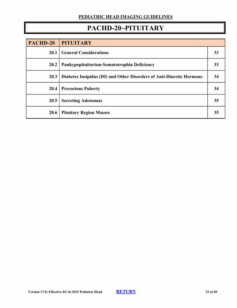
PACHD-20 PITUITARY
20.1 General Considerations 33
20.2 Panhypopituitarism-Somatotrophin Deficiency 33
20.3 Diabetes Insipidus (DI) and Other Disorders of Anti-Diuretic Hormone 34
20.4 Precocious Puberty 34
20.5 Secreting Adenomas 35
20.6 Pituitary Region Masses 35

Version 17.0; Effective 02-16-2015 Pediatric Head RETURN 33 of 49
PEDIATRIC HEAD IMAGING GUIDELINES
PACHD-20~PITUITARY
PACHD-20.1 General Considerations
The initial step in the evaluation of all potential pituitary masses is a detailed history, recent physical examination, and thorough neurological exam, including evaluation of the visual fields.
Endocrine laboratory studies should be performed prior to considering advanced imaging.
When pituitary imaging is indicated, brain MRI without and with contrast (CPT®70553) is preferred. o One study (either brain MRI [CPT®70553] or MRI Orbit, Face, Neck
[CPT®70543]) is adequate to image the pituitary. The ordering physician should specify that the study is specifically to evaluate the pituitary gland. The reporting of two CPT® codes, to image the pituitary, is not indicated.
PACHD-20.2 Panhypopituitarism/somatotrophin (growth hormone) Deficiency Endocrine testing should be performed initially, and will reveal growth hormone deficiency with or without associated deficiency of other pituitary hormones. For isolated growth hormone deficiency, two measurements of growth hormone with stimulation are performed. In children, normal stimulated values are greater than 10 ng/ml
Laron syndrome is caused by a rare inherited mutation of the growth hormone receptor. Affected children have the features of growth hormone deficiency, but have normal or elevated growth hormone levels
Primary failure: 15%-20% of all cases, with autosomal recessive, X-linked, and dominant patterns of transmission. Many specific DNA defects are known.
Secondary failure:causes can include mass of the sellar region, histiocytosis, trauma, hemorrhage, sarcoidosis, bacterial meningitis, etc.
Radiation of the skull base for brain tumor or leukemia requires special mention. Growth hormone deficiency can be a result of dose-related radiation damage and these children must be monitored for this.
Initial Imaging
Brain MRI, contrast as requested, with special attention to the pituitary, can be performed when cause of the clinical presentation is in doubt.
Version 17.0; Effective 02-16-2015 Pediatric Head RETURN 34 of 49
Re-imaging
The need for re-imaging depends on the particular condition and should be at the preference of the specialist following the patient.
PACHD-20.3 Diabetes Insipidus (DI) and Other Disorders of Anti-Diuretic Hormone
The principal evaluation of ADH deficiency is by urine and blood electrolyte and osmolality testing - serum osmolality greater than 300 with urine osmolality less than 300. Deficiencies in ADH can either be central or nephrogenic.
Central Diabetes Insipidus (DI)
Brain MRI without and with contrast (CPT®70553) is usually appropriate initially
Head CT without contrast (CPT®70450) with attention to the skull base may be useful in traumatic cases
If the cause remains uncertain based on the initial brain MRI, serial brain MRI scans (CPT®70553) at 6 month to one year intervals can be performed, since germinomas and other hypothalamic region tumors can initially be difficult to identify on MRI
Nephrogenic DI
Once this diagnosis is firmly established, further advanced imaging is usually not indicated
Syndrome of Inappropriate Antidiuretic Hormone Secretion (SIADH)
Laboratory studies should be obtained prior to considering advanced imaging—urine osmolality should be high and serum osmolality low
PACHD-20.4 Precocious Puberty
Defined as the appearance of secondary sexual characteristics before age 8 in girls and before age 9 in boys. Most cases, especially in girls greater than 2 years old, are of no known cause. However, brain tumors, especially those of the diencephalon, need to be excluded. The most common tumor is a hypothalamic hamartoma.
When Precocious Puberty is noted in the Pediatric population on Physical Examination by an Endocrinology Specialist, Endocrine Lab studies are not necessary prior to advanced imaging when criteria are otherwise met.
Brain MRI without and with contrast (CPT®70553) is appropriate and usually sufficient.
Version 17.0; Effective 02-16-2015 Pediatric Head RETURN 35 of 49
PACHD-20.5 Secreting Adenomas
Pituitary Adenomas Endocrine work-up should be performed initially. If the indications for imaging are met, (see: HD-19~Pituitary in the adult Head Imaging Guidelines):
Brain MRI without and with contrast with attention to the pituitary (CPT®70553) can be performed.
Prolactinomas and Galactorrhea in Adolescent Girls
Brain MRI without and with contrast with attention to the pituitary (CPT®70553) is appropriate if there is unexplained, confirmed elevation of serum prolactin, or if galactorrhea persists for at least 6 months, regardless of serum prolactin level
ACTH-producingTumors
Endocrine work-up to measure hormone levels should be performed initially and usually point to pituitary Cushing’s disease rather than primary adrenal over-activity. If endocrine work-up supports pituitary Cushing’s disease:
Brain MRI without and with contrast with attention to the pituitary (CPT®70553) is appropriate
Pituitary Gigantism
Endocrine work-up should be performed initially - failure of growth hormone levels to be depressed to below 5 ng/ml by glucose loading is diagnostic of pituitary hyperactivity. Since growth hormone is normally released in pulses, a random blood level of growth hormone is not sufficient.
Brain MRI without and with contrast with attention to the pituitary (CPT®70553) can be performed to identify or exclude a pituitary adenoma if endocrine work-up supports the diagnosis of pituitary hyperactivity
Soto’s Syndrome (cerebral gigantism)
Brain MRI without and with contrast with attention to the pituitary (CPT®70553) can be performed if this diagnosis is suspected
Thyrotropin-producing Tumors
These are rare tumors in which both TSH and thyroid hormone levels are elevated (ordinary hyperthyroidism presents with elevated thyroid hormones and low TSH levels). Gonadotropin-producing tumors are not known to occur in children
PACHD-20.6 Pituitary Region Masses
See: Pediatric Oncology Imaging Guidelines, PACONC-4.10 Pituitary Tumors
References
1. Endocr Pract 2003;9:65-76 2. N Engl J Medicine 2000;343:998-1007 3. N Engl J Medicine 2008;358:2366-2377
Version 17.0; Effective 02-16-2015 Pediatric Head RETURN 36 of 49
PEDIATRIC HEAD IMAGING GUIDELINES
PACHD-21~Pseudotumor Cerebri (Benign or Idiopathic Intracranial Hypertension
Pseudotumor cerebri present with headaches, a normal neurological examination, and papilledema. Pediatric cases are often associated with veno-occlusive disorders such as lateral sinus thrombosis, or with the use of certain drugs (vitamin A, isoretinoin, tetracyclines, steroid withdrawal)
Brain MRI, contrast as requested (to exclude a mass) and brain MRV (CPT®70544) can be performed initially in children
o Brain MRV may not be needed in overweight females who are age 17 or older
MRI of the orbits, contrast as requested, can be performed if there is concern about orbital pseudotumor. This concern should be documented, but it can by assumed if the request is from an ophthalmologist
Repeat imaging studies are generally not necessary
Reference 1. Headache Currents 2005 Jan/Feb;2(1):1-10

Version 17.0; Effective 02-16-2015 Pediatric Head RETURN 37 of 49
PEDIATRIC HEAD IMAGING GUIDELINES
PACHD-22~SINUS and FACIAL IMAGING
PACHD-22 Sinus and Facial Imaging
22.1 Sinusitis in Immune-Competent Children 38
22.2 Sinusitis in Immune-Compromised Children 38
22.3 Repeat Sinus Imaging 38
22.4 Additional Uses of Sinus Imaging 39
22.5 Stereotactic CT Localization (CPT®77011) 39
22.5 Requests for Both Head and Sinus Imaging 40
Version 17.0; Effective 02-16-2015 Pediatric Head RETURN 38 of 49
PEDIATRIC HEAD IMAGING GUIDELINES
PACHD-22~SINUS and FACIAL IMAGING
PACHD-22.1 Sinusitis in Immune-Competent Children
Suspected sinus infections should be treated empirically, generally with some combination of steroids, and antibiotics
CT of the sinuses without contrast (CPT®70486) can be performed if any of the following is present: o No improvement after 10 days of treatment (7 days if the child also has asthma) o Recurrence of a treated infection within 8 weeks of treatment o Fungal sinusitis o Evidence of spread of sinusitis such as orbital or facial cellulitis, or features
suggesting intracranial extension—sinus CT either without (CPT®70486) or without and with contrast (CPT®70488) can be performed
o NOTE: children are not generally referred to see ENT or Allergy specialists unless the problem is persistent, so sinus imaging is often a reasonable part of the specialist’s initial evaluation
Mild mucosal thickening in the paranasal sinuses or mastoids is often incidentally noted on head imaging studies done for other indications. If there are no other abnormalities of facial structures noted, this finding is not an indication for advanced imaging of the sinuses or temporal bone
PACHD-22.2 Sinusitis in Immune-Compromised Children
Suspected sinus infections in children with compromised immune systems require a more aggressive diagnostic approach, since occult neoplasm and spread of infection are possibilities. Immune-compromised children include children with cystic fibrosis, known malignancies, long-term treatment with steroids or other immune suppressants, HIV disease, hypogammaglobulinemias, ataxia telangiectasia, severe congenital heart disease, etc.
Initial imaging can include sinus CT without contrast (CPT®70486) or without and with contrast (CPT®70488)
PACHD-22.3 Repeat Sinus Imaging
Repeat sinus CT is appropriately ordered to address a specific issue in management, and the reasons for the repeat study should be documented.
Generally, re-imaging of a patient who has responded satisfactorily to treatment is not appropriate unless needed for preoperative planning of an interventional procedure
Version 17.0; Effective 02-16-2015 Pediatric Head RETURN 39 of 49
PACHD-22.4 Additional Uses of Sinus Imaging
See: PACHD-29.2 Facial Trauma
Facial trauma - CT sinus without contrast (CPT®70486)
Congenital anomalies of facial structures - CT without contrast (CPT®70486)
Tumors or other disorders of facial structures - CT without and with contrast (CPT®70488)
Obstructive sleep apnea—see PACHD-23~Sleep Disorders of Childhood
Repeated attacks of sinusitis - CT of the sinuses without contrast (CPT®70486) or if immune-compromised, CT of the sinuses without and with contrast (CPT®70488)
CT sinus or orbit with contrast only for orbital cellulitis
PACHD-22.5 Stereotactic CT Localization (CPT®77011)
Stereotactic CT localization frequently obtained prior to sinus surgery. The dataset is then loaded into the navigational workstation in the operating room for use during the surgical procedure. The information provides exact positioning of surgical instruments with regard to the patient’s 3D CT images. In most cases, the preoperative CT is a technical-only service that does not require interpretation by a radiologist.
The imaging facility should report CPT®77011 when performing a scan not requiring interpretation by a radiologist.
If a diagnostic scan is performed and interpreted by a radiologist, the appropriate diagnostic CT code (e.g. CPT®70486) should be used.
It is not appropriate to report both CPT®70486 and CPT®77011 for the same CT stereotactic localization imaging session.
3D Rendering (codes CPT®76376 or CPT®76377) should not be reported in conjunction with CPT®77011 (or CPT®70486 if used). The procedure inherently generates a 3D dataset.
Such operative studies are appropriate when ordered by the operating surgeon for this purpose.
Version 17.0; Effective 02-16-2015 Pediatric Head RETURN 40 of 49
PACHD-22.6 Requests for both Head and Sinus Imaging
Head CT does not visualize all of the sinuses.
Head MRI provides excellent visualization of the sinuses sufficient to recognize sinusitis, and addition of sinus CT for this purpose is unnecessary.
In patients being evaluated for potential sinus surgery, separate sinus CT is often appropriate even after a head MRI in order to visualize obstructions to spontaneous mucous flow.
Separate head imaging is not generally indicated in patients with a normal neurological examination who have headaches associated with sinus symptoms.
Sinus CT or MRI is not indicated for the evaluation of headaches or neurological complaints without a more specific indication pointing to a sinus etiology.
References
1. Pediatrics 2001;108:798-808 2. ACR Appropriateness Criteria, Sinusitis – child, Last review date: 2012 3. Otolaryngol Clin N Am 2005;38:1279-1299 4. N Engl J Med 1997;337:254-259 5. N Engl J Med 2004;351:902-910 6. Otolaryngol Clin N Am 2005;38:1137-1141 7. Prim Care Clin Office Pract 2007;34:39-58 8. BMJ 2007;334:358-61
Version 17.0; Effective 02-16-2015 Pediatric Head RETURN 41 of 49
PEDIATRIC HEAD IMAGING GUIDELINES
PACHD-23~Sleep Disorders of Childhood
Advanced imaging is not indicated for the following: o Parasomnias o Bed wetting (if child is otherwise neurologically normal) o Insomnia o Narcolepsy o Restless Leg Syndrome (polysomnography is useful)
Sleep Apnea
Polysomnography should be performed initially for all sleep apnea.
For Obstructive Sleep Apnea, endoscopic examination of the upper airway should be performed initially. Lateral upper airway x-rays or maxillofacial CT (CPT®70486) may be helpful if requested by the specialist evaluating the patient
For Central Sleep Apnea, brain MRI, contrast as requested, can be performed if the clinical picture and polysomnography study suggests central sleep apnea
Version 17.0; Effective 02-16-2015 Pediatric Head RETURN 42 of 49
PEDIATRIC HEAD IMAGING GUIDELINES
PACHD-24~STROKE
PACHD-24.1 General Considerations
Neonatal Strokes
Imaging is the same as in older children (see PACHD-24.2 and PACHD-24.3)
Brain MRI without and with contrast (CPT®70553) is appropriate even if an initial head CT to exclude hemorrhage was performed.
Brain and neck MRA, or CTA will generally be indicated as well.
Neck MRA should be without and with contrast (CPT®70549) when dissection is suspected.
PACHD-24.2 Initial Imaging
Noncontrast head CT (CPT® 70450) can be performed if requested, but brain MRI, contrast as requested, and MRA of the head and neck (CPT®70544 and CPT®70548) or CTA of the head and neck (CPT®70496 and CPT®70498) are preferred and can be performed even if an initial head CT was done.
PACHD-24.3 Subsequent Imaging
Repeat brain MRI, contrast as requested, is indicated if seizures or new neurological findings appear once the acute phase of the stroke is past
Surveillance imaging is generally not appropriate except for the following: o Children with sickle cell disease or known Moya Moya disease from another cause
(See: PACHD-24.6 Sickle Cell Disease and PACHD-24.5 Moya Moya Disease)
o Progressive vasculitis—brain MRI without and with contrast (CPT®70553) can be performed every few months or as course and treatment dictate (preference of the specialist following the patient)
o Veno-occlusive disease—brain MRI, contrast as requested, and/or MRV (CPT®70544) can be performed at 3 months and one year
PACHD-24.4 Kawasaki Syndrome
Kawasaki syndrome (mucocutaneous lymph node syndrome) can cause aseptic meningitis and occasionally pediatric strokes. Most patients with this disease are under age 12. Coronary aneurysms are the most feared complications
See: CD-8.3 CCTA – Additional Indications in the adult Cardiac Imaging Guidelines
Version 17.0; Effective 02-16-2015 Pediatric Head RETURN 43 of 49
PACHD-24.5 Moya Moya Disease
Initial imaging Brain MRI, contrast as requested, and MRA of the head (CPT®70544) or MRA of the
head and neck (CPT®70544 and CPT®70548)
Surveillance imaging Head MRA (CPT®70544) once a year
Noncontrast brain MRI (CPT®70551) can also be performed once a year if requested
PACHD-24.6 Sickle Cell Disease
Noncontrast brain MRI (CPT® 70551) and brain MRA (CPT®70544) can be performed once a year for surveillance imaging
Many centers follow the cerebral circulation of children with sickle cell disease (SS) with transcranial Doppler (CPT®93886 for complete study, CPT®93888 for limited study, CPT®93890 for vasoreactive study). Positive findings are further evaluated with brain MRI/MRA.
PACHD-24.7 Takayasu’s Arteritis (“pulseless disease”)
Suspected in patients under age 40 with loss of at least one peripheral pulse, symptoms of limb claudication, and blood pressure asymmetries between limbs. About half of patients have recurrent syncope. Strokes, transient ischemic attacks (TIA’s), amaurosis fugax, and cardiovascular events are common. The site of involvement is the aorta and its major branches, including the coronary arteries (see: CD-8.3 CCTA – Additional Indications in the adult Cardiac Imaging Guidelines).
1. MRA or CTA is useful for diagnosis and follow-up, and multiple studies (brain to lower limbs) are commonplace.
2. Brain MRI (CPT®70553) is appropriate if there are focal neurological complaints or substantial changes on head or cervical MRA or CTA.
3. Periodic re-evaluation with extensive MRA of the aorta and its primary branches is standard (annual studies are acceptable).
References
1. J Child Neurol 2005;20:194-197 2. Ann Neurol 2003;53:167-173 3. Rowland LP (Ed.). Merritt's Neurology.12th Ed. Philadelphia, Lippincott, 2010, pp.296-299 4. N Engl J Med 2005;352:1791-1798 5. Practical Neurology 2002;2:80-93 6. N Engl J Med 2009;360:1226-1237 7. Practical Neurology 2002;2:80-93
Version 17.0; Effective 02-16-2015 Pediatric Head RETURN 44 of 49
PEDIATRIC HEAD IMAGING GUIDELINES
PACHD-25~Sturge-Weber Syndrome
Brain MRI without and with contrast (CPT®70553). Head CT without contrast (CPT®70450) can be useful in children greater than 2 years old to detect calcifications, which appear after that age and are cortical, not meningeal.
Cortical resections can be used to treat intractable seizures, and brain PET (CPT®78606) can be useful for preoperative mapping.
This condition is not familial and familial screening is not indicated
Reference
1. Maria BD, Menkes JH. Neurocutaneous syndromes. In Menkes JH, Sarnat HB, Maria BL. Child Neurology. 7th Ed. Philadelphia, Lippincott, 2006, pp.803-824
PACHD-26~SYNCOPE
If the neurological examination is normal, advanced imaging is not indicated. Syncope provoked by exercise should lead to cardiac evaluation for conditions such as short QT syndrome and aberrant coronary artery (see CD-11~Syncope in the adult Cardiac Imaging Guidelines). Syncopal episodes from cardiac disease can be provoked by emotional stress.
Version 17.0; Effective 02-16-2015 Pediatric Head RETURN 45 of 49
PEDIATRIC HEAD IMAGING GUIDELINES
PACHD-27~Temporomandibular Joint (TMJ) Imaging in Children
There is a paucity of clinical symptoms and poor sensitivity of conventional x-rays in diagnosing TMJ arthritis in this population.
TMJ MRI (CPT®70336) can be useful in detecting silent TMJ arthritis in children with juvenile rheumatoid arthritis (JRA), juvenile idiopathic arthritis (JIA), or psoriatic arthritis.
References
1. Current Opinion in Rheumatology 2006;18:490-495 2. AJR 2007;188:182-186
PACHD-28~Tourette’s Syndrome
The diagnosis of Tourette’s syndrome is made clinically and advanced neuroimaging is generally not of value for either diagnosis or management.
If the presentation is atypical and there is an unresolved differential diagnostic issue, MRI brain, generally done without contrast (CPT®70551) may be helpful. CT is not often helpful and MRA/CTA is generally not indicated unless justified by specific MRI findings.
Repeat or surveillance imaging for this disorder is not generally necessary.
Reference
1. Bloch MH, Leckman JF, Zhu H, Peterson BS. Caudate volumes in childhood predict symptom severity in adults with Tourette syndrome. Neurology. Oct 25 2005;65(8):1253-8.
Version 17.0; Effective 02-16-2015 Pediatric Head RETURN 46 of 49
PEDIATRIC HEAD IMAGING GUIDELINES
PACHD-29~Trauma – Head and Face
PACHD-29.1 Head Trauma
In patients with recent head trauma, a history focused on the accident itself and careful examination of the head, neck, and neurological function should be performed prior to considering advanced imaging.
Head CT without contrast (CPT®70450) is the primary advanced imaging study in patients with acute head trauma. o CT of the facial structures (CPT®70486 or CPT®70480) or cervical spine CT
without contrast (CPT®72125) may also be needed if there has been associated injury to those structures.
Brain MRI without contrast (CPT®70551) can be useful as a secondary test to evaluate the following: o Children with an abnormal neurological exam that is not explained by the CT
findings. o Children suspected of being the victims of repeated assaults (battered child)
Under age 2, brain MRI without contrast (CPT®70551) is superior in confirming the presence of lesions of varying ages in these children.
History can be used in older children to determine the timeline of injuries, so head CT without contrast (CPT®70450) is usually sufficient.
During the six months following a head injury, a repeat head CT without contrast (CPT®70450) or brain MRI (CPT®70551) can be performed if the child develops fixed or fluctuating diminished mental acuity or alertness, or new abnormalities on neurological examination.
For Follow-up of known or treated subdural or epidural hematoma, imaging should be dictated by the neurosurgeon or neurologist who has treated and is following the patient.
Currently there is no well-validated pediatric version of the Canadian or New Orleans Head CT Rule to aid in deciding which children seen after recent head trauma would benefit from head CT.
It is generally unnecessary to obtain advanced imaging if there is no history of concussion, no clinical sign of skull fracture, no period of abnormal mental status, no very large scalp hematoma, no persistent symptoms, and no focal deficits on neurological examination.
Version 17.0; Effective 02-16-2015 Pediatric Head RETURN 47 of 49
PACHD-29.2 Facial Trauma
CT without contrast is the preferred imaging study in facial trauma.
Coding of Facial Imaging Both orbital/facial bone CT (CPT®70480, and CPT®70482) and maxillofacial CT (CPT®70486, and CPT®70488) cover the structures of the orbits, sinuses, and face. Unless there is a grounded suspicion of simultaneous involvement of more posterior lesions, especially of the region involving the middle or inner ear, one of these studies only should be sufficient.
Maxillofacial CT (CPT®70486) is the usual study (except in obvious orbital or temporal bone trauma), but the preference of a requesting ENT or neurologist/neurosurgeon should be honored.
References
1. CMAJ 2010;182:341-348 2. Arch Pediatr Adolesc Med 2008;162:439-445 3. Pediatrics 2009;124:e145-e154 4. Zee CS, et al. Computed tomography and magnetic resonance imaging in traumatic brain injury.
(Ch 3). In Evans RW. Neurology and Trauma. 2nd Ed. Oxford, 2006 5. Gutierrez-Cadavid JE. Imaging of head trauma. (Ch 43). In Latchaw RE, Kucharczyk J, Moseley
ME. Imaging of the Nervous System. Philadelphia, Elsevier, 2005
Version 17.0; Effective 02-16-2015 Pediatric Head RETURN 48 of 49
PEDIATRIC HEAD IMAGING GUIDELINES
PACHD-30~Tuberous Sclerosis
A genetic disorder characterized by epilepsy, variable degrees of mental retardation, and skin lesions. It is transmitted as an autosomal dominant with prevalence of ~1 per 7500. Autism and pervasive developmental disorder are common in children with this disorder. Tuberous sclerosis is a common cause of infantile spasms (a severe epilepsy occurring during the first year of life) and other types of epilepsy. Adenoma sebaceum (angiofibroma), the characteristic facial cutaneous lesion of tuberous sclerosis, appears between ages 1 to 5 years old. De-pigmented areas of skin are common
Initial Imaging
Tuberous sclerosis is a clinical diagnosis, but advanced imaging is appropriate to confirm the diagnosis. Brain MRI without and with contrast (CPT®70553) is preferred, but noncontrast head CT (CPT®70450) can also be performed in order to identify typical calcifications.
Extra-cranial neoplasms are common, especially cardiac rhabdomyomas, renal cysts, and various benign tumors
o Echocardiogram (CPT®93306) and/or CT abdomen with contrast (CPT®74160) can be performed if there is concern for these neoplasms
Pulmonary complications: females often develop lymphangiomatosis of the lungs. High resolution chest CT (CPT®71250) can be performed as a screening study in adult female patients with tuberous sclerosis
Repeat Imaging
Repeat imaging is needed to follow the status of giant cell astrocytomas, and is also indicated if clinical deterioration occurs. The frequency of repeat imaging should be individualized and based on the preference of the specialist following the patient. o Periodic surveillance imaging with brain MRI without and with contrast
(CPT®70553) can be performed to evaluate for the presence of giant cell astrocytomas. The frequency of surveillance imaging should be based on the preference of the specialist following the patient.
Familial Screening
Careful clinical evaluation without imaging is generally sufficient. Screening neuroimaging studies have not been shown to be helpful.
References
1. N Engl J Med 2006;355:1345-1356 2. Semin Neurol 1988;8:83-96
Version 17.0; Effective 02-16-2015 Pediatric Head RETURN 49 of 49
PEDIATRIC HEAD IMAGING GUIDELINES
PACHD-31~von Hippel Lindau Disease
This is an autosomal dominant disorder with a frequency of about 1/50,000. Rarely manifests itself before the teenage years. Principal features are retinal angiomas and hemangioblastoma of the cerebellum.
Pheochromocytomas (10%) and renal carcinoma are also relatively frequent. The hemangioblastomas are benign cystic tumors and may be associated with secondary polycythemia. Just over half are cerebellar, but spinal and brainstem hemangioblastomas do occur. Hemangiomas in other regions and benign renal and hepatic cysts occur.
Screening Studies for Those at Risk
DNA testing can identify family members not at risk, and no screening imaging is needed for those members
Abdominal screening by ultrasound (CPT®76700 or CPT®76705) should be done during the teenage years. o If the ultrasound is abnormal, CT of the abdomen with contrast (CPT®74160) can
be performed
MRI of the brain and spine without and with contrast are recommended annually during the teenage years and then every two years.
Temporal bone CT (CPT®70482) or MRI (CPT®70543) to rule out tumors of the endolymphatic sac is appropriate if hearing loss is present.
References
1. Frantzen C, Links TP, Giles RH. Von Hippel-Lindau Disease. 2000 May 17 [Updated 2012 Jun 21]. In: Pagon RA, Adam MP, Bird TD, et al., editors. GeneReviews™ [Internet]. Seattle (WA): University of Washington, Seattle; 1993-2014. Available from: http://www.ncbi.nlm.nih.gov/books/NBK1463/
2. von Hippel Lindau Alliance, Screening recommendation. http://www.vhl.org/handbook/vhlhb4/htm. 3. von Hippel Lindau syndrome, Online Mendelian Inheritance in Man.
http://www.ncbi.nlm.nih.gov/entrez/query.fcgi?cmd=Retrieve&db=OMIM&dopt=Detailed.