Pediatric cataract
49
PEDIATRIC CATARACT Presenter: Dr. Ajay Kumar Singh Moderator: Dr. Wangchuk Doma Venu Eye Institute & Research Centre, New Delhi
-
Upload
dr-ajay-kumar-singh -
Category
Health & Medicine
-
view
468 -
download
3
Transcript of Pediatric cataract

PEDIATRIC
CATARACTPresenter: Dr. Ajay Kumar Singh
Moderator: Dr. Wangchuk Doma
Venu Eye Institute & Research
Centre, New Delhi
INTRODUCTION
Pediatric cataract
Congenital cataract
(present at birth)
Developmental cataract
(develops soon after birth)
Traumatic cataract
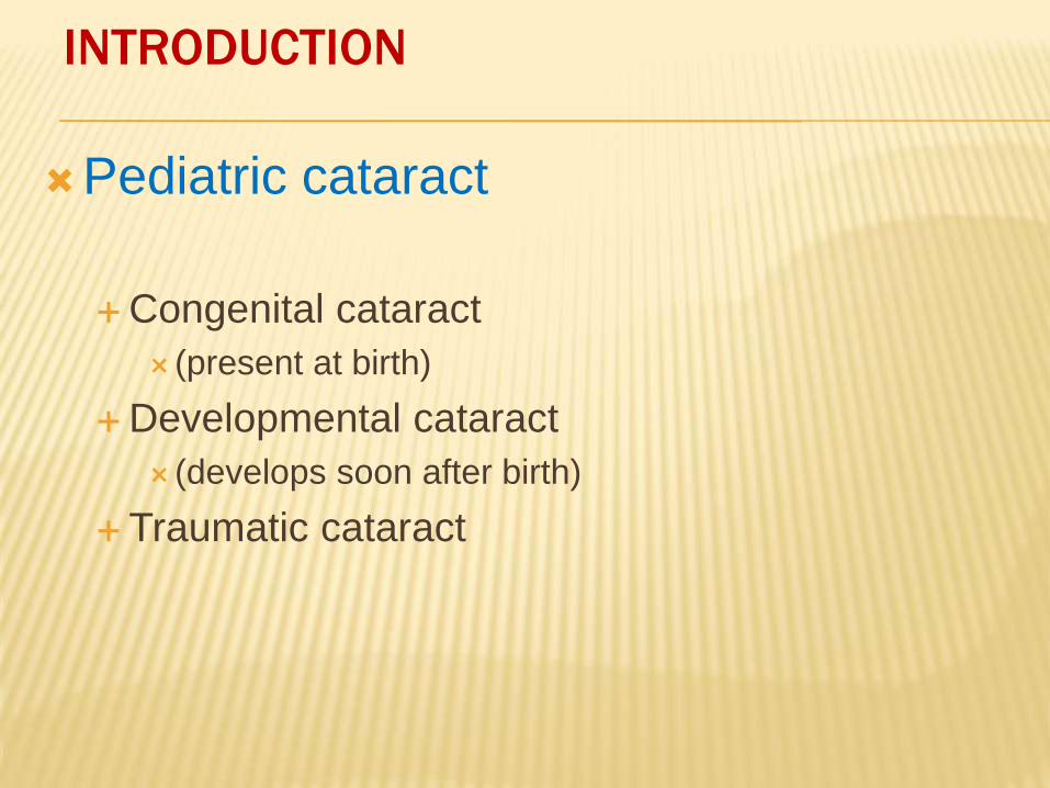
INTRODUCTION
Incidence - 6/10,000
10% of childhood blindness
Congenital or Acquired
Visually significant or not
Stable or Progressive
Unilateral or Bilateral
Partial or Complete
Developmental opacity are usually partial & stationary
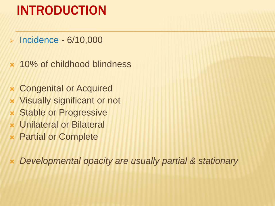
ANATOMY
Lens - A transparent,biconvex,crystalline structure
between iris & vitreous in a saucer shaped
depression called patellar fossa.
Components Anterior capsule
Anterior capsular epithelium
Cortex
Nucleus
Posterior capsule
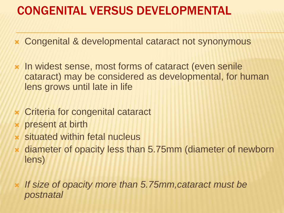
CONGENITAL VERSUS DEVELOPMENTAL
Congenital & developmental cataract not synonymous
In widest sense, most forms of cataract (even senile cataract) may be considered as developmental, for human lens grows until late in life
Criteria for congenital cataract
present at birth
situated within fetal nucleus
diameter of opacity less than 5.75mm (diameter of newborn lens)
If size of opacity more than 5.75mm,cataract must be postnatal
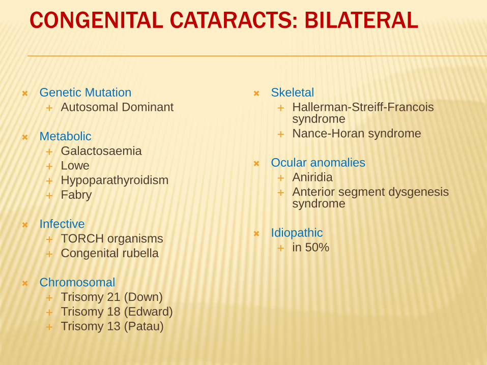
Genetic Mutation
Autosomal Dominant
Metabolic
Galactosaemia
Lowe
Hypoparathyroidism
Fabry
Infective
TORCH organisms
Congenital rubella
Chromosomal
Trisomy 21 (Down)
Trisomy 18 (Edward)
Trisomy 13 (Patau)
Skeletal
Hallerman-Streiff-Francois syndrome
Nance-Horan syndrome
Ocular anomalies
Aniridia
Anterior segment dysgenesissyndrome
Idiopathic
in 50%
CONGENITAL CATARACTS: BILATERAL
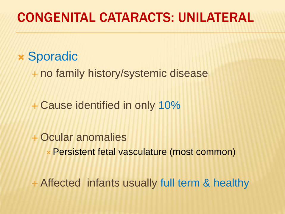
CONGENITAL CATARACTS: UNILATERAL
Sporadic
no family history/systemic disease
Cause identified in only 10%
Ocular anomalies
Persistent fetal vasculature (most common)
Affected infants usually full term & healthy
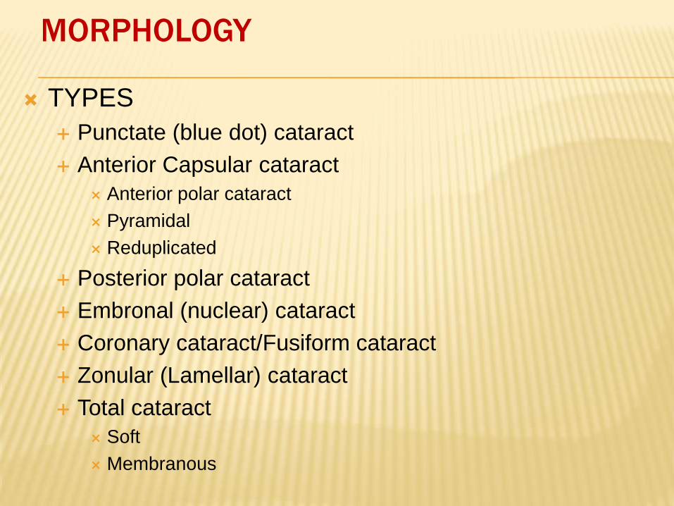
MORPHOLOGY
TYPES
Punctate (blue dot) cataract
Anterior Capsular cataract
Anterior polar cataract
Pyramidal
Reduplicated
Posterior polar cataract
Embronal (nuclear) cataract
Coronary cataract/Fusiform cataract
Zonular (Lamellar) cataract
Total cataract
Soft
Membranous
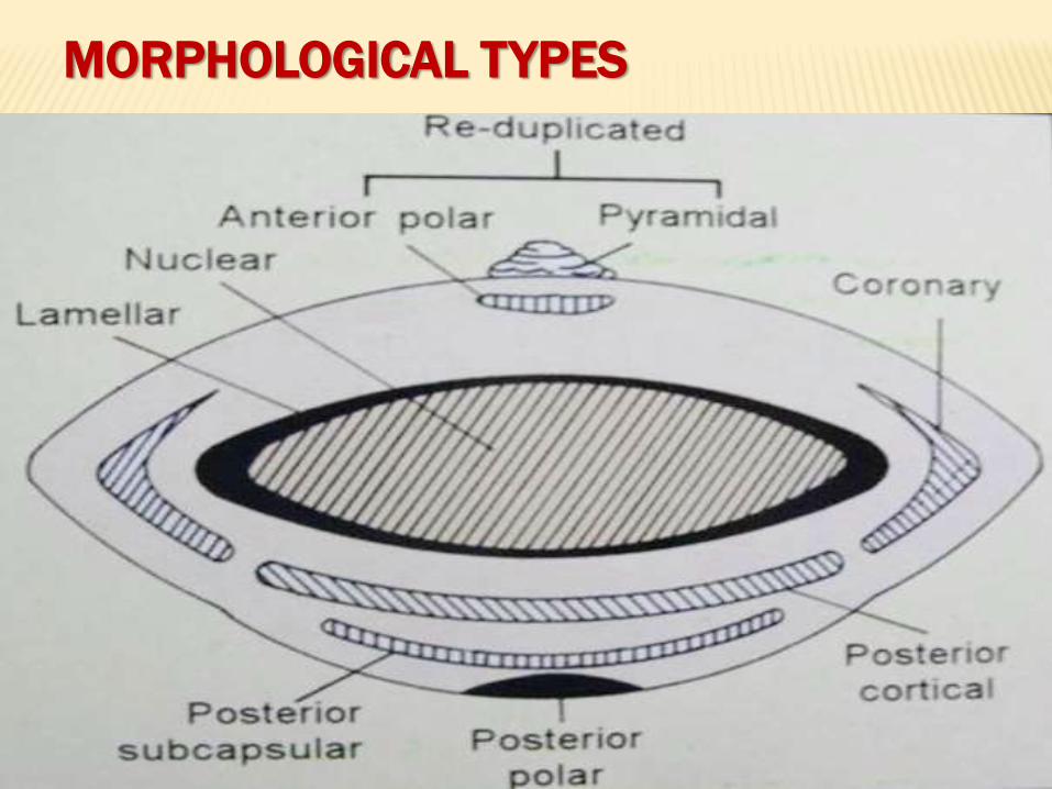
MORPHOLOGICAL TYPES
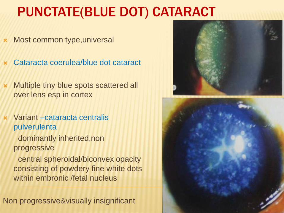
PUNCTATE(BLUE DOT) CATARACT
Most common type,universal
Cataracta coerulea/blue dot cataract
Multiple tiny blue spots scattered all
over lens esp in cortex
Variant –cataracta centralis
pulverulenta
dominantly inherited,non
progressive
central spheroidal/biconvex opacity
consisting of powdery fine white dots
within embronic /fetal nucleus
Non progressive&visually insignificant
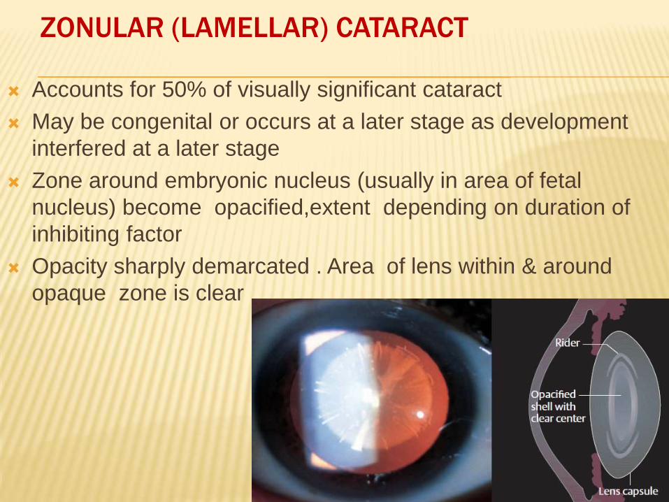
ZONULAR (LAMELLAR) CATARACT
Accounts for 50% of visually significant cataract
May be congenital or occurs at a later stage as development
interfered at a later stage
Zone around embryonic nucleus (usually in area of fetal
nucleus) become opacified,extent depending on duration of
inhibiting factor
Opacity sharply demarcated . Area of lens within & around
opaque zone is clear
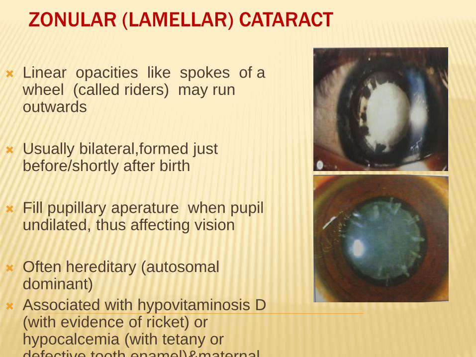
Linear opacities like spokes of a wheel (called riders) may run outwards
Usually bilateral,formed just before/shortly after birth
Fill pupillary aperature when pupil undilated, thus affecting vision
Often hereditary (autosomaldominant)
Associated with hypovitaminosis D (with evidence of ricket) or hypocalcemia (with tetany or defective tooth enamel)&maternal
ZONULAR (LAMELLAR) CATARACT
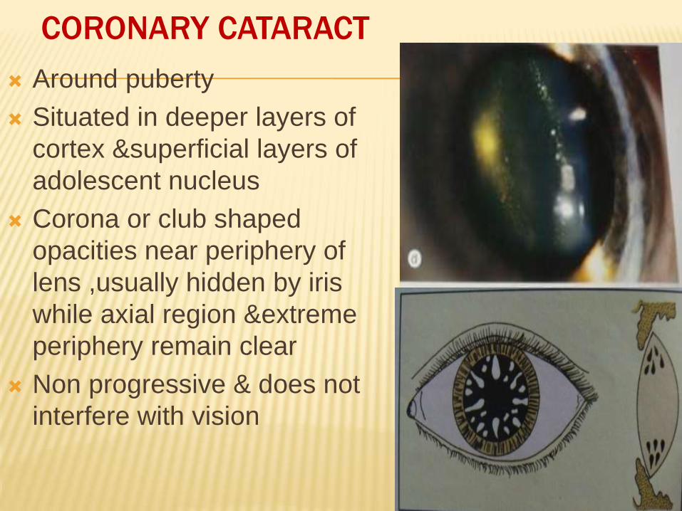
CORONARY CATARACT
Around puberty
Situated in deeper layers of
cortex &superficial layers of
adolescent nucleus
Corona or club shaped
opacities near periphery of
lens ,usually hidden by iris
while axial region &extreme
periphery remain clear
Non progressive & does not
interfere with vision
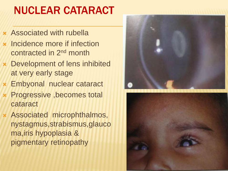
NUCLEAR CATARACT
Associated with rubella
Incidence more if infection
contracted in 2nd month
Development of lens inhibited
at very early stage
Embyonal nuclear cataract
Progressive ,becomes total
cataract
Associated microphthalmos,
nystagmus,strabismus,glauco
ma,iris hypoplasia &
pigmentary retinopathy
ANTERIOR CAPSULAR CATARACT
May be due to
1. Delayed formation of anterior chamber (Developmental)
2. Acquired (commonly)-follows contact of capsule with cornea,
usually after perforation of ulcer in ophthalmia neonatorum
Three types-
Anterior polar (flat type)
Anterior pyramidal
Reduplicated
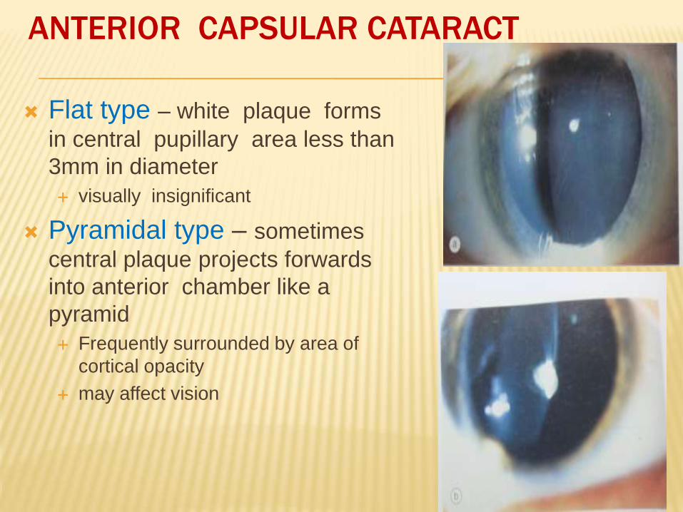
Flat type – white plaque forms
in central pupillary area less than
3mm in diameter
visually insignificant
Pyramidal type – sometimes
central plaque projects forwards
into anterior chamber like a
pyramid
Frequently surrounded by area of
cortical opacity
may affect vision
ANTERIOR CAPSULAR CATARACT
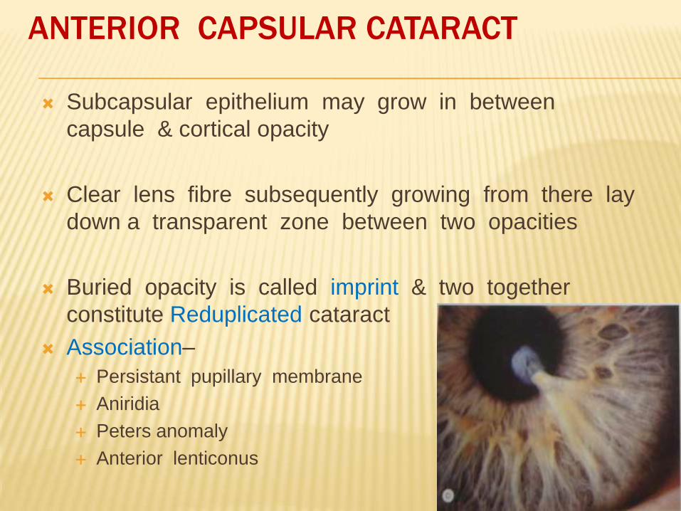
Subcapsular epithelium may grow in between
capsule & cortical opacity
Clear lens fibre subsequently growing from there lay
down a transparent zone between two opacities
Buried opacity is called imprint & two together
constitute Reduplicated cataract
Association–
Persistant pupillary membrane
Aniridia
Peters anomaly
Anterior lenticonus
ANTERIOR CAPSULAR CATARACT
POSTERIOR CAPSULAR ( POLAR) CATARACT
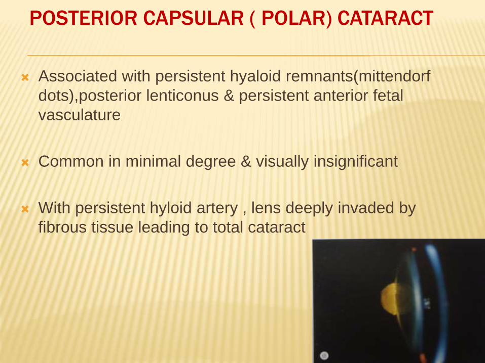
Associated with persistent hyaloid remnants(mittendorf
dots),posterior lenticonus & persistent anterior fetal
vasculature
Common in minimal degree & visually insignificant
With persistent hyloid artery , lens deeply invaded by
fibrous tissue leading to total cataract
OTHER TYPES
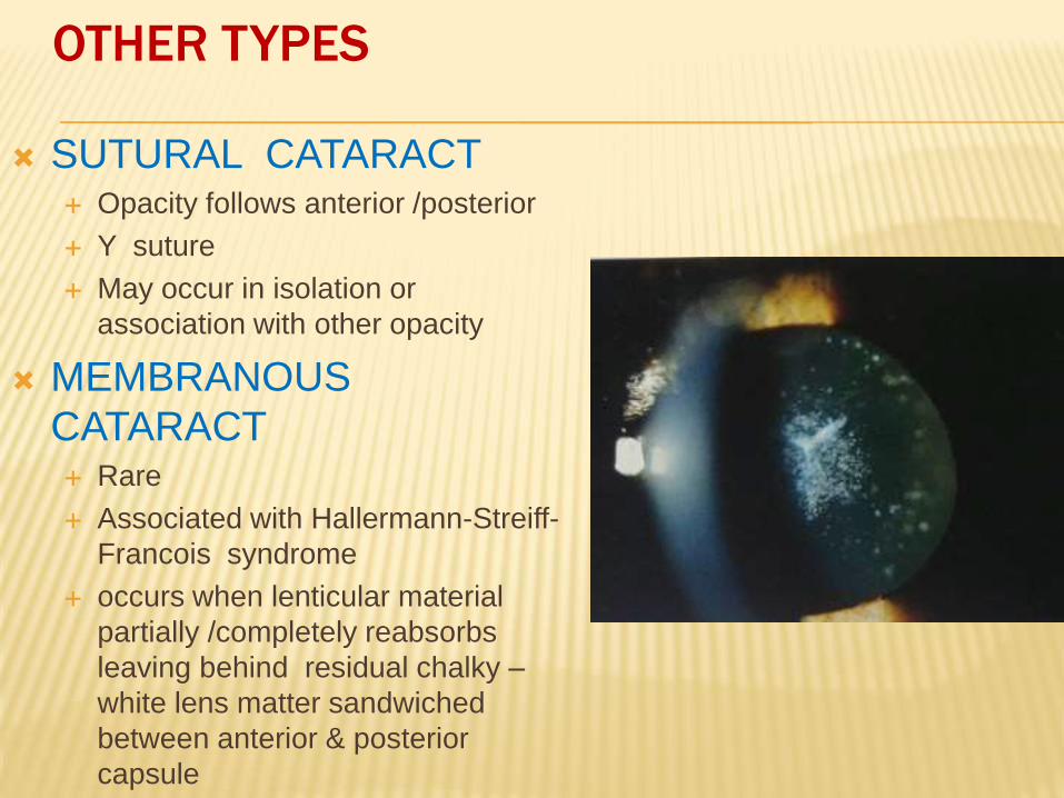
SUTURAL CATARACT Opacity follows anterior /posterior
Y suture
May occur in isolation or
association with other opacity
MEMBRANOUS
CATARACT Rare
Associated with Hallermann-Streiff-
Francois syndrome
occurs when lenticular material
partially /completely reabsorbs
leaving behind residual chalky –
white lens matter sandwiched
between anterior & posterior
capsule
SYMPTOMS OF DEVELOPMENTAL CATARACT
Informant – usually parents
History of white spot in pupillary area
Child is usually brought with history of diminution of vision / does not recognize objects and parents
Unsteady eyes
Deviation of eye
Associated symptoms of systemic disease, if present
SIGNS
Diminished vision (at times it is difficult to establish in very young children)
Lenticular opacity
Nystagmus
Deviation of eye
There may be other ocular and systemic abnormalities in cases of rubella nuclear cataract
MANAGEMENT OF DEVELOPMENTAL CATARACT
Investigations
I. Detailed history
II. Complete ocular examination-I. UCVA, BCVA, pupillary reaction intra-ocular tension, fundus
examination
II. B scan ultrasonography to exclude posterior segment
abnormality like growth/ retinoblastoma
III. A scan to determine axial length of the eye
IV. Retinoscopy, to deremine the refractive status
V. cover test to exclude squint
VI. Early photographs to know onset of cataract
Laboratory investigations:
(For bilateral cases)
Blood Test
Full blood count,Blood glucose(FBS/RBS)
Serum calcium and phosphorus
RBC transferase and Galactokinase levels
TORCH test
Hepatitis B virus
Urine analysis:
For reducing substance for galactosaemia
For amino acids (to exclude Lowe syndrome in suspected cases)
U/L Pediatric cases are mostly idiopathic.
No need of lab investigation
MANAGEMENT OF DEVELOPMENTAL CATARACT
Cataract in childhood not only reduce vision but also interfere
with normal visual development
Timing of surgery ,surgical technique,choice of aphakic
correction & amblyopia management are of utmost
importance to achieve good &long standing results
The inflammatory response to surgical results are more
pronounced in children
Post operative amblyopia management forms an integral part
of visual rehabilitation in children
MANAGEMENT OF DEVELOPMENTAL CATARACT
??????
To operate or not?
If yes, when to operate?
If no, how to manage?
If operating, should one place the IOL?
If not placing IOL, how to manage and follow the child?
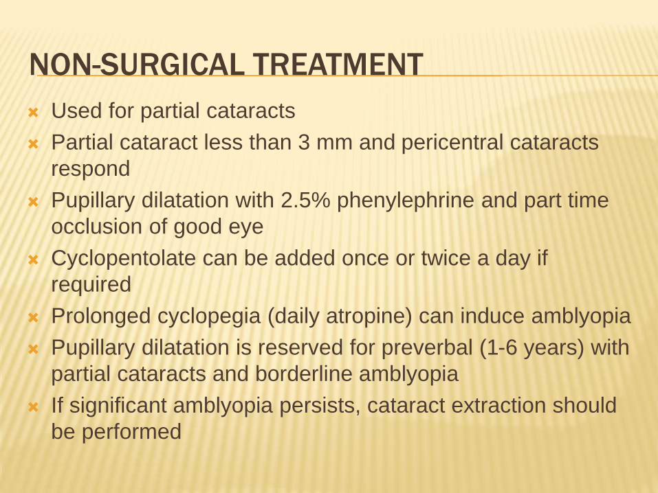
NON-SURGICAL TREATMENT
Used for partial cataracts
Partial cataract less than 3 mm and pericentral cataracts
respond
Pupillary dilatation with 2.5% phenylephrine and part time
occlusion of good eye
Cyclopentolate can be added once or twice a day if
required
Prolonged cyclopegia (daily atropine) can induce amblyopia
Pupillary dilatation is reserved for preverbal (1-6 years) with
partial cataracts and borderline amblyopia
If significant amblyopia persists, cataract extraction should
be performed
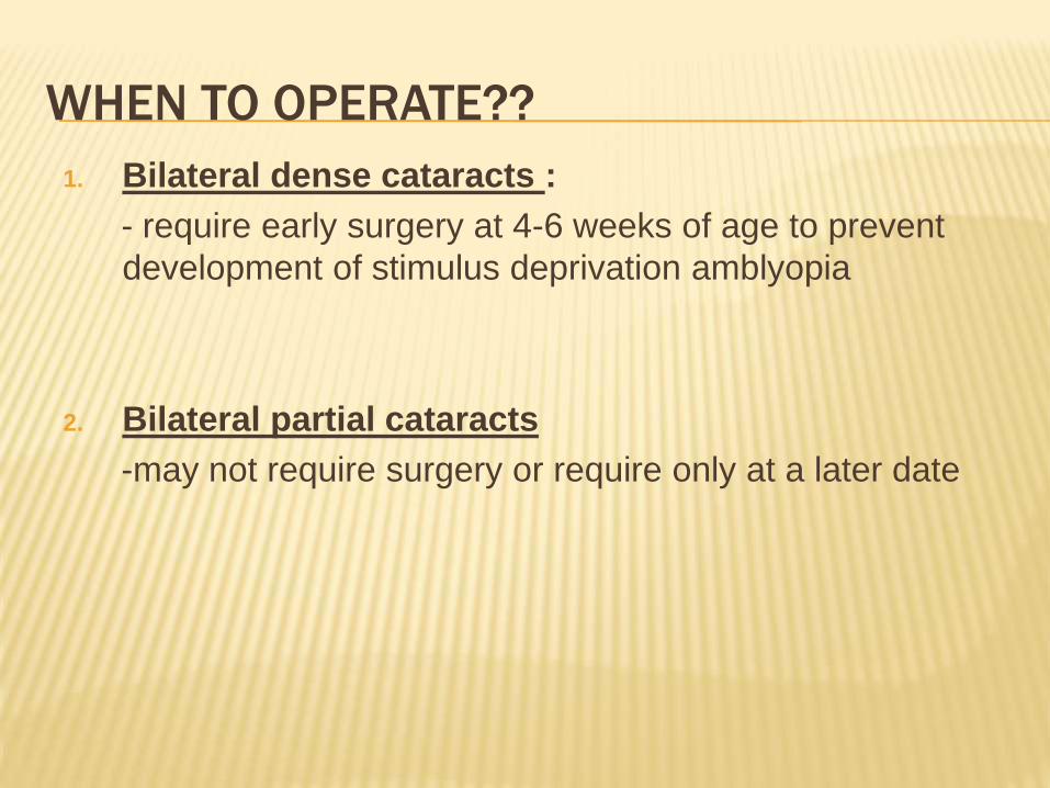
WHEN TO OPERATE??
1. Bilateral dense cataracts :
- require early surgery at 4-6 weeks of age to prevent
development of stimulus deprivation amblyopia
2. Bilateral partial cataracts
-may not require surgery or require only at a later date
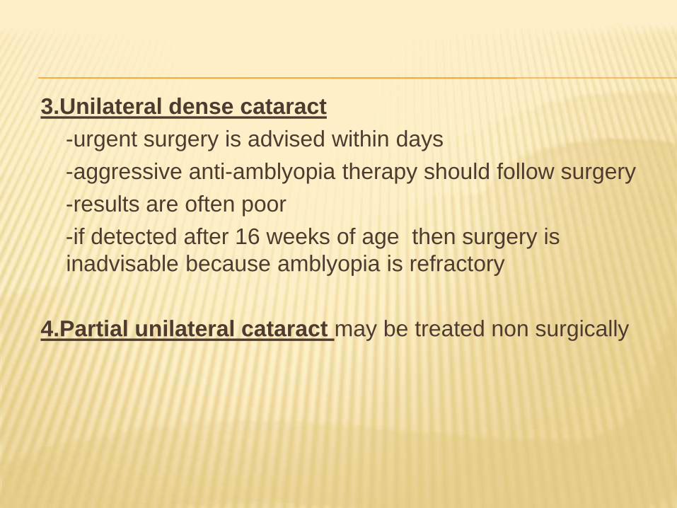
3.Unilateral dense cataract
-urgent surgery is advised within days
-aggressive anti-amblyopia therapy should follow surgery
-results are often poor
-if detected after 16 weeks of age then surgery is
inadvisable because amblyopia is refractory
4.Partial unilateral cataract may be treated non surgically

GENERAL TECHNIQUES
Deep general anaesthesia is required
Pediatric cataracts are soft – lens material can be aspirated
through incisions that are 1-1.5mm long at the limbus ;
can be subjected to lensectomy through pars plana
A larger wound is needed to introduce IOL
Tunnel should be securely sutured to prevent dehiscence of
wound with iris incarceration
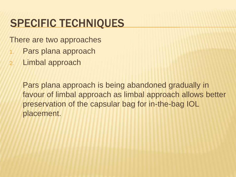
SPECIFIC TECHNIQUES
There are two approaches
1. Pars plana approach
2. Limbal approach
Pars plana approach is being abandoned gradually in
favour of limbal approach as limbal approach allows better
preservation of the capsular bag for in-the-bag IOL
placement.
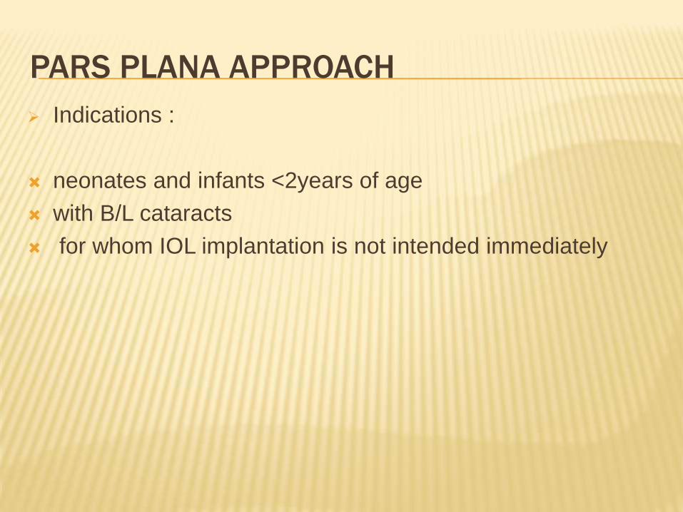
PARS PLANA APPROACH
Indications :
neonates and infants <2years of age
with B/L cataracts
for whom IOL implantation is not intended immediately
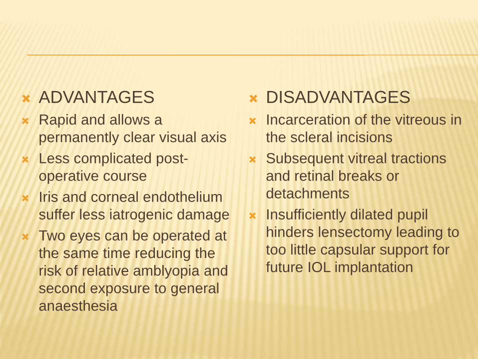
ADVANTAGES
Rapid and allows a
permanently clear visual axis
Less complicated post-
operative course
Iris and corneal endothelium
suffer less iatrogenic damage
Two eyes can be operated at
the same time reducing the
risk of relative amblyopia and
second exposure to general
anaesthesia
DISADVANTAGES
Incarceration of the vitreous in
the scleral incisions
Subsequent vitreal tractions
and retinal breaks or
detachments
Insufficiently dilated pupil
hinders lensectomy leading to
too little capsular support for
future IOL implantation
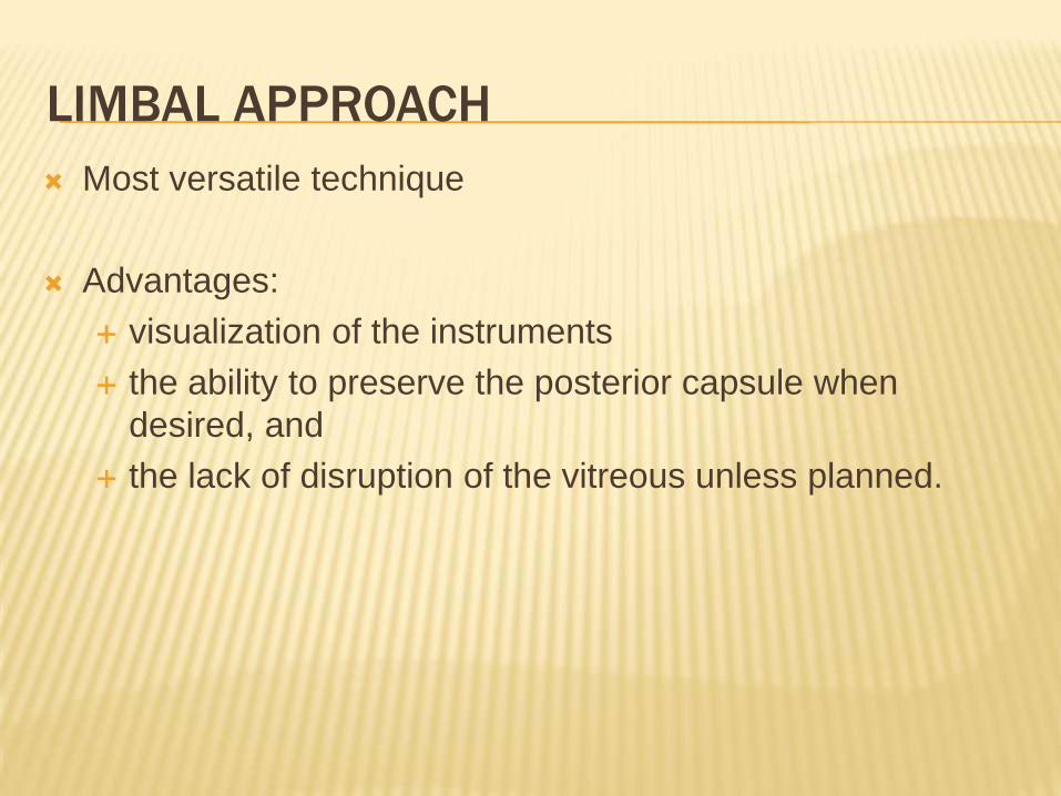
LIMBAL APPROACH
Most versatile technique
Advantages:
visualization of the instruments
the ability to preserve the posterior capsule when
desired, and
the lack of disruption of the vitreous unless planned.
APHAKIC CORRECTION IN CHILDREN
Spectacles
Contact lenses
IOLs
SPECTACLES
Satisfactory only in cases of B/L aphakia
Most develop good visual acuity
Cosmetically not acceptable
Poor optical quality of high –plus lenses
CONTACT LENSES
Better optical correction than spectacles
Dioptric power can be adjusted throughout the life
Difficult to manage and costly
Loss of lenses
Frequent infections
Poor follow up
Thus most impractical
INTRAOCULAR LENSES
The IOL facilitates amblyopia management by providing
a more permanent correction
Implanting an IOL in a growing eye is not an ideal
solution, but it is currently the most practical one.
The aim in the IOL option is to correct most, but not all,
of the aphakia
the residual refractive error has to be corrected using
spectacles, which can be adjusted throughout life.
Posterior chamber IOL implantation is the safe method
Both the biometry and the age of the child determine the
choice of the IOL dioptric power.
Age <2 years- axial length and the keratometric (K)
readings change rapidly
Age 2-8 years- changes are slower and more moderate.
Expected large Myopic Shift
AIM FOR UNDERCORRECTION
SELECTION OF IOL
GUIDELINES FOR THE CHOICE OF
INTRAOCULAR LENS DIOPTRIC POWER
CHILDREN LESS THAN 2 YEARS OLD
• Do biometry and undercorrect by 20%
• Use axial length measurements only:
- 17mm, 25 D
-18 mm, 24 D
-19 mm, 23 D
-20mm, 21 D
-21 mm, 19 D
CHILDREN BETWEEN 2 AND 8 YEARS OLD
• Do biometry and undercorrect by 10%
IOL Implantation in children
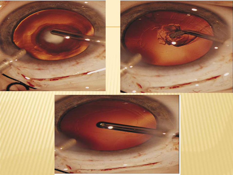
After the cataract has been aspirated, an elective posterior capsulectomy-anterior vitrectomy is performed.
Sulcus implantation is easier and also allows an easier explantation- may be done in neonates and infants less than 1 year of age. But with the newer foldable IOLs, in the bag implantation is the preferred technique.
An in-the-bag IOL is more difficult to explant, this option should be chosen for infants above 1 year of age because they are less likely to need an IOL exchange, provided they are undercorrected by 20%.
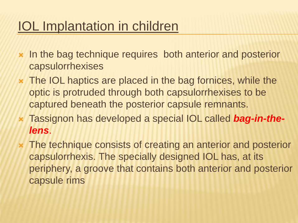
IOL Implantation in children
In the bag technique requires both anterior and posterior
capsulorrhexises
The IOL haptics are placed in the bag fornices, while the
optic is protruded through both capsulorrhexises to be
captured beneath the posterior capsule remnants.
Tassignon has developed a special IOL called bag-in-the-
lens.
The technique consists of creating an anterior and posterior
capsulorrhexis. The specially designed IOL has, at its
periphery, a groove that contains both anterior and posterior
capsule rims
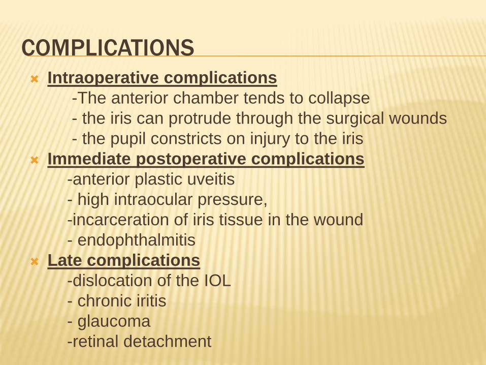
COMPLICATIONS
Intraoperative complications
-The anterior chamber tends to collapse
- the iris can protrude through the surgical wounds
- the pupil constricts on injury to the iris
Immediate postoperative complications
-anterior plastic uveitis
- high intraocular pressure,
-incarceration of iris tissue in the wound
- endophthalmitis
Late complications
-dislocation of the IOL
- chronic iritis
- glaucoma
-retinal detachment
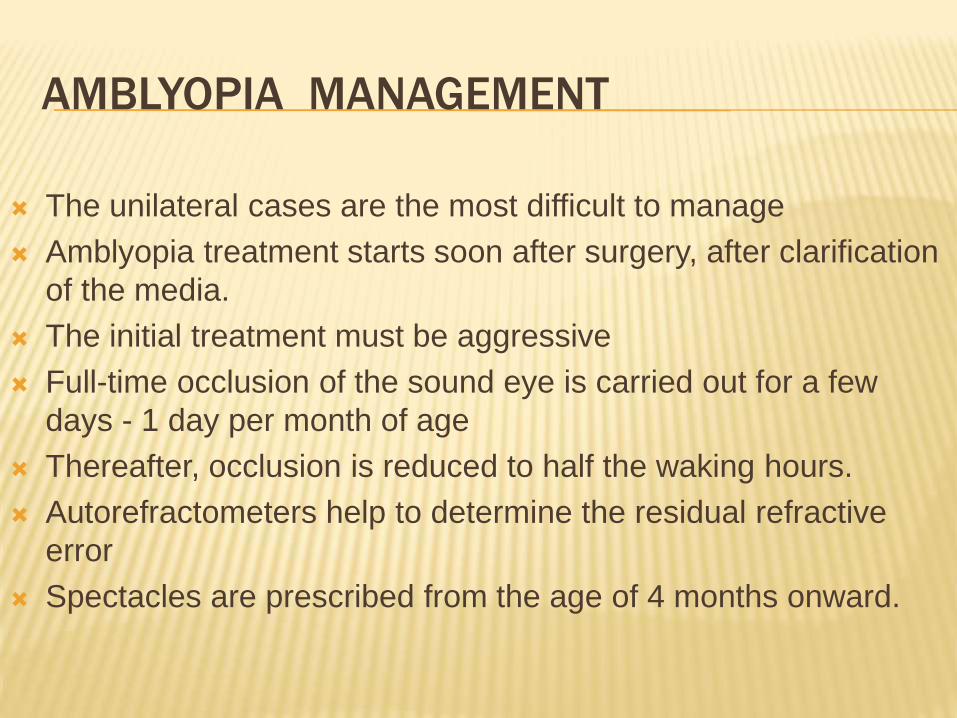
AMBLYOPIA MANAGEMENT
The unilateral cases are the most difficult to manage
Amblyopia treatment starts soon after surgery, after clarification
of the media.
The initial treatment must be aggressive
Full-time occlusion of the sound eye is carried out for a few
days - 1 day per month of age
Thereafter, occlusion is reduced to half the waking hours.
Autorefractometers help to determine the residual refractive
error
Spectacles are prescribed from the age of 4 months onward.
A bifocal lens with an add of +3.00 is prescribed in the
pseudophakic eye from the age of 3 years, when the
child becomes verbal.
Unilateral pseudophakes should continue with half-day
patches until 4-5 years of age.
Thereafter, the patch time can be reduced gradually, but
should not be abandoned until 10–12 years of age.
After that age, amblyopia management is practically
superfluous.
Cases of bilateral pseudophakia should be followed
closely to detect and treat relative amblyopia.
Intraocular Lens Exchange and Alternative
Options
Exchange of IOLs should be considered when a great
myopic shift has occurred
When the pseudophakic eye becomes 7 D more myopic
than the sound eye, the IOL should be exchanged
Refractive surgery in children is not yet an acceptable
option
An alternative to IOL exchange is to implant an additional
negative dioptric power IOL in posterior chamber to correct
the myopia.
This procedure is easily performed when the primary IOL
was inserted in the bag.
PROGNOSIS Visual outcome depends on
- Type of cataract
- Timing of intervention
- Quality of life
- Amblyopia management
Near normal vision can be achieved in U/L congenital
cataract, provided amblyopia management is aggressive
Binocularity is usually poor
Aphakic & pseudophakic children should be followed
throughout childhood &preferably throughout life

THANK YOU


![Overview of Congenital, Senile and Metabolic Cataractrelated cataract [7] and metabolic cataract [8]. Congenital & Senile Cataract Cataract is a clouding of the eye’s natural lens](https://static.fdocuments.net/doc/165x107/5f361b7a353bcc123d74d127/overview-of-congenital-senile-and-metabolic-cataract-related-cataract-7-and-metabolic.jpg)