PDH Kinase Inhibitor Screening Base on PDH...
21
PDH Kinase I PDH Activity Inhibitor Screening y 1 g Base on
Transcript of PDH Kinase Inhibitor Screening Base on PDH...

PDH Kinase Inhibitor Screening Base on
PDH Activity
PDH Kinase Inhibitor Screening Base on
PDH Activity
1
PDH Kinase Inhibitor Screening Base on

PDH Protocol #3: PDH Kinase Inhibitor Screening Base on PDH Activity
2
Table of contents
1. Introduction 3
2. Regulation of PDH activity 4
3. Materials 5
4. Additional materials required 6
5. Assay protocol 6
6. Protocol notes 11
7. Data analysis 11
8. Appendix A. Recipes for required components 12
9. Supporting data 14
10. Flow chart 19

PDH Protocol #3: PDH Kinase Inhibitor Screening Base on PDH Activity
3
1. Introduction
This protocol describes high throughput screens for inhibitors (or activators) of pyruvate
dehydrogenase kinase (PDK) isoforms PDK1, PDK2, PDK3 or PDK4 by measuring the activity of
PDH after exposure of immunocaptured PDH to each kinase in the presence and absence of test
compounds. Each set of measurements can be performed on 96 samples.
Although a variety of generic approaches can be used to measure protein kinase activity, the protocol
described here is unique as it allows one to screen rapidly for inhibitors/activators of each of the 4
isoforms of PDK by monitoring the functional effects of PDK phosphorylation on the activity of PDH
purified from any tissue or cell type of interest, including whole tissue/cell extracts of human, bovine,
rat or mouse cells, tissues or mitochondria. The PDH of interest is first quickly purified and
immobilized in microplate arrays by microscale immunocapture and then exposed to the PDK of
choice in the presence or absence of PDK inhibitors/activators. Residual PDH activity in each
microwell is then measured to determine whether or not test compounds inhibited, activated, or had
no effect on PDK phosphorylation of PDH. Unaltered PDKs phosphorylate PDH at a defined rate,
resulting in quantitative inactivation of PDH activity. In contrast, drug-induced inhibition of PDK
activity blocks PDK-mediated inhibition of PDH activity. Thus, compounds of interest can be added to
the PDK phosphorylation reaction and their effect on PDH phosphorylation can be analyzed, making
this product an ideal screening tool for inhibitors or activators of PDKs. Optimized conditions of the
PDK-mediated phosphorylation reactions are described that result in fully phosphorylated one (or
more) of PDH E1α Serines (232, 293 and 300), and fully inactivated PDH (Figures 2, 3, 4 and 5) when
unaltered PDK is used.
The main advantage of this protocol is that PDH is purified by immunocapture in individual microwells
and the endogenous specific and non-specific kinases originally associated with the enzyme are
washed away. Therefore, the subsequent PDK-mediated phosphorylation reaction is dependent only
on the activity of exogenously added PDK. Further advantages are as follows: (1) a variety of PDH
sources can be used and there is no need to biochemically purify PDH laboriously or to depend on a
commercial enzyme, (2) the phosphorylation reaction is stopped rapidly by a simple wash, (3) the
wash removes reactants of the phosphorylation reaction that would otherwise interfere with the PDH
activity assay, (4) the wash also removes traces of test compounds (potential PDK
inhibitors/activators) that might otherwise interfere directly with the PDH activity assay. Finally, if
desired, the immunocaptured PDH can first be fully dephosphorylated after immunocapture, but prior
to exposure to PDKs to maximize the PDK effect. Dephosphorylation can be accomplished with the
use of specific recombinant PDH phosphatases (PDP1 or PDP2) available from Abcam
(ab110357/MSP45 and ab110358/MSP46, respectively), and then the PDPs can be washed away
prior the PDK-mediated phosphorylation of immunocaptured PDH.

PDH Protocol #3: PDH Kinase Inhibitor
2. Regulation of PDH activity
The pyruvate dehydrogenase compl
metabolism, in that it links the TCA cycle and subsequent oxidative
and gluconeogenesis as well as with both l
of three enzymes, pyruvate dehydrogenase (E
dehydrogenase (E3), and one structural protein (E
composed of five different polypeptide chains,
stoichiometry. PDH catalyzes irreversible oxidative decarboxylation of pyruvate to acetyl coenzyme
A. The activity of PDH is negatively regulated by
phosphorylation is catalyzed by
dephosphorylation that activates
and 2.
The PDH kinases are Serine/Threonine
bound to the E2 domain of PDH
subunit (the phospho-Serine positions are given
Site 1 (Ser 293), Site 2 (Ser 300) and Site 3 (Ser 232),
Each of the kinases has different reactivity towards the three
normal dietary conditions PDK2 is the predominant isoform in skeletal muscle, the
by exercise and diet as well as by starvation
diabetes. The levels of PDK4 are sensitive to inhibitors of the PPAR transcription factors.
the levels of PDK1 are sensitive to O
increase in the level of PDK1 is a key part of
production that characterizes cancer cells.
disease, there is considerable interest in
Figure 1. Schematic representation of the PDdependent dephosphorylation of PDH.human E1α subunits. Phosphorylation results in inactivationactivation of PDH.
PDH Protocol #3: PDH Kinase Inhibitor Screening Base on PDH Activity
Regulation of PDH activity
complex (PDH) (E.C.1.2.4.1) is the key regulatory enzyme
, in that it links the TCA cycle and subsequent oxidative phosphorylation with glycolysis
and gluconeogenesis as well as with both lipid and amino acid metabolism. PD
pyruvate dehydrogenase (E1), dihyrolipoyl acyltransferase (E
one structural protein (E2/E3 binding protein). In total
polypeptide chains, E1α, E1β, E2, E3 and E2/E3BP
catalyzes irreversible oxidative decarboxylation of pyruvate to acetyl coenzyme
is negatively regulated by reversible phosphorylation of the E
phosphorylation is catalyzed by four PDH kinase isozymes (E.C.2.7.11.2), PDK
that activates PDH is catalyzed by two PDH phosphatases (E.C.
hreonine protein kinases. They are ATP-dependent
H. The PDH kinases phosphorylate three specific
Serine positions are given through this text with respect to the human protein):
Site 2 (Ser 300) and Site 3 (Ser 232), thus inhibiting the enzyme activity
has different reactivity towards the three phosphorylation sites
normal dietary conditions PDK2 is the predominant isoform in skeletal muscle, the
se and diet as well as by starvation. This kinase is aberrantly upregulated in insulin resistant
diabetes. The levels of PDK4 are sensitive to inhibitors of the PPAR transcription factors.
the levels of PDK1 are sensitive to O2 levels and under regulation by the transcription factor HIF1. An
1 is a key part of Warburg effect, a switch from oxidative to glycolytic ATP
production that characterizes cancer cells. Because of the manifold roles of PDKs in physiology
disease, there is considerable interest in identifying drugs that regulate PDK activities
Schematic representation of the PDK-dependent phosphorylationdependent dephosphorylation of PDH. Phosphorylation occurs at Serines 232
Phosphorylation results in inactivation of PDH, dephosphorylation results in
Screening Base on PDH Activity
4
key regulatory enzyme of cellular
phosphorylation with glycolysis
PDH is a large complex
dihyrolipoyl acyltransferase (E2), and dihydrolipoyl
n total the enzyme is
BP with 30:30:60:12:12
catalyzes irreversible oxidative decarboxylation of pyruvate to acetyl coenzyme
of the E1α subunit. The
PDK 1, 2, 3 and 4. The
(E.C.3.1.3.43), PDP 1
dependent enzymes that are
specific sites of the E1 α
text with respect to the human protein):
thus inhibiting the enzyme activity (Figure 1).
phosphorylation sites. While under
normal dietary conditions PDK2 is the predominant isoform in skeletal muscle, the PDK4 is induced
upregulated in insulin resistant
diabetes. The levels of PDK4 are sensitive to inhibitors of the PPAR transcription factors. In contrast,
nder regulation by the transcription factor HIF1. An
Warburg effect, a switch from oxidative to glycolytic ATP
Because of the manifold roles of PDKs in physiology and
regulate PDK activities.
phosphorylation and PDP-232, 293, and 300 of the
H, dephosphorylation results in

PDH Protocol #3: PDH Kinase Inhibitor Screening Base on PDH Activity
5
Abcam offers a comprehensive line of PDH-related assays and tools, including all four PDH kinases,
both PDH phosphatases, PDH activity microplate assays and PDH protein quantity microplate assays.
For convenience, these tools are available combined in kits and described in additional protocols.
See the PDH Playbook for an overview of applications possible using Abcam PDH assays and tools.
This protocol (PDH Protocol #3) describes how to screen for inhibitors and/or activators of each of the
four PDH kinases, using PDK-regulated PDH activity as a reporter. A complementary protocol (PDH
Protocol #4) describes how to screen for inhibitors and/or activators of the 4 PDKs by measurement of
PDK-mediated phosphorylation of PDH subunit E1α at serines 232, 293 and 300. The two protocols
(PDH activity-based and PDH phosphorylation-based) can be performed in parallel to obtain a
comprehensive analysis of the effects of potential inhibitors and/or activators of PDKs.
3. Materials
Most of the components required in this protocol are provided in the PDH Enzyme Activity Microplate
Assay Kit (ab109902/MSP18). The recipes for the components not included in ab109902/MSP18 are
provided in Appendix A of this protocol. Note – When using this protocol with ab109902/MSP18, it
REPLACES the standard ab109902/MSP18 protocol.
Item Quantity Storage
Detergent (component of ab109902/MSP18) 2 x 1 mL 4°C
20X Buffer (component of ab109902/MSP18) 15 mL 4°C
96-well Pre-coated Microplate (component of ab109902/MSP18)
1 EA 4°C
100X Reagent Dye (component of ab109902/MSP18) 0.25 mL -20°C
100X Coupler (component of ab109902/MSP18) 0.25 mL -80°C
20X Reagent Mix (component of ab109902/MSP18) 2 x 0.6 mL -80°C
50X ATP 500 µL -20°C
5X Stabilizer 13 mL 4°C
Phosphatase Inhibitor (PI) 1.5 mL 4°C
Choice of PDKs:
PDK1 (ab110359/MSP41) 50 µg -80°C
PDK2 (ab110354/MSP42) 50 µg -80°C
PDK3 (ab110355/MSP43) 50 µg -80°C
PDK4 (ab110356/MSP44) 50 µg -80°C
Avoid repeated freeze/thaw cycles of frozen components and keep them on ice when not in storage.

PDH Protocol #3: PDH Kinase Inhibitor Screening Base on PDH Activity
6
4. Additional materials required
• Spectrophotometer plate reader capable of measuring absorbance at 450 nm, preferably in a
kinetic mode.
• Method for determining protein concentration
• Multichannel pipette
• Deionized water
• PBS (phosphate buffered saline) – for recipe see Appendix A
5. Assay protocol
This protocol has four steps:
A. Sample Preparation.
PDH-containing extracts are prepared.
B. PDH Immunocapture.
PDH from the sample of interest is immunocaptured to a desired number of wells.
C. Phosphorylation Reactions.
The immunocaptured PDH is phosphorylated with PDK of choice in the presence (and in the
absence) of compound being tested as a modulator of PDK activity.
D. Activity Measurement.
Below are recommended amounts of materials. These amounts were carefully chosen to fit within
the linear range of PDH activity signal (Figure 6). For a control sample, purified bovine heart
mitochondria (ab110338/MS802) are available from Abcam.
Sample type Recommended amount
Purified mitochondria 50 µg/well (0.25 mg/mL)
Tissue homogenates 125 µg/well (0.625 mg/mL)
Cultured cells 500 µg/well (2.50 mg/mL)

PDH Protocol #3: PDH Kinase Inhibitor Screening Base on PDH Activity
7
This protocol describes the use of sufficient PDK1, 2, 3 or 4 to perform 96 phosphorylation
reactions with each enzyme, with each able to phosphorylate and fully inhibit PDH
immunocaptured using recommended amounts of materials (50 µg of bovine heart mitochondria
(available individually as ab110338/MS802, Bovine Heart Mitochondria IP Control), corresponding
to 1 milliunit of porcine PDH, 125 µg of rat or mouse heart tissues, or 500 µg of HepG2 cells).
The ab110338/MSP18 immunocapture microplate can be broken into 12 separate 8-well strips,
allowing 12 independent experiments. For convenience, considering the above, unless specified
otherwise, the volumes in this protocol are given for analysis of multiples of three strips (24 wells).
If different numbers of strips are used, simply adjust the volumes proportionally.
Note – Be completely familiar with protocol and protocol notes before beginning the assay.
Do not deviate from the specified protocol steps or optimal results may not be obtained.
A. Sample Preparation
1. Determine the sample protein concentration using a standard method such as BCA method.
2. Adjust the protein concentration of the sample, according to the sample type used, by dilution
in PBS, as specified below. See Note 1 for addition of protease inhibitors.
Sample type Protein concentration
(mg/mL)
Purified mitochondria 5.3
Tissue homogenates 23.7
Cultured cells 15.0
3. Prepare the extraction according to the table below. Mix components immediately after the
addition of the detergent. It is imperative to keep the ratio of components of extraction as
specified below. Below are suggested volumes for preparation of sufficient amounts for
loading of three strips (24 wells).
Component Purified
mitochondria Tissue
homogenates Cultured cells
Sample (µL) 285 237.5 1,350
Detergent (µL) 15 12.5 150
Total Volume (µL) 300 250 1,500
Final Protein Concentration (mg/mL) 5.0 22.5 13.5
4. Incubate on ice for 10 minutes.

PDH Protocol #3: PDH Kinase Inhibitor Screening Base on PDH Activity
8
5. Centrifuge in a tabletop centrifuge for 10 minutes at 4°C as specified below. Carefully collect
and save the supernatant. Discard the pellet.
Sample type RCF (x g)
Purified mitochondria 5,000
Tissue homogenates 1,000
Cultured cells 1,000
6. Prepare the Dilution Buffer as specified below.
No. of Plate Strips
dH2O (mL)
20X Buffer (mL)
Total (mL)
3 5.7 0.3 6
6 11.4 0.6 12
9 17.1 0.9 18
12 22.8 1.2 24
7. Dilute the samples’ supernatants in the Dilution Buffer to recommended concentration
according to the table below.
Sample type Recommended concentration
(mg/mL)
Purified mitochondria 0.25 (20X dilution)
Tissue homogenates 0.625 (36X dilution)
Cultured cells 2.50 (5.4X dilution)
B. PDH Immunocapture
1. Plate loading. Add 200 µL/well of diluted sample prepared in Step 7 of Section A.
2. Incubate microplate for 2.5 hours at room temperature.
C. Phosphorylation Reactions
1. Prepare 1X Reaction/Wash Buffer according to the table below.
1X Reaction/Wash Buffer
No. of Plate Strips
dH2O (mL)
20X Buffer (mL)
5X Stabilizer (mL)
PI (mL)
Total (mL)
3 29 2 8 0.8 40
6 58 4 16 1.6 80
9 88 6 24 2.4 120
12 117 8 32 3.2 160

PDH Protocol #3: PDH Kinase Inhibitor Screening Base on PDH Activity
9
2. Prepare 2X Compound of Interest Working Stock. Below are suggested volumes for
preparation of sufficient amounts for loading of two wells, allowing analysis in duplicates.
2X Compound of Interest Working Stock
No. of wells
dH2O (µL)
20X Buffer (µL)
5X Stabilizer (µL)
Compound of Interest (µL)
Total (µL)
2 187.5-X 12.5 50 X 250
3. Prepare PDK Working Stock following the appropriate table below. Prepare only the amount
needed. NOTE – It is important to load the PDKs at sub-saturating levels so reductions in
PDK activity (due to the action of a potential PDK inhibitor) can be detected easily. The
formulation below yields final amounts of PDK1, 2 or 4 at 1 µg/well, and PDK3 at 0.05 µg/well,
as these are rate-limiting amounts for many sample types (see Figures 2-5). It is
recommended that PDK titration curves as shown in Figures 2-5 be run for any novel source
of PDH used as a capture target in this assay.
2X PDK1, 2 or 4 Working Stock
No. of Plate Strips
dH2O (mL)
20X Buffer (mL)
5X Stabilizer (mL)
PI (µL)
PDK1, 2 or 4 at 1.5 µg/µL
(µL)
50X ATP (µL)
Total (mL)
3 1.73 0.13 0.52 104 17 104 2.6
6 3.45 0.26 1.04 208 35 208 5.2
9 5.17 0.39 1.56 312 52 312 7.8
12 6.90 0.52 2.08 416 69 416 10.4
2X PDK3 Working Stock
No. of Plate Strips
dH2O (mL)
20X Buffer (mL)
5X Stabilizer (mL)
PI (µL)
*PDK3 at 0.15 µg/µL
(µL)
50X ATP (µL)
Total (mL)
3 1.72 0.13 0.52 104 9 104 2.6
6 3.44 0.26 1.04 208 17 208 5.2
9 5.15 0.39 1.56 312 26 312 7.8
12 6.86 0.52 2.08 416 35 416 10.4
*Note – To ensure stability, PDK3 (an110355/MSP43) is shipped at 0.5 µg/µL and must first
be diluted to 0.15 µg/µL before being diluted again to make a “2X PDK3 Working Stock” as
described in the table. See Appendix A, Recipes for Required Components, page 12 “PDK of
choice” for more details.
4. Empty the wells of the microplate by turning it over and shaking out any remaining liquid. Blot
the plate face down on paper towel.

PDH Protocol #3: PDH Kinase Inhibitor Screening Base on PDH Activity
10
5. Add 300 µL/well of 1X Reaction/Wash Buffer to wash the wells.
6. Repeat the steps 6. and 7. one more time for a total of 2 wash steps.
7. Again, empty the wells of the microplate by turning it over and shaking out any remaining
liquid. Blot the plate face down on paper towel.
8. Add promptly 100 µL of 2X Compound of Interest Working Stock and 100 µL of 2X PDK
Working Stock into each well. Mix immediately. See Note 2 for phosphorylation reaction
controls.
9. Incubate the plate for 10 min at 30°C.
D. Activity measurement
1. Prepare the PDH Assay Solution, according to the table below. Warm up the PDH Assay
Solution to 30°C.
PDH Assay Solution
No. of Plate Strips
dH2O (mL)
20X Buffer (mL)
20X Reagent Mix
(mL)
100X Coupler (µL)
100X Reagent Dye
(µL)
Total Volume
(mL)
3 4.75 0.25 0.25 50 50 5.35
6 9.50 0.50 0.50 100 100 10.70
9 14.25 0.75 0.75 150 150 16.05
12 19.00 1.000 1.00 200 200 21.40
2. Empty the wells of the microplate by turning it over and shaking out any remaining liquid. Blot
the plate face down on paper towel.
3. Add 300 µL/well of 1X Reaction/Wash Buffer to wash the wells.
4. Repeat the steps 2. and 3. two more times for a total of 3 wash steps.
5. Again, empty the wells of the microplate by turning it over and shaking out any remaining
liquid. Blot the plate face down on paper towel.
6. Carefully add to each well 200 µL of PDH Assay Solution. Avoid bubbles. Any bubble should
be popped with a fine needle as rapidly as possible.
7. Promptly place the plate into a spectrophotometer and begin microplate reading using the
following parameters. (Alternatively, an endpoint measurement can be made at a user
defined time by recording absorbance at 450 nm.)

PDH Protocol #3: PDH Kinase Inhibitor Screening Base on PDH Activity
11
Mode: Kinetic
Wavelength: 450 nm
Time: 15 min
Interval: 20-30 sec
Shaking: Shake between readings
Temperature: 30°C
8. Save data and analyze them as described in the Data Analysis section.
6. Protocol notes
1. If desired, samples can be supplemented with protease inhibitors, such as Protease Inhibitor
Cocktail to minimize nonspecific proteolysis during the sample preparation.
2. Each experiment should include following two phosphorylation reaction controls using the
PDH source of interest. Control 1 (mock-PDK phosphorylated sample), this is a sample
treated in the absence of PDK and in the absence of compound of interest. Control 2 (mock-
compound of interest phosphorylated sample), this is a sample treated with PDK in the
absence of compound of interest.
3. If a compound of interest (included in the phosphorylation reaction) increases the PDH activity
and thus is suspected to be a PDK inhibitor, the direct toxic effect of the compound on PDH
activity should be assayed. This can be accomplished by including the compound directly into
the PDH assay solution.
7. Data analysis
1. As explained in the Introduction, the PDK activity is inversely proportional to the PDH activity.
2. The maximum PDH activity is equal the activity of the mock-PDK phosphorylated sample
(Control 1). Control 1 is the sample treated in the absence of PDK and in the absence of
compound of interest.
3. The residual PDH activity is equal the activity of the mock-compound of interest
phosphorylated sample (Control 2). Control 2 is a sample treated with PDK in the absence of
compound of interest and its PDH activity is fully inhibited due to the phosphorylation of one
(or more) PDH E1α Serines (232, 293 and 300).
4. If a compound of interest (included in the phosphorylation reaction) does not inhibit the PDK-
catalyzed phosphorylation of PDH E1α, the PDH activity of this sample is equal the activity of
Control 2.

PDH Protocol #3: PDH Kinase Inhibitor Screening Base on PDH Activity
12
5. If a compound of interest (included in the phosphorylation reaction) inhibits the PDK-catalyzed
phosphorylation of PDH E1α, the PDH activity of this sample is higher than the activity of
Control 2.
6. If a compound of interest (included in the phosphorylation reaction) fully inhibits the PDK-
catalyzed phosphorylation of PDH E1α, the PDH activity of this sample is equal the activity of
Control 1.
7. A direct inhibitory effect of a compound on PDH activity should be excluded. See Note 3.
8. Then the inhibitory effect of a compound of interest on the PDK-catalyzed phosphorylation of
PDH E1 α is proportional to the PDH activity measured and can be expressed as follows:
Effect of COI (%) = 100 X (ActivityCOI
-ActivityCon2
) / (ActivityCon1
– ActivityCon2
)
Abbreviation are as follows:
COI = Compound of Interest
Activity = PDH activity
Con1 = Control 1
Con2 = Control 2
9. Thus, a compound of interest will have no inhibitory effect on PDK-catalyzed phosphorylation
when the Effect of COI is 0 %. A compound of interest will have maximum inhibitory effect on
PDK-catalyzed phosphorylation when the Effect of COI is 100 %.
8. Appendix A. Recipes for required components
Phosphatase Inhibitor (PI)
• NaF FW 41.99
• Deionized Water
Component Grams per 100 mL Concentration (M)
NaF 2.10 0.5
Dissolve NaF in deionized water. Adjust volume to 100 mL. Store at -20°C.

PDH Protocol #3: PDH Kinase Inhibitor Screening Base on PDH Activity
13
5X Stabilizer
• Albumin, from bovine serum
• Deionized water
Component Grams per Liter Concentration (mg/mL)
Albumin, from bovine serum 50 50
Dissolve Albumin in water. Adjust volume to 1000 mL. Filter Sterilize and store at 4°C
50X ATP
• Adenosine 5’-triphosphate, disodium salt, FW 551.1
• Deionized water
Component Grams per 10 mL Concentration (mM)
Adenosine 5’-triphosphate 0.551 100
Dissolve Adenosine 5’-triphosphate in deionized water. Adjust volume to 10 mL.
Store aliquoted at -20°C.
PDK of choice (Abcam)
• PDK3, ab110355/MSP43. Note – This stock PDK3 is at 0.5 µg/µL and must be diluted to make the component PDK3 at 0.15 µg/µL
• PDK1, ab110359/MSP41. Note – This stock PDK1 is at 1.5 µg/µL and can be used as is for the component PDK1 at 1.5 µg/µL
• PDK2, ab110354/MSP42. Note – This stock PDK2 is at 1.5 µg/µL and can be used as is for the component PDK2 at 1.5 µg/µL
• PDK4, ab110356/MSP44. Note – This stock PDK4 is at 1.5 µg/µL and can be used as is for the component PDK4 at 1.5 µg/µL
• Dilution Buffer (10 mM Na-PO4, 250 mM NaCl, 30% glycerol, 5 mM DTT, 0.5 mM EDTA, 0.05% Triton X-100, pH 7.5 with HCl)
PDK of choice Grams per mL Concentration (µg/µL)
PDK1 NA 1.50
PDK2 NA 1.50
PDK3 NA 0.15
PDK4 NA 1.50
Avoid repeated freeze/thaw cycles of the enzymes and keep on ice when not in storage.
Thaw on ice the stock of the Human Recombinant PDK.
Equilibrate the Dilution Buffer to 4°C.
Dilute the Human Recombinant PDK in the Dilution Buffer to the desired concentration.
Aliquot to pre-chilled tubes.
Store at -80°C.

PDH Protocol #3: PDH Kinase Inhibitor Screening Base on PDH Activity
14
9. Supporting data
Figure 2. The PDKs phosphorylate and fully inactivate bovine PDH. As described in this protocol, PDH immunocaptured from 50 µg of bovine heart mitochondria was treated with indicated amounts of recombinant PDKs in the presence of ATP (in red) or in the absence of ATP (in blue). PDH activities (first row) were measured following PDH Protocol #3. PDH E1α Phospho-Serine 232 (second row), 293 (third row), and 300 (fourth row) quantities were measured following PDH Protocol #4. Note that: (1) 2 µg of PDK1 are sufficient to fully phosphorylate Serine 232, 293 and 300, and fully inactivate the PDH, (2) 2 µg of PDK2 are sufficient to fully phosphorylate Serine 293 and 300, and fully inactivate the PDH, (3) 0.1 µg of PDK3 is sufficient to fully phosphorylate Serine 293 and 300, and fully inactivate the PDH, and (4) 2 µg of PDK4 are sufficient to fully phosphorylate Serine 293 and 300, and fully inactivate the PDH. Also note that only PDK1 can phosphorylate the Serine 232 of PDH E1α. In addition, note that the treatment with ATP alone (in the absence of PDKs) when compared to the treatment in the absence of ATP does not substantially reduce the PDH activity, indicating absence of endogenous kinases.
PDH Activity
0 1 2 3 40
10
20
30
40
PDK1 (µµµµg/well)
∆∆ ∆∆ m
OD
450/m
in
PDH Activity
0 1 2 3 40
10
20
30
40
PDK2 (µµµµg/well)∆∆ ∆∆
mO
D4
50/m
in
PDH Activity
0.00 0.05 0.10 0.15 0.200
10
20
30
40
PDK3 (µµµµg/well)
∆∆ ∆∆ m
OD
450/m
in
PDH Activity
0 1 2 3 40
10
20
30
40
PDK4 ( µµµµg/well)
∆∆ ∆∆ m
OD
450/m
in
Phospho-Serine 232
0 1 2 3 40
100
200
300
400
PDK1 (µµµµg/well)
∆∆ ∆∆ m
OD
650/m
in
Phospho-Serine 232
0 1 2 3 40
100
200
300
400
PDK2 (µµµµg/well)
∆∆ ∆∆ m
OD
650/
min
Phospho-Serine 232
0.00 0.05 0.10 0.15 0.200
100
200
300
400
PDK3 (µµµµg/well)
∆∆ ∆∆ m
OD
650/
min
Phospho-Serine 232
0 1 2 3 40
100
200
300
400
PDK4 (µµµµg/well)
∆∆ ∆∆ m
OD
650/
min
Phospho-Serine 293
0 1 2 3 40
50
100
150
200
PDK1 (µµµµg/well)
∆∆ ∆∆ m
OD
650/m
in
Phospho-Serine 293
0 1 2 3 40
50
100
150
200
PDK2 (µµµµg/well)
∆∆ ∆∆ m
OD
650/
min
Phospho-Serine 293
0.00 0.05 0.10 0.15 0.200
50
100
150
200
PDK3 (µµµµg/well)
∆∆ ∆∆ m
OD
650/
min
Phospho-Serine 293
0 1 2 3 40
50
100
150
200
PDK4 (µµµµg/well)
∆∆ ∆∆ m
OD
650/
min
Phospho-Serine 300
0 1 2 3 40
100
200
300
PDK1 (µµµµg/well)
∆∆ ∆∆ m
OD
650/m
in
Phospho-Serine 300
0 1 2 3 40
100
200
300
PDK2 (µµµµg/well)
∆∆ ∆∆ m
OD
650/
min
Phospho-Serine 300
0.00 0.05 0.10 0.15 0.200
100
200
300
PDK3 (µµµµg/well)
∆∆ ∆∆ m
OD
650/
min
Phospho-Serine 300
0 1 2 3 40
100
200
300
PDK4 ( µµµµg/well)
∆∆ ∆∆ m
OD
650/
min

PDH Protocol #3: PDH Kinase Inhibitor Screening Base on PDH Activity
15
Figure 3. The PDKs phosphorylate and fully inactivate rat PDH. Extracts of rat heart homogenate were dephosphorylated with PDP1 to ensure maximum activity. Then, as described in this protocol, PDH was immunocaptured from 125 µg of the dephosphorylated rat heart homogenate and it was treated with indicated amounts of recombinant PDKs in the presence of ATP. PDH activities (first row) were measured following PDH Protocol #3. PDH E1α Phospho-Serine 232 (second row), 293 (third row), and 300 (fourth row) quantities were measured following PDH Protocol #4. Note that: (1) 2 µg of PDK1 are sufficient to fully phosphorylate Serine 232 and 293, and fully inactivate the PDH, (2) 2 µg of PDK2 are sufficient to fully phosphorylate Serine 293 and fully inactivate the PDH, (3) 0.1 µg of PDK3 is sufficient to fully phosphorylate Serine 293 and 300, and fully inactivate the PDH, and (4) 2 µg of PDK4 are sufficient to fully phosphorylate Serine 293 and 300, and fully inactivate the PDH. Also note that only PDK1 can phosphorylate the Serine 232 of PDH E1α.
PDH Activity
0 1 2 3 40
2
4
6
8
10
PDK1 (µµµµg/well)
∆∆ ∆∆ m
OD
450/m
inPDH Activity
0 1 2 3 40
2
4
6
8
10
PDK2 (µµµµg/well)
∆∆ ∆∆ m
OD
450/m
in
PDH Activity
0.00 0.05 0.10 0.15 0.200
2
4
6
8
10
PDK3 (µµµµg/well)
∆∆ ∆∆ m
OD
450/m
in
PDH Activity
0 1 2 3 40
2
4
6
8
10
PDK4 ( µµµµg/well)
∆∆ ∆∆ m
OD
45
0/m
in
Phospho-Serine 232
0 1 2 3 40
40
80
120
160
PDK1 (µµµµg/well)
∆∆ ∆∆ m
OD
650/m
in
Phospoho-Serine 232
0 1 2 3 40
40
80
120
160
PDK2 (µµµµg/well)
∆∆ ∆∆ m
OD
650/m
in
Phospho-Serine 232
0.00 0.05 0.10 0.15 0.200
40
80
120
160
PDK3 (µµµµg/well)
∆∆ ∆∆ m
OD
650/m
in
Phospho-Serine 232
0 1 2 3 40
40
80
120
160
PDK4 (µµµµg/well)
∆∆ ∆∆ m
OD
650/m
in
Phospho-Serine 293
0 1 2 3 40
40
80
120
160
PDK1 (µµµµg/well)
∆∆ ∆∆ m
OD
650/m
in
Phospho-Serine 293
0 1 2 3 40
40
80
120
160
PDK2 (µµµµg/well)
∆∆ ∆∆ m
OD
650/m
in
Phospho-Serine 293
0.00 0.05 0.10 0.15 0.200
40
80
120
160
PDK3 (µµµµg/well)
∆∆ ∆∆ m
OD
650/m
in
Phospho-Serine 293
0 1 2 3 40
40
80
120
160
PDK4 (µµµµg/well)
∆∆ ∆∆ m
OD
650/
min
Phospho-Serine 300
0 1 2 3 40
100
200
300
400
500
PDK1 (µµµµg/well)
∆∆ ∆∆ m
OD
650/m
in
Phospho-Serine 300
0 1 2 3 40
100
200
300
400
500
PDK2 (µµµµg/well)
∆∆ ∆∆ m
OD
650/m
in
Phospho-Ser 300
0.00 0.05 0.10 0.15 0.200
100
200
300
400
500
PDK3 (µµµµg/well)
∆∆ ∆∆ m
OD
650/m
in
Phospho-Serine 300
0 1 2 3 40
100
200
300
400
500
PDK4 (µµµµg/well)
∆∆ ∆∆ m
OD
650/m
in

PDH Protocol #3: PDH Kinase Inhibitor Screening Base on PDH Activity
16
Figure 4. The PDKs phosphorylate and fully inactivate mouse PDH. Extracts of mouse heart homogenate were dephosphorylated with PDP1 to ensure maximum activity. Then, as described in this protocol, PDH was immunocaptured from 125 µg of the dephosphorylated mouse heart homogenate and it was treated with indicated amounts of recombinant PDKs in the presence of ATP (in red) or in the absence of ATP (in blue). PDH activities (first row) were measured following PDH Protocol #3. PDH E1α Phospho-Serine 232 (second row), 293 (third row), and 300 (fourth row) quantities were measured following PDH Protocol #4. Note that: (1) 2 µg of PDK1 are sufficient to fully phosphorylate Serine 232 and 293, and fully inactivate PDH, (2) 2 µg of PDK2 are sufficient to fully phosphorylate Serine 293 and 300, and fully inactivate PDH, (3) 0.1 µg of PDK3 is sufficient to fully phosphorylate Serine 293 and fully inactivate the PDH, and (4) 2 µg of PDK4 are sufficient to fully phosphorylate Serine 293 and 300, and fully inactivate the PDH. Also note that only PDK1 can phosphorylate the Serine 232 of PDH E1α. In addition, note that the treatment with ATP alone (in the absence of PDKs) when compared to the treatment in the absence of ATP does not substantially reduce the PDH activity, indicating absence of endogenous kinases.
PDH Activity
0 1 2 3 40
20
40
60
PDK1 (µµµµg/well)
∆∆ ∆∆ m
OD
450/m
in
PDH Activity
0 1 2 3 40
20
40
60
PDK2 (µµµµg/well)
∆∆ ∆∆ m
OD
450/m
in
PDH activity
0.00 0.05 0.10 0.15 0.200
20
40
60
PDK3 ( µµµµg/well)
∆∆ ∆∆ m
OD
45
0/m
in
PDH Activity
0 1 2 3 40
20
40
60
PDK4 (µµµµg/well)
∆∆ ∆∆ m
OD
450/m
in
Phospho-Serine 232
0 1 2 3 40
200
400
600
PDK1 ( µµµµg/well)
∆∆ ∆∆ m
OD
650/m
in
Phospho-Serine 232
0 1 2 3 40
200
400
600
PDK2 (µµµµg/well)
∆∆ ∆∆ m
OD
650/m
in
Phospho-Serine 232
0.00 0.05 0.10 0.15 0.200
200
400
600
PDK3 (µµµµg/well)
∆∆ ∆∆ m
OD
65
0/m
in
Phospho-Serine 232
0 1 2 3 40
200
400
600
PDK4 (µµµµg/well)
∆∆ ∆∆ m
OD
650/m
in
Phospho-Serine 293
0 1 2 3 40
100
200
300
400
500
PDK1 ( µµµµg/well)
∆∆ ∆∆ m
OD
650/m
in
Phospho-Serine 293
0 1 2 3 40
100
200
300
400
500
PDK2 (µµµµg/well)
∆∆ ∆∆ m
OD
650/m
in
Phospho-Serine 293
0.00 0.05 0.10 0.15 0.200
100
200
300
400
500
PDK3 ( µµµµg/well)
∆∆ ∆∆ m
OD
65
0/m
in
Phospho-Serine 293
0 1 2 3 40
100
200
300
400
500
PDK4 (µµµµg/well)
∆∆ ∆∆ m
OD
650/m
in
Phospho-Serine 300
0 1 2 3 40
200
400
600
PDK1 ( µµµµg/well)
∆∆ ∆∆ m
OD
650/m
in
Phospho-Serine 300
0 1 2 3 40
200
400
600
PDK2 (µµµµg/well)
∆∆ ∆∆ m
OD
650/m
in
Phospho-Serine 300
0.00 0.05 0.10 0.15 0.200
200
400
600
PDK3 (µµµµg/well)
∆∆ ∆∆ m
OD
65
0/m
in
Phospho-Serine 300
0 1 2 3 40
200
400
600
PDK4 (µµµµg/well)
∆∆ ∆∆ m
OD
650/m
in
PDH Protocol #3: PDH Kinase Inhibitor Screening Base on PDH Activity
17
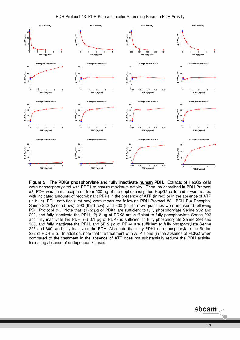
Figure 5. The PDKs phosphorylate and fully inactivate human PDH. Extracts of HepG2 cells were dephosphorylated with PDP1 to ensure maximum activity. Then, as described in PDH Protocol #3, PDH was immunocaptured from 500 µg of the dephosphorylated HepG2 cells and it was treated with indicated amounts of recombinant PDKs in the presence of ATP (in red) or in the absence of ATP (in blue). PDH activities (first row) were measured following PDH Protocol #3. PDH E1α Phospho-Serine 232 (second row), 293 (third row), and 300 (fourth row) quantities were measured following PDH Protocol #4. Note that: (1) 2 µg of PDK1 are sufficient to fully phosphorylate Serine 232 and 293, and fully inactivate the PDH, (2) 2 µg of PDK2 are sufficient to fully phosphorylate Serine 293 and fully inactivate the PDH, (3) 0.1 µg of PDK3 is sufficient to fully phosphorylate Serine 293 and 300, and fully inactivate the PDH, and (4) 2 µg of PDK4 are sufficient to fully phosphorylate Serine 293 and 300, and fully inactivate the PDH. Also note that only PDK1 can phosphorylate the Serine 232 of PDH E1α. In addition, note that the treatment with ATP alone (in the absence of PDKs) when compared to the treatment in the absence of ATP does not substantially reduce the PDH activity, indicating absence of endogenous kinases.
PDH Activity
0 1 2 3 40
5
10
15
PDK1 (µµµµg/well)
∆∆ ∆∆ m
OD
450/m
inPDH Activity
0 1 2 3 40
5
10
15
PDK2 (µµµµg/well)
∆∆ ∆∆ m
OD
45
0/m
in
PDH Activity
0.00 0.05 0.10 0.15 0.200
5
10
15
PDK3 (µµµµg/well)
∆∆ ∆∆ m
OD
450/m
in
PDH Activity
0 1 2 3 40
5
10
15
PDK4 (µµµµg/well)
∆∆ ∆∆ m
OD
45
0/m
in
Phospho-Serine 232
0 1 2 3 40
50
100
150
PDK1 (µµµµg/well)
∆∆ ∆∆ m
OD
650/m
in
Phospho-Serine 232
0 1 2 3 40
50
100
150
PDK2 (µµµµg/well)
∆∆ ∆∆ m
OD
65
0/m
in
Phospho-Serine 232
0.00 0.05 0.10 0.15 0.200
50
100
150
PDK3 (µµµµg/well)
∆∆ ∆∆ m
OD
650/m
in
Phospho-Serine 232
0 1 2 3 40
50
100
150
PDK4 (µµµµg/well)
∆∆ ∆∆ m
OD
65
0/m
in
Phospho-Serine 293
0 1 2 3 40
50
100
150
200
PDK1 (µµµµg/well)
∆∆ ∆∆ m
OD
650/m
in
Phospho-Serine 293
0 1 2 3 40
50
100
150
200
PDK2 (µµµµg/well)
∆∆ ∆∆ m
OD
65
0/m
in
Phospho-Serine 293
0.00 0.05 0.10 0.15 0.200
50
100
150
200
PDK3 (µµµµg/well)
∆∆ ∆∆ m
OD
650/m
in
Phospho-Serine 293
0 1 2 3 40
50
100
150
200
PDK4 (µµµµg/well)
∆∆ ∆∆ m
OD
65
0/m
in
Phospho-Serine 300
0 1 2 3 40
100
200
300
PDK1 (µµµµg/well)
∆∆ ∆∆ m
OD
650/m
in
Phospho-Serine 300
0 1 2 3 40
100
200
300
PDK2 (µµµµg/well)
∆∆ ∆∆ m
OD
65
0/m
in
Phospho-Serine 300
0.00 0.05 0.10 0.15 0.200
100
200
300
PDK3 (µµµµg/well)
∆∆ ∆∆ m
OD
650/m
in
Phospho-Serine 300
0 1 2 3 40
100
200
300
PDK4 (µµµµg/well)
∆∆ ∆∆ m
OD
65
0/m
in

PDH Protocol #3: PDH Kinase Inhibitor Screening Base on PDH Activity
18
Figure 6. Determination of a linear range of PDH activity signal of bovine, rat, mouse and human PDH. Indicated amounts of extracts of bovine heart mitochondria, rat heart homogenate, mouse heart homogenate and human HepG2 cells were immunocaptured and PDH activities were measured following PDH Protocol #3. Prior the immunocapture, the extracts of rat heart homogenate, mouse heart homogenate and human HepG2 cells were dephosphorylated with PDP1 to ensure maximum activity.
Bovine Heart Mitochondria
0 20 40 60 80 1000
20
40
60
80
100
Bovine Heart Mitochondria ( µµµµg/well)
∆∆ ∆∆ m
OD
45
0/m
inRat Heart Homogenate
0 50 100 150 200 2500
10
20
30
Rat Heart Homogenate (µµµµg/well)
∆∆ ∆∆ m
OD
45
0/m
in
Mouse Heart Homogenate
0 50 100 150 200 2500
20
40
60
80
100
Mouse Heart Homogenate ( µµµµg/well)
∆∆ ∆∆ m
OD
45
0/m
in
HepG2 Cells
0 100 200 300 400 5000
10
20
30
HepG2 cells (µµµµg/well)
∆∆ ∆∆ m
OD
45
0/m
in

PDH Protocol #3: PDH Kinase Inhibitor Screening Base on PDH Activity
19
10. Flow chart
For quick reference only. Be completely familiar with previous details of this document before
performing the assay.
Sample Preparation (1-3 hours)
• Dilute the sample to appropriate concentration with PBS
• Prepare the extraction
• Incubate samples 10 min on ice
• Centrifuge samples at appropriate g for 10 min
• Prepare Dilution Buffer
• Dilute the supernatant in the Dilution Buffer to appropriate concentration
PDH Immunocapture (2.5 hours)
• Load 200 µL of the diluted sample per well
• Incubate the plate for 2.5 hours at room temperature
Activity Measurement (30 minutes)
• Prepare PDH Assay Solution
• Wash wells trice with 1X Reaction/Wash Buffer
• Add 200 µL/ well of PDH Assay Solution
• Measure OD450 nm at 20 to 30 seconds intervals for 15 minutes at 30°C with shaking between the readings
Modification Reactions (20 minutes)
• Prepare 1X Reaction/Wash Buffer
• Prepare 2X Compound of Interest Working Stock
• Prepare PDK Working Stock
• Wash wells twice with 1X Reaction/Wash Buffer
• Add 100 µL/well of 2X Compound of Interest Working Stock and 100 µL/well PDK Working Stock
• Incubate the plate for 10 min at 30°C

PDH Protocol #3: PDH Kinase Inhibitor Screening Base on PDH Activity
20
For further technical questions please do not hesitate to contact us by email
([email protected]) or phone (select “contact us” on www.abcam.com for
the phone number for your region).

PDH Protocol #3: PDH Kinase Inhibitor Screening Base on PDH Activity
21
Abcam in the USA Abcam in Japan
Abcam Inc Abcam KK
1 Kendall Square, Ste B2304 41-16-8 Nihonbashi
Cambridge, Kakigaracho,
MA 02139-1517 Chuo-ku, Tokyo
USA 103-0014
Japan
Toll free: 888-77-ABCAM (22226) Fax: 866-739-9884 Tel: +81-(0)3-6231-094 Fax: +81-(0)3-6231-0941
Abcam in Europe Abcam in Hong Kong
Abcam plc Abcam (Hong Kong) Ltd
330 Cambridge Science Park Unit 225A & 225B, 2/F
Cambridge Core Building 2
CB4 0FL 1 Science Park West Avenue
UK Hong Kong Science Park
Hong Kong
Tel: +44 (0)1223 696000
Fax: +44 (0)1223 771600 Tel: (852) 2603-682 Fax: (852) 3016-1888
Copyright © 2011 Abcam, All Rights Reserved. The Abcam logo is a registered trademark. All information / detail is correct at time of going to print.