Standard Safety Practices in the Microbiology … Safety Practices in the Microbiology Laboratory...
56
Laboratory Safety | 163 L aboratorians working with infectious agents are subject to laboratory-acquired infections as a result of accidents or unrecognized incidents. The degree of hazard depends upon the virulence of the biological agent concerned and host resistance. Laboratory-acquired infections occur when microorganisms are inadvertently ingested, inhaled, or introduced into the tissues. Laboratorians are relatively safe when working with Haemophilus influenzae and Streptococcus pneumoniae; however, persons who work with aerosolized Neisseria meningitidis are at increased risk of acquiring a meningococcal infection. The primary laboratory hazard associated with enteric pathogens such as Shigella, Vibrio or Salmonella is accidental ingestion. Biosafety Level 2 (BSL-2) practices are suitable for work involving these agents that present a moderate potential hazard to personnel and the environment. The following requirements have been established for laboratorians working in BSL-2 facilities. • Laboratory personnel must receive specific training in handling pathogenic agents and be directed by competent scientists. • Access to the laboratory must be limited when work is being conducted. • Extreme precautions must be taken with contaminated sharp items. • Certain procedures involving the creation of infectious aerosols or splashes must be conducted by personnel who are wearing protective clothing and equipment. Standard microbiological safety practices The following safety guidelines listed below apply to all microbiology laboratories, regardless of biosafety level. Limiting access to laboratory Sometimes, people who do not work in the laboratory attempt to enter the laboratory to look for test results they desire. Although this occurs more frequently in clinical laboratories, access to the laboratory should be limited, regardless of the setting. Standard Safety Practices in the Microbiology Laboratory APPENDIX 1
Transcript of Standard Safety Practices in the Microbiology … Safety Practices in the Microbiology Laboratory...

Laboratory Safety | 163
Laboratorians working with infectious agents are subject to laboratory-acquiredinfections as a result of accidents or unrecognized incidents. The degree ofhazard depends upon the virulence of the biological agent concerned and host
resistance. Laboratory-acquired infections occur when microorganisms areinadvertently ingested, inhaled, or introduced into the tissues. Laboratorians arerelatively safe when working with Haemophilus influenzae and Streptococcuspneumoniae; however, persons who work with aerosolized Neisseria meningitidisare at increased risk of acquiring a meningococcal infection. The primarylaboratory hazard associated with enteric pathogens such as Shigella, Vibrio orSalmonella is accidental ingestion. Biosafety Level 2 (BSL-2) practices are suitablefor work involving these agents that present a moderate potential hazard topersonnel and the environment. The following requirements have been establishedfor laboratorians working in BSL-2 facilities.
• Laboratory personnel must receive specific training in handling pathogenicagents and be directed by competent scientists.
• Access to the laboratory must be limited when work is being conducted.
• Extreme precautions must be taken with contaminated sharp items.
• Certain procedures involving the creation of infectious aerosols or splashes mustbe conducted by personnel who are wearing protective clothing and equipment.
Standard microbiological safety practices
The following safety guidelines listed below apply to all microbiology laboratories,regardless of biosafety level.
Limiting access to laboratory
Sometimes, people who do not work in the laboratory attempt to enter thelaboratory to look for test results they desire. Although this occurs more frequentlyin clinical laboratories, access to the laboratory should be limited, regardless of thesetting.
Standard Safety Practices in the Microbiology Laboratory
APPENDIX 1

164 | Manual for Identification and Antimicrobial Susceptibility Testing
Biohazard signs or stickers should be posted near all laboratory doors and on allequipment used for laboratory work (e.g., incubators, hoods, refrigerators, andfreezers). Children under 12 years of age and pets are not allowed in laboratoryareas. All laboratories should be locked when not in use. In addition, all freezersand refrigerators located in corridors should be locked.
Handwashing
Each laboratory should contain a sink for handwashing. Hands should be washedfor at least one minute. Frequent handwashing is one of the most effectiveprocedures for avoiding laboratory-acquired infections. Hands should be washedwith an appropriate germicidal soap before exiting the laboratory and afterinfectious materials are handled. (Laboratorians working with gram-positiveorganisms should use alcohol (70%) to cleanse their hands if germicidal soap isunavailable.)
Eating
Eating, drinking, and smoking are not permitted in laboratory work areas. Foodmust be stored and eaten outside of the work area in designated areas used for thatpurpose only. Personal articles (e.g., handbags, eyeglasses, or wallets) should not beplaced on the workstations.
Mouth pipetting
Mouth pipetting is strictly prohibited in the laboratory. Rubber bulbs ormechanical devices should be used.
Sharps
A high degree of precaution must always be taken with any contaminated sharpitems, including needles and syringes, slides, pipettes, capillary tubes, and scalpels.Dispose of sharps in designated containers. To minimize finger sticks, useddisposable needles must not be bent, sheared, broken, recapped, removed from disposable syringes, or otherwise manipulated by hand before disposal.Non-disposable sharps, including syringes, should be placed in a labeled discardpan for decontamination before cleaning. Broken glassware should not be handleddirectly by hand but should be removed by mechanical means (e.g., a brush anddustpan, tongs, or forceps).
Aerosols
All procedures must be carefully performed to minimize splashes or aerosolization.Techniques that tend to produce aerosols should be avoided. Inoculating wires and

Laboratory Safety | 165
loops should be cooled by holding them still in the air for 5 – 10 seconds beforethey touch colonies or clinical material. Loops containing infectious materialshould be dried in the hot air above a burner before flaming. Vortexing andcentrifugation should be done in closed containers. (If safety capped tubes are notavailable, sealed tubes should be used.) Gauze should be used to remove the topson blood specimens and should be placed around the top of blood culture bottlesto minimize aerosol production during removal of the needle. Needles shouldnever be cut or removed from the syringe before autoclaving. All body fluidsshould be centrifuged in carriers with safety caps only.
When procedures with a high potential for creating infectious aerosols areconducted or when a procedure is used that can result in splashing or spraying ofthe face with infectious or other hazardous materials, laboratory work should beconducted in a safety cabinet or by laboratorians wearing the appropriate face-protection equipment (e.g., goggles, mask, face shield, or other splatter guards).Procedures that pose a risk may include centrifuging, grinding, blending, vigorousshaking or mixing, sonic disruption, opening containers of infectious materialswhose internal pressures may be different from ambient pressures, inoculatinganimals intranasally, and harvesting infected tissues from animals or eggs. Faceprotection should also be used when working with high concentrations or largevolumes of infectious agents.
Decontaminating bench tops and other surfaces
Bench tops should be wiped with a disinfectant (a phenolic disinfectant, 1%sodium hypochlorite [bleach], or 70% isopropyl alcohol) routinely after workingwith infectious agents or clinical specimens or after spills, splashes, or other contamination by infectious materials. Solutions of disinfectants should bemaintained at the work station (see Disinfectants).
Disposal of contaminated materials
All discarded plates, tubes, clinical samples, and other contaminated materialsshould be placed in disposal containers at each bench. Special disposal boxes mustbe used for sharps (e.g., syringes or broken glass) to minimize the risk of injury.Avoid overfilling such containers. Containers of contaminated material should becarefully transported to the autoclave room and autoclaved before disposal.
Autoclaving
An autoclave must be available for the BSL-2/3 laboratory and must be operatedonly by personnel who have been properly trained in its use. To verify that eachautoclave is working properly, spore strips or other biological indicators designedto test for efficiency of sterilization should be included in autoclave loads on a

166 | Manual for Identification and Antimicrobial Susceptibility Testing
regular basis. Each autoclave load should be monitored with temperature-sensitivetape, thermograph, or by other means (e.g., biological indicators).
General laboratory policies
All areas of the laboratory must be kept clean and orderly. Dirt, dust, crowding, orclutter is a safety hazard and is not consistent with acceptable biological research.Floors should be kept clean and free of unnecessary clutter. They should be washedwith a germicidal solution on a regular basis and after any spill of infectiousmaterial has occurred.
Refrigerators and freezers
Refrigerators and freezers should be regularly inspected for the presence of brokenvials or tubes containing infectious agents. When removing and discarding brokenmaterial, laboratorians should wear gloves and proper protective attire (e.g.,laboratory coat, goggles, or face-shield). Refrigerators and freezers should beregularly cleaned with a disinfectant and defrosted to prevent possiblecontamination and temperature failure.
Fire prevention
Burners should be used away from lamps and flammable materials. Bulkflammable material must be stored in the safety cabinet. Small amounts of theseflammable materials (e.g., ethyl acetate, ethyl alcohol, and methanol) can be storedin safety containers. Burners must be turned off when not in use. All laboratoriansmust know the location of fire extinguishers, fire blankets, and showers, and firesafety instructions and evacuation routes should be posted.
Special practices
Transport of biohazardous materials
Transport of biohazardous materials from one building to another increases therisk of breakage and spills. If transport is necessary, the primary infectious agentcontainer (regardless of size) must be placed in an unbreakable second containerthat can be sealed (e.g., using a screw-top tube or a plastic bag).
Disinfectants
Organisms may have different susceptibilities to various disinfectants. As a surfacedisinfectant, 70% alcohol is generally effective for the Enterobacteriaceae, but otherorganisms are more resistant. However, 70% isopropyl alcohol is not the

Laboratory Safety | 167
disinfectant of choice for decontaminating spills. Phenolic disinfectants, althoughexpensive, are usually effective against many organisms. Always read disinfectantlabels for manufacturers’ recommendations for dilution and for exposure times forefficacy, especially before use on BSL-3 organisms (e.g., Mycobacteriumtuberculosis). A effective general disinfectant is a 1:100 (1%) dilution ofhousehold bleach (sodium hypochlorite) in water; at this dilution, bleach can beused for wiping surfaces of benches, hoods and other equipment. A 1:10 (10%)dilution of bleach is corrosive and will pit stainless steel and should not be usedroutinely; however, the 10% bleach solution may be used to clean up spills ofcultured or concentrated infectious material where heavy contamination hasoccurred. If sodium hypochlorite is used as a disinfectant, the standard 1%dilutions should be made daily from a stock solution.
Decontamination of spills
The following procedure is recommended for decontaminating spills.
• Isolate the area to prevent anyone from entering.
• Wear gloves and protective clothing (e.g., a gown or lab coat, shoes, and a mask[if the spill may contain a respiratory agent or if the agent is unknown]).
• Absorb or cover the spill with disposable towels.
• Saturate the towels with an appropriately diluted intermediate or high-leveldisinfectant (e.g., a phenolic formulation or household bleach).
• Place disinfectant-soaked towels over the area and leave them in place for atleast 15 minutes before removing and discarding them.
• Wipe area using clean disinfectant-soaked towels and allow area to air dry.
• Place all disposable materials used to decontaminate the spill into a biohazardcontainer.
• Handle the material in the same manner as other infectious waste.
Accidents
All injuries or unusual incidents should be reported immediately to the supervisor.When cuts or puncture wounds from potentially infected needles or glasswareoccur, the affected area should be promptly washed with disinfectant soap andwater for 15 minutes. In the event of a centrifuge accident in which safety carriershave not been used, other personnel in the area should be warned immediatelyand the area isolated to prevent anyone from entering.
Protective clothing and equipment
Laboratory coats
Protective coats, gowns, smocks, or uniforms designated for laboratory use mustbe worn while working in the laboratory. Laboratory coats should fit properly andshould cover arms to the wrist. This protective clothing should be removed and leftin the laboratory before leaving for non-laboratory areas. All protective clothing iseither disposed of in the laboratory or laundered by the institution; it should neverbe taken home by personnel.
Gloves
Regardless of the type of infectious material, gloves should be worn whenperforming potentially hazardous procedures (e.g., slide agglutination) in whichthere is a risk of splashing or skin contamination or when the laboratory workerhas cuts or broken skin on his or her hands. Gloves should always be worn whenhandling clinical specimens, body fluids, and tissues from humans and animals.These tissues should be assumed to be positive for hepatitis B virus, humanimmunodeficiency virus (HIV), other bloodborne pathogens, and M. tuberculosis.Gloves must be removed when contaminated by splashing or spills or when workwith infectious materials is completed. Gloves should not be worn outside thelaboratory. Personnel should not use the telephone or open doors with gloves thathave been used in laboratory procedures. All used gloves should be disposed of bydiscarding them with other disposable materials and autoclaving. Hands should bewashed immediately after removing gloves.
Barrier precautions
Clinical specimens, body fluids, and tissues from humans and animals should beassumed to be positive for hepatitis B virus, HIV, other bloodborne pathogens, andM. tuberculosis. These materials should be handled in a safety cabinet or usingother barrier precautions (e.g., goggles, mask, face shield, or other splatter guards)whenever a procedure is performed that can potentially create an aerosol.
Useful biosafety references for the laboratory
• Centers for Disease Control and Prevention, National Institutes of Health.Biosafety in microbiological and biomedical laboratories. Washington, DC: U.S.Government Printing Office; 1999: stock no. 017-040-00547-4.
• World Health Organization. Laboratory Biosafety Manual, 2nd edition. Geneva:WHO; 1993: ISBN 92 4 154450 3.
168 | Manual for Identification and Antimicrobial Susceptibility Testing

Each laboratory must ensure adequate control of the media and reagents it uses.Quality control (QC) includes the selection of satisfactory reagents, thepreparation of media according to approved formulations or specific
manufacturer’s instructions, and the use of well-characterized reference strains tocheck prepared media. The World Health Organization (WHO) encourages centralpublic health laboratories to participate in at least three external quality assessmentsurveys per year; for reference laboratories, this may involve an internationaltesting scheme. National central laboratories (reference laboratories) should workto standardize procedures of regional and local laboratories to their own, so thatobservations can be interpreted in the same manner across sites.
Quality control of media
A summary of considerations for quality control of media, methods, and sourcesof quality control strains follows:
1) Considerations for quality control of media
Each batch of medium prepared from individual ingredients or each differentmanufacturer’s lot number of dehydrated medium should be tested for sterility, theability to support growth of the target organism(s), and/or the ability to produceappropriate biochemical reactions, as appropriate.
Sterility
• Incubate one tube or plate from each autoclaved or filter-sterilized batch ofmedium overnight at 35˚–37˚C and examine it for contaminants.
Ability to support growth of the target organism(s)
• Use at least one strain to test for ability of selective media to support growth of the target pathogen (e.g., for MacConkey agar, a Shigella strain such as S. flexneri). Documentation should be made regarding whether this strainproduces the appropriate biochemical reactions / color on the test medium.
Media, Reagents, and Quality Control
APPENDIX 2
Media, Reagents, and Quality Control | 169

170 | Manual for Identification and Antimicrobial Susceptibility Testing
(Discussions of specific biochemical reactions are included in the media sectionof this appendix.)
Ability to produce appropriate biochemical reactions
• For selective media: Use at least one organism that will grow on the mediumand at least one organism that will not grow on the selective medium to test forthe medium’s ability to differentiate target organisms from competitors. If themedium is both selective and differential, it may be useful to include twoorganisms that will grow on the medium and produce different reactions (e.g.,for MacConkey agar: a lactose-nonfermenting organism such as S. flexneri; alactose-fermenting organism such as E. coli; and, S. aureus, which should notgrow).
• For biochemical media: Use at least one organism that will produce a positivereaction and at least one organism that will produce a negative reaction (e.g.,for urea medium, a urease-positive organism such as Proteus and a urease-negative organism such as E. coli).
2) Methods for quality control of media
When testing for ability of a medium to support growth, a small inoculum willgive greater assurance that the medium is adequate for recovery of a small numberof organisms from a clinical specimen; therefore, use a dilute suspension of controlorganisms to inoculate the medium for QC. An example of a protocol for qualitycontrol of media follows here:
a) Inoculate the control strain to nonselective broth (e.g., a tryptone-based soybroth [TSB]) and incubate / grow overnight.
b) Prepare a standardized inoculum for testing the medium. The appropriatestandard dilution differs for selective and nonselective media.
• If testing selective or inhibitory media: To prepare a standardized inoculumfor testing selective and inhibitory media, make a 1:10 dilution of theovernight nonselective broth culture.
• If testing nonselective media: To prepare a standardized inoculum fortesting nonselective media, make a 1:100 dilution of the nonselective brothculture.
c) Using a calibrated loop, if available, inoculate one tube or plate of each mediumwith a loopful of the standardized inoculum of the control strain(s). (Ifperforming QC of plating medium, streak for isolation.) The same loop shouldbe used for all QC of all media; it is more important to have the consistency ofthe same inoculating loop every time than it is to use a calibrated loop.

Media, Reagents, and Quality Control | 171
• If testing selective or inhibitory media: A nonselective plating medium (e.g.,heart infusion agar [HIA]) should be inoculated at the same time as theselective medium for comparison purposes.
3) Sources of quality control strains
Suitable QC strains may be obtained in the following ways.
• A laboratory may use strains isolated from clinical specimens or qualityassurance specimens, provided the strains have been well characterized by allavailable methods (e.g., biochemical, morphologic, serologic, and molecular).
• Many laboratories purchase QC strains from official culture collections (e.g., the American Type Culture Collection [ATCC] and the NationalCollection of Type Cultures [NCTC]).34 (Addresses for ATCC and NCTC are included in Appendix 13.)
Quality control strains appropriate for antimicrobial susceptibility testing asincluded in this manual
The following ATCC numbers can be used to identify the appropriateantimicrobial susceptibility testing QC organisms included in this laboratorymanual.
Haemophilus influenzae H. influenzae ATCC 49247 *(* for testing of the antimicrobial agents included in this
laboratory manual)
Neisseria meningitidis S. pneumoniae ATCC 49619
Streptococcus pneumoniae S. pneumoniae ATCC 49619
Neisseria gonorrhoeae N. gonorrhoeae ATCC 49226 (other reference strains, available from CDC, may be
used when testing antimicrobial agents not included in
NCCLS criteria)
Salmonella serotype Typhi E. coli ATCC 25922
Shigella E. coli ATCC 25922
Vibrio cholerae E. coli ATCC 25922
34 This manual presents the ATCC numbers for quality control organisms, but these ATCC strains may also beobtained from the NCTC.

172 | Manual for Identification and Antimicrobial Susceptibility Testing
Quality control of reagents
As with all other products used in testing, reagents (whether purchased orprepared in the laboratory) should be clearly marked to indicate the date on whichthey were first opened and the expiration date, if appropriate. Each reagent shouldbe tested to make sure the expected reactions are obtained.
If the reagent is a rare, expensive, or difficult-to-obtain product (e.g., diagnosticantiserum) it does not necessarily have to be discarded on the expiration date.If satisfactory sensitivity and specificity can still be verified by normal QCprocedures, the laboratory may indicate on the vial label the date of verification of quality of the reagent. All reagents should be tested for quality at intervalsestablished by each laboratory to ensure that no deterioration has occurred; if thequality of the reagent is being verified after the expiration date, testing should beperformed more frequently.
Slide agglutination method for quality control of antisera
For QC of antiserum, two or more control strains (one positive and one negative)should be used to test the agglutination characteristics of the antiserum. The resultsof all reactions should be recorded. Following is an example of a typical QCprocedure.
• Place a drop (about 0.05 ml, though as little as 10 µl can be used) of eachantiserum on a slide or plate. Also, place a drop of 0.85% saline on each slide orplate to test each antigen for roughness or autoagglutination.
• Prepare a densely turbid suspension (2 or 3 McFarland turbidity standard,see Table 21) of each control isolate in 0.85% saline with growth asepticallyharvested from an 18- to 24-hour culture from nonselective agar (e.g., HIA ortryptone soy agar [TSA]).
• Add one drop of the antigen suspension to the antiserum and the saline. Mixthoroughly with an applicator stick, glass rod, or inoculating loop. Rock theslide back and forth for 1 minute.
• Read the agglutination reaction over a light box or an indirect light source witha dark background. The saline control must be negative for agglutination forthe test to be valid.
The degree of agglutination should be read and recorded as follows:
Percentage of agglutination: Record reaction as:100% 4+75% 3+50% 2+25% 1+0% negative

Media, Reagents, and Quality Control | 173
Advantages of centralized acquisition of media and reagents
Centralizing acquisition of media and reagents in the national reference laboratoryor Ministry of Health can provide several benefits:
• Large amounts of a single lot of medium or reagent can be purchased andsubsequently divided into smaller aliquots for distribution to provincial/districtlaboratories. This may be more cost effective because of, e.g., discounts forlarger orders, lower shipping costs, less waste because of product going pastexpiration date, etc.
• Quality control can be performed in the central laboratory, avoidingduplication of effort among provincial and district laboratories. Anunsatisfactory medium or reagent can then be returned to the manufacturerbefore the lot is distributed to other laboratories.
• The standardization of methods among laboratories at all levels is facilitated byuse of single lots of media.
Preparation of media and reagents
Each manufacturer’s lot number of commercial dehydrated medium and eachbatch of medium prepared from individual ingredients should be qualitycontrolled before use. Immediately after preparation, each medium should betested, as appropriate, with a reference strain of H. influenzae, N. meningitidis,S. pneumoniae, N. gonorrhoeae, S. Typhi, Shigella, and/or V. cholerae O1/O139 for proper growth characteristics as described for each medium.
A record of all media preparation or purchase dates and quality control test resultsshould be kept, and any unusual characteristic (e.g., the color of the medium orslow growth of test bacteria) should be noted.
Many media call for the use of defibrinated blood. Defibrinated blood can beprepared mechanically by swirling 30 ml blood and (e.g.) sterile glass beads or awooden-stick device in a 125–250 ml Erlenmeyer flask at approximately 90 rpmfor 7–9 minutes. (Sterile paper clips in a flask can also serve to assist indefibrination.) Blood is defibrinated when clotting factors have been removed;they will be visible in the flask as a translucent, fibrous “web.” (A useful referencefor “low-technology” methods to defibrinate blood is in a publication by Kay et al.[1986], and is included in Appendix 15.)
Agar media should be dispensed into 15 x 100-mm or 15 x 150-mm Petri dishes to a uniform depth of 3–4 mm; approximately 20-ml of liquid agar medium willachieve this depth in a 15 x 100-mm plate. If agar is cooled to 50˚C prior topouring, condensation is minimized. After pouring, the plates should be kept at

room temperature for several hours to prevent excess condensation from formingon the covers of the dishes. Another means by which condensation will be reducedis if plates are stacked so that they cool more slowly. Alternatively, if whenpreparing selective media (e.g., MacConkey [MAC], xylose lysine desoxycholate[XLD], thiosulfate citrate bile salts [TCBS] agar, etc.), conditions are such thatthere is little chance that the cooling media will be contaminated, after the agar ispoured into the plates, the lids can be placed on the dish so that a small opening isleft to let the heat out, resulting in the formation of less condensation on the upperlid; the lid should remain slightly open like this for approximately 30 minutes,while the agar solidifies. If, however, it is likely that the agar will be contaminated ifthe lid is left partly open, the agar should be allowed to solidify with the lid closed.
Note: Covering the agar while it is still hot will allow for the formation of asubstantial amount of condensation on the upper lid. If the plates containcondensation, the plates should be covered at room temperature for 24 hours to allow the condensation to evaporate. After condensation has evaporated, theplates should be placed in an inverted position and stored in a plastic bag in aninverted position at 4˚C.
Media for enrichment, identification, and antimicrobial susceptibility testing
Some of the culture media included in this manual are commonly referred to bytheir abbreviations rather than their full names. Therefore, when these media arementioned in the methods for the preparation or quality control of other media inthis section, they are referred to by their abbreviations.
Name of commonly abbreviated culture medium Abbreviation
Alkaline peptone water APW
Bismuth sulfite agar BS
Cystine trypticase agar CTA
Desoxycholate citrate agar DCA
Gram-negative broth GN
Hektoen enteric agar HE
Heart infusion agar HIA
Haemophilus test medium HTM
Kligler iron agar KIA
Lysine iron agar LIA
MacConkey agar MAC
Martin-Lewis agar ML
Modified Thayer-Martin agar MTM
174 | Manual for Identification and Antimicrobial Susceptibility Testing

Name of commonly abbreviated culture medium, continued Abbreviation
Phosphate buffered saline PBS
Selenite broth SEL
Sulfide-indole-motility medium SIM
Salmonella-Shigella agar SS
Thiosulfate citrate bile salts sucrose agar TCBS
Tryptone (Trypticase) soy agar TSA
Tryptone (Trypticase) soy broth TSB
Triple sugar iron agar TSI
Xylose lysine desoxycholate agar XLD
Methods for the preparation of media (from individual ingredients orcommercially available preparations) follow, in alphabetical order.
Acidometric agar
This agar may be used to test H. influenzae for ß-lactamase if nitrocefin is notavailable.
Distilled water 100 ml
Agar 1.5 g
0.5% phenol red solution 0.2 ml
NaOH (1 Normal)
Penicillin G powder (to prepare a 5000 unit/ml concentration)
Combine distilled water, agar, and phenol red solution and boil until the agardissolves. Adjust the pH of the solution with NaOH until the pH is in the range of8.5 – 9.0. Dispense into 20-ml tubes and cool in a water bath to 50˚–55˚C. Addpenicillin G powder (to yield a concentration of 5000 unit/ml) to the tube andvortex (or mix well). Re-check the pH for the proper range, pour the contents ofthe tube into a Petri plate and allow it to solidify.
Quality control:
• The acidometric agar surrounding ß-lactamase-positive colonies will be yellowin color after incubation for one hour at 35˚C.
• The acidometric agar surrounding ß-lactamase-negative colonies will exhibit nocolor change after incubation for one hour at 35˚C.
Alkaline peptone water (APW)
Alkaline peptone water (APW) can be used to enhance the recovery of V. choleraewhen there are few organisms present. [Note: There are several different publishedformulations for this medium.]
Media, Reagents, and Quality Control | 175

Peptone 10.0 g
NaCl 10.0 g
Distilled water 1000.0 ml
Add ingredients to the water and adjust to pH 8.5 with 3 M NaOH solution.Distribute into jars, bottles, or tubes, and autoclave at 121˚C for 15 minutes.Store at 4˚C for up to 6 months, making sure containers’ caps are tightly closed toprevent a drop in pH or evaporation.
• Peptone water differs from APW in that it does not have added salt, nor is the pH adjusted to 8.5. Add 10 g of peptone to 1000 ml of distilled water;distribute, autoclave and store as previously described for APW.
Quality control: When inoculated into alkaline peptone water for quality control,V. cholerae O1 should show good growth at 6–8 hours.
Bismuth sulfite agar (BS)
Bismuth sulfite agar (BS), which is highly selective, is the preferred medium forisolation of S. Typhi. However, bismuth sulfite agar should not be used forisolation of S. Typhi if it has been stored for more than 24–36 hours. BS has beenreported to inhibit Salmonella serotypes other than S. Typhi unless it is refrigeratedat 4˚C for at least 24 hours before use. When culturing fecal specimens fromsuspected typhoid carriers, the use of a BS pour plate may enhance isolation.
Prepare according to manufacturer’s instructions. [Note: Several commercialbrands of bismuth sulfite agar are available. This medium can also be preparedfrom individual ingredients, but results may be much more variable than with acommercial dehydrated formulation.] Heat to boiling to dissolve, but avoidoverheating (i.e., once dissolved, remove from heat). Do not autoclave.
For preparation of BS streak plates:When cool enough to pour, dispense the BS medium into plates. Plates can bestored at 4˚C for up to 1 week if BS will not be used to isolate S. Typhi.
For preparation of BS pour plates:For pour plates, the BS agar must be boiled and cooled to 50˚C in a water bath.A 5-ml quantity of fecal suspension is added to a Petri plate, after whichapproximately 20 ml of cooled BS is immediately poured into the plate. Theplate is swirled to mix the fecal suspension and the BS agar, and the plate is leftto harden.
Quality control: The following organisms should be adequate for quality controlof BS agar:
• S. Typhi should produce excellent growth of black colonies with a metallicsheen;
176 | Manual for Identification and Antimicrobial Susceptibility Testing

• E. coli should grow poorly, if at all, and will appear as brown to green colonies.
Blood agar: TSA with 5% sheep blood
Sheep blood agar is used as a general blood agar medium, and consists of TSA plus5% sheep’s blood. The sheep blood agar plate should appear a bright red color. Ifthe plates appear dark red, the blood has been added when the agar was too hot;if this happens, the medium should be discarded and a new batch prepared.
a) Prepare TSA according to the instructions given on the label of the dehydratedpowder. For convenience, 500 ml of molten agar can be prepared in a l-literflask. Add 20 g of agar into 500 ml of water. Heat to dissolve.
b) Autoclave at 121˚C for 20 minutes. Cool to 60˚C.
c) Add 5% sterile, defibrinated sheep blood (i.e., add 25 ml sheep blood to 500 mlof agar). If a different volume of basal medium is prepared, the amount ofblood added must be adjusted accordingly to 5% (e.g., 50 ml of blood per literof medium).
d) Dispense 20 ml into 15 x 100-mm Petri dishes. Allow the medium to solidifyand dry out, place in a plastic bag, and store at 4˚C.
Quality control: Test each new, freshly prepared or purchased batch of blood agarplates for growth and hemolytic reaction with a strain of S. pneumoniae. Thecolonies are small and should appear grey to grey-green surrounded by a distinctgreenish halo in the agar (Figure 56).
Blood culture broth
Blood culture broth contains TSB and sodium polyanetholesulfonate (SPS).
a) Follow the instructions of the manufacturer on the label of each bottle ofdehydrated trypticase soy broth.
b) Add 0.25 g SPS per liter of medium. SPS is especially important for recovery ofH. influenzae since it prevents the inoculated blood from clotting.
c) Dispense in 20-ml (pediatric blood culture bottle) and 50-ml (adult bloodculture bottle) amounts into suitable containers (e.g., tubes or bottles) withscrew-caps with rubber diaphragms. The amount of liquid in the containersshould comprise at least two-thirds of the total volume of the container.
d) Sterilize by autoclaving at 121˚C for 15 minutes. Cool and store medium atroom temperature.
Quality control: Each new batch of freshly prepared or purchased blood culturemedium should be tested for supporting the growth of a variety of pathogens
Media, Reagents, and Quality Control | 177

including, e.g., H. influenzae, N. meningitidis, S. pneumoniae, and S. Typhi. Add 1–3ml of sterile rabbit, horse, or human blood to three bottles of freshly preparedblood culture medium. A fresh culture of each of the three bacteria should beinoculated into separate blood culture bottles. Make a dilute suspension of growthfrom an agar plate in broth by collecting a loopful of the growth and suspending itin 1–2 ml of broth. Inoculate this suspension into the blood culture broth to betested. Incubate the broths at 35˚C for 7 days; observe for growth and subculture at14 hours and 48 hours. All four bacteria mentioned above should be recovered onsubculture after 14 and 48 hours.
Chocolate agar with TSA base and growth supplement
Chocolate agar with growth supplements is a medium that supports the specialgrowth requirements needed for the isolation of fastidious organisms (whenincubated in a 5% CO2 atmosphere). Chocolate agar contains a reducedconcentration of agar, which increases the moisture content of the medium.Supplemented chocolate agar should support the growth of H. influenzae.Chocolate agar slants for transport and short-term storage can be prepared in thesame manner as that described for agar plates, except that the medium is dispensedin 16 x 125-mm screw-cap tubes and slanted before solidifying.
a) Use TSA as the basal medium. Prepare double strength (i.e., 20 g in 250 mldistilled water). Autoclave, and then cool to 50˚C. Use the thermometer to verifythe cooling temperature.
b) Prepare a solution of 2% hemoglobin (i.e., 5 g in 250 ml distilled water).Mix the hemoglobin in 5–6 ml of the distilled water to form a smooth paste.Continue mixing as the rest of the water is added. Autoclave, and cool to 50˚C.
c) Add the hemoglobin solution to the double-strength TSA and continue to holdat 50˚C.
• Alternative to steps a-c: If a hemoglobin solution is unavailable, analternative is to add 5% sterile defibrinated sheep, rabbit, guinea pig, orhorse blood (i.e., 5 ml blood per 100-ml agar) to full-strength TSA (i.e., 20 gin 500 ml distilled water). DO NOT use human blood. After the basemedium has been autoclaved and cooled to 50˚C, add the blood and place ina hot water bath at no more than 80˚C for 15 minutes or until a chocolatecolor is achieved. Then cool to 50˚C.
d) After the hemoglobin solution or the defibrinated blood has been added to thebase medium and the medium has cooled to 50˚C, add growth supplement(e.g., IsoVitaleX or Vitox) to a final concentration of 1%. Mix the ingredients bygently swirling the flask; avoid forming bubbles.
e) Dispense 15–20 ml in each 15 x 100-mm Petri dish.
178 | Manual for Identification and Antimicrobial Susceptibility Testing

Quality control: All freshly prepared or purchased chocolate agar media should betested to determine the medium’s capacity to support growth of the bacteria to beisolated, particularly H. influenzae. If the medium does not support the growth ofone or all of the bacteria, the medium should be discarded, and a new batch ofmedium should be prepared or purchased.
• Chocolate agar should look brown to brownish-red in color. N. meningitidisand H. influenzae should appear as a greyish, almost translucent film on theslant’s surface with no discoloring of the medium after 24 hours of incubation;S. pneumoniae should appear as small grey to grey-green colonies with a very distinct greenish discoloring of the medium (see Figures 61 and 62).If H. influenzae does not grow, the growth supplement (IsoVitaleX or itsequivalent) may have been inadvertently omitted.
Chocolate agar with bacitracin
This medium is used for primary isolation of H. influenzae from respiratorysources. Bacitracin-chocolate agar should not be used for subcultures.
A stock solution of bacitracin can be made by suspending 3 g of bacitracin in 20 ml of distilled water. The solution should be sterilized by filtration. Dispense in l-ml amounts and store in a -20˚C to -70˚C freezer.
a) Prepare chocolate agar suspension according to the instructions for thepreparation of chocolate agar outlined above (in steps a–d).
b) After adding the hemoglobin and growth supplement to the TSA base medium,cool the medium to 50˚C, and add 1.0 ml of stock solution of bacitracin per 500 ml of chocolate agar.
c) Dispense 15–20 ml in each 15 x 100-mm Petri dish (so that agar is a uniform3–4 mm); this volume makes 25 to 30 bacitracin-chocolate agar plates.
d) After quality control testing, the plates should be placed in plastic bags andstored at 4˚C.
The final concentration of bacitracin in the chocolate agar is 300 µg/ml. Thebacitracin does not change the color of the medium, and it should appear brownlike standard supplemented chocolate agar (similar to the medium shown inFigure 58).
Quality control: For quality control of bacitracin-chocolate agar, a strain ofH. influenzae (e.g., ATCC 49247) should be tested for proper growth characteristics.
Cystine trypticase agar (CTA) with 1% carbohydrate
CTA medium is a semi-solid medium presented in this manual for the biochemicaltesting of N. meningitidis with glucose, maltose, lactose, and sucrose. It can bepurchased as a ready-made medium or prepared from dehydrated media with the
Media, Reagents, and Quality Control | 179

addition of sugars. (Follow the manufacturer’s instructions when using dehydratedmedium.)
a) Suspend 28.0 g of cystine trypticase agar medium in 900 ml of distilled water.Mix thoroughly, heat with frequent agitation, and bring to a boil for 1 minuteto completely dissolve the powder.
b) Autoclave the flask at 121˚C for 15 minutes. Cool to 50˚C.
c) Prepare a 10% glucose solution using 10 g of glucose in 100 ml of distilledwater. Filter-sterilize the solution using a 0.22-micron filter.
d) Aseptically add this entire solution (100 ml of 10% glucose solution) to the 900ml of the CTA medium to obtain a final 1% concentration of the glucose.
e) Dispense 7 ml into each 16 x 125-mm glass screw-capped tube.
f) Store at 4˚C.
g) Repeat this procedure for the remaining three carbohydrates (in place ofglucose in step c): maltose, lactose, and sucrose.
Quality control: Testing of CTA agar with a reference strain of N. meningitidis willyield an acid reaction (indicated by a color change from red to yellow) in glucoseand maltose (but not in lactose or sucrose). Reactions typical of other organismsare included in Table 3. It should be noted that because N. gonorrhoeae typicallyhas a weak glucose reaction difficult to detect in CTA media, this laboratorymanual suggests using a rapid acid-detection test (in place of CTA) forpresumptive N. gonorrhoeae.
Desoxycholate citrate agar (DCA)
Desoxycholate citrate agar (DCA) is a differential selective plating medium for the isolation of enteric pathogens, particularly Shigella and Salmonella. Lactose-fermenting organisms produce pink colonies surrounded by a zone of bileprecipitation. Colonies of lactose-nonfermenting strains are colorless. Severalformulations of DCA, which may vary in selectivity, are available from differentmanufacturers.
Prepare according to manufacturer’s instructions. [Note: DCA may also beprepared from individual ingredients, but doing so can result in much greater lot-to-lot variation than when prepared from commercial dehydrated preparations.]DCA medium is very heat-sensitive, and overheating during boiling should beavoided; do not autoclave DCA medium. Plates can be stored at 4˚C for up to aweek.
Quality control: For quality control of DCA, the following organisms should beadequate for confirmation of selective and inhibitory growth characteristics:
180 | Manual for Identification and Antimicrobial Susceptibility Testing

• E. coli may be somewhat inhibited, depending on the particular formulationused, but will produce pink colonies surrounded by a zone of precipitated bile;and,
• S. flexneri and S. dysenteriae 1 will produce fair-to-good growth of colorlesscolonies.
Formalinized physiological saline
(Refer to “Physiological saline,” listed later in this Appendix.)
GC-Chocolate agar (gonococcus agar medium [GC] base plus hemoglobin and 1% definedgrowth supplement)
GC-chocolate agar is a nonselective medium used to grow pure cultures ofNeisseria gonorrhoeae. Although chocolate agar is often made with fresh sheepblood, it is not recommended that fresh blood products be used in gonococcalmedium because they vary in their ability to support the growth of N. gonorrhoeae.Hemoglobin powder must therefore be used to prepare a standardized GC-chocolate agar medium.
The following methods allow for the production of 100 ml of medium (five 15 x100-mm diameter plates); adjust quantities proportionately for the production oflarger volumes of media. It is suggested that laboratorians should prepare no morethan 500 ml of media per individual container, because it is difficult to properlymix larger amounts of media.
a) Suspend 7.2 g of GC agar base in 100 ml of distilled water. Mix thoroughly, heatwith frequent agitation, bring to a boil, and gently swirl to completely suspendthe powder (approximately 1 minute). Autoclave the medium at 121˚ C for 15minutes. Cool in a water bath set to 50˚ C.
b) Add 2 g of soluble hemoglobin powder to 5–10 ml of warm distilled water in ascrew-cap bottle. Gradually add warm distilled water (to a total volume of 100ml) and gently agitate the bottle until a smooth suspension is achieved. (Thisprocess is made easier if the solution can be stirred using a magnetic stirrer.)Autoclave the hemoglobin solution at 121˚C for 15 minutes. Cool in a water bathset to 50˚C.
• If ready-made sterile hemoglobin solution is available, instead of followingthe methods described here in step b, use 100 ml of ready-made 2% sterilehemoglobin solution warmed in a water bath set at 50˚C.
c) Reconstitute the growth supplement (e.g., IsoVitaleX).
d) Aseptically open the vial containing the lyophilized growth supplement.
e) Use a sterile needle and syringe to aseptically transfer 10 ml of theaccompanying diluent to the vial.
Media, Reagents, and Quality Control | 181
f) Shake to ensure complete solution. After reconstitution, use growth supplementimmediately, or store at 4˚C and use within 2 weeks.
g) Aseptically add 100 ml sterile hemoglobin solution and 2 ml of reconstitutedgrowth supplement to 100 ml of GC agar base medium. Mix gently, butthoroughly, to avoid the formation of air bubbles in the agar (i.e., foam).Holding the bottle in an upright position, bottle neck in the hand, and gentlyswirling it three times in one direction (e.g., clockwise) and then three times in the other direction (e.g., counter-clockwise) is a good, gentle mixingtechnique.
h) Dispense 15–20 ml volumes of medium into each sterile 15 x 100-mm Petridish to achieve a uniform depth of 3–4 mm in the plate.
i) Replace the lid on the plate, and allow the medium to stay at room temperaturefor several hours. Place plates in a plastic bag and store at 4˚C.
Quality control: Inoculated GC-chocolate agar should support the growth ofN. gonorrhoeae (e.g., the fastidious strain 14AHU [available from CDC, seeAppendix 14] as well as ATCC 49226) after incubation at 35˚–36.5˚C in a 5% CO2,humid atmosphere for 18–24 hours. QC strains may be inoculated onto eitherwhole- or half-plates.
GC- susceptibility test medium (GC-agar base medium plus 1% defined growth supplement)
Mueller-Hinton agar does not support growth of N. gonorrhoeae, and thereforemust not be used for antimicrobial susceptibility testing of this organism.Antimicrobial susceptibility testing of N. gonorrhoeae is performed on a GC-susceptibility test medium, a simple nonselective medium of GC agar baseplus 1% defined supplement (e.g.,IsoVitaleX). GC-susceptibility test medium doesnot contain hemoglobin (i.e., it is not a chocolate medium), unlike other GC testmedia.
GC-susceptibility test medium is similar to but simpler than the standard GC-chocolate medium; it is prepared in the same manner with the exception of theexclusion of hemoglobin from the GC-susceptibility testing medium. The methodsdescribed here allow for the production of 100 ml of medium (five 15 x 100-mmdiameter plates); adjust quantities proportionately for the production of largervolumes of media. If a large number of plates is required, prepare no more than500 ml of media per individual container (because it is difficult to mix largerquantities of the ingredients properly).
a) Suspend 7.2 g of GC agar base in 100 ml of distilled water. Mix thoroughly, heatwith frequent agitation, bring to a boil, and gently swirl to completely suspendthe powder (approximately 1 minute). Autoclave the medium at 121˚ C for 15minutes. Cool in a water bath set to 50˚ C.
182 | Manual for Identification and Antimicrobial Susceptibility Testing

b) Reconstitute the growth supplement (e.g., IsoVitaleX).
1) Aseptically open the vial containing the lyophilized growth supplement.
2) Use a sterile needle and syringe to aseptically transfer 10 ml of theaccompanying diluent to the vial.
3) Shake to assure complete solution. After reconstitution, use growthsupplement immediately, or store at 4˚C and use within 2 weeks.
c) Aseptically add 2 ml of reconstituted growth supplement to 100 ml of GC agarbase medium. Mix gently, but thoroughly, to avoid the formation of air bubblesin the agar (i.e., foam). Holding the bottle in an upright position, bottle neck inthe hand, and gently swirling it three times in one direction (e.g., clockwise)and then three times in the other direction (e.g., counter-clockwise) is a good,gentle mixing technique.
d) Dispense 15–20 ml volumes of medium into each sterile 15x100-mm Petri dishto achieve a uniform depth of 3–4 mm in the plate.
e) Replace the lid on the plate, and allow the medium to stay at room temperaturefor several hours. Place plates in a plastic bag and store at 4˚C.
Quality control: Each new lot of GC-susceptibility test medium should be qualitycontrolled by testing both the NCCLS-recommended N. gonorrhoeae strain (ATCC49226) and the other reference strains to be used when performing antimicrobialsusceptibility testing of N. gonorrhoeae. Appropriate ranges of antimicrobialsusceptibility test values for these QC strains are listed in Tables 9 and 10.
Gonococcal selective media
Gonococcal selective media support adequate growth of N. gonorrhoeae fromclinical specimens while inhibiting commensal species and fungi. A variety ofmedia share the same supplemented GC-chocolate base and antibacterial agents(vancomycin and colistin), and vary by antifungal agents (e.g., anisomycin ornystatin) and additional antibacterial agents (e.g., amphotericin B or trimethoprimlactate). N. gonorrhoeae, N. meningitidis, N. lactamica, and K. denitrificans areroutinely able to grow on gonococcal selective media whereas strains of mostcommensal species cannot. Two commonly used selective gonococcal mediamentioned in this manual are Modified Thayer-Martin and Martin-Lewis.
Modified Thayer-Martin (MTM) is GC-chocolate agar containing 3 µg/mlvancomycin, 7.5 µg/ml colistin, 12.5 units/ml nystatin and sometimes also 5 µg/mltrimethoprim lactate. Similarly, Martin-Lewis (ML) is GC-chocolate agarcontaining 4 µg/ml vancomycin, 7.5 µg/ml colistin, 20 µg/ml anisomycin andsometimes 5 µg/ml trimethoprim lactate. The antimicrobial/antifungal agents arecommercially available as combined inhibitory supplements (e.g., “VCA”, “VCN”,“VCNT”, and GC-supplement).
Media, Reagents, and Quality Control | 183

Laboratories able to access inhibitory supplements can prepare their owngonococcal selective media by following the instructions included in this manualfor the supplemented GC-chocolate base and following the manufacturer’sinstructions for the addition of the inhibitory supplement to the base medium.Alternatively, laboratories can purchase prepared gonococcal selective media fromcommercial suppliers.
Quality control: Gonococcal selective media should support the growth ofN. gonorrhoeae (e.g., ATCC 49226) after incubation at 35˚–36.5˚C in a 5% CO2,humid atmosphere for 18–24 hours. N. cinerea is colistin-susceptible and shouldnot grow on gonococcal selective media. Additional QC strains may be suggestedon the manufacturer’s package insert.
• Note: Because some gonococcal strains are vancomycin-sensitive, part of thequality assurance program should include comparing the rate of positiveculture results with the rate of observation of gram-negative diplococci inurethral smears from the corresponding men with uncomplicated symptomaticgonorrhea. If a discrepancy is observed between these rates, laboratoriansshould culture the next (approximately) 50 specimens first on nonselective GC-chocolate agar and then on the selective medium. If cultures grow on the nonselective medium and exhibit poor growth or no growth on the selective medium, laboratorians should suspect that isolates are susceptible to vancomycin and consider sending strains to a reference lab for confirmation.
Gram-negative (GN) broth
Gram-negative (GN) broth is a selective / inhibitory medium for isolation ofgram-negative organisms. It may be used for the enrichment of fecal specimenssuspected to contain Salmonella serotype. Prepare GN broth according tomanufacturer’s instructions. [Note: GN broth may also be prepared fromindividual ingredients, but doing so can result in much greater lot-to-lot variationthan when prepared from commercial dehydrated preparations.]
Quality control: After overnight enrichment in GN broth, S. Typhi should producegood growth of colorless colonies on MacConkey agar.
Haemophilus test medium (HTM)
The unsupplemented Mueller-Hinton agar used for antimicrobial susceptibilitytesting of many bacteria included in this manual does not support growth ofH. influenzae and therefore must not be used for the antimicrobial susceptibilitytesting of this organism. Antimicrobial susceptibility testing of H. influenzae isperformed on Haemophilus test medium (HTM).
184 | Manual for Identification and Antimicrobial Susceptibility Testing

HTM can be prepared by supplementing thymidine-free Mueller-Hinton agar with 15 µg/ml ß-NAD (ß- nicotinamide adenine dinucleotide), 15 µg/ml bovinehaematin and 5 mg/ml yeast extract. However, to decrease lot-to-lot variation,it is suggested that laboratories prepare HTM from commercially availabledehydrated preparations when possible (or purchase ready-made plated media).
Prepare HTM according to manufacturer’s instructions.
Quality control: Incubate HTM for 48 hours at 35˚C, and then for 120 hours atroom temperature to ensure purity in the closed medium container. Each new lotof HTM should also be quality controlled by testing the NCCLS-recommended H. influenzae strain (ATCC 49247). Appropriate ranges of antimicrobialsusceptibility test values for this QC strain are listed in Table 2.
Heart infusion agar (HIA)
Heart infusion agar (HIA) is a general-purpose medium used with or withoutblood for isolating and cultivating a number of microorganisms. The mediumshould appear straw colored (a yellowish to gold coloring). HIA can also be usedfor determining the X- and V-factor requirements of H. influenzae.
a) Prepare the HIA according to the instructions on the label of the dehydratedmedium. Prepare the volume needed in flasks. These media should be fullydissolved with no powder on the walls of the vessel before autoclaving; stirringover heat may help the powder dissolve more rapidly.
b) Autoclave at 121˚C for 20 minutes.
c) Cool to 50˚C and pour into 15 x 100-mm Petri dishes.
d) Allow medium to solidify and condensation to dry out before placing plates inplastic bags and storing at 4˚C until used.
Quality control: Each freshly prepared or purchased batch of HIA should bequality control tested by determining the X and V requirements of H. influenzae.Inoculate a fresh plate of HIA with a control strain, such as H. influenzae ATCC49247 (which should be readily available in laboratories performing antimicrobialsusceptibility testing of H. influenzae); X, V, and XV disks should be placed on theinoculated plate identical to that shown in Figure 3. H. influenzae should growonly around the XV disk.
Heart infusion rabbit blood agar (HIA-Rabbit blood)
HIA-rabbit blood is used for determining the hemolytic reaction of Haemophilusspecies. This medium should appear bright red and look very similar to blood agarplates. (Be sure to label the prepared medium carefully.) If the medium is dark red,discard and prepare a new batch. (Horse blood may be substituted for rabbit blood
Media, Reagents, and Quality Control | 185

in this medium; the preparation is exactly the same, with the exception of theblood source.)
a) Prepare the HIA according to the instructions on the label of the dehydratedmedium. Prepare the volume needed in flasks and autoclave at 121˚C for 20 minutes. Cool to 50˚C in a water bath.
b) Add 5% sterile, defibrinated rabbit blood (5 ml/100 ml of medium) anddispense into 15x100-mm Petri dishes. Allow to solidify and dry for a fewhours. Then, place in a plastic bag and store at 4˚C.
Quality control: A strain of H. haemolyticus should be used to quality control the proper growth and hemolytic reactions of the HIA-rabbit blood medium.H. haemolyticus should grow well and be surrounded by a distinct zone ofcomplete hemolysis that appears as a clear halo surrounding the colonies.
Hektoen enteric agar (HE)
Hektoen enteric (HE) agar is a differential selective agar that is useful for isolationof Salmonella and Shigella. It has an H2S-indicator system for selecting H2S-producing Salmonella, which produce blue-green colonies with a black center.Shigella colonies are green wheras rapid lactose-fermenters (e.g., E. coli) are pink to orange with a zone of bile precipitation.
Prepare HE according to manufacturer’s instructions. [Note: Several commercialbrands of Hektoen enteric agar are available. This medium can also be preparedfrom individual ingredients, but results may be much more variable when themedium is prepared in this manner than with a commercial dehydratedformulation.] Heat to boiling to dissolve, but avoid overheating (i.e., remove fromheat after the powder has dissolved); do not autoclave. When cool enough to pour,dispense the HE into plates. Plates can be stored at 4˚C for up to 1 week.
Quality control: For quality control of Hektoen enteric agar, the followingorganisms should be adequate for confirmation of selective and inhibitory growthcharacteristics:
• E. coli should produce colonies that are pink to orange surrounded by a bileprecipitate; and,
• S. flexneri should produce fair to good growth of green colonies, but S. dysenteriae 1 colonies should be smaller.
Horse blood agar (blood agar base)
Horse blood agar is a highly nutritive medium that may be used for the primaryisolation of H. influenzae and for the determination of the hemolysis with H. haemolyticus or other bacteria.
186 | Manual for Identification and Antimicrobial Susceptibility Testing

a) Prepare blood agar base according to the instructions on the label of thedehydrated medium. Oxoid number 2 base is best, but other blood agar basesmay be substituted.
b) Autoclave at 121˚C for 15 minutes, and cool to 50˚C in a water bath.
c) Add horse blood (5 ml per 100 ml of medium).
d) Mix well, dispense in 15 x 100-mm Petri dishes. Allow to solidify and dry outexcess moisture before placing in plastic bags and storing at 4˚C.
Quality control: The quality control testing of this medium is the same as thatdescribed for HIA-rabbit blood: a strain of H. haemolyticus should be used toquality control the proper growth and hemolytic reactions of horse blood agarmedium. H. haemolyticus should grow well and be surrounded by a distinct zoneof complete hemolysis that appears as a clear halo surrounding the colonies.
Kligler iron agar and triple sugar iron agar
Kligler iron agar (KIA) and triple sugar iron (TSI) agar are carbohydrate-containing screening media widely used for identification of enteric pathogens.Both media differentiate lactose fermenters from nonfermenters and have ahydrogen sulfide indicator. H2S-producing organisms will cause blackening of themedium in both KIA and TSI.
KIA contains glucose and lactose. Organisms that ferment glucose cause the buttof the tube to become acid (yellow); some also produce gas. Lactose-fermentingorganisms will produce an acid (yellow) slant; lactose-nonfermenting organismswill have an alkaline (red) slant.
TSI contains sucrose in addition to the ingredients in KIA. Organisms that fermenteither lactose or sucrose will produce an acid (yellow) slant while organisms thatferment neither carbohydrate will have an alkaline (red) slant. As in KIA, in TSIglucose-fermenters produce an acid (yellow) reaction in the butt (sometimes withgas produced).
a) Prepare according to manufacturer’s instructions. [Note: There are severalcommercially available dehydrated formulations of KIA and TSI. These mediacan also be prepared from individual ingredients, but doing so may result inlot-to-lot variation.]
b) Dispense a quantity of medium in appropriate tubes with a sufficient volume to give a deep butt and a long slant (e.g., dispense 6.5 ml of medium into 16 x125-mm screw-cap tubes).
c) Leave screw-caps loose, and autoclave the medium.
d) After autoclaving, allow the slants to solidify in a manner such that the mediumin the butt of the tube is about 3.5-cm deep and the slant is about 2.5-cm long.
Media, Reagents, and Quality Control | 187

e) Tighten the screw-cap tops of the tubes and store at 4˚C for up to 6 months.
Quality control: For quality control of KIA or TSI, the following organisms shouldbe adequate for confirmation of biochemical response characteristics:
• E. coli should give an acid slant and butt, with the production of gas but noH2S;
• S. flexneri should give an alkaline slant, acid butt, without production of gas orH2S (as shown in Figure 38);
• an H2S-producing Salmonella may be used to control this reaction, whichwould appear as blackening of the medium in a positive reaction.
Lysine iron agar (LIA)
Organisms that produce lysine decarboxylase in lysine iron agar (LIA) cause analkaline reaction (purple color) in the butt of the medium and also on the slant(Figure 40). H2S production is indicated by a blackening of the medium.Organisms lacking lysine decarboxylase (e.g., Shigella) typically produce an alkalineslant (purple), an acid butt (yellow), no gas, and no H2S (see Table 13). Proteusand Providencia species will often produce a red slant caused by deamination ofthe lysine. LIA must be prepared so that the volume of medium in the tube issufficient to give a deep butt. LIA tubes must have a deep butt because thedecarboxylation reaction occurs only in anaerobic conditions.
a) Prepare LIA medium according to manufacturer’s instructions on the bottle.[Note: There are several commercially available formulations of dehydratedLIA. LIA may also be prepared from individual ingredients, but doing so mayresult in lot-to-lot variation.]
b) Dispense a quantity of medium in appropriate containers such that the volumeof medium is sufficient to give a deep butt and a long slant (e.g., dispense 6.5 mlof medium into 16 x 125-mm screw-cap tubes).
c) Leave the screw-caps loose), and autoclave the medium.
d) After autoclaving, allow the slants to solidify in a manner such that the mediumin the butt of the tube is approximately 3.5-cm deep and the slant isapproximately 2.5-cm long.
e) Tighten the screw-top caps and store at 4˚C for up to 6 months.
Quality control: For quality control of LIA, the following organisms should beadequate to confirm the biochemical response properties of the medium:
• S. flexneri should produce an alkaline slant and an acid butt without productionof H2S;
188 | Manual for Identification and Antimicrobial Susceptibility Testing

• an H2S-producing Salmonella strain may be used to control the H2S reaction.Salmonella strains will most likely be lysine-positive and give an alkalinereaction in the butt of the tube.
MacConkey agar (MAC)
MacConkey agar (MAC) is a differential plating medium recommended for use inthe isolation and differentiation of lactose-nonfermenting, gram-negative entericbacteria from lactose-fermenting organisms. Colonies of Shigella on MAC appearas convex, colorless colonies about 2–3 mm in diameter. S. dysenteriae 1 coloniesmay be smaller. S. Typhi colonies are flat, colorless and usually 2–3 mm indiameter.
Several commercial brands of MAC are available. Most manufacturers prepareseveral formulations of MAC, which may vary in selectivity and thereby affect theisolation of Shigella. For example, some formulations of MAC do not containcrystal violet, a selective agent; these types are not as selective and should not beused for isolation of Shigella. Oxoid MacConkey Agar No. 3, Difco BactoMacConkey Agar, and BBL MacConkey Agar are all suitable.
a) Prepare MAC according to manufacturer’s instructions. [Note: MAC can also beprepared from individual ingredients, but this produces more lot-to-lot variationthan preparation of a commercially available dehydrated formulation.]
b) Sterilize the medium by autoclaving at 121˚C for 15 minutes.
c) Cool to 50˚C and pour into Petri plates (to a uniform depth of 3–4 mm).
d) Leave lids ajar for about 20 minutes so that the surface of the agar will dry.Close lids and store at 4˚C for up to 1 month. If plates are to be stored for morethan a few days, put them in a sealed plastic bag to prevent drying.
Quality control: For quality control of MAC, the following organisms should beadequate for confirmation of selective and inhibitory growth characteristics:
• E. coli should produce pink to red colonies with good to excellent growth; and,
• S. flexneri should produce colorless colonies with fair to good growth, but S. dysenteriae 1 colonies may be smaller.
Martin-Lewis (ML) agar medium
(Refer to ”Gonococcal Selective Media,” listed earlier in this Appendix.)
Modified Thayer-Martin (MTM) agar medium
(Refer to “Gonococcal Selective Media,” listed earlier in this Appendix.)
Media, Reagents, and Quality Control | 189

Motility medium
Because Shigella are always nonmotile, motility medium is a useful biochemicalscreening test. Motility is indicated by the presence of diffuse growth (appearing asclouding of the medium) away from the line of inoculation (Figure 39).Nonmotile organisms do not grow out from the line of inoculation.
a) Follow manufacturer’s instructions to weigh out and suspend the dehydratedmedium. [Note: Several commercial dehydrated formulations of motility agarare available. This medium can also be prepared from individual ingredients,but this results in more lot-to-lot variation than commercial preparations.]
b) Heat to boiling to make sure medium is completely dissolved.
c) Dispense into tubes with screw-caps (or other types of containers), leaving thecaps loose, and sterilize at 121˚C for 15 minutes.
d) Allow the medium to solidify upright, forming a deep butt with no slant (e.g.,about 4–5 ml of medium per 13 x 100-mm screw-cap tube). When the mediumis solidified and cooled, leave caps loose until the surface of the medium hasdried.
e) Tighten caps and store at 4˚C for up to 6 months.
Quality control: For quality control of motility medium, the following organismsshould be adequate:
• E. coli is motile;
• Shigella spp. are nonmotile.
The surface of the medium should be dry when used. If moisture has accumulatedin the tube, carefully pour it out before the tube is inoculated. Moisture can causea nonmotile organism to grow down the sides of the agar, creating a haze ofgrowth and making it appear to be motile.
Mueller-Hinton agar
Mueller-Hinton agar is the NCCLS-recommended medium used for standardizedantimicrobial susceptibility testing of certain bacteria; the organisms in thisdocument for which it is appropriate to use this formulation of Mueller-Hintonmedium (i.e., unsupplemented Mueller-Hinton) are S. Typhi, Shigella spp., and V. cholerae.
[Note: Several formulations of Mueller-Hinton agar are commercially available.This laboratory manual suggests that Mueller-Hinton agar medium should not be prepared from individual ingredients because this can diminish the quality.Commercial dehydrated Mueller-Hinton is carefully quality controlled beforebeing released for sale.]
190 | Manual for Identification and Antimicrobial Susceptibility Testing

a) Follow manufacturer’s instructions to prepare medium.
b) After autoclaving, cool medium to 50˚C in a water bath.
c) Measure 60–70 ml of medium per plate into 15 x 150-mm plates, or measure25–30 ml per plate into 15 x 100-mm plates. Agar should be poured into flat-bottom glass or plastic Petri dishes on a level pouring surface to a uniformdepth of 3–4 mm. Using more or less agar will affect the susceptibility results.Agar deeper than 4 mm may cause false-resistance results, whereas agar lessthan 4 mm deep may be associated with a false-susceptibility report.
d) Freshly prepared plates may be used the same day or stored in a refrigerator (at 2˚–8˚C) for up to 2 weeks. If plates are not used within 7 days ofpreparation, they should be wrapped in plastic to minimize evaporation.Just before use, if excess moisture is on the surface, plates should be placed inan incubator (35˚–37˚C) until the moisture evaporates (usually 10–30 min).Do not leave lids ajar because the medium is easily contaminated.
Quality control: Each new lot of Mueller-Hinton agar should be quality controlledbefore use by testing the E. coli ATCC 25922 standard strain for antimicrobialsusceptibility testing. (This formulation of Mueller-Hinton agar can also be usedfor testing of gram-positive aerobes, in which case S. aureus ATCC 25923 can beused as a quality control strain.) The pH of each new lot of Mueller-Hintonshould be between 7.2 and 7.4; if the pH is outside this range, the pH of themedium should not be adjusted by the addition of acid or base, i.e., the batch ofMueller-Hinton plates should be discarded and a new batch of plates prepared. Ifthe pH for every batch is too high or low, the entire lot of dehydrated mediummay have to be returned to the manufacturer as unsatisfactory. Inhibition zonesizes / minimal inhibitory concentration (MIC) values for quality control areincluded in the antimicrobial susceptibility testing section of each pathogen-specific chapter.
Mueller-Hinton agar plus 5% sheep (or horse) blood
Mueller-Hinton agar plus 5% sheep (or horse) blood is the NCCLS-recommendedmedium used for standardized antimicrobial susceptibility testing of certainbacteria; the organisms in this document for which it is appropriate to use thisformulation of Mueller-Hinton medium supplemented with sheep (or horse)blood are S. pneumoniae and N. meningitidis.
[Note: Several commercial formulations of Mueller-Hinton agar are available.This medium should not be prepared from individual ingredients because this can diminish the quality and result in increased lot-to-lot variation. Commercialdehydrated Mueller-Hinton is carefully quality controlled before being released for sale.]
a) Follow manufacturer’s instructions to prepare medium.
Media, Reagents, and Quality Control | 191

b) After autoclaving, cool medium to 50˚C in a water bath.
c) Add 5% sterile, defibrinated sheep (or horse) blood, i.e., 50 ml of blood per literof medium. (If a different volume of base medium is prepared, the amount ofblood must be adjusted accordingly to 5%, e.g., 25 ml of blood would be addedto 500 ml of base medium.)
d) Measure 60–70 ml of medium per plate into 15 x 150-mm plates, or measure25–30 ml per plate into 15 x 100-mm plates. Agar should be poured into flat-bottom glass or plastic Petri dishes on a level pouring surface to a uniformdepth of 3–4 mm. Using more or less agar will affect the susceptibility results.Agar deeper than 4 mm may cause false-resistance results, whereas agar lessthan 4 mm deep may be associated with a false-susceptibility report.
e) Freshly prepared plates may be used the same day or stored in a refrigerator(2˚–8˚C) for up to 2 weeks. If plates are not used within 7 days of preparation,they should be wrapped in plastic to minimize evaporation. Just before use,if excess moisture is on the surface, plates should be placed in an incubator(35˚–37˚C) until the moisture evaporates (usually 10–30 minutes). Do notleave lids ajar because the medium is easily contaminated.
Quality control: Each new lot of Mueller-Hinton plus sheep blood agar (or horseblood, if preparing Mueller-Hinton for antimicrobial susceptibility testing ofS. pneumoniae with trimethoprim-sulfamethoxazole [cotrimoxazole]) should bequality controlled before use by testing the S. pneumoniae standard strain (forquality control of S. pneumoniae and N. meningitidis). The pH of each new lot ofMueller-Hinton should be between 7.2 and 7.4; if outside this range, the pH ofthe medium should not be adjusted by the addition of acid or base; the batch ofplates should be discarded and a new batch of plates prepared. If the pH forevery batch is too high or low, the entire lot of dehydrated medium may have to bereturned to the manufacturer as unsatisfactory. Inhibition zone sizes and MICvalues for quality control are included in the antimicrobial susceptibility testingsection of each pathogen-chapter.
Phosphate buffered saline (PBS)
The formula for this medium is:
Sodium dihydrogen phosphate 7.0 gDisodium hydrogen phosphate 7.0 gDistilled water 1000.0 ml
To prepare 0.1 M PBS, pH 7.2: Dissolve ingredients in distilled water. Adjust pH to7.2 with 1 N acid or base. Dispense buffer in 500-ml bottles, and autoclave at121˚C for 15 minutes. Label bottles with the reagent name, date prepared, and theexpiration date.
PBS has a shelf-life of one year if stored at room temperature (25˚C).
192 | Manual for Identification and Antimicrobial Susceptibility Testing

Physiological saline
(0.85% saline, also referred to as “Physiologic” saline or “Normal” saline)
Physiological saline is used in many different microbiological techniques.The formula for this saline is:
NaCl 8.5 gDistilled water 1 liter
Dissolve the NaCl in water, heating if necessary. Physiological saline may besterilized by autoclaving or membrane filtration. Store physiological saline atambient temperature for up to 6 months with caps tightened to preventevaporation.
Formalinized physiological saline is physiological saline with the addition offormalin (formaldehyde). Follow the instructions above for preparation ofphysiological saline, and after autoclaving add 5 ml of 36% – 38% formaldehydesolution. Do not autoclave after the addition of formaldehyde to the saline.
Polysaccharide medium
Polysaccharide medium is used to detect the production of polysaccharide fromsucrose, and consists of TSA with 1% sucrose. The medium is included in thislaboratory manual to assist in the identification of N. gonorrhoeae, which has anegative reaction. The formula for this medium is:
Tryptone soy agar (TSA) 40 grams Distilled water (endotoxin-free; ETF) 1000.0 mlReagent-grade sucrose (10% solution, preferably in
distilled water)
If reagent-grade sucrose is not available, white table sugar may be an acceptablesubstitute, but brown sugar is not appropriate for preparation of this medium.
a) Suspend TSA in the distilled, ETF water.
b) Autoclave at 121˚C for 15 minutes. Cool to 50˚C.
c) Prepare a 10% sucrose solution using the reagent-grade sucrose, and filter-sterilize it using a 0.45-micron filter.
d) Aseptically add sucrose solution to agar to give a final concentration of 1%(wt/vol).
e) Dispense 20–25 ml volumes in 100-mm Petri dishes.
Store medium refrigerated (at 4˚–10˚C) until used. Pre-warm the medium to roomtemperature prior to inoculation.
Quality control: For quality control of polysaccharide medium, the followingorganisms may be used:
Media, Reagents, and Quality Control | 193

• N. gonorrhoeae and N. meningitidis are two examples of organisms that do notproduce polysaccharide from sucrose, and therefore exhibit a negative reaction(no color change) with the addition of Gram’s iodine to the incubatedinoculated medium.
• N. polysaccharea and N. mucosa are two examples of polysaccharide-positiveorganisms, and will exhibit a color change to dark brown to blue-black with theaddition of Gram’s iodine to the incubated inoculated medium.
Salmonella-Shigella agar (SS agar)
SS agar is a highly selective medium for isolation of Salmonella and Shigella,although it should not be used for isolation of Shigella dysenteriae type 1because some strains are inhibited. S. Typhi, which is lactose-negative, producessmooth, colorless, transparent or translucent colonies that may or may not haveblack centers indicating production of H2S. Lactose-positive colonies are pinksurrounded by a zone of bile precipitation.
Quality control: To quality control SS agar, Salmonella should produce goodgrowth of colorless colonies that may have black centers, whereas E. coli shouldgrow poorly and appear as pink colonies.
Selenite broth (SEL)
Selenite broth (SEL) is a frequently used as an enrichment broth for Salmonella,including S. Typhi. It may be advantageous for a laboratory to use SEL because itcan also be used for enrichment for Shigella. SEL should only be incubated for14–16 hours at 35˚–37˚C. After incubation, selenite broth should be streaked toselective agar (e.g., HE or XLD).
Quality control: After overnight enrichment in SEL, Salmonella spp. typicallyproduce good to excellent growth when streaked on MacConkey agar.
Sulfide-indole-motility medium (SIM)
Sulfide-indole-motility medium (SIM) is a commercially available combinationmedium that combines three tests in a single tube: hydrogen sulfide (H2S)production, indole production, and motility. The indole reaction is not useful forscreening suspected Shigella isolates because strains vary in their reactions in thistest. SIM is inoculated in the same way as motility agar (i.e., by using a needle tostab about 1–2 cm down into the medium, and is incubated overnight at35˚–37˚C). The motility reaction in SIM is read the same as for motility medium.As in Kligler iron agar or triple sugar iron agar, H2S production is indicated byblackening of the medium. Indole production can be tested by either the filterpaper method or by adding Kovac’s reagent to the tube.
194 | Manual for Identification and Antimicrobial Susceptibility Testing

a) Follow manufacturer’s instructions to weigh out and suspend dehydrated SIMmedium.
b) Heat to boiling to make sure the medium is completely dissolved.
c) Dispense into tubes and sterilize by autoclaving for 15 minutes at 121˚C.
Quality control: For quality control of SIM medium, the following organisms maybe used:
• E. coli is indole positive, H2S-negative, and motility positive;
• an H2S-producing Salmonella strain may be used to control the H2S reactionand will most likely be motile and indole negative;
• Shigella are motility negative and H2S-negative but are variable for the indolereaction.
Thiosulfate citrate bile salts sucrose agar (TCBS)
TCBS is a selective medium used to isolate V. cholerae from fecal specimens.
a) Follow manufacturer’s instructions to weigh out and suspend the dehydratedmedium. [Note: Several commercial brands of thiosulfate citrate bile saltssucrose agar (TCBS) agar are available. This medium can also be prepared fromindividual ingredients, but results may be much more variable than with acommercial dehydrated formulation.]
b) Heat with agitation, until the medium is completely dissolved.
c) Cool agar in a water-bath until cool enough to pour (50˚–55˚C).
d) Pour into Petri plates, leaving lids ajar about 20 minutes so that the surface ofthe agar will dry. Close lids and store at 4˚C for up to 1 week.
Quality control: Each new lot should be quality controlled before use becauseTCBS is subject to lot-to-lot and brand-to-brand variations in selectivity.
• V. cholerae O1 should show good growth of yellow colonies; and,
• E. coli should have none to poor growth of translucent colonies.
Todd-Hewitt broth
Todd-Hewitt broth is used (in the context of this laboratory manual) to incubateS. pneumoniae before re-testing when a Quellung reaction is not observed withgrowth in cell suspension from a blood agar plate. It is suggested that laboratoriesuse a commercially available dehydrated formulation to prepare Todd-Hewittbroth, when possible.
a) Prepare the Todd-Hewitt broth according to the instructions on the label of thedehydrated medium.
Media, Reagents, and Quality Control | 195

b) Dispense 1 ml into 15 x 125-mm tubes, autoclave at 121˚C for 20 minutes, cool,and store at 4˚C.
Quality control: For quality control of Todd-Hewitt broth, inoculate a tube ofmedium with a loop of freshly growing strain of S. pneumoniae; incubateovernight at 35˚C; the broth should be turbid the next day. Subculture the brothonto a blood agar plate to test for proper growth characteristics of S. pneumoniae.
Triple sugar iron agar (TSI)
(Refer to “Kligler iron agar and triple sugar iron agar,” listed earlier in thisAppendix.)
Tryptone-based soy agar (TSA)
TSA is a general-purpose tryptone-based agar medium (also commonly referred toas Trypticase soy agar or Tryptic soy agar) used with or without blood for isolatingand cultivating a number of microorganisms. The medium should appear strawcolored (i.e., a yellowish to gold coloring). TSA is also used for determining the X- and V-factor requirements of H. influenzae (as is HIA).
a) Prepare the TSA according to the instructions on the label of the dehydratedmedium. Prepare the volume needed in flasks. The medium should be fullydissolved with no powder on the walls of the vessel before autoclaving; stirringover heat may help the powder dissolve more rapidly.
b) Autoclave at 121˚C for 20 minutes.
c) Cool to 50˚C and pour into 15x100-mm Petri dishes.
d) Allow medium to solidify and condensation to evaporate before placing platesin plastic bags and storing at 4˚C until they are used.
Quality control: Each freshly prepared or purchased batch of TSA should undergoquality control testing; follow instructions provided by the manufacturer.
• In general, E. coli is an organism that should show good growth on TSA.
• If a laboratory is using TSA to test suspect H. influenzae for growth factorrequirements, it is suggested that the quality control include testing of a knownH. influenzae isolate for X and V- factor requirements. To test the X and Vrequirements, inoculate a fresh plate of TSA with a control strain ofH. influenzae (e.g., ATCC 49247, which should be in stock for laboratoriesperforming antimicrobial susceptibility testing): X, V, and XV disks should beplaced on the inoculated plate identical to that shown in Figure 3. H. influenzaeshould grow only around the XV disk.
196 | Manual for Identification and Antimicrobial Susceptibility Testing

Tryptone soy broth (TSB)
TSB (also commonly referred to as Trypticase soy broth or Tryptic soy broth) isused for making suspensions of H. influenzae prior to testing for X- and V- factorrequirements. (HIA, sterile saline or phosphate-buffered saline [PBS] may besubstituted for TSB when making the H. influenzae suspension for X and V factortesting.)
a) Prepare the TSB according to the instructions on the label of the dehydratedmedium.
b) Dispense 5 ml into 15 x 125-mm tubes, autoclave at 121˚ C for 20 minutes,cool, and store at 4˚ C.
Quality control: Inoculate a tube of medium with a loop of freshly growing strainof S. pneumoniae; incubate overnight at 35˚C; the broth should be turbid the nextday. Use a blood agar plate to subculture the broth to test for proper growthcharacteristics of S. pneumoniae. [Note: H. influenzae is not an appropriateorganism for quality control of TSB because TSB lacks the X and V factors H. influenzae requires to grow.]
Tryptone soy sheep blood agar with gentamicin
Sheep blood agar with gentamicin is used for the primary isolation of S. pneumoniaefrom nasopharyngeal swabs.
Prepare a stock solution of gentamicin by adding 80 mg of gentamicin in 32 ml ofdistilled water; the stock solution contains 2.5 mg of gentamicin/ml. Sterilize byfiltration and dispense in 1.0-ml amounts, store at -20˚C to -70˚C.
To prepare sheep blood agar with gentamicin:
a) Add 1 ml of the stock solution of gentamicin to 500 ml of molten agar(prepared according to manufacturer directions). Add the gentamicin at thesame time the defibrinated sheep blood is added.
b) Place the plates in plastic bags and store them at 4˚C after they solidify. The agarshould appear only slightly darker than the medium without gentamicin. If theagar is not bright red, discard and prepare a new batch.
Quality control: The quality control testing of freshly prepared or purchasedgentamicin-blood medium is the same as for the (TSA-sheep) blood agar withoutgentamicin.
Urea medium
Urease-positive cultures produce an alkaline reaction in the medium, evidenced bya pinkish-red color (Figure 40). Urease-negative organisms do not change the color
Media, Reagents, and Quality Control | 197
of the medium, which is a pale yellowish-pink. Shigella are always urease-negative(Table 15).
a) Follow manufacturer’s instructions for preparation of urea medium. [Note:Several commercial brands of urea medium are available, some of whichrequire the preparation of a sterile broth that is added to an autoclaved agarbase. Some manufacturers have sterile prepared urea concentrate available forpurchase.] Prepare urea agar base as directed on the bottle.
b) Sterilize at 121˚C for 15 min.
c) Cool to 50˚–55˚C, then add urea concentrate according to manufacturer’sdirections. (Before adding the urea to the agar base, make sure the agar base iscool since the urea is heat labile.)
d) Mix and distribute in sterile tubes. Slant the medium during cooling so that adeep butt is formed.
Quality control: For quality control of urea medium, the following organisms areadequate:
• Proteus species produce urease;
• E. coli is urease negative.
Xylose lysine desoxycholate agar (XLD)
Xylose lysine desoxycholate agar (XLD) is a selective differential medium suitablefor isolation of Shigella and Salmonella from stool specimens. Differentiation ofthese two species from non-pathogenic bacteria is accomplished by xylose andlactose fermentation, lysine decarboxylation, and hydrogen sulfide production.Shigella colonies on XLD agar are transparent pink or red smooth colonies 1–2-mm in diameter (Figure 86). S. dysenteriae 1 colonies on XLD agar arefrequently very tiny, unlike other Shigella species (Figure 85). Coliforms appearyellow (Figure 87). Salmonella colonies are usually red with black centers but canalso be yellow with black centers.
a) Prepare according to manufacturer’s instructions. [Note: Several commercialbrands of XLD agar are available. This medium can also be prepared fromindividual ingredients, but results exhibit more lot-to-lot variation than acommercially available dehydrated formulation.]
b) Mix thoroughly.
c) Heat with agitation just until the medium boils. Do not overheat; overheatingwhen boiling XLD or allowing the medium to cool too long may cause themedium to precipitate.
d) Cool flask under running water until just cool enough to pour; avoid coolingthe medium too long.
198 | Manual for Identification and Antimicrobial Susceptibility Testing

e) Pour the XLD into Petri plates, leaving the lids ajar for about 20 minutes so thatthe surface of the agar will dry.
f) Plates can be stored at 4˚C for up to a week.
Quality control: For quality control of XLD, the following organisms should beadequate for confirmation of selective and inhibitory growth characteristics:
• S. flexneri should produce fair to good growth of transparent pink or redsmooth colonies that are 1–2 mm in diameter;
• S. dysenteriae 1 may produce very small transparent or red colonies;
• E. coli should produce poor to fair growth of yellow colonies.
Transport and storage media
Cary-Blair medium, Amies medium, and Stuarts medium
Prepare each of these transport media according to the manufacturer’sinstructions. [Note: Several dehydrated formulations of Cary-Blair arecommercially available; some require the addition of calcium chloride and somedo not.] These media can also be prepared from individual ingredients; however, itis very difficult to make a well quality-controlled batch and so this manualrecommends purchasing them from a manufacturer.
When the Cary-Blair medium is prepared, it should be dispensed into containersin sufficient volume so that swabs will be covered by at least 4 cm of medium. Forexample, 5- to 6-ml amounts may be dispensed into 13 x 100-mm screw cap tubes.With the caps loosened, sterilize the medium by steaming (not by autoclave) at100˚C for 15 minutes. Tighten the caps after sterilization, and store the medium at15˚–30˚C.
These media are quite stable if stored in tightly sealed containers in a cool darkplace so that the medium does not dry out. Each may be used for up to 1 year aslong as no loss of volume, visible contamination (e.g., foreign objects or bacterialgrowth), or color change is observed. Prepared Amies medium that has been storedfor longer than 9 months, however, should be freshly steamed and the charcoal re-suspended before use.
Dorset egg medium
Dorset egg medium (DE) is a good choice for the long-term survival of S.pneumoniae isolates (up to 44 days), H. influenzae isolates (up to 21 days) andN. meningitidis isolates (up to 21 days) at room temperature. The formula for thismedium includes physiologic (normal) saline and whole hen’s eggs.
a) Combine sterile 0.85% (normal) saline solution with beaten whole hen’s eggsin a 1:3 ratio.
Media, Reagents, and Quality Control | 199

b) Inspissate (i.e., thicken) the mixture in an electric inspissator at 80˚C for 60minutes.
Greaves solution
Greaves solution can be used in the process of preparation of isolates for frozenstorage, as described in Appendix XI of this manual. The formula for thismedium is:
Albumin, bovine, fraction V 10.0 g
L-glutamic acid, sodium salt 10.0 g(Fluka, Buchs, Switzerland, 49621)
Glycerol 20.0 ml
Distilled water 200.0 ml
a) Mix all ingredients and let them dissolve for 2–3 hours.
b) Filter-sterilize the solution.
c) Transfer the solution to a sterile tube.
d) Incubate for 2 days at 35˚–37˚C (to control the sterility of the medium).
• If contamination is observed, discard the solution and prepare a new batch.
e) Store at 4˚C.
JEMBEC ® Plates
Jembec® plates are commercially available kits containing a CO2-generating systemand a medium that will support the growth of gonococcus.
Skim-milk tryptone glucose glycerol (STGG) transport medium
STGG medium is used for transport (and sometime storage) of nasopharyngealsecretions on swabs. The formula for this medium is:
Skim milk powder 2 g(from grocery or, e.g., Difco)
TSB (from, e.g., Oxoid) 3 gGlucose 0.5 gGlycerol 10 mlDistilled water 100 ml
a) Mix to dissolve all ingredients.
b) Dispense in 1.0 ml amounts in screw-capped 1.5-ml vials.
c) Loosen the screw-cap tops and autoclave for 10 minutes (at 15 pounds).
d) Tighten caps after autoclaving.
200 | Manual for Identification and Antimicrobial Susceptibility Testing

e) Store STGG frozen at -20˚C or refrigerate until use.
• Use STGG medium within 6 months of preparation.
Transgrow medium
Transgrow medium is a selective medium for the transport and isolation ofN. gonorrhoeae. It should be prepared according to manufacturer’s instructions.[Note: Transgrow medium is a chocolate agar plus three antibiotics and may alsobe prepared from individual ingredients, but doing so can result in much greaterlot-to-lot variation than when the medium prepared from commercially availabledehydrated preparations.]
Trans-Isolate (T-I) Medium
T-I medium is a biphasic medium that is useful for the primary culture ofN. meningitidis, S. pneumoniae, and H. influenzae from cerebrospinal fluid (CSF)samples. It can be used as a growth medium as well as a holding and transportmedium.
When preparing T-I medium, use glass serum bottles with flange-type, slottedrubber plugs and aluminum crimp seals. Any size serum bottle that has at least a20-ml capacity can be used, provided that the combined volume of solid andliquid phases equals approximately one-half the capacity of the bottle.
T-I medium includes solid and liquid phases; 0.1 M MOPS buffer (i.e., 3- (N-morpholino) propanesulfonic acid buffer) with a pH of 7.2 is used in thepreparation of both the solid and liquid phases of T-I medium. NaOH can be usedto adjust the pH, and distilled water should be used to create the appropriatevolume of 0.1 M solution (approximately 21 g MOPS: 1000 ml distilled water).
a) Solid phase:Activated charcoal 2.0 gSoluble starch 2.5 gAgar agar (e.g., from Difco) 10.0 g0.1 M MOPS buffer, pH 7.2 500 ml
1. Suspend the activated charcoal, the soluble starch and the agar agar in 500 ml of MOPS buffer and add a magnetic bar to the flask.
2. Heat on a magnetic stirrer-heater to dissolve the starch and the agar.
3. With continued mechanical stirring to keep the charcoal in suspension,dispense 5.0 ml to each 20-ml serum bottle.
4. Cap each bottle with a piece of aluminum foil and autoclave in metal basketsat 121˚C for 20 minutes.
5. Remove from the autoclave and slant the baskets until the bottles cool, sothat the apex of the agar reaches the shoulder of each bottle.
Media, Reagents, and Quality Control | 201

b) Liquid phase:
TSB 30.0 gGelatin (e.g., from Difco) 10.0 gMOPS buffer [0.1 M, pH 7.2] 500.0 ml
1. Heat the medium to dissolve the gelatin and avoid coagulation.
2. Autoclave at 121˚C for 15 minutes.
3. Optional: the addition of growth supplement (e.g., IsoVitaleX or Vitox) to the liquid phase of T-I medium can help support the growth ofH. influenzae.
• To add growth supplement to the entire batch of liquid phase medium,use aseptic technique to add a total of 10 ml of the growth supplement tothe cooled liquid medium.
• To add growth supplement to individual bottles, add 0.1 ml of thesupplement to the liquid phase contained in an individual T-I bottle (1% of the volume of both phases) or to a limited number of bottles,as needed.
c) Addition of the liquid phase to the solid phase
1. Dispense 5 ml of the broth aseptically into each of the bottles containingthe solid-phase slants.
2. Seal with sterile rubber stoppers and aluminum caps.
• Use a hand-crimping tool to fasten the aluminum caps if anautomated system is not available.
T-I bottles can be stored and used for at least 2 years if tightly capped and stored at4˚C. In the refrigerator, the liquid phase becomes gelatinous but re-liquefies atroom temperature.
Before use, perform quality control of the T-I medium: check several uninoculatedbottles for sterility at 35˚C. Inoculate several bottles with N. meningitidis and checktheir ability to support meningococcal growth at 35˚C.
Before inoculation, the bottles should be pre-warmed in the incubator (at 35˚–37˚C) or allowed to reach room temperature (i.e., 25˚–30˚C).
Miscellaneous reagents
Gram stain (Hucker Modification) reagents
The Gram stain (Hucker modification) requires the use of two stains (e.g., crystalviolet and safranin or carbol-fuchsin), Gram’s iodine, and a decolorizing agent(e.g., ethyl alcohol). Individual reagents and Gram stain kits are availablecommercially from several laboratory supply sources. Alternatively, follow the
202 | Manual for Identification and Antimicrobial Susceptibility Testing

methods for preparation of the individual reagents (as presented in steps a–dbelow).
a) Ammonium oxalate-crystal violet contains two solutions (solution a andsolution b).
Solution a Crystal violet (certified) 2.0 gDissolve in 95% ethyl alcohol 20.0 ml
Solution b Ammonium oxalate 0.8 gDistilled water 80.0 ml
1. Mix solutions a and b.
2. Let stand overnight.
3. Filter through coarse filter paper before use.
b) Gram’s iodine must be protected from light.Iodine (crystalline) 1.0 gPotassium 2.0 gDistilled water 300.0 ml
1. Combine the crystalline iodine, potassium, and distilled water to preparean iodine solution.
• Grinding the dry chemicals in a mortar with small additions ofdistilled water may be helpful in preparing the iodine solution.
2. Store the Gram’s iodine solution in a dark bottle and protected from lightso it does not degrade.
c) Decolorizer is commonly ethyl alcohol. (Some kits use acetone or an acetone-alcohol combination.)
95% ethyl alcohol
d) Counterstain is commonly either safranin or carbol-fuchsin. Ziehl-Nielsencarbol-fuchsin is considered by many to be a more effective counter-stain thancarbol-fuchsin.1. Safranin
Stock solution:Safranin-O (certified) 2.5 g95% ethyl alcohol 100.0 ml
Working solution:Safranin stock solution 10.0 mlDistilled water 90.0 ml
Media, Reagents, and Quality Control | 203

a. Prepare the stock safranin solution by combining the Safranin-O with the95% ethyl alcohol.
b. Combine 10-ml of the stock solution with 90-ml of distilled water.
OR
2. Ziehl-Nielsen carbol-fuchsinBasic fuchsin 0.3 g95% ethyl alcohol 10.0 mlPhenol crystals, melted 5.0 mlDistilled water 95.0 ml
a. Dissolve fuchsin in alcohol.
b. Add the 5% phenol solution.
c. Let stand overnight.
d. Filter through coarse filter paper.
• This solution can be used as described or diluted 1:10.
When limited by resource availability, counterstain can be prepared as a 0.3–0.5aqueous solution of the basic fuchsin.
Loeffler’s methylene blue stain
Loeffler’s methylene blue provides a simple staining method for visualize the shape of bacterial cells; it does not determine whether bacteria are gram-positiveor gram-negative. If determining whether an organism is gram-positive or gram-negative is essential, smears must be stained by Gram’s method (i.e., usingreagents as described in “Gram stain”, earlier in this Appendix). Because of thecharacteristic shape and arrangement of cells in Neisseria species, the methyleneblue stain may provide an inexpensive, rapid method for detecting diplococci.(This laboratory manual recommends use of Loeffler’s methylene blue stain inplace of Gram stain for staining of suspect N. gonorrhoeae from specimens orcultures of non-sterile sites, but not for staining of N. meningitidis from sterile-sitespecimens.)
To prepare the Loeffler’s methylene blue stain, add the components in the orderpresented in the following two steps to first prepare saturated ethanolic methyleneblue and then the staining solution.
a) Saturated ethanolic methylene blueMethylene blue powder 1.0 gEthanol (95%) 100 ml
204 | Manual for Identification and Antimicrobial Susceptibility Testing

b) Staining solutionKOH (1% aqueous solution) 1 mlDistilled water 99 mlEthanolic methylene blue solution (step 1) 30 ml
Loeffler’s methylene blue reagent must be ‘ripened by oxidation,’ and the ripenedreagent is called polychrome methylene blue. Normally, oxidation takes severalmonths, but it can be hastened by aerating the reagent: Place the reagent in bottlesfilled no more than half-full and shake the bottle frequently.
Loeffler’s methylene blue stain improves with age, and the shelf-life of this reagentis 5–10 years; thus, the reagent can be prepared in batches large enough to last forthis time period.
Nitrate reduction test reagents
These media and reagents are used to perform the nitrate reduction test for theconfirmation of an isolate as N. gonorrhoeae. The test is performed in a nitratebroth composed of heart infusion broth containing 0.2% potassium nitrate.
The formula for the nitrate reduction test medium is:Heart infusion broth 25.0 gPotassium nitrate 2.0 gDistilled water 1000.0 ml
Reagents for developing the nitrate reduction tests are as follows:
Nitrate Reagent A (Sulfanilic acid solution): 0.8% in 5 N acetic acid*
4-aminobenzene sulfonic acid 0.5 gAcetic acid, glacial 20 mlDistilled water 100 ml
1) Dissolve 0.5 g of 4-aminobenzene sulfonic acid in 30 ml of glacial acetic acid.
2) Add 100 ml of distilled water and filter.
Store Nitrate Reagent A at room temperature (15˚–30˚C) in the dark. Reagentsmay be stored in dark brown glass bottles or in clear bottles wrapped inaluminum foil to ensure darkness. Nitrate Reagent A is stable for one month.
Nitrate Reagent B (alpha-naphthylamine solution): 0.6% in 5 N acetic acid*
N,N-dimethyl-1 naphthylamine 0.1 gDistilled water, boiling 100 mlAcetic acid, glacial 30 ml
1) Dissolve 0.1 g of N,N-dimethyl-1 naphthylamine in 100 ml of boilingdistilled water. Cool to room temperature.
Media, Reagents, and Quality Control | 205

2) Add 30 ml of glacial acetic acid.
3) Filter.
Store Nitrate Reagent B at room temperature (15˚–30˚C). Reagents may bestored in dark brown glass bottles or in clear bottles wrapped in aluminum foilto ensure darkness. Nitrate Reagent B is stable for up to one week (7 days).
Zinc powder: reagent grade. Store at room temperature.
* Warning: 5 Normal (glacial) acetic acid is corrosive. Contact with skin may causeblisters and burns. In case of contact, flush eyes and skin immediately with plentyof water for at least 15 minutes.
Nitrocefin reagent for ß-lactamase (penicillinase) test
The nitrocefin test is used to detect ß-lactamase. Reagents should be warmed toroom temperature prior to use. There are two formulations of the liquid reagentfor the nitrocefin test: one has a nitrocefin powder concentration of 500-µg/ml,and the other has a nitrocefin powder concentration of 25-µg/ml. The reagent usedfor the plate test contains 500 µg of nitrocefin powder/ml and is dropped directlyonto colonies on culture medium; in contrast, the reagent used for the tube test (in which bacterial cells are suspended in the reagent) contains only 25 µg ofnitrocefin/ml. Nitrocefin disks are also commercially available.
Because the nitrocefin reagent is expensive, this laboratory manual suggests that acommercially available nitrocefin disk be used because it is a more cost-effectivemeans of performing the test than use of the liquid reagent (unless a laboratory isconducting surveillance for penicillin resistance in N. gonorrhoeae and will beperforming the nitrocefin test on large numbers of isolates). If, however, alaboratory wants to prepare its own liquid nitrocefin reagent, instructions arebelow. (Methods for performing the nitrocefin test with liquid reagent are includedin the N. gonorrhoeae chapter of this manual.) Because the reagent used for thetube method is more dilute than that used for the plate test, performing thenitrocefin test by the tube method using the liquid reagent is more cost-effectivethan the plate or disk method for testing large numbers of isolates.
Note that preparation of the nitrocefin solution requires dimethyl sulfoxide(DMSO; CH2SO4), and because of the hazardous nature of DMSO some suppliersmay require a letter of justification for its purchase.
Materials for preparation of nitrocefin solution include:
Nitrocefin powder (0.5 g for stock solution of 100 ml)0.1 M phosphate buffer, pH 7.2 (100 ml for stock solution; 1:20 dilution
for tube test)
206 | Manual for Identification and Antimicrobial Susceptibility Testing

DMSO (dimethyl sulfoxide)Graduated cylinder (50 ml)Screw-cap/snap-capped tubes (5 ml capacity)Pasteur pipets (sterile)
Nitrocefin solution for plate test (“stock solution”; 500 µg/ml)
a) Weigh 0.5 g nitrocefin powder in a weigh boat or beaker. Transfer nitrocefinpowder to graduated cylinder.
b) Using a sterile glass pipette, add a few drops of DMSO to the nitrocefin powder.Swirl until the powder dissolves.
c) Make volume up to 100 ml with 0.1 M phosphate buffer, pH 7.2.
d) Dispense nitrocefin reagent in 5 ml volume into screw-cap/snap-capped tubes.
e) Label tubes with the following information: Reagent name, date prepared, theexpiration date when moved to storage at 4˚–10˚C, and hazard code for DMSO.(This information should also be logged in the QC log.)
Nitrocefin solution for tube test (25 µg/ml) (Growth suspended in nitrocefinreagent contained in tubes / microtiter plate wells)
a) Prepare the stock nitrocefin solution (500 µg nitrocefin/ml) as described in the“Plate test nitrocefin solution, stock solution” section above.
b) Dilute the stock solution 1:20 with 0.1 M phosphate buffer, pH 7.2.
c) Dispense 3-ml volumes of the diluted nitrocefin solution into screw-cap orsnap-capped tubes.
d) Label tubes with the following information: reagent name, date prepared, theexpiration date, and hazard code for DMSO. (This information should also belogged in the work record.)
Nitrocefin reagents may be prepared in bulk, dispensed in small aliquots (1–2 ml),and stored at -20˚C or -70˚C indefinitely if no color change (from colorless/yellowto pink) is observed. If a tube of the reagent is ‘in-use,’ the reagent may be storedfor up to one year at 4˚–10˚C if no color change is observed.
Quality control: Perform quality control with each newly prepared batch ofnitrocefin reagent or each newly purchased batch of nitrocefin disks.
• An example of a ß-lactamase negative control strain is N. gonorrhoeae ATCC49226.
• Examples of ß-lactamase positive control strain are H. influenzae ATCC 49247and N. gonorrhoeae P681E (available from the CDC’s Gonorrhea ResearchLaboratory, see Appendix 14).
Media, Reagents, and Quality Control | 207

Oxidase reagent (Kovac’s oxidase)
Kovac’s oxidase reagent is used to test for the presence of cytochrome oxidase;N. gonorrhoeae, N. meningitidis, and V. cholerae are all oxidase-positive and exhibita purple reaction when exposed to this reagent. The formula for Kovac’s oxidasefollows:
N,N,N’,N’ -Tetramethyl-ρ-phenylenediamine dihydrochloride 0.05 gDistilled water 5.0 ml
Dissolve the reagent in purified water. (Do not heat to dissolve.)
Preparation of 1% Kovac’s oxidase reagent from powder
To prevent deterioration of stock oxidase-reagent powder, store in a tightly closedbottle in a desiccator kept in a cool dark area. Prepare 10 ml of a 1.0% tetramethyl-ρ-phenylenediamine hydrochloride solution in distilled water. Dispense thereagent in 1-ml aliquots and store frozen at -20˚C.
For use, thaw a 1-ml vial and either use the liquid reagent to moisten filter paperor a swab or prepare dried strips of filter paper.
• To prepare dried treated filter paper, immediately after the vial is thawed, wet asmany strips of filter paper as possible on a nonporous surface (i.e., Petri dish,glass plate). Let the strips dry in air or in the incubator. When the strips arecompletely dry, place them in a tightly capped tube/bottle and refrigerate at4˚C. The strips can then be used as needed.
Note: Oxidase reagent is intended only for in vitro diagnostic use; avoid contactwith the eyes and skin because it can cause irritation. In case of contact,immediately flush eyes or skin with plenty of water for at least 15 minutes.
Instead of Kovac’s oxidase reagent (described above), some laboratories may useGordon and McLeod’s reagent. Gordon and McLeod’s reagent is prepared to a 1%solution (as is Kovac’s oxidase), but instead of the tetramethyl- reagent used forKovac’s reagent, Gordon and McLeod’s reagent uses dimethyl-ρ-phenylenediamedihydrochloride. Gordon and McLeod’s oxidase is a more stable reagent butoxidase reactions take up to 30 minutes to occur, instead of 5 minutes; it shouldalso be noted that oxidase-positive reactions with Gordon and McLeod’s reagentare blue (not purple). This laboratory manual suggests using Kovac’s oxidasereagent if it is available.
Quality control: Positive and negative controls should be tested every time thereagent is prepared.
• V. cholerae, N. meningitidis, and N. gonorrhoeae are oxidase-positive• E. coli and S. pneumoniae are oxidase negative.
208 | Manual for Identification and Antimicrobial Susceptibility Testing

Sodium desoxycholate reagent (0.5%) for string test
The string test is used to help identify V. cholerae. The formula for this reagentfollows:
Sodium desoxycholate (also seen as “deoxycholate”) 0.5 gSterile distilled water 100.0 ml
Add sterile distilled water to sodium desoxycholate and mix well. Store at roomtemperature for up to 6 months.
Quality control: Each new batch of sodium desoxycholate should be qualitycontrolled before use.• Use a V. cholerae O1 strain as a positive control.• E. coli may be used as a negative control.
Preparation of turbidity standards
Turbidity standards (McFarland turbidity standards)
Commercially prepared 0.5 McFarland turbidity standards are available fromvarious manufacturers. Alternately, the 0.5 McFarland turbidity standard may beprepared by adding 0.5 ml of a 1.175% (wt/vol) barium chloride dihydrate(BaCl2•2H20) solution to 99.5 ml of 1% (vol/vol) sulfuric acid (H2SO4). Theturbidity standard is then aliquoted into test tubes identical to those used toprepare the inoculum suspension. Seal the McFarland turbidity standard tubeswith wax, Parafilm, or some other means to prevent evaporation. McFarlandturbidity standards may be stored for up to 6 months in the dark at roomtemperature (i.e., 22˚–25˚C); discard after 6 months or sooner if any volume is lost.(Mark the tube to indicate the level of liquid, and check before use to be sure thatevaporation has not occurred; if it has, a fresh turbidity standard should beprepared.) Before each use, shake the tube containing the turbidity standard well,so that the fine white precipitate of barium sulfate is mixed in the tube.
The composition of McFarland turbidity standards and the correspondingdensities of bacteria (/ml) are presented in Table 23.
The accuracy of the density of a prepared McFarland turbidity standard should bechecked by using a spectrophotometer with a 1-cm light path; for the 0.5McFarland turbidity standard, the absorbance at a wavelength of 625 nm shouldbe 0.08–0.1. Alternately, the accuracy of the McFarland turbidity standard may beverified by adjusting a suspension of a control strain (e.g., E. coli ATCC 25922) tothe same turbidity, preparing serial 10-fold dilutions, and then performing platecounts of colonies (Figure 50). The adjusted suspension should give a count of108 colony forming units/ml. Figures 51 and 52 are helpful guides for how to readand compare the McFarland turbidity standard to a newly prepared cellsuspension.
Media, Reagents, and Quality Control | 209
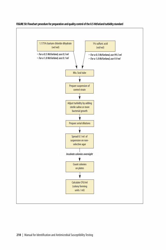
210 | Manual for Identification and Antimicrobial Susceptibility Testing
1.175% barium chloride dihydrate (wt/vol)
1% sulfuric acid (vol/vol)
• For a 0.5 McFarland, use 0.5 ml• For a 1.0 McFarland, use 0.1 ml
• For a 0.5 McFarland, use 99.5 ml• For a 1.0 McFarland, use 9.9 ml
Mix. Seal tube
Prepare suspension ofcontrol strain
Adjust turbidity by addingsterile saline or more
bacterial growth
Prepare serial dilutions
Spread 0.1 ml of suspension on non-
selective agar
Count colonies on plates
Calculate CFU/ml (colony forming
units / ml)
Incubate colonies overnight
FIGURE 50: Flowchart procedure for preparation and quality control of the 0.5 McFarland turbidity standard

Media, Reagents, and Quality Control | 211
FIGURE 51: Comparison of the 0.5 McFarland turbidity standard with inoculum suspension
TABLE 23: Composition of McFarland turbidity standards
Turbidity Barium chloride Sulfuric acid Corresponding approximate standard number dihydrate (1.175%) (1%) density of bacteria
0.5 0.5 ml 99.5 ml 1 x 10⁸
1 0.1 ml 9.9 ml 3 x 10⁸
2 0.2 ml 9.8 ml 6 x 10⁸
3 0.3 ml 9.7 ml 9 x 10⁸
4 0.4 ml 9.6 ml 12 x 108
5 0.5 ml 9.5 ml 15 x 108
6 0.6 ml 9.4 ml 18 x 108
7 0.7 ml 9.3 ml 21 x 108
8 0.8 ml 9.2 ml 24 x 108
9 0.9 ml 9.1 ml 27 x 108
10 1.0 ml 9.0 ml 30 x 108

Plate count method for testing a 0.5 McFarland turbidity standard
The goal of this procedure is to determine the number of bacteria per ml of fluid.A bacterial suspension equivalent in turbidity to a 0.5 McFarland turbiditystandard contains approximately 108 bacteria per ml.
1) Prepare 0.5 McFarland turbidity standard, as described above.
2) Prepare a suspension of a test organism (e.g., E. coli ATCC 25922) to match thedensity of the McFarland turbidity standard.
3) Make serial, 10-fold dilutions of the bacterial suspension in a suitable brothmedium. (Examples of suitable broth media include Mueller Hinton broth,TSB, or PBS.) The following steps a – i describe the procedure for making theserial dilutions.
Materials necessary for testing of the 0.5 McFarland turbidity standard include:seven sterile screw-capped tubes, seven agar plates (with medium to supportgrowth of the organism you’re testing), and pipettes capable of measuring out4.5 ml and 0.5 ml, respectively. In addition, a vortex machine is useful forvigorous mixing in tubes.
a) Make sure you have seven screw-capped tubes, each capable of holding atleast 10 ml of fluid. Prepare dilution tubes by adding 4.5 ml of sterile brothto each of the seven 10-ml tubes.
b) Label the tubes from 1 to 7, indicating the dilution the tube will hold. Alsolabel agar plates of the appropriate medium from 1 to 7.
c) Add 0.5 ml of the bacterial suspension made up to 0.5 McFarland turbiditystandard to the tube labeled 7 and mix vigorously.
212 | Manual for Identification and Antimicrobial Susceptibility Testing
FIGURE 52: Background lines for viewing turbidity of a suspension in comparison to a turbidity standard

d) Using the same pipette as in step C, draw up and release the suspensionseveral times into the pipette and then transfer 0.5 ml from tube 7 to tube 6and mix vigorously.
e) Continue this process of transferring 0.5 ml to each successive tube, usingthe same pipette, until you have completed the dilutions with tube 1. Aftervigorously mixing tube 1, use the pipette to draw up and release thesuspension in the tube several times.
f) Using the same pipette, transfer 0.1 ml from tube 1 to the plate labeled 1.
g) Using the same pipette, transfer 0.1 ml from tube 2 to the plate labeled 2.Continue this process for tube 3 to plate 3, and tube 4 to plate 4. (Iflaboratorians unfamiliar with making bacterial suspensions to match aMcFarland turbidity standard are responsible for the procedure, the processmay be continued through tube 7. However, plating out the tubes with ahigher concentrations of medium is not mandatory, because doing so wouldresult in too many colonies to count when they grow.)
h) Using a bent rod and starting with plate 1, spread the fluid on each plateover the entire surface of the plate. A bent rod can be made by using heat tobend a 2–5 mm diameter glass rod to an approximately 60˚-angle, with theshort end measuring approximately 5 cm. A bent, stainless steel metal rod ofsimilar size can be used as an alternative to a glass rod. (The fluid can also bespread with a wire inoculating loop or needle bent to a 60˚-angle, butspreading the fluid evenly is more difficult using these methods.)
i) Incubate the plates overnight and count the number of colonies on eachplate. It may be difficult to count the colonies on plates 4 through 7, and ifthere are more than 300 colonies per plate it should not be counted.
Interpretation of plate count results
A 0.5 McFarland turbidity standard is equivalent to approximately 108 bacteria perml. The original bacterial suspension that resembles the 0.5 McFarland turbiditystandard could have a range of 1.0 x 10⁸ bacteria/ml to 9.0 x 10⁸ bacteria/ml.Within this range, the 0.5 turbidity standard is accurate; the difference will beevident in the number of bacteria that grow out on the plates.
After 0.5 ml of the original bacterial suspension (i.e., which is equivalent to the 0.5 McFarland turbidity standard) is added to the 4.5 ml of broth in tube 7, asuspension of bacteria is produced that contains approximately 107 bacteria per ml.Then 0.1 ml of this suspension has been transferred to the plate marked 7, whichtranslates to approximately 106 (1,000,000–9,000,000) bacteria present on thatplate. If the bacteria were diluted correctly: approximately 105 (or 100,000 –
Media, Reagents, and Quality Control | 213
34 All lists are limited and incomplete. Note that inclusion of a company or specific product does not imply endorse-ment by CDC or WHO.

900,000) bacteria should be present on the plate labeled 6; approximately10,000–90,000 bacteria on the plate labeled 5; approximately 1,000–9,000 bacteriapresent on plate 4; approximately 100 to 900 bacteria present on plate 3;approximately 10 to 90 bacteria on plate 2; and, approximately 1 to 9 bacterialcolonies would be present on plate 1. Each plate should have one-tenth thebacteria as the plate with the next higher number. Generally, the plate labeled 3will be the plate that is counted; however, if there are more than 300 coloniespresent on plate 3, then plate 2 should be counted.
Sources of prepared media and reagents
Although commercially prepared media and reagents are more expensive thanmedia or reagents that can be prepared locally, commercially available items can beused (and may be preferable) in certain situations. Dehydrated media, for example,are often preferable to media prepared from individual components because ofreduced lot-to-lot variation. It may also be desirable to purchase the supply ofmedia and reagents to perform short-term studies rather than attemptformulation. The following media and reagents are available in most parts of theworld from suppliers including, but not limited to: BBL (available from Becton,Dickinson and Company), bioMérieux, Difco (available from Becton, Dickinsonand Company), Merck, Oxoid, and Quélab (Table 24); a partial listing ofmanufacturers, suppliers, and distributors with contact information is included inAppendix 13.35 (The listing of supplies, media and reagents in this laboratorymanual is not exhaustive, and availability of products from specific companies orsuppliers may change. Inclusion of a company or product does not implyendorsement by CDC or WHO.) It is essential that each lot of materials has asatisfactory expiration date and that the date of expiration and lot number forcommercial media are recorded in the laboratory.
In addition to media and reagents, laboratories must maintain their supplies (e.g., glassware) and equipment; Developing Health Technology is a company that provides low-cost laboratory equipment for developing countries,nongovernmental organizations (NGOs) and aid agencies. Furthermore, as notedelsewhere in this document, the manufacturer of the Etest® (AB Biodisk) maymake materials available at a reduced price to laboratories in developing countrysettings. Contact information for these companies is available in Appendix 13.
214 | Manual for Identification and Antimicrobial Susceptibility Testing
35 All lists are limited and incomplete. Note that inclusion of a company or specific product does not implyendoresement by CDC or WHO.

Media, Reagents, and Quality Control | 215
TABLE 24: Partial listing of materials and suppliers / manufacturers
Description of item a Sample listing of manufacturers b
a This is not intended to be comprehensive b Inclusion does not imply endorsement of commercial products or catalog of materials and suppliers. suppliers by CDC or WHO.
Acetone-alcohol (decolorizing agent) Remel; Difco (BD); BBL (BD)
Agar agar Remel; Oxoid; BBL (BD)
Ampicillin disks Remel; Oxoid; BBL (BD)
Azithromycin disks Remel; Oxoid; BBL (BD)
Bile salts Quélab; Remel; Oxoid; BBL (BD)
Bismuth sulfite (BS) agar Quélab; Difco (BD); Oxoid; BBL (BD)
Blood culture medium - prepared bioMérieux; Oxoid
Bovine hematin Quélab; BBL (BD)
Brain-heart infusion (BHI) Quélab; Difco (BD); Oxoid; BBL (BD); Remel
Cary-Blair medium Quélab; Difco (BD); Oxoid; BBL (BD)
Cefixime disks Remel; Oxoid; BBL (BD)
Ceftriaxone disks Remel; Oxoid; BBL (BD)
Chloramphenicol disks Remel; Oxoid; BBL (BD)
Chocolate blood agar - prepared bioMérieux; Remel; BBL (BD)
Chocolate blood agar + bacitracin - prepared Remel; BBL (BD)
Ciprofloxacin disks Remel; Oxoid; BBL (BD)
CO2-generating systems Oxoid; Remel
Colistin disks Quélab; Remel; Oxoid; BBL (BD)
Crystal violet Quélab; Difco (BD); Remel; BBL (BD)
Cystine trypticase agar (CTA) Quélab; Difco (BD); BBL (BD)
Defined growth supplement BBL (BD); Oxoid; Difco (BD)(e.g., IsoVitaleX,Vitox,VX Supplement)
Desoxycholate Quélab; Remel
Desoxycholate citrate agar (DCA) Quélab; Difco (BD); BBL (BD)
Etest® antimicrobial gradient strips AB Biodisk; Remel; Fisher Scientific
Ethanol Remel; Merck
Formalin (formaldehyde) Remel; Merck
Furazolidone disks Remel; Oxoid; BBL (BD)
Gonochek II® (enzyme substrate test) TCS Microbiology
Gonococcus (GC) agar base medium Quélab; Difco (BD); Oxoid; BBL (BD)
Gram stain kit bioMérieux; Difco (BD); Remel; BBL (BD)
Gram-negative broth Difco (BD); BBL (BD); Renek
Gram’s iodine Quélab; Difco (BD); Remel; BBL (BD)
H. influenzae serotyping antisera Difco (BD); Remelcontinued

TABLE 24: Partial listing of materials and suppliers / manufacturers, continued
Description of item a Sample listing of manufacturers b
a This is not intended to be comprehensive b Inclusion does not imply endorsement of commercial products or catalog of materials and suppliers. suppliers by CDC or WHO.
Haemophilus test medium (HTM) Oxoid; BBL (BD)
Heart infusion agar / broth Quélab; Difco (BD); BBL (BD)
Hektoen enteric (HE) agar Quélab; Difco (BD); Oxoid; BBL (BD)
Hemoglobin powder Quélab; Difco (BD); Oxoid; BBL (BD)
Horse blood Difco (BD); Oxoid
Jembec® plates BBL (BD); Quélab
Kligler iron agar (KIA) Quélab; Difco (BD); Oxoid; BBL (BD)
Kovac’s oxidase reagent Quélab; Merck(N,N,N’,N’-tetramethyl-ρ-phenylenediamine dihydrochloride)
Lysine iron agar (LIA) Quélab; Difco (BD); Oxoid; BBL (BD)
MacConkey agar (MAC) Quélab; Difco (BD); Oxoid; BBL (BD)
Martin-Lewis medium - prepared Quélab; BBL (BD)
Methylene blue Quélab; Remel
Modified Thayer-Martin medium - prepared Quélab; Remel; BBL (BD)
Motility medium Quélab; Difco (BD); BBL (BD)
Mueller-Hinton agar - prepared bioMérieux; Remel; BBL (BD)
Mueller-Hinton agar plus 5% sheep blood - prepared bioMérieux; Remel; BBL (BD)
Mueller-Hinton agar/broth Quélab; Difco (BD); Oxoid; Remel
N. meningitidis serogrouping antisera Difco (BD); Remel
Nalidixic acid disks Oxoid; BBL (BD)
Nicotinamide adenine dinucleotide (NAD; V factor) Quélab; Merck
Nitrate broth Quélab; Difco (BD)
Nitrate Reagents A and B Remel; BBL (BD)
Nitrocefin (beta-lactamase) Remel; Difco (BD); Oxoid; BBL (BD)
Optochin (p-disks) Oxoid; BBL (BD)
Oxacillin disks Oxoid; BBL (BD)
Penicillin disks Oxoid; BBL (BD)
Peptone Difco (BD); Oxoid
Permeable membrane screw-caps (Biomedical Polymers, Inc., via) Fisher Scientific;(for short-term storage of N. meningitidis at 4˚C) VWR International
Phosphate buffered saline (PBS) Quélab; Oxoid
Quad ID plate Quélab; BBL (BD)
Quality control strains (type cultures) American Type Culture Collection (ATCC);National Culture Type Collection (NCTC)
S. pneumoniae serotyping antisera Omniserum; Remel; Difco (BD)
216 | Manual for Identification and Antimicrobial Susceptibility Testing
continued

TABLE 24: Partial listing of materials and suppliers / manufacturers, continued
Description of item a Sample listing of manufacturers b
a This is not intended to be comprehensive b Inclusion does not imply endorsement of commercial products or catalog of materials and suppliers. suppliers by CDC or WHO.
Safranin Quélab; Difco (BD); Remel; BBL (BD)
Salmonella (ser.Typhi) serotyping antisera Remel; Difco (BD)
Salmonella-Shigella (SS) agar Quélab; Difco (BD); Oxoid; BBL (BD)
Selenite broth (SEL) Quélab; Difco (BD); Oxoid; BBL (BD)
Sheep blood Remel; Quélab
Sheep blood agar (TSA + 5% sheep blood) - prepared bioMérieux; BBL (BD)
Sheep blood agar + gentamicin - prepared Quélab; BBL (BD)
Shigella antisera Remel; Difco (BD)
Silica gel packets Scientific Device Laboratory, Inc.(for transport and short-term storage of some pathogens)
Skim milk powder Quélab; Difco (BD); BBL (BD)
Sodium polyanethol sulfonate (SPS) Quélab; Oxoid
Spectinomycin disks Oxoid; BBL (BD)
Sucrose (reagent-grade) Quélab; BBL (BD)
Sulfide-indole-motility medium (SIM) Remel; Oxoid; BBL (BD)
Superoxol reagent (30% H2O2) Quélab; Merck
Tetracycline disks Remel; Oxoid; BBL (BD)
Thiosulfate citrate bile salts sucrose agar (TCBS) Quélab; Oxoid; BBL (BD)
Todd-Hewitt broth Difco (BD); Oxoid; BBL (BD)
Trimethoprim-sulfamethoxazole (cotrimoxazole) disks Remel; Oxoid; BBL (BD)
Triple sugar iron agar (TSI) Quélab; Difco (BD); BBL (BD)
Tryptone (Trypticase) soy agar/broth Quélab; Difco (BD); Oxoid; BBL (BD); bioMérieux
Tryptone (Trypticase) soy agar - prepared BBL (BD); bioMérieux
Urea medium Quélab; Difco (BD); Oxoid; BBL (BD)
V. cholerae antisera Remel; Difco (BD)
VCA(T) supplement (to prepare Martin-Lewis medium) Quélab; Oxoid; BBL (BD)
VCN(T) supplement (to prepare Modified Thayer-Martin) Quélab; Oxoid; BBL (BD)
V-factor disks (NAD) Remel; Oxoid
X-factor disks (haemin) Remel; Oxoid; Quélab
XV-factor disks Remel; Oxoid; Quélab
Xylose lysine desoxycholate agar (XLD) Quélab; Difco (BD); Oxoid; BBL (BD); Remel
Zinc dust Quélab; BBL (BD)
Media, Reagents, and Quality Control | 217