PET-CT for radiotherapy planning in lung cancer: … for radiotherapy planning in lung cancer:...
68
PET-CT for radiotherapy planning in lung cancer: current recommendations and future directions Gerry Hanna Centre for Cancer Research and Cell Biology Queen’s University of Belfast @gerryhanna
Transcript of PET-CT for radiotherapy planning in lung cancer: … for radiotherapy planning in lung cancer:...
PET-CT for radiotherapy planning in lung cancer: current
recommendations and future directions
Gerry Hanna Centre for Cancer Research and Cell Biology
Queen’s University of Belfast
@gerryhanna
Talk Outline
• Key principles underlying PET/CT acquisition
• PET/CT for staging in NSCLC • Background of PET/CT for RTP in NSCLC • How to acqiure a PET/CT for RTP? • Displaying a PET/CT for RTP • Guidance on PET/CT based TVD • 4D PET/CT
KEY PRINCIPLES OF PET
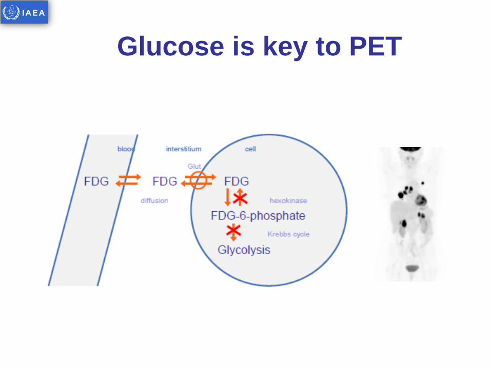
Glucose is key to PET
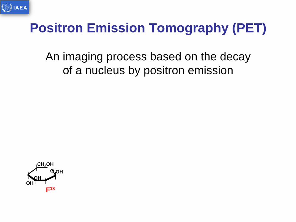
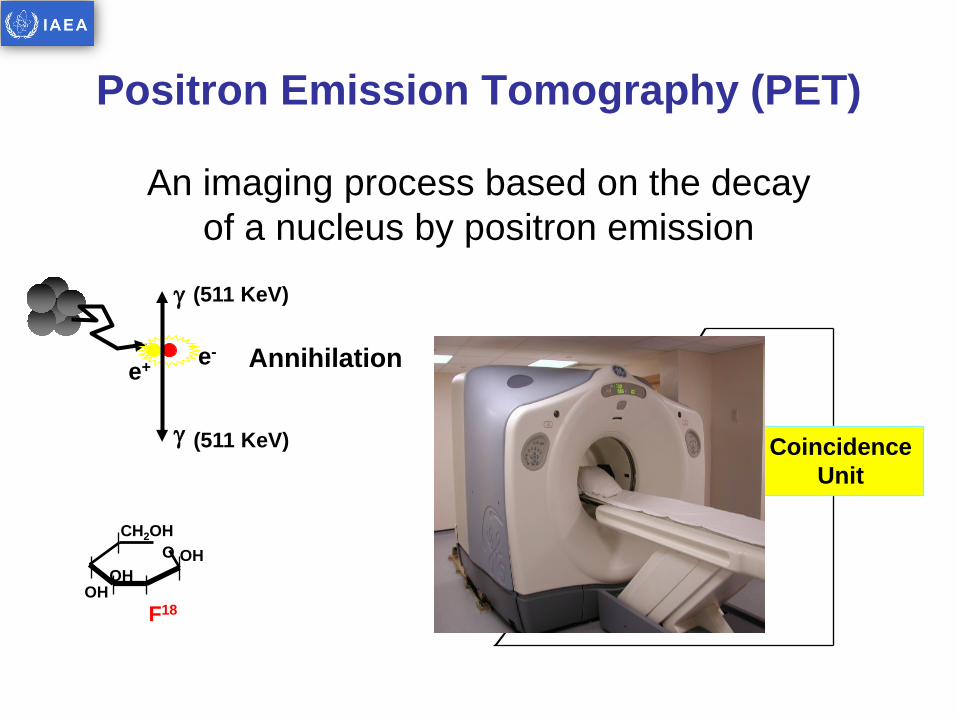
Positron Emission Tomography (PET)
An imaging process based on the decay of a nucleus by positron emission
O OH
OH OH
CH2OH
F18
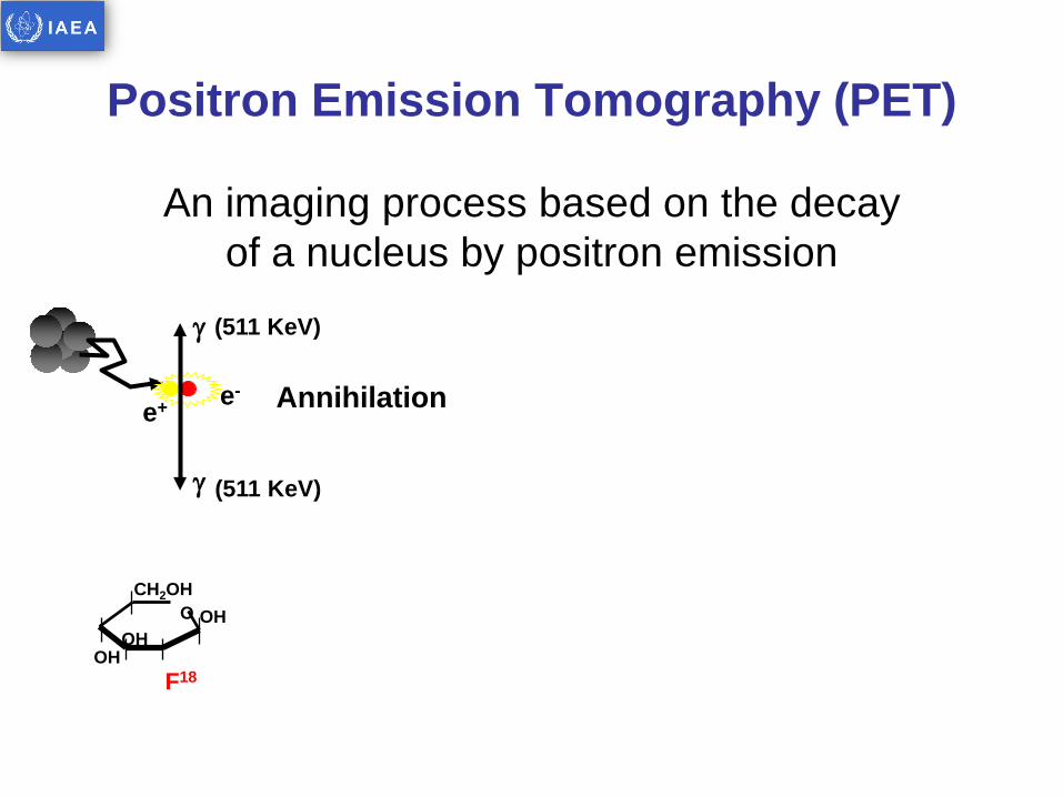
Positron Emission Tomography (PET)
An imaging process based on the decay of a nucleus by positron emission
γ (511 KeV)
Annihilation e+ e-
γ (511 KeV)
O OH
OH OH
CH2OH
F18
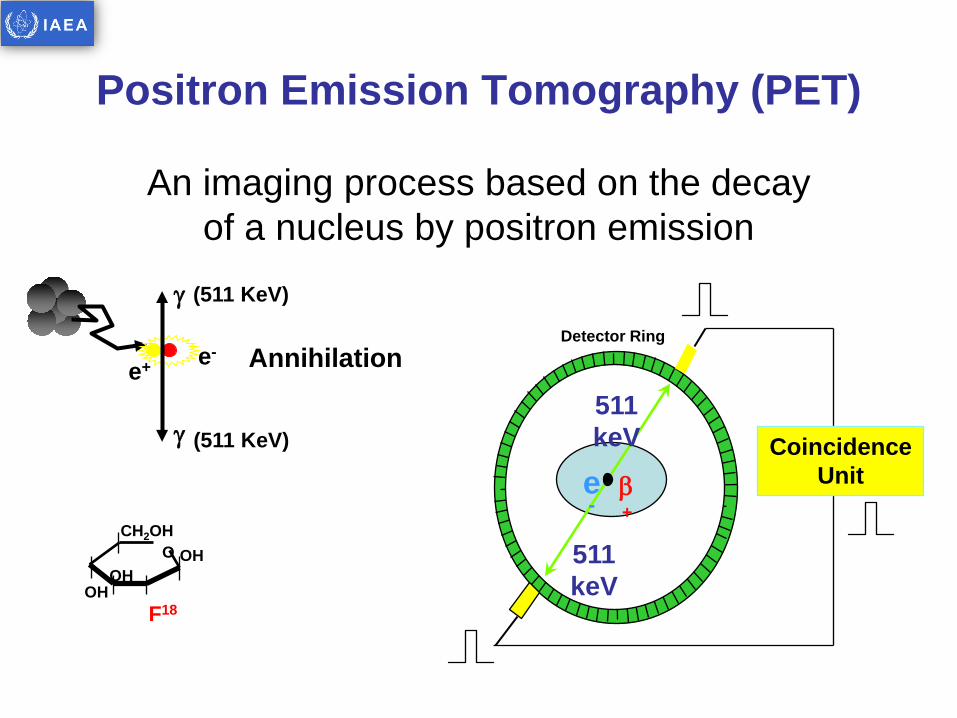
Positron Emission Tomography (PET)
An imaging process based on the decay of a nucleus by positron emission
γ (511 KeV)
Annihilation e+ e-
γ (511 KeV)
β+
511 keV
511 keV
e-
Coincidence Unit
Detector Ring
O OH
OH OH
CH2OH
F18
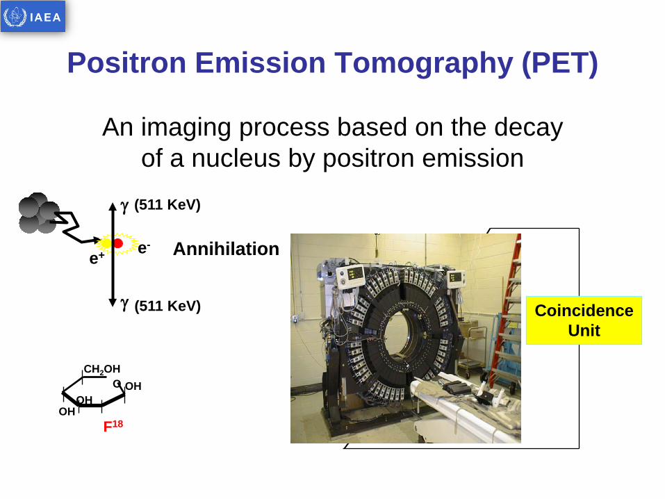
Positron Emission Tomography (PET)
An imaging process based on the decay of a nucleus by positron emission
γ (511 KeV)
Annihilation e+ e-
γ (511 KeV)
β+
511 keV
511 keV
e-
Coincidence Unit
O OH
OH OH
CH2OH
F18
Positron Emission Tomography (PET)
An imaging process based on the decay of a nucleus by positron emission
γ (511 KeV)
Annihilation e+ e-
γ (511 KeV)
β+
511 keV
511 keV
e-
Coincidence Unit
O OH
OH OH
CH2OH
F18
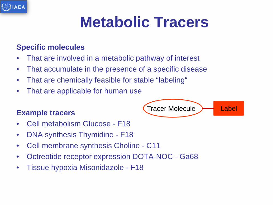
Metabolic Tracers Specific molecules • That are involved in a metabolic pathway of interest • That accumulate in the presence of a specific disease • That are chemically feasible for stable “labeling“ • That are applicable for human use
Example tracers • Cell metabolism Glucose - F18 • DNA synthesis Thymidine - F18 • Cell membrane synthesis Choline - C11 • Octreotide receptor expression DOTA-NOC - Ga68 • Tissue hypoxia Misonidazole - F18
Tracer Molecule Label
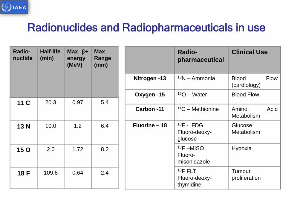
Radio-nuclide
Half-life (min)
Max β+ energy (MeV)
Max Range (mm)
11 C 20.3 0.97 5.4
13 N 10.0 1.2 6.4
15 O 2.0 1.72 8.2
18 F 109.6 0.64 2.4
Radio-pharmaceutical
Clinical Use
Nitrogen -13 13N – Ammonia Blood Flow (cardiology)
Oxygen -15 15O – Water Blood Flow
Carbon -11 11C – Methionine Amino Acid Metabolism
Fluorine – 18 18F - FDG Fluoro-deoxy-glucose
Glucose Metabolism
18F –MISO Fluoro-misonidazole
Hypoxia
18F FLT Fluoro-deoxy-thymidine
Tumour proliferation
Radionuclides and Radiopharmaceuticals in use
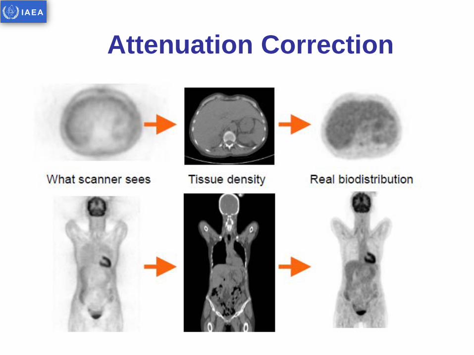
Attenuation Correction
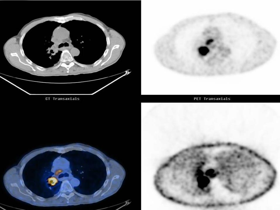
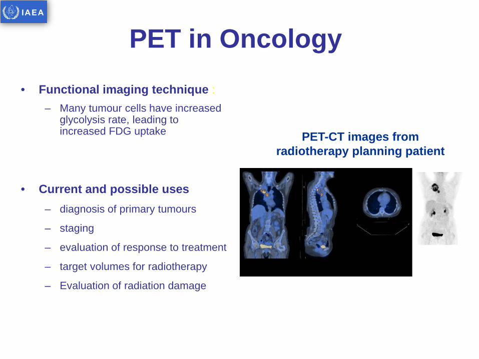
PET in Oncology • Functional imaging technique :
– Many tumour cells have increased glycolysis rate, leading to increased FDG uptake
• Current and possible uses – diagnosis of primary tumours
– staging
– evaluation of response to treatment
– target volumes for radiotherapy
– Evaluation of radiation damage
PET-CT images from radiotherapy planning patient
PET/CT FOR STAGING IN NSCLC
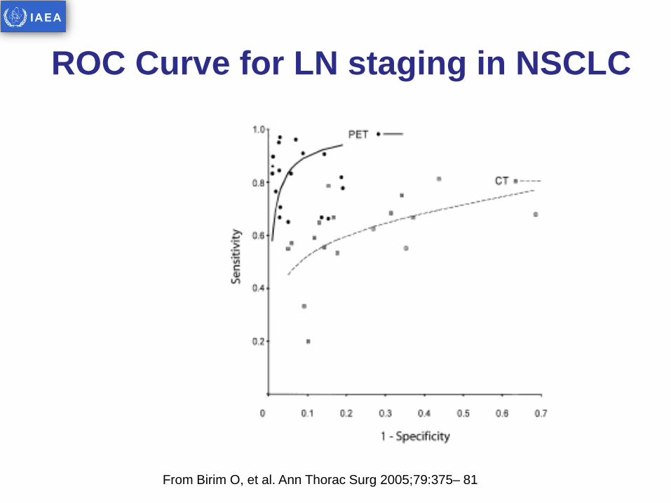
From Birim O, et al. Ann Thorac Surg 2005;79:375– 81
ROC Curve for LN staging in NSCLC
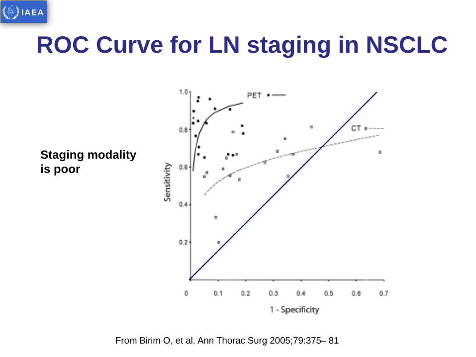
From Birim O, et al. Ann Thorac Surg 2005;79:375– 81
Staging modality is poor
ROC Curve for LN staging in NSCLC
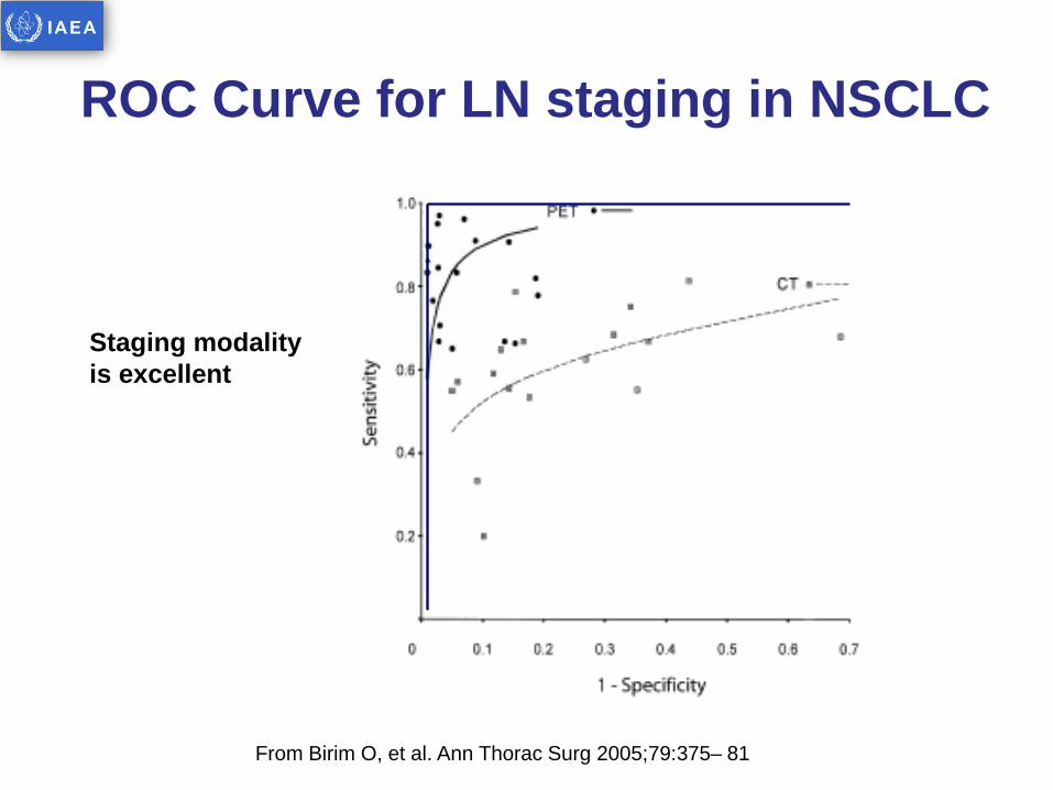
From Birim O, et al. Ann Thorac Surg 2005;79:375– 81
Staging modality is excellent
ROC Curve for LN staging in NSCLC
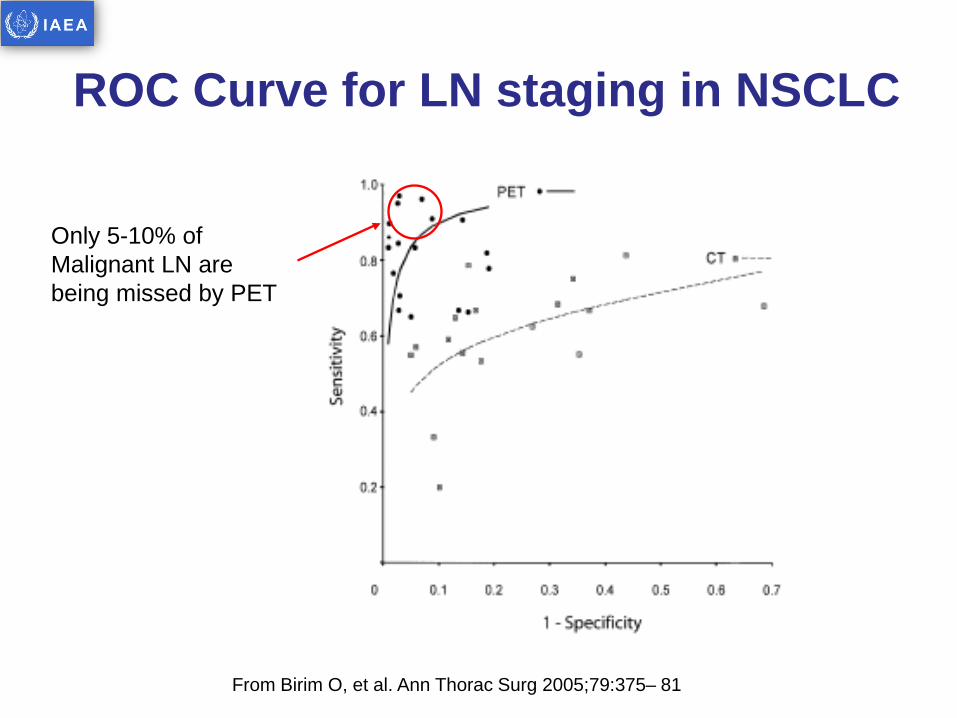
From Birim O, et al. Ann Thorac Surg 2005;79:375– 81
Only 5-10% of Malignant LN are being missed by PET
ROC Curve for LN staging in NSCLC
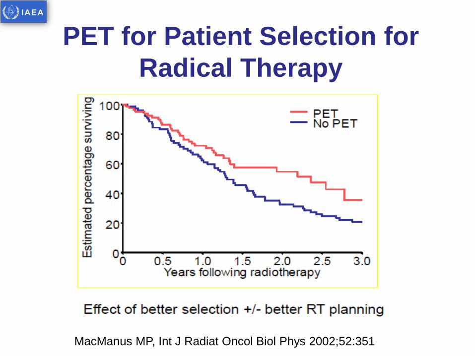
MacManus MP, Int J Radiat Oncol Biol Phys 2002;52:351
PET for Patient Selection for Radical Therapy
PET/CT for RT planning in NSCLC
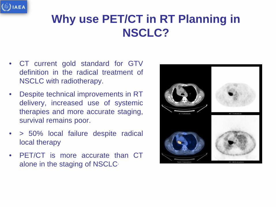
Why use PET/CT in RT Planning in NSCLC?
• CT current gold standard for GTV definition in the radical treatment of NSCLC with radiotherapy.
• Despite technical improvements in RT delivery, increased use of systemic therapies and more accurate staging, survival remains poor.
• > 50% local failure despite radical local therapy
• PET/CT is more accurate than CT alone in the staging of NSCLC.
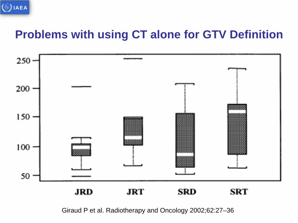
Problems with using CT alone for GTV Definition
Giraud P et al. Radiotherapy and Oncology 2002;62:27–36
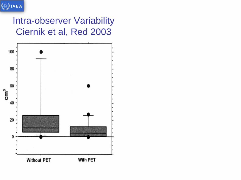
Intra-observer Variability Ciernik et al, Red 2003
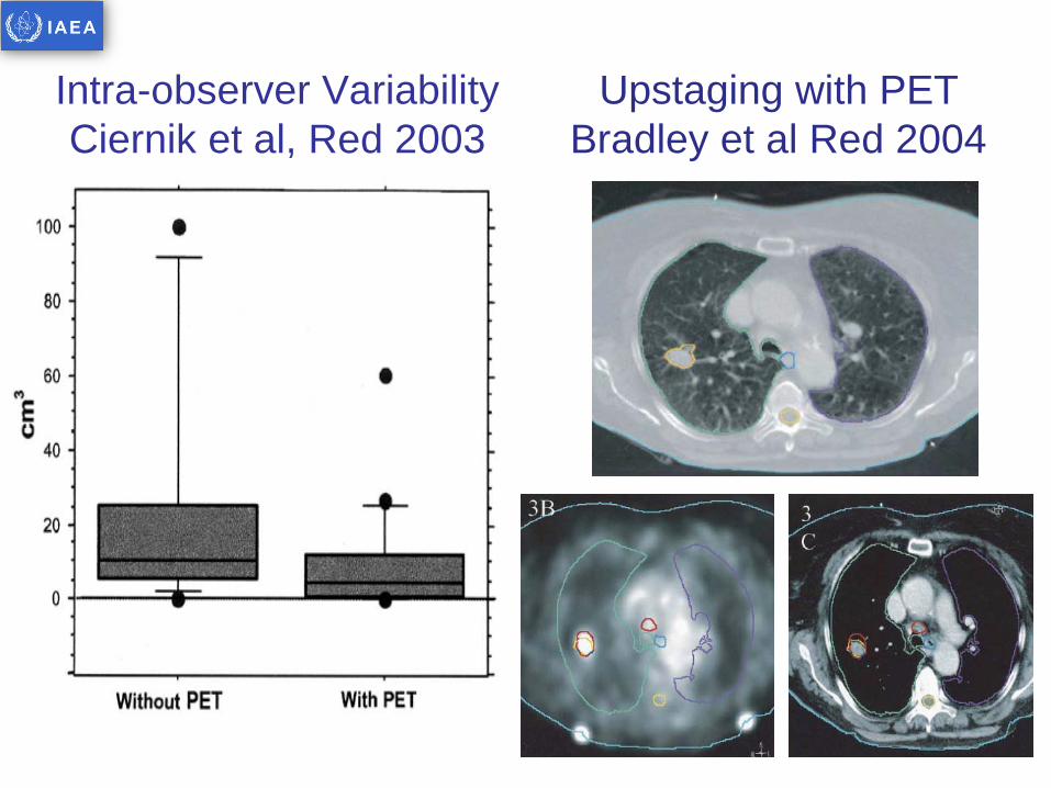
Intra-observer Variability Ciernik et al, Red 2003
Upstaging with PET Bradley et al Red 2004
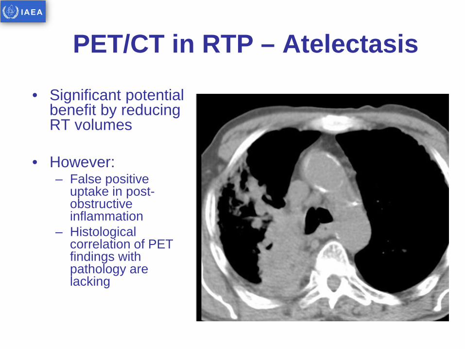
PET/CT in RTP – Atelectasis
• Significant potential benefit by reducing RT volumes
• However: – False positive
uptake in post-obstructive inflammation
– Histological correlation of PET findings with pathology are lacking
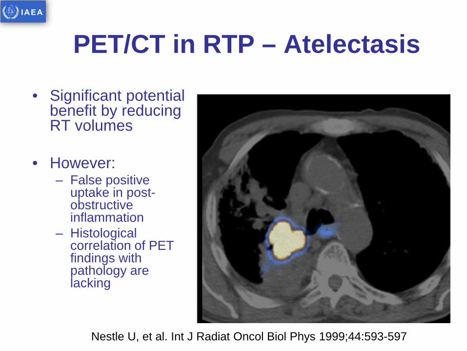
PET/CT in RTP – Atelectasis
• Significant potential benefit by reducing RT volumes
• However: – False positive
uptake in post-obstructive inflammation
– Histological correlation of PET findings with pathology are lacking
Nestle U, et al. Int J Radiat Oncol Biol Phys 1999;44:593-597
HOW TO ACQIURE A PET/CT FOR RTP
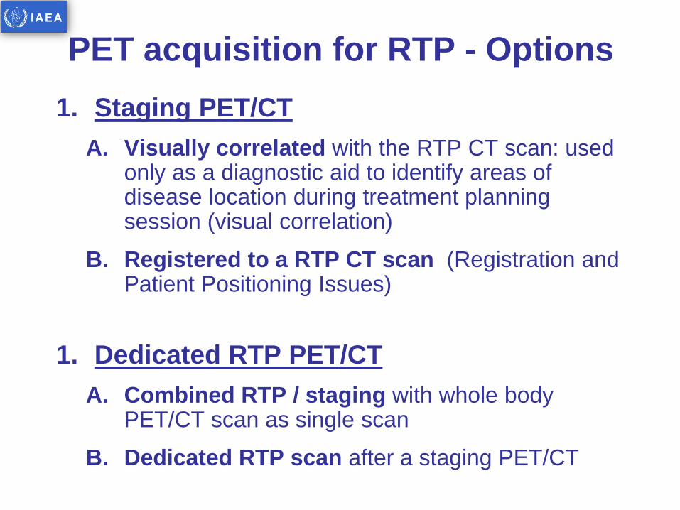
PET acquisition for RTP - Options 1. Staging PET/CT
A. Visually correlated with the RTP CT scan: used only as a diagnostic aid to identify areas of disease location during treatment planning session (visual correlation)
B. Registered to a RTP CT scan (Registration and Patient Positioning Issues)
1. Dedicated RTP PET/CT A. Combined RTP / staging with whole body
PET/CT scan as single scan
B. Dedicated RTP scan after a staging PET/CT
Recommendations about registering a PET/CT to a RTP CT
• PET/CT images should be registered using a rigid registration (for 4DCT – register to the average intensity projection (Ave-IP) scan)
• Registration should focus on bony anatomy which is not affected by respiratory motion (e.g. spinal column)
• It is advised that this approach should not be used routinely for gated treatments
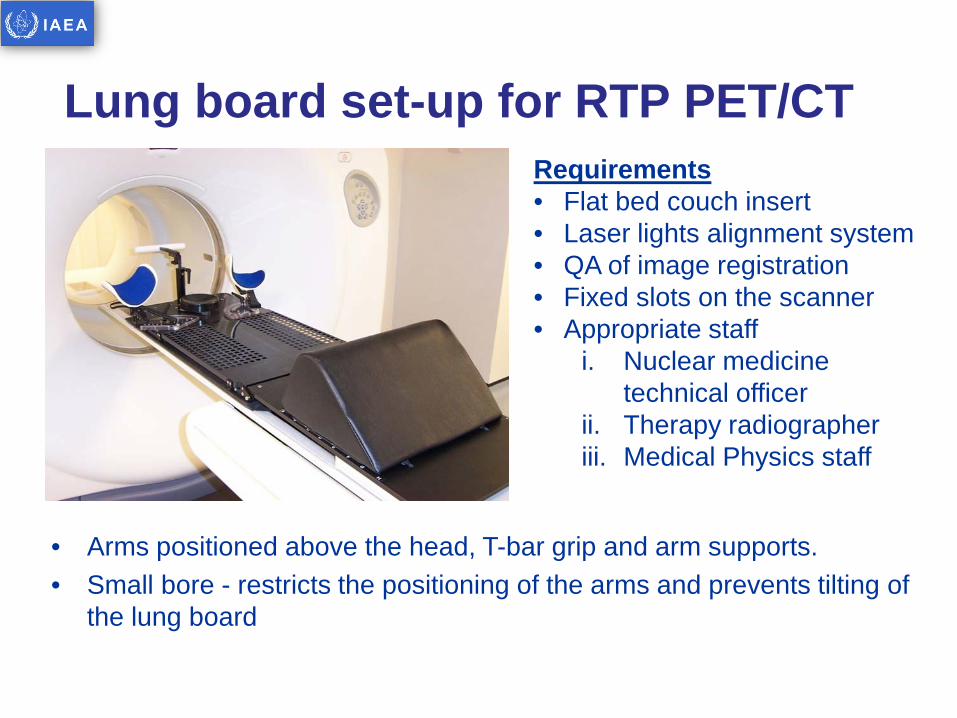
Lung board set-up for RTP PET/CT
• Arms positioned above the head, T-bar grip and arm supports. • Small bore - restricts the positioning of the arms and prevents tilting of
the lung board
Requirements • Flat bed couch insert • Laser lights alignment system • QA of image registration • Fixed slots on the scanner • Appropriate staff
i. Nuclear medicine technical officer
ii. Therapy radiographer iii. Medical Physics staff
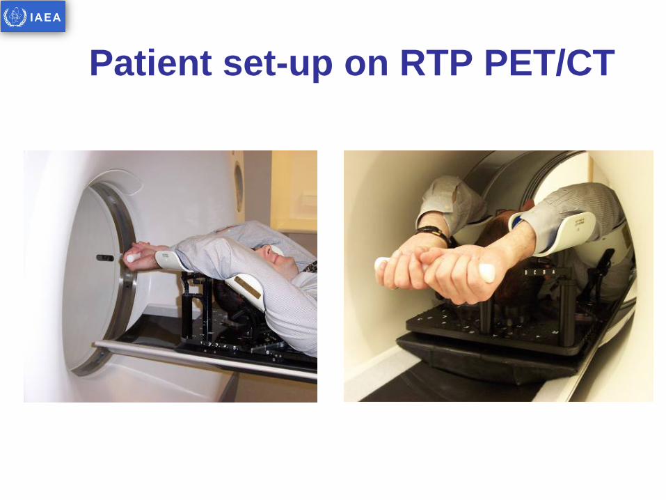
Patient set-up on RTP PET/CT
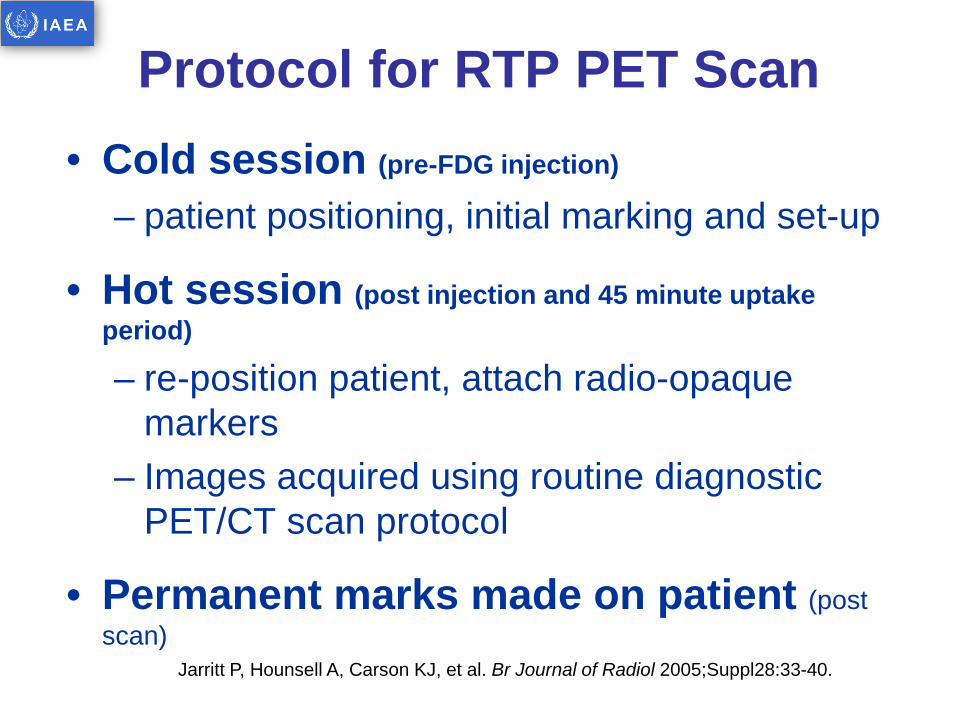
Protocol for RTP PET Scan • Cold session (pre-FDG injection)
– patient positioning, initial marking and set-up
• Hot session (post injection and 45 minute uptake period)
– re-position patient, attach radio-opaque markers
– Images acquired using routine diagnostic PET/CT scan protocol
• Permanent marks made on patient (post scan)
Jarritt P, Hounsell A, Carson KJ, et al. Br Journal of Radiol 2005;Suppl28:33-40.
PET/CT BASED TARGET VOLUME DELINEATION
DISPLAYING A PET/CT FOR RTP
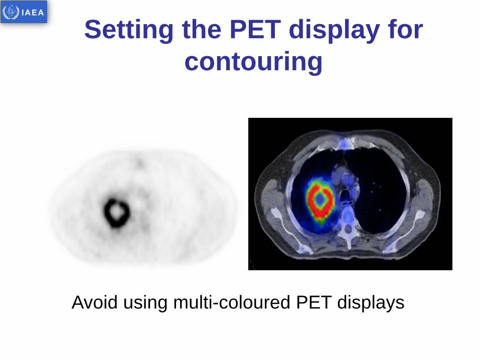
Setting the PET display for contouring
Avoid using multi-coloured PET displays
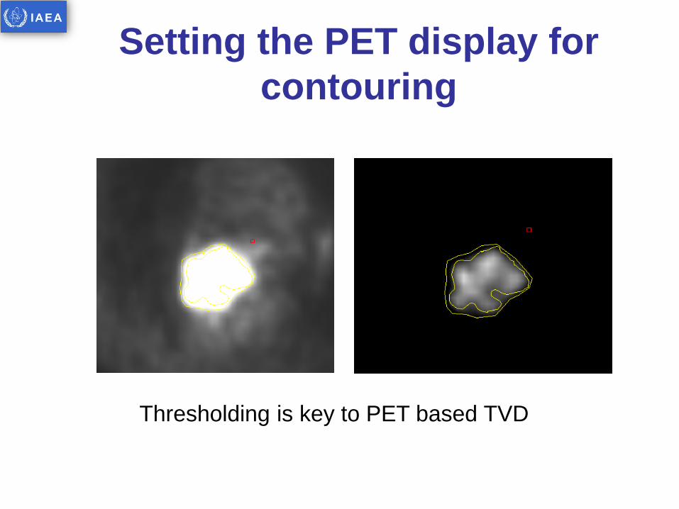
Setting the PET display for contouring
Thresholding is key to PET based TVD
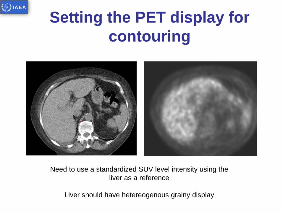
Setting the PET display for contouring
Need to use a standardized SUV level intensity using the liver as a reference
Liver should have hetereogenous grainy display
Setting the PET display for contouring
• Use a standardised display • Use no more that two colours in any colour wash
display • Navigate to the liver • Adjust the brightness and contrast levels in the
PET window in a way that you can still see the shape of anatomical structures e.g. the skin,
• The liver should contain almost no white pixels

WHAT TO DELINEATE?
Reproducibility of Tumour contouring in NSCLC
• Several studies have shown that tumour contouring by “expert” physicians on CT is not reproducible
• Physician variability is the biggest pitfall • Peter Mac study showed that use of a rigorous
protocol improved reproducibility • Reproducibility is better for PET/CT but inter and
intra clinician variability still exists
Image processing and display
• Image Processing - Most centre use similar image processing protocols for a diagnostic/staging PET/CT as for a RTP PET/CT (attenuation correction, image reconstruction etc…)
• RTP PET display - Ensure that when images are exported to RT planning software they appear the same (need to undertake phantom studies)
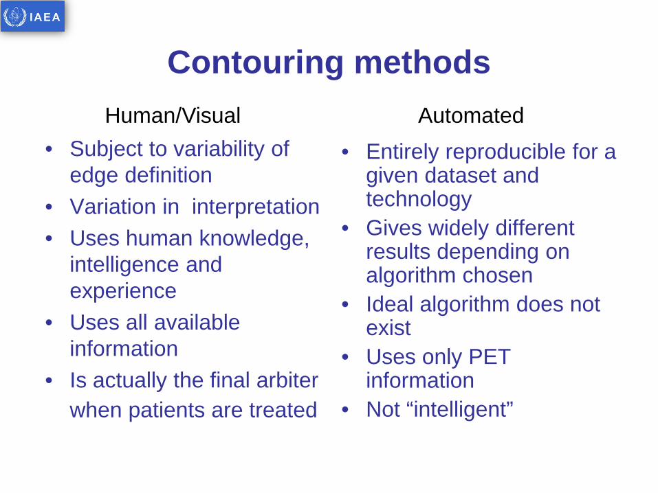
Contouring methods
• Subject to variability of edge definition
• Variation in interpretation • Uses human knowledge,
intelligence and experience
• Uses all available information
• Is actually the final arbiter when patients are treated
• Entirely reproducible for a given dataset and technology
• Gives widely different results depending on algorithm chosen
• Ideal algorithm does not exist
• Uses only PET information
• Not “intelligent”
Human/Visual Automated

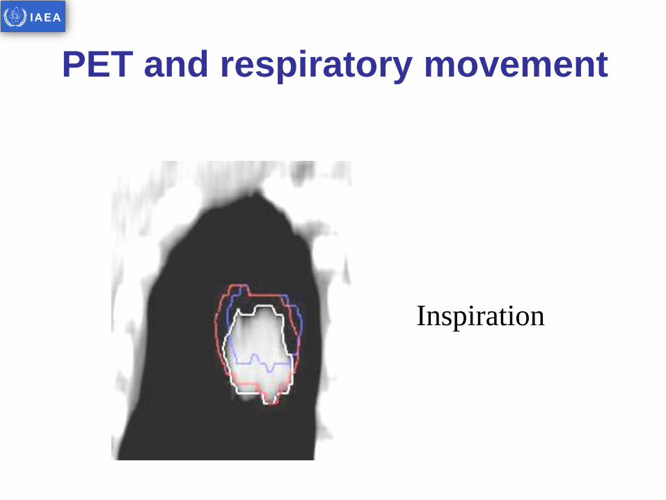
PET and respiratory movement
Expiration
Presenter
Presentation Notes
These images in which the tumour was captured in both expiration and inspiration on CT illustrate the ability of a PET derived GTV to encompass the tumour throughout respiration
PET and respiratory movement
Inspiration
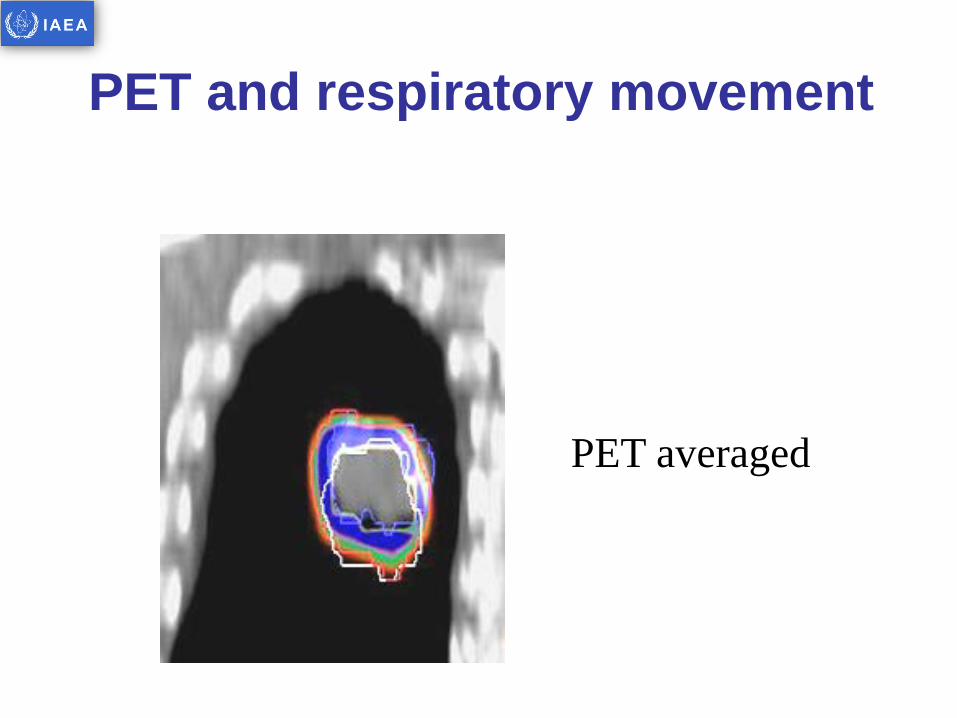
PET and respiratory movement
PET averaged
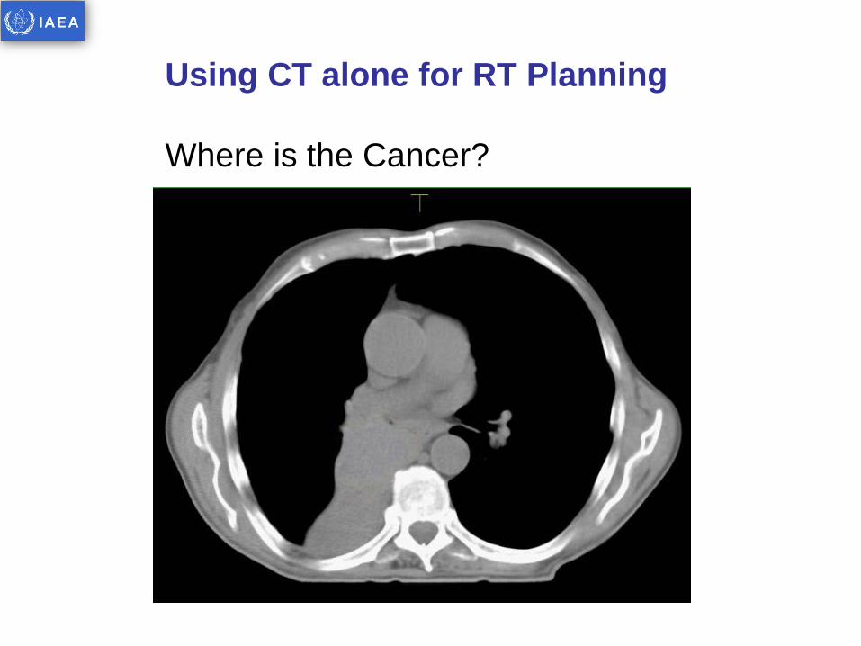
Using CT alone for RT Planning Where is the Cancer?
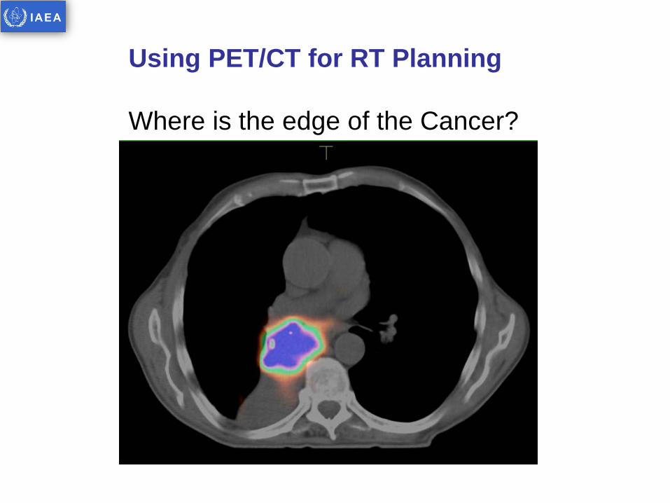
Using PET/CT for RT Planning Where is the edge of the Cancer?
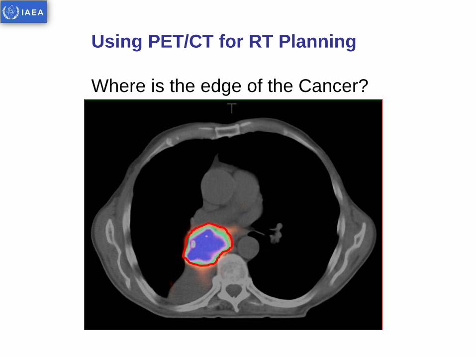
Using PET/CT for RT Planning Where is the edge of the Cancer?
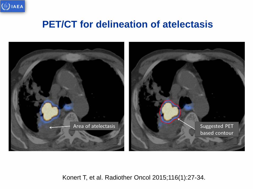
PET/CT for delineation of atelectasis
Konert T, et al. Radiother Oncol 2015;116(1):27-34.
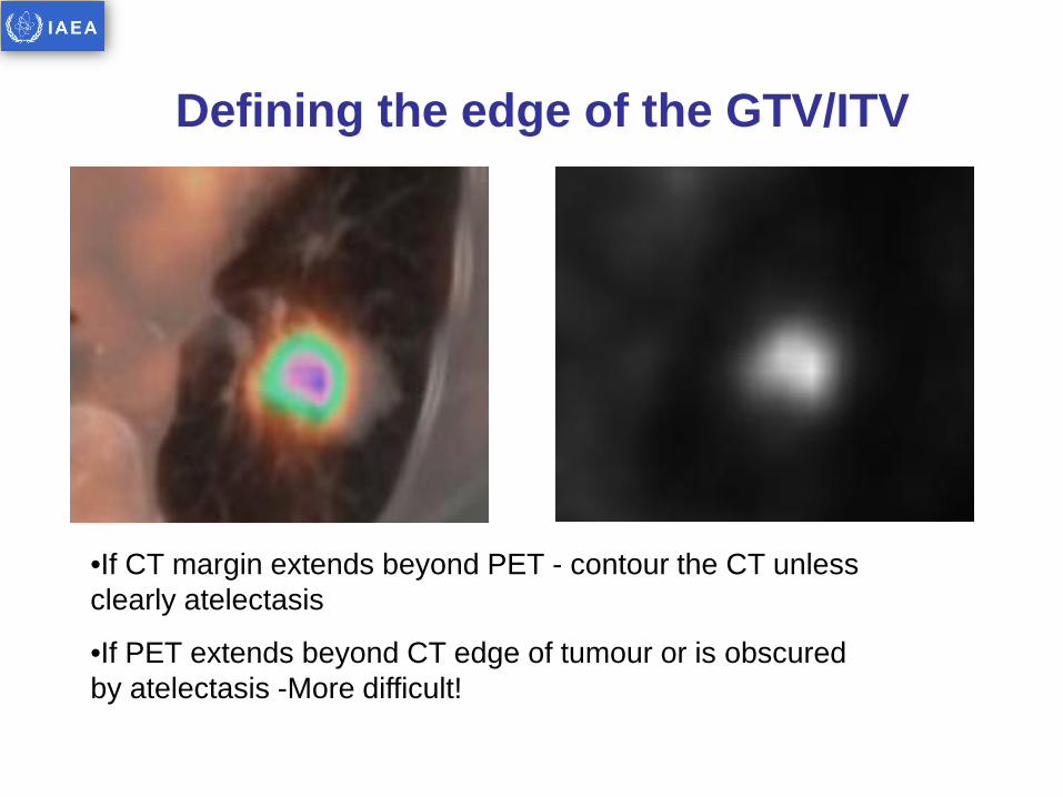
Defining the edge of the GTV/ITV
•If CT margin extends beyond PET - contour the CT unless clearly atelectasis
•If PET extends beyond CT edge of tumour or is obscured by atelectasis -More difficult!
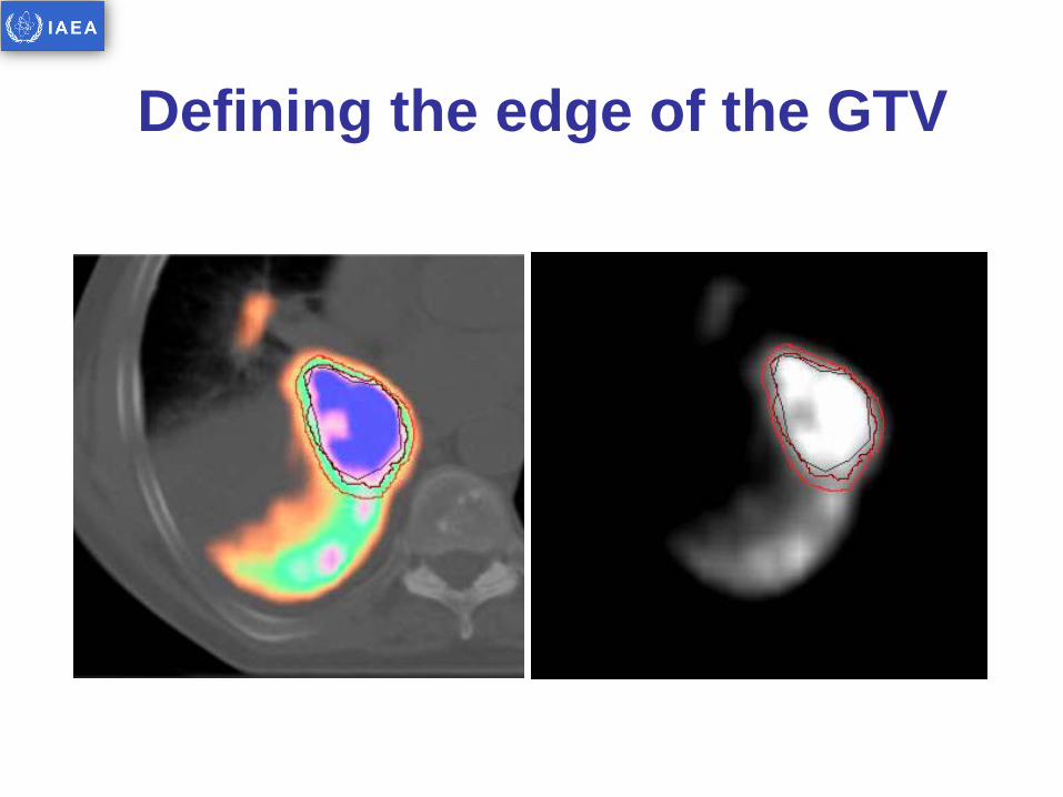
Defining the edge of the GTV

Peer Review
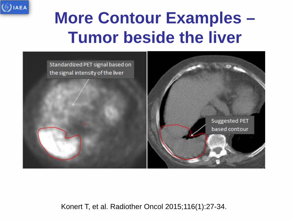
More Contour Examples – Tumor beside the liver
Konert T, et al. Radiother Oncol 2015;116(1):27-34.
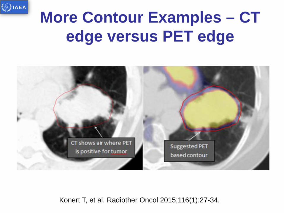
More Contour Examples – CT edge versus PET edge
Konert T, et al. Radiother Oncol 2015;116(1):27-34.
PET/CT RTP Margins to use…
• When delineating on PET – structure incorporates a respiratory motion
• Consensus statement refers to this as a respiratory expanded GTV (reGTV)
• Suggest reGTV CTV 6mm or 8mm
• Suggest CTV PTV at least 5mm (dependent on local set-up error)
Konert T, et al. Radiother Oncol 2015;116(1):27-34.

HOW TO INCORPORATE GATING?
How to incorporate gating?
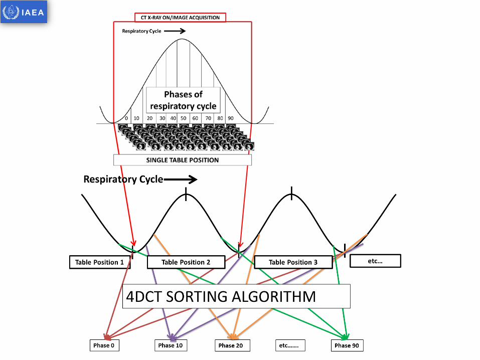
• 4DCT now standard technique to account for tumour motion
• 4DCT gives reliable information about tumour morphology and movement
• Where 4DCT acquisition is used alongside PET/CT the GTV edge should be based on the 4DCT
• The PET being used to discriminate tumor and non-tumor sites to adapt the GTV where appropriate
4D PET/CT
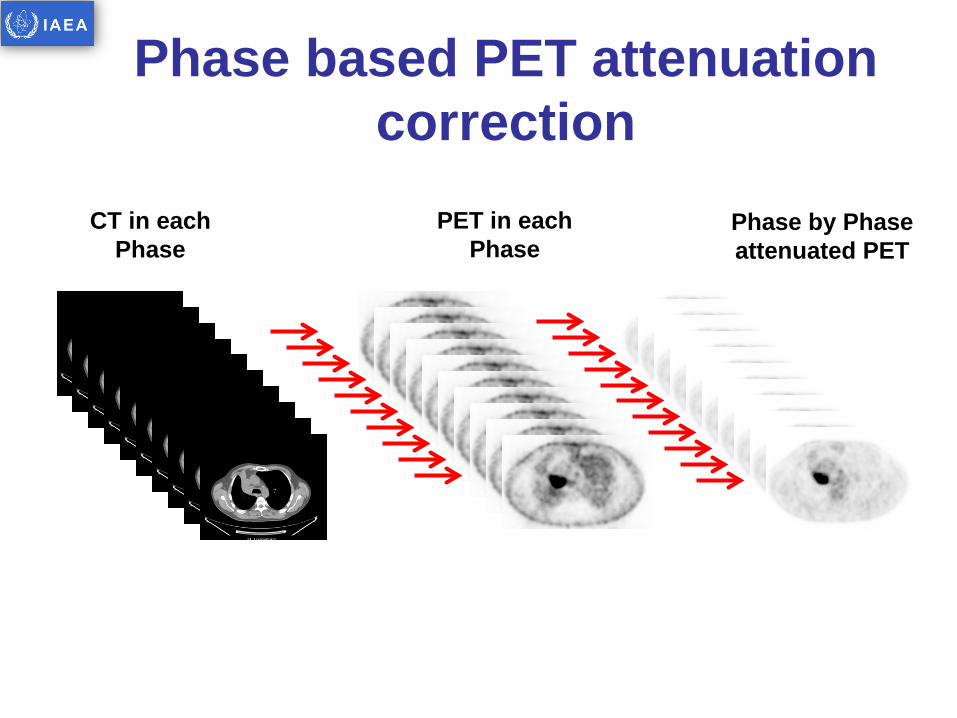
CT in each Phase
PET in each Phase
Phase by Phase attenuated PET
Phase based PET attenuation correction
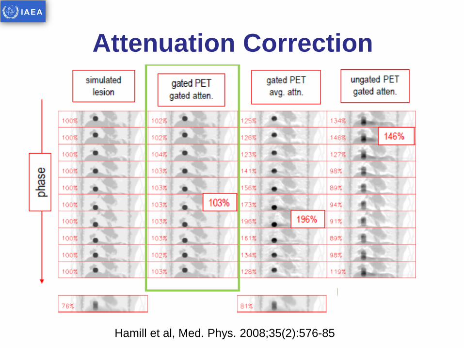
Attenuation Correction
Hamill et al, Med. Phys. 2008;35(2):576-85
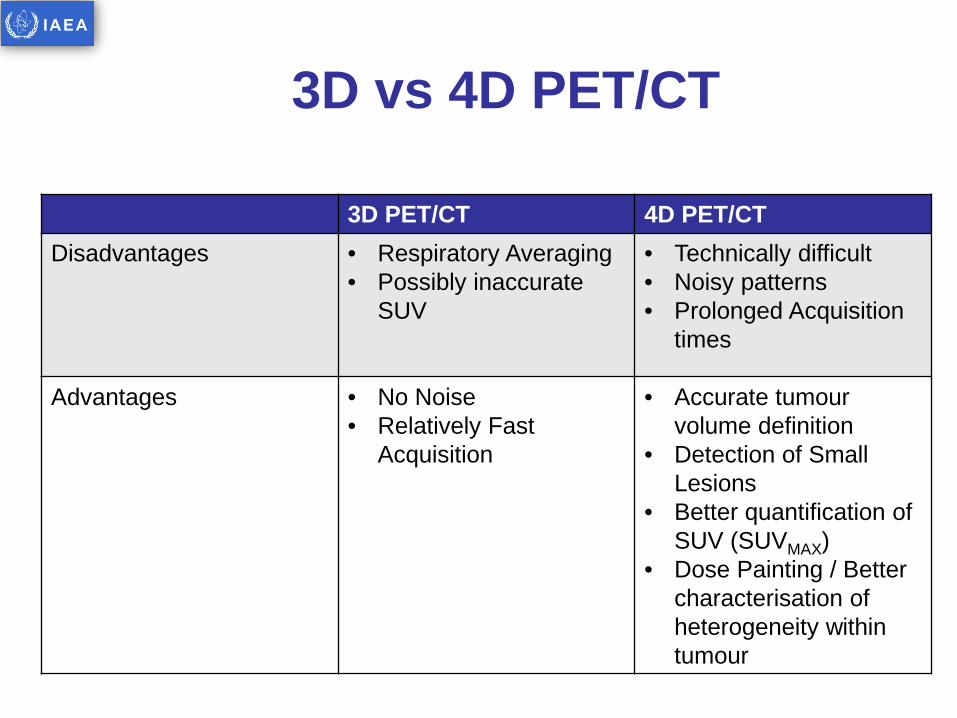
3D vs 4D PET/CT
3D PET/CT 4D PET/CT Disadvantages • Respiratory Averaging
• Possibly inaccurate SUV
• Technically difficult • Noisy patterns • Prolonged Acquisition
times
Advantages • No Noise • Relatively Fast
Acquisition
• Accurate tumour volume definition
• Detection of Small Lesions
• Better quantification of SUV (SUVMAX)
• Dose Painting / Better characterisation of heterogeneity within tumour
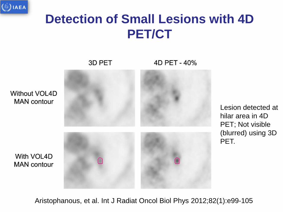
Detection of Small Lesions with 4D PET/CT
Aristophanous, et al. Int J Radiat Oncol Biol Phys 2012;82(1):e99-105
Lesion detected at hilar area in 4D PET; Not visible (blurred) using 3D PET.
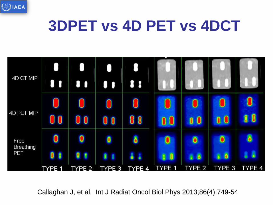
3DPET vs 4D PET vs 4DCT
Callaghan J, et al. Int J Radiat Oncol Biol Phys 2013;86(4):749-54
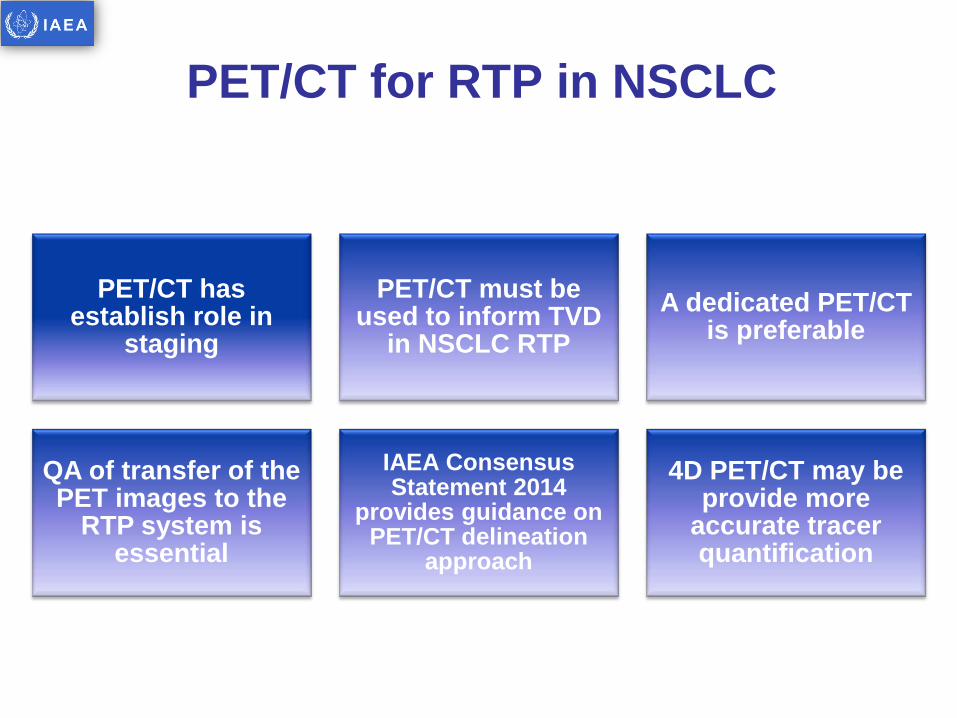
PET/CT for RTP in NSCLC
PET/CT has establish role in
staging
PET/CT must be used to inform TVD
in NSCLC RTP A dedicated PET/CT
is preferable
QA of transfer of the PET images to the
RTP system is essential
IAEA Consensus Statement 2014
provides guidance on PET/CT delineation
approach
4D PET/CT may be provide more
accurate tracer quantification