Detection of mutations and gene fusions in pediatric ... Distinguish relapse or metastasis from...
18
Detection of mutations and gene fusions in pediatric hematologic malignancies and solid tumors with the OncoKids SM next generation sequencing based assay Jaclyn Biegel, Timothy Triche, D. Gigi Ostrow, Jonathan Buckley, Tracy Busse, Xiaowu Gai, Jianling Ji, Alexander Judkins, Dennis Maglinte, Gordana Raca, and Matthew Hiemenz
Transcript of Detection of mutations and gene fusions in pediatric ... Distinguish relapse or metastasis from...

Detection of mutations and gene fusions in pediatric hematologic malignancies and solid tumors with the OncoKidsSM next generation
sequencing based assay
Jaclyn Biegel, Timothy Triche, D. Gigi Ostrow, Jonathan Buckley, Tracy Busse, Xiaowu Gai, Jianling Ji, Alexander Judkins, Dennis Maglinte, Gordana Raca, and Matthew
Hiemenz
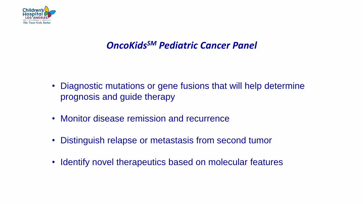
OncoKidsSM Pediatric Cancer Panel
• Diagnostic mutations or gene fusions that will help determine
prognosis and guide therapy
• Monitor disease remission and recurrence
• Distinguish relapse or metastasis from second tumor
• Identify novel therapeutics based on molecular features

OncoKidsSM Pediatric Cancer Panel
– Primer-based target enrichment (Ampliseq)
– Interrogate DNA and RNA isolated from fresh, frozen, and FFPE tissue
– Small input (20 ng DNA and RNA)
– Sequencing on the Ion Torrent S5XL (2 hours)
– Analysis with Ion Reporter and custom tool for indels
– 3 day turn-around time

OncoKidsSM was Designed Specifically for Use in Pediatric Cancers
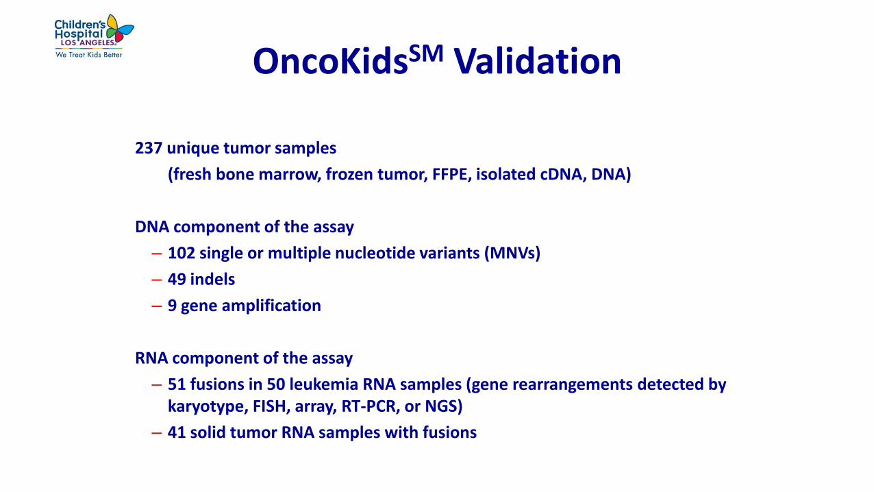
OncoKidsSM Validation
237 unique tumor samples
(fresh bone marrow, frozen tumor, FFPE, isolated cDNA, DNA)
DNA component of the assay
– 102 single or multiple nucleotide variants (MNVs)
– 49 indels
– 9 gene amplification
RNA component of the assay
– 51 fusions in 50 leukemia RNA samples (gene rearrangements detected by karyotype, FISH, array, RT-PCR, or NGS)
– 41 solid tumor RNA samples with fusions
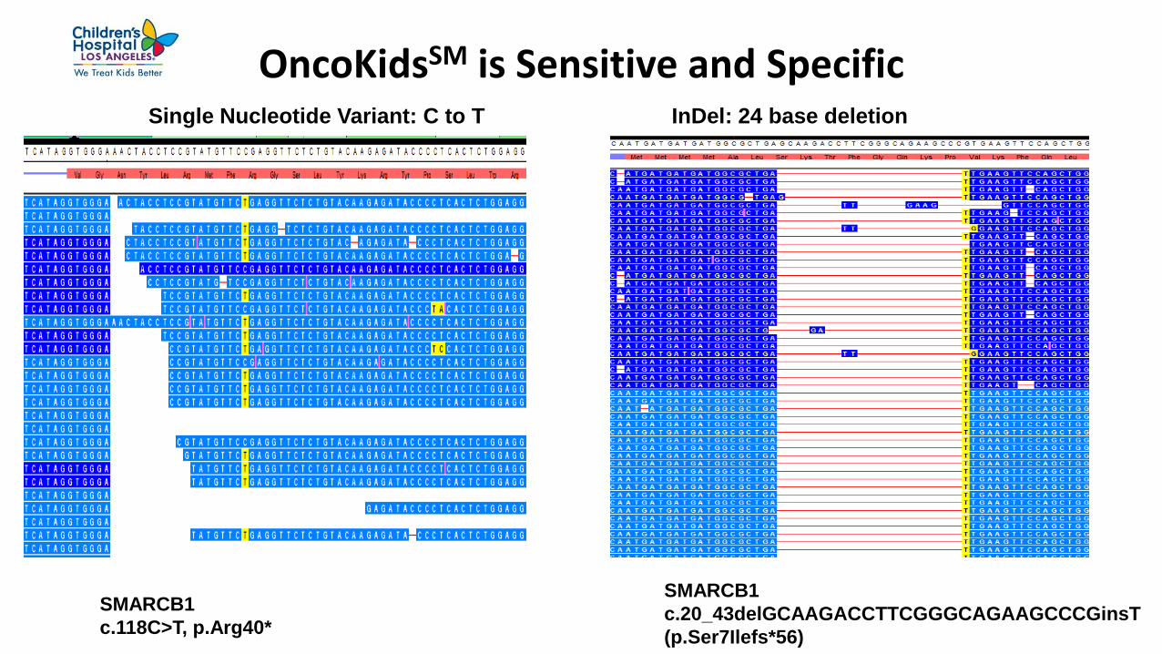
OncoKidsSM is Sensitive and Specific
SMARCB1
c.20_43delGCAAGACCTTCGGGCAGAAGCCCGinsT
(p.Ser7Ilefs*56)
SMARCB1
c.118C>T, p.Arg40*
Single Nucleotide Variant: C to T InDel: 24 base deletion

Detection of DNA Amplifications is Highly Specific MYCN
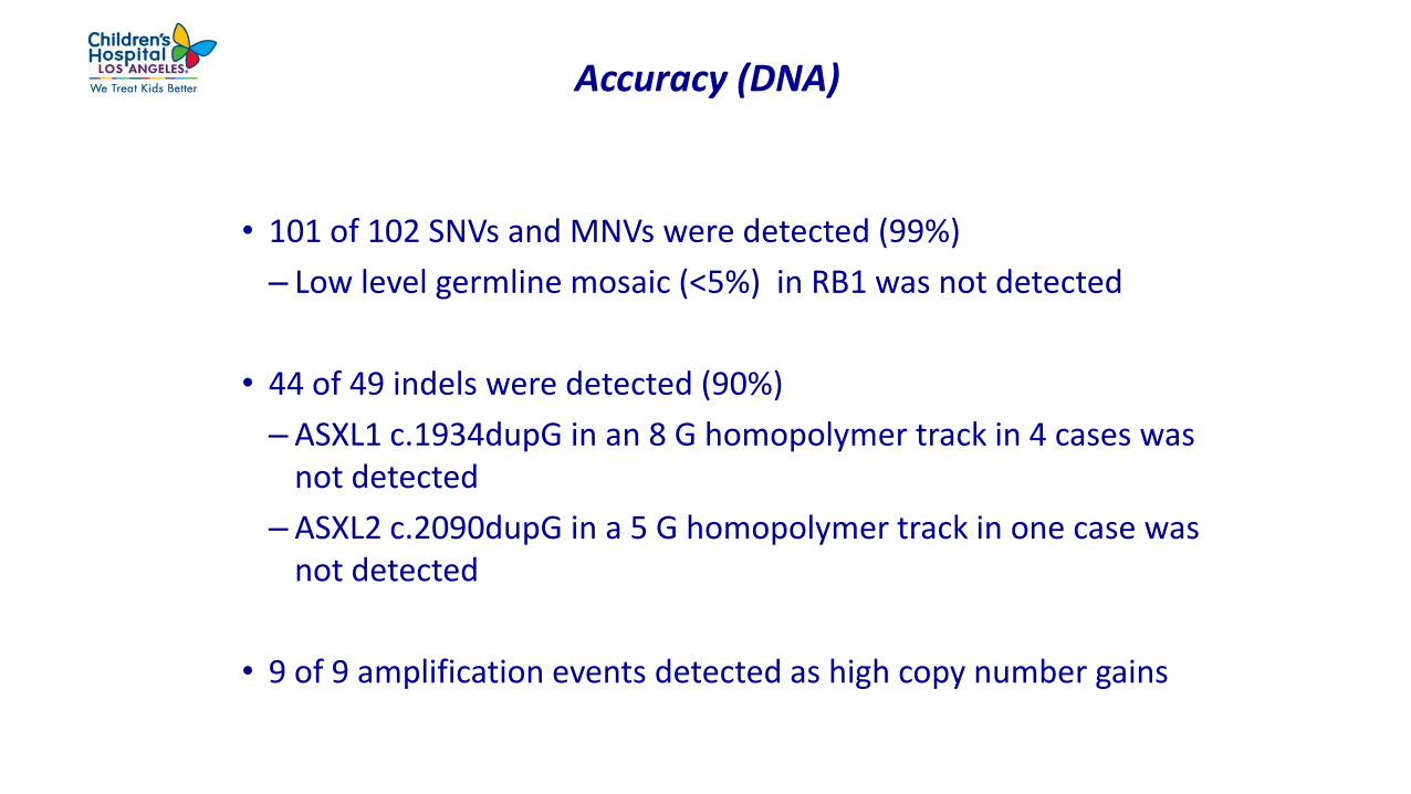
Accuracy (DNA)
• 101 of 102 SNVs and MNVs were detected (99%)
– Low level germline mosaic (<5%) in RB1 was not detected
• 44 of 49 indels were detected (90%)
– ASXL1 c.1934dupG in an 8 G homopolymer track in 4 cases was not detected
– ASXL2 c.2090dupG in a 5 G homopolymer track in one case was not detected
• 9 of 9 amplification events detected as high copy number gains
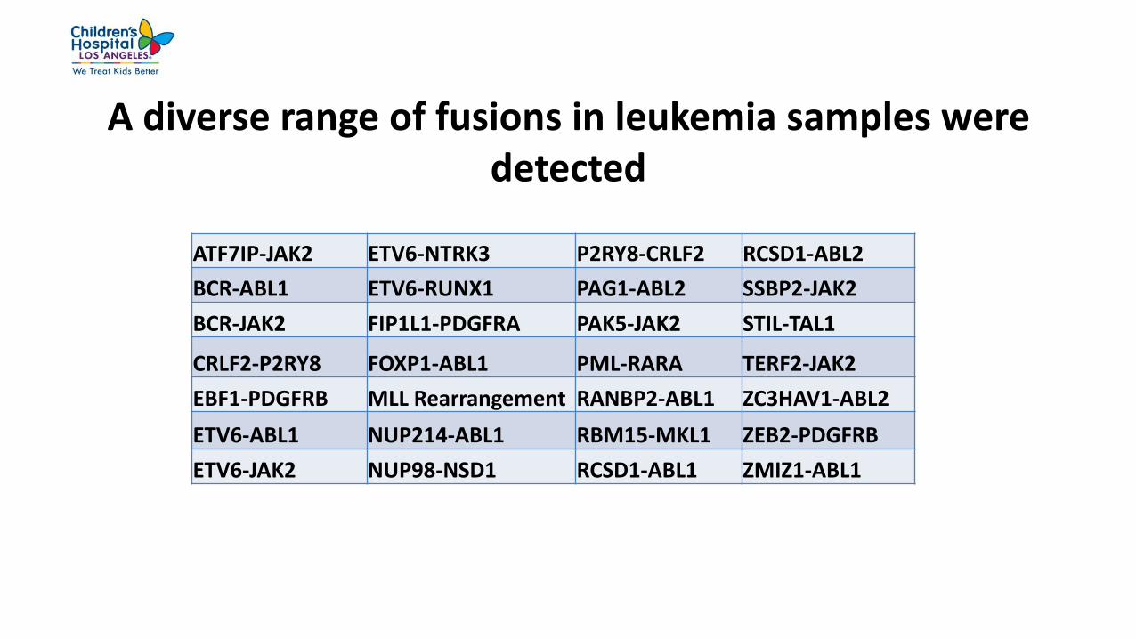
A diverse range of fusions in leukemia samples were detected
ATF7IP-JAK2 ETV6-NTRK3 P2RY8-CRLF2 RCSD1-ABL2
BCR-ABL1 ETV6-RUNX1 PAG1-ABL2 SSBP2-JAK2
BCR-JAK2 FIP1L1-PDGFRA PAK5-JAK2 STIL-TAL1
CRLF2-P2RY8 FOXP1-ABL1 PML-RARA TERF2-JAK2
EBF1-PDGFRB MLL Rearrangement RANBP2-ABL1 ZC3HAV1-ABL2
ETV6-ABL1 NUP214-ABL1 RBM15-MKL1 ZEB2-PDGFRB
ETV6-JAK2 NUP98-NSD1 RCSD1-ABL1 ZMIZ1-ABL1

Representative solid tumor fusions
CCDC6-RET Lung adenocarcinoma
Cllorf95-RELA Ependymoma
EML4-ALK Lung adenocarcinoma
ETV6-NTRK3 Congenital mesoblastic nephroma
EWSR1 Rearrangement Ewing sarcoma
FUS-CREB3L2 Fibromyxoid sarcoma
GOPC-ROS1 High grade glioma
KIAA1549-BRAF Pilocytic astrocytoma
NPM1-ALK Anaplastic large cell lymphoma
PAX3-FOXO1 Alveolar rhabdomyosarcoma
SS18-SSX1 Synovial sarcoma
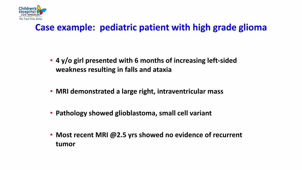
Case example: pediatric patient with high grade glioma
• 4 y/o girl presented with 6 months of increasing left-sided weakness resulting in falls and ataxia
• MRI demonstrated a large right, intraventricular mass
• Pathology showed glioblastoma, small cell variant
• Most recent MRI @2.5 yrs showed no evidence of recurrent tumor

ROS1 GOPC (FIG)
Single interstitial deletion with breakpoints in ROS1 and GOPC(FIG)
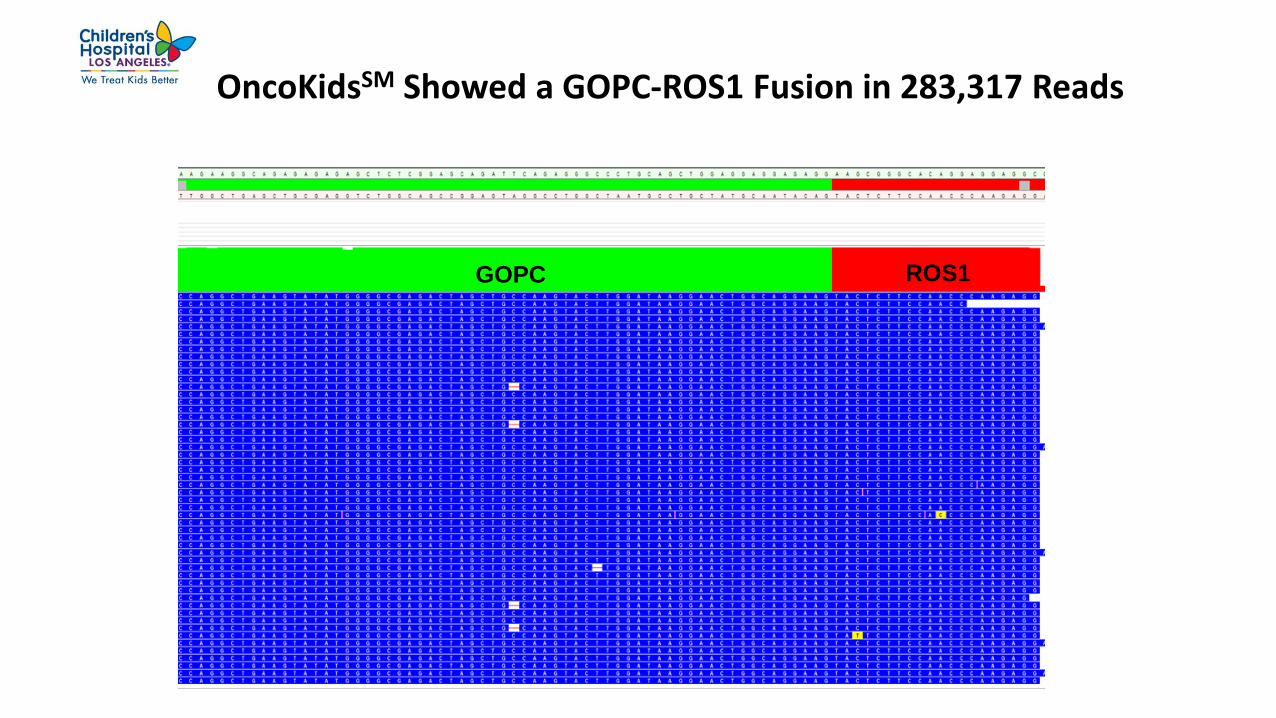
OncoKidsSM Showed a GOPC-ROS1 Fusion in 283,317 Reads
GOPC ROS1

GOPC-ROS1 Fusion Positive Glioblastoma May Respond to Crizotinib
Cancer Growth Metastasis. 2015; 8:51-60.
…..GAT AAG GAA CTG GCA GGA AGT ACT CTT CCA ACC
CAA GAG…..
…..Asp Lys Glu Leu Ala Gly Ser Thr Leu Pro Thr Gln
Glu…….
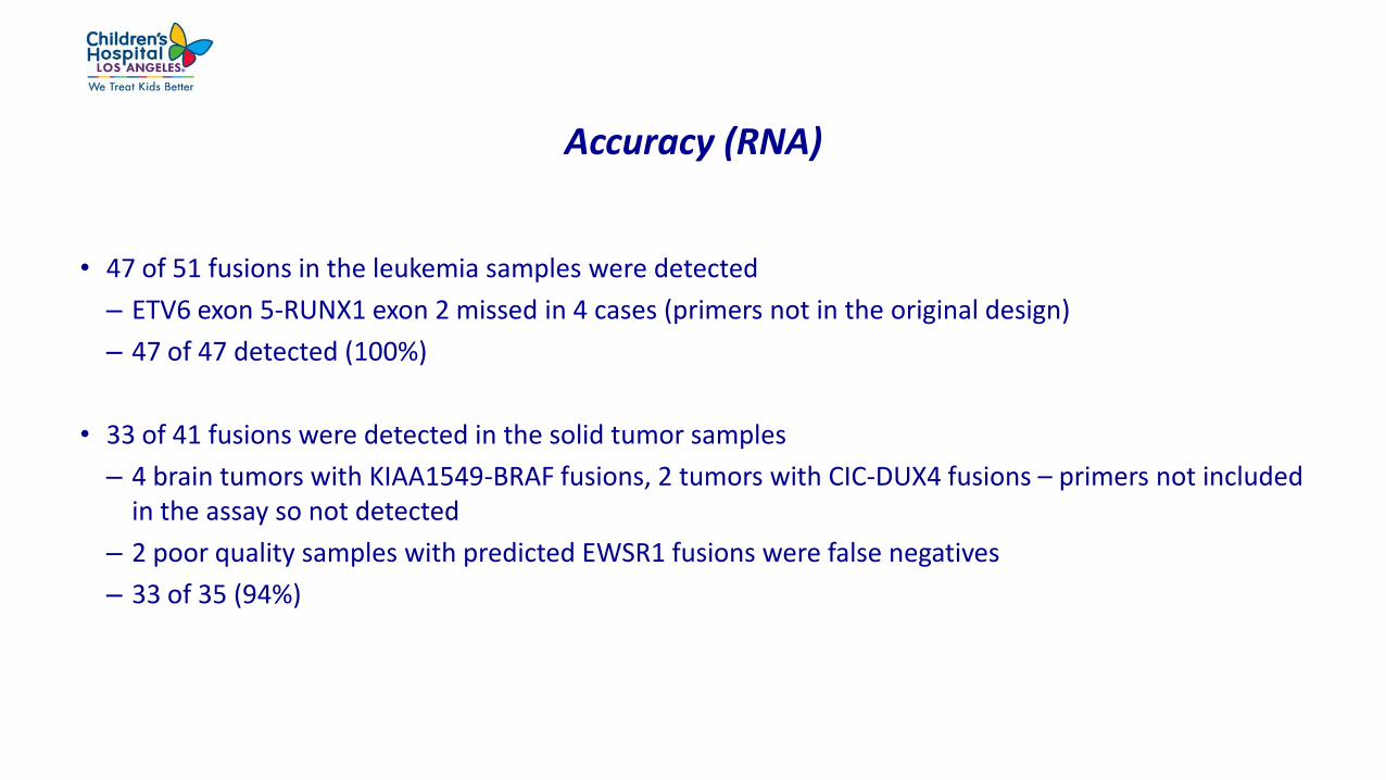
Accuracy (RNA)
• 47 of 51 fusions in the leukemia samples were detected
– ETV6 exon 5-RUNX1 exon 2 missed in 4 cases (primers not in the original design)
– 47 of 47 detected (100%)
• 33 of 41 fusions were detected in the solid tumor samples
– 4 brain tumors with KIAA1549-BRAF fusions, 2 tumors with CIC-DUX4 fusions – primers not included in the assay so not detected
– 2 poor quality samples with predicted EWSR1 fusions were false negatives
– 33 of 35 (94%)

Performance and reporting criteria
• DNA
– Average read depth 5,000 X
– Average read length 120 bp
– 98% coverage at 250X
– Report SNVs at >5% allele frequency
– Report indels (<20 bp) at >10% allele frequency
– Report amplification at >8 copies
• RNA
– Evaluate expression of targeted and non-targeted fusions compared with 4 internal control genes
– Optimum read length 115 bp
– Read count for fusions varies from 600 – 300,000
– All novel fusions are confirmed by RT-PCR

Clinical test launched June 20, 2017
• 19 samples analyzed to date
• 11 cases with Tier 1 variants of strong clinical significance
– Metastatic solid pseudopapillary carcinoma with B-catenin mutation
– Angiomatoid fibrous histiocytoma with EWSR1-ATF1 fusion
– Diffuse midline glioma with H3F3A, ATRX, KRAS, PIK3CA and PPM1D mutations
– Sertoli leydig cell ovarian tumor with DICER1 mutation

Acknowledgements
• Jon Sherlock, Susan Ewald, John Bishop, Scott Myrand, Nick Khazanov, Jeff Smith, Janice Au–
Young, Chaitali Parikh, Jingwei Ni, Seth Sadis, Vinay Mittal and Dinesh Cyanam from Thermo
Fisher Scientific.
• Cindy Fong, Jennifer Han and Sherry Yen for technical assistance.
• Moiz Bootwalla and Alex Ryutov for bioinformatics support.
• Clinical collaborators from University of Chicago, COG Biopathology Center, UCLA, University of
Pennsylvania, Denver Children’s Hospital, Children’s Healthcare of Atlanta, Lucille Packard
Hospital, OmniSeq, and ARUP who provided samples for analysis.