Radicular cyst (Periapical cyst, Apical periodontal cyst, Dental cyst)

Choledochal Cyst
Zachary Stiles, PGY2 UTHSC
Department of Surgery

History
1723: First described by Vater and Ezler
1924: First choledochal cyst excision described by MacWorter
original procedures involved external or internal drainage along with cholecystectomy
1959: Alonso-Lej and colleagues proposed first classification system
1969: Babbitt described anomalous pancreaticobiliary duct union (APBDU)
1977: Todani et al described classification system based on site of cystic change
1995: Farello first described laparoscopic resection of a choledochal cyst

Epidemiology
~80% diagnosed in infants and young children within first decade of life
Incidence
1 in 100,000 to 1 in 150,000 in western countries
Much higher in Asian populations, as high as 1 in 1,000 to 1 in 13,000 in Japan
Four times more common in females
Pathophysiology
Anomalous pancreaticobiliary duct union (APBDU)
common bile duct and pancreatic duct junction occurs outside the duodenum
reflux of pancreatic fluid into the biliary tree
believed to be secondary to arrest in migration of the choledochopancreatic junction into the duodenal wall
Long common channel
defined as insertion of the CBD farther than 15 mm from the ampulla of vater

APBDU
Source: radiologyassistant.nl

APBDU
Source: Soares KC, et al.

Pathophysiology
80-96% of pediatric choledochal cysts are associated with APBDU
Nagi et al:
series of 2,885 patients undergoing ERCP
~90% of patients diagnosed with APBDU had a choledochal cyst
Other mechanistic hypotheses for choledochal cysts include a weak bile duct wall, sustained increased intrabiliary pressure, inadequate autonomic innervations, sphincter of Oddi dysfunction, and distal obstruction of the CBD

Classification
Multiple classification systems
Todani (1977): most widely accepted
based on site of cystic change
five types
Type I (80-90% of all choledochal cysts)
Type IV (15-20%)

Classification

Source: Soares et al.

Source: radiopaedia.org

Classification
Source: Gonzales KD, Lee H.

Type I
Most common (at least 90% of cases)
Cystic/saccular or fusiform dilation of common bile duct
Three subtypes
Ia - cystic dilation of the entire CBD (gallbladder arises from the choledochal cyst)
Ib - cystic dilation of a segment of the CBD
Ic - Fusiform dilation of the CBD

Type I
Typically appear as anechoic cystic lesions which communicate with the biliary tract
May be associated with mild enlargement of the intrahepatic bile ducts secondary to bile stasis

Case courtesy of Dr Aneesh km, Radiopaedia.org, rID: 17160

Type I (CT, MRCP, ERCP) Source: Soares et al.

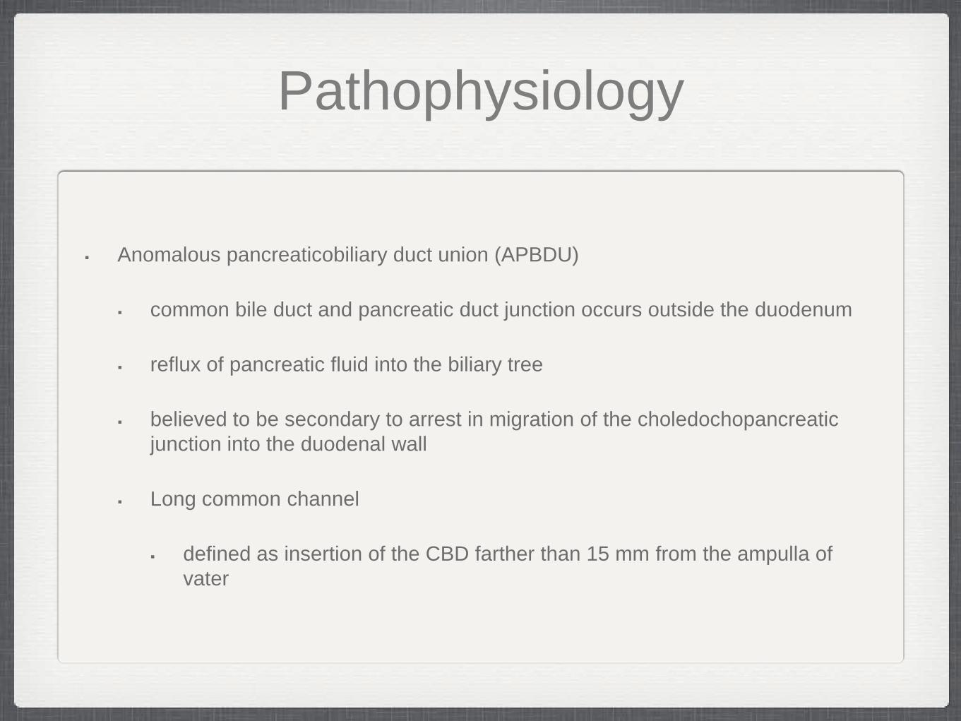
Type II
True diverticula of the CBD
No dilation of the common bile, extrahepatic, or intrahepatic ducts
Represent ~2% of reported cases
Appear as anechoic cysts juxtaposed to the CBD with a normal appearing gallbladder and CHD
Can resemble gallbladder duplication
Type II (CT, MRCP, MRCP) Source: Soares KC, et al.

Type III
Also known as choledochoceles
Comprise 1-4% of cases
Characterized by intraduodenal location at the pancreaticobiliary junction
More evenly distributed between sexes (as opposed to other CC)
Much lower incidence of malignant transformation
Biliary tract symptoms less common; pancreatitis commonly seen
APBDU much less commonly seen with choledochoceles

Type III
Source: Gonzales KD, Lee H.

Type III (MRCP, ERCP) Source: Cha SW, Park MS, Kim KW, et al.

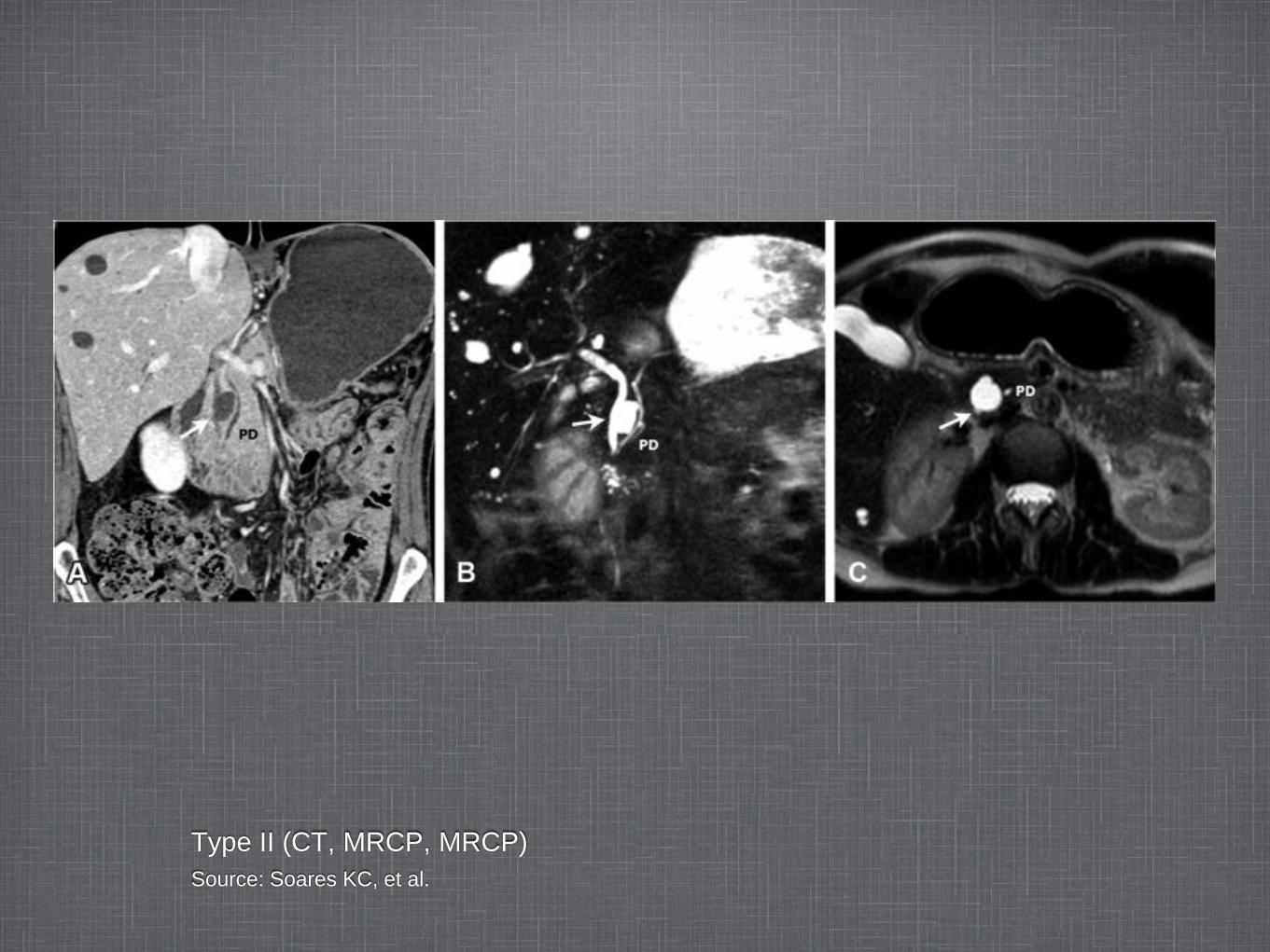
Type IV
Multiple cystic dilations of biliary tree
Can include both intra- and extrahepatic duct involvement
Two subtypes:
IVA - extends from the CBD and CHD into the intrahepatic biliary tree
primary ductal stricture around hepatic hilum commonly seen
IVB - multiple dilations of the extrahepatic biliary tree (classically described as ‘string of beads’ appearance) without intrahepatic involvement

Type IVA - CT Case courtesy of Dr Mohammad Taghi Niknejad, Radiopaedia.org, rID: 20611
Type IVA - CT (cont’d) Case courtesy of Dr Mohammad Taghi Niknejad, Radiopaedia.org, rID: 20611

Type V
Also known as Caroli’s disease
Intrahepatic saccular or fusiform dilation with no underlying obstruction or extrahepatic biliary tree involvement
Thought to arise from ductal plate malformation
Can be associated with polycystic kidney disease
When associated with congenital hepatic fibrosis - Caroli’s syndrome
Enhancement of portal vein surrounded by dilated intrahepatic bile ducts (“central dot sign”)

Type V (MRCP) Case courtesy of Dr Frank Gaillard, Radiopaedia.org, rID: 8362

Type V (US, MRCP, MRCP) Source: Soares KC, et al.

Caroli disease, “central dot sign” Source: Levy, AD

Clinical Presentation
Usually diagnosed in childhood (in utero and adult diagnosis also)
Classic triad (only seen in ~20%)
abdominal pain
jaundice
RUQ mass
Rare to see complete triad, but more commonly seen in pediatric patients as opposed to adults
May present with cholangitis, pancreatitis, portal hypertension, LFT abnormalities

Clinical Presentation
• Patients can manifest symptoms at any point in life
• 80% of patients are symptomatic before age 10
• Classification based on age at presentation:
• ‘Infantile’ (<12 mo) – obstructive jaundice, acholic stools, hepatomegaly (all similar to biliary atresia)
• ‘Adult’ (>12 mo) – more symptoms including fever, nausea/vomiting, jaundice

Adult vs Pediatric
ADULTS
more likely to present with biliary or pancreatic symptoms and abdominal pain
CHILDREN
more likely to present with abdominal mass and jaundice
Cyst rupture - rare, typically only seen in neonates and infants

Diagnosis
More choledochal cysts are being diagnosed as incidental findings secondary to increased use of imaging modalities
Diagnosis usually established using multimodality approach (US, CT, MRI)
ultrasound most frequently used
cholangiography (ERCP, PTC) is the most sensitive technique to define anatomy - may be difficult to perform in pediatric population
MRCP gaining popularity
highly sensitive (70-100%) and specific (90-100%) in choledochal cyst diagnosis and classification

Pathology
Fibrosis of cyst wall lined with columnar epithelium and lymphocytic infiltration typical of pediatric CC
Adult CC includes evidence of inflammation and hyperplasia
Most show some degree of change in liver including portal fibrosis, central venous distention, parenchymal inflammation, and bile duct proliferation

Pathology
Type I (and sometimes Type IV) lack biliary mucosa
Type II closely resembles gallbladder duplication
Type III lined by duodenal mucosa
Type V can have extensive hepatic fibrosis
Increased risk of malignant transformation with age
half of patients >50 years old have invasive biliary neoplasm
less than 1% have invasive biliary neoplasm before the age of 10

Pathology
Malignancy
most commonly associated with Type I and IV cysts
II, III, and V have minimal neoplastic risk
believed to occur through multistep genetic events (early K-ras and p53, late DPC-4)
most cases of malignant transformation are cholangiocarcinoma (gallbladder carcinoma reported in 10-25% of CC-related malignancies)

Management
Risk of malignant transformation warrants complete and total excision whenever possible
Fetal and newborn diagnosis is associated with early progression to liver fibrosis (particularly type IV)
RCT has shown that early excision in prenatally diagnosed asymptomatic cysts resulted in significantly less hepatic fibrosis
Early excision recommended

Type I & IV
Management:
complete extrahepatic bile duct excision down to level of communication with the pancreatic duct
cholecystectomy
restoration of bilioenteric continuity
hepatectomy warranted in type IVA cysts with a significant intrahepatic component likely to result in complications if not removed

Bilioenteric reconstruction
Roux-en-Y hepaticojejunostomy preferred
Hepaticoduodenostomy has been reported but no longer reconstruction of choice
associated with increased rates of gastric cancer (due to bile reflux) and biliary cancer
significantly more cases of postoperative reflux and gastrititis
wide anastomosis imperative - prevent stricture

Type II & III
Extremely low risk of malignant transformation
Type II
diverticulectomy
primary CBD closure at the diverticulum
Small choledochoceles
endoscopic sphincterotomy
Large choledochoceles
trans-duodenal excision, especially if associated with complications

Type V
Liver resection or orthotopic liver transplant (OLT)
Localized or unilobar cystic disease - hepatic resection
incomplete resection leads to poor long-term outcumes
Asymptomatic bilobar disease - non-operative with aggressive surveillance
Complicated bilobar disease (cholangitis, portal hypertension, suspicion of malignancy) - OLT

Laparoscopic Approach
Laparoscopic resection with roux-en-Y hepaticojejunostomy shown to be safe with comparable outcomes to open approach in restrospective studies
Reported advantages:
improved intra-operative visualization of deeper structures
decreased post-operative pain
shorter hospital stay
improved cosmetic result
decreased post-operative ileus

Tang et al. (2011, China)
Retrospective analysis of 62 children undergoing laparoscopic resection
8.2% morbidity (bile leak, adhesive SBO, intestinal necrosis, cholangitis, anastomotic narrowing)
Ono et al. (2010, Japan)
Retrospective review of 56 patients undergoing open resection and reconstruction
16/56 late complications within 10 years (biochemical liver dysfunction, persistent dilation of intrahepatic ducts, recurrent abdominal pain, recurrence of CBD adenocarcinoma)

Outcomes
Post operative morbidity and mortality typically lower in children compared to adults
Late complications occur in up to 40% of adult patients (stricture, cancer, cholangitis, cirrhosis)
Type IVA cysts most commonly associated with post-operative complications

Prognosis
• Overall, resection has good prognosis
• 89% event free rate
• 5 year OS rates >90%
• Risk of malignancy
• Remains elevated, even after excision
• Choledochal cyst associated biliary malignancy associated with extremely unfavorable outcomes (median survival 6-21 months)

Surveillance
• Long term surveillance indicated, especially given risk of malignancy
• Particularly important in cases with persistent intrahepatic biliary dilatation
• Regular biochemical evaluation and abdominal US or cross-sectional imaging

Sources
Cha SW, Park MS, Kim KW, Byun JH, Yu JS, Kim MJ, Kim KW. Choledochal cyst and anomalous pancreaticobiliary ductal union in adults: radiologic spectrum and complications. J Comp Assist Tomography 2008; 32(1): 17-22
Gonzales KD, Lee H. Choledochal Cyst. from Pediatric Surgery, 7th Ed. Coran A, Adzick NS, Krummel TM, et al.
Nagi B, Kochhar R, Bhasin D, Singh K. Endoscopic retrograde cholangiopancreatography in the evaluation of anomalous junction of the pancreaticobiliary duct and related disorders. Abdom Imaging 2003; 28:847-852
Levy AD. Biliary Tract - Pathology. www.radiologyassistant.nl
Ono S, Fumino S, Shimadera S, Iwai N. Long term outcomes after hepaticojejunostomy for choledochal cyst: a 10- to 27-year follow up

sources
Soares KC, Arnaoutakis DJ, Kamel I, et al. Choledochal Cysts: Presentation, Clinical Differentiation, And Management. J Am Coll Surg. 2014 Dec; 219(6): 1167-80
Tang ST, Yang Y, Wang Y, Mao YZ, Li SW, Tong QS, Cao GQ, Zhao ZX. Laparoscopic choledochal cyst excision, hepaticojejunostomy, and extra-corporeal Roux-en-Y anastomosis: a technical skill and intermediate-term report in 62 cases. Surg Endosc. 2011; 25: 416-422.
Weerakkody Y, et al. Todani classification of bile duct cysts. www.radiopaedia.org
Yamataka A, Lane GJ, Cazares, J. Laparoscopic surgery for biliary atresia and choledochal cyst. Seminars in Pediatric Surgery. 2012; 21: 201-210