Dr. Brian Payne - PCV2: Diagnostics, Control, Protection, and Efficacy Measures

PCV2 and reproduction failure in sows
Authors
Vincent DEDET, DVMAuzalide Santé Animale, La Chouannière, Le Cannée 35380 Paimpont (France)[email protected]
François JOISEL, DVM, MScMERIAL , 29, avenue Tony Garnier, BP 7123, 69348 Lyon Cedex 07 (France) [email protected]
Thaïs VILA, DVMMERIAL, 29, avenue Tony Garnier, BP 7123, 69348 Lyon Cedex 07 (France)[email protected]
PCV2_sept08 1/10/08 14:48 Page 1
PCV2 and reproduction failure in sows
Summary
Porcine Circovirus type 2 (PCV2) related diseases (PCVD) have been reported throughout the world, and reproduc-tive failure can clearly be associated with PCV2 infection in sows. Following the guidelines set by evidence-basedmedicine, this bibliographic work highlights the PCV2-related reproductive failure, which has been describedworldwide. It also draws attention to the increasing set of epidemiological studies, field trials and reports that showan association between the subclinical PCV2 infection of gilts and sows and their lifelong productivity.
PCV2_sept08 1/10/08 14:48 Page 2

PCV2 and reproduction failure in sows
INTRODUCTION 4
1. MATERIAL AND METHODS 4
2. EXPERIMENTAL DATA 5
2. 1. Trans-uterine inoculation of fetuses with PCV1 5
2. 2. Trans-uterine inoculations of fetuses with PCV2 5
2. 3. Oronasal inoculation of non-pregnant SPF gilts 7
2. 4. Oronasal inoculations of pregnant sows with PCV2 8
2. 5. Intra-uterine inoculation of PCV2-negative sows 10
2. 6. Embryo exposure to PCV2 11
3. EPIDEMIOLOGICAL DATA 12
3. 1. Indirect proofs 12
3. 2. Direct proofs 14
124. CLINICAL REPORTS 16
4. 1. In cesarean derived-colostrum deprived (CDCD) piglets 16
4. 2. In conventional farms 16
5. ANECDOTAL REPORTS 22
6. CONCLUSION 23
REFERENCES 24
PCV2_sept08 1/10/08 14:48 Page 3

PCV2 and reproduction failure in sows
Introduction
Post-weaning Multisystemic Wasting Syndrome (PMWS) has emerged as a herd disease in Canada and in Europein 1995/1996, and has been identified as linked to Porcine Circovirus type 2 (PCV2) infection in 1998. For the 10following years, other PCV2-related diseases (PCVD) have been reported throughout the world. In a recent reviewabout PCVD, reproductive failure is the last of the six items listed (Gillespie, 2007). It is described as including“abortions, stillbirths and foetal mummification”. The object of the present review is to screen the available publi-shed data in order to assess whether the mechanisms underlying this reproductive outcome of PCV2 infection arepresently understood. We chose to follow the guidelines set by evidence-based medicine while performing thisbibliographic work.
1. Material and methods
The method used through the process of writing this review has followed the experience-based medicine1 recom-mendations regarding systematic reviews2. The question that guided our searching was structured according to the PICO method: Population, Indicator,Comparator, Outcome. This question was formulated as: “Can PCV2 infection be considered as a cause for repro-ductive failure in sows?”. Population considered were sow herds, or sow groups in experimental infection works (in some cases, the popu-lation considered were the fetuses/stillborn submitted to the diagnostic labs). Indicator was a reproductive failure (this inferred that the description of lesions such as myocardial necrosis in abor-ted foetuses/deadborn piglets was also an indicator of a reproductive failure).Comparator was taken either as the average reproductive performances of sow herds at regional/national levels,or as the “control” sows included in the case-control studies or in the experimental infection trials.Outcome is the presence of recent PCV2 infection in dams and/or the exclusive or nearly systematic presence ofPCV2 in fetuses/stillborn piglets.
The data that were consulted were:
� The Pubmed database3, with the only key word “PCV2” in the first step of our search. This retrieved 320 refe-rences. It is to be noted that this search had not to be limited in time, since PCV2 has been first described in 1998,which is the year of publication of the oldest references retrieved by this search. It is to be noted that the Pubmedsearch with “PCV2 AND sows” only retrieved five references; which did not correspond to the amount of articlesbrought to our attention before beginning this extensive search. Consequently, each of the 320 abstracts availablefrom the first search were read; those dealing with reproductive failure, experimental inoculation of sows and/orPCV2 lesions in aborted/stillborn piglets were selected for full article reading.
� The table of contents of the proceedings of different Swine congresses (Leman Conference, ISU Swine diseaseConference for Swine practitioners, George A. Young Swine Health and management Conference, IPVS and AASV)have also been screened, from 1996 to 2007. From 1996 to 1998, our screening was mostly focusing on reports ofreproductive failure while from 1998 onwards, we focused on presentations dealing with PCV2.
� We also consulted non-referenced journals with a peer-reviewed section, such as the Journal of Swine4 Healthand Practice, the Pig Journal. Folia Veterinaria5 had a more erratic online presentation of its contents, none after2003 being available online.Finally, two PhD thesis from Ghent University (Belgium) were available on the subject. Both were extensively read.
In total, 59 references were found relevant to our question, and have been read in full. The reliability of the asso-ciation between the presence of the agent (PCV2) and the occurrence of the outcome (reproductive failure) is herepresented through the hierarchy retained in the evidence-based medicine approach: experimental studies with ino-culation and re-isolation/sequencing of the agent come first. Case-control studies prevail on clinical reports. Finally,anecdotal reports are mentioned, although they should not be considered as providing conclusive answers to ourquestion.
4
1 For more detailed information, see the UK Centre for evidence-based Medicine at http://www.cebm.net/?o=1016 2 See : “Teaching EBM with Learners' questions”, by Scott Richardson, on the same website (download section).3 http://www.ncbi.nlm.nih.gov/sites/entrez?db=pubmed 4 Journal of the American Association of Swine Veterinarians : www.aasv.org5 This Slovakian Journal has been presented to us by Dr G. Bilkei, which we acknowledge. More details are available on: http://www.uvm.sk/dept/journals/folia/contents.html
PCV2_sept08 1/10/08 14:48 Page 4

2. Experimental data
2. 1. Trans-uterine inoculation of fetuses with PCV1
Late-term pregnant sows underwent laparotomy, the uterine horns were exposed to air and each fetus was injec-ted through the uterine wall intramuscularly with PCV1 (Johnson et al, 1999). One of the sows died prior to farro-wing. The remaining two sows farrowed as expected. One gave birth to nine liveborn pigs and one stillborn pig.The other sow gave birth to eight liveborn, one stillborn and five mummified fetuses. No gross lesions were obser-ved in any of the animals that died or were euthanised. PCV1 was successfully isolated from some fetuses, all still-born pigs and other piglets euthanised at birth. Prior to suckling colostrum, all piglets had anti-PCV1 antibodies.This demonstrates that PCV1 is capable of infecting and replicating in late-term fetuses. The presence of stillborn,weak-born and mummified fetuses “suggests that reproductive failure could result following foetal infection withPCV1”.
2. 2. Trans-uterine inoculations of fetuses with PCV2
� Late-term pregnant sows underwent laparotomy, the uterine horns were exposed to air and each fetus wasinjected through the uterine wall intramuscularly with PCV2 (Johnson et al, 1999). One sow farrowed three stillborn,two partial mummies, five weakborn and two liveborn piglets. The other two sows farrowed 22 liveborn and two still-born piglets. Piglets at farrowing of within their first week of age had relatively large lymph nodes. PCV2 was successfully isola-ted from the partial mummies, stillborn and weakborn piglets. The piglets that did not receive colostrum had anti-PCV2 antibodies. This demonstrates that PCV2 is capable of infecting and replicating in late-term fetuses. The presence of stillborn,weak-born and mummified fetuses “suggests that reproductive failure could result following foetal infection withPCV2”. The absence of clinical disease after weaning suggests that the development of PMWS may not occur fol-lowing congenital infection.
� The Virology unit at Ghent Veterinary Faculty (Belgium) reproduced this work, with inoculation at different ges-tation stages for conventional (PCV2 seropositive) sows (Sanchez, 2003a). The PCV2 1121-isolate passaged in PK-15 cells (4th passage) was used. In each gravid sow, two foetuses were inoculated via trans-uterine route, with 104.3
TCID50 PCV2. This procedure was applied for two sows at 57, 75 and 92 days of gestation. Twenty-one days aftervirus inoculation, sows were sacrificed, hysterectomies were performed and the fetuses were collected. As a posi-tive control, day-old CDCD piglets received the same inoculum (intranasally and intratracheally) (Sanchez et al,2003b). All inoculated fetuses provided a positive PCV2 isolation, but the heart was the only organ to be positiveregardless of gestational age at inoculation. Viral antigens were localized in cardiomyocytes, hepatocytes andmacrophages during foetal life and only in macrophages postnatally. Infected macrophages were observed in alltypes of tissues examined (heart, liver, lungs, spleen and inguinal lymph nodes) both in fetuses and in piglets. Theabsolute number of PCV2-positive cardiomyocytes was highest in 57-day fetuses and changed in decreasing orderin 75- and 92-day fetuses (p<0.05). Also, the highest absolute number of PCV2-positive hepatocytes and macro-phages was observed in 57-day fetuses (p<0.05) and changed in decreasing order in fetuses inoculated at laterstages of gestation. Gross abnormalities were only observed in fetuses inoculated on d576, which taken togetherwith higher replication in these fetuses, indicates these lesions are due to PCV2. Non-inoculated fetuses adjacentto the inoculated ones were all PCV2 negative. This study shows that cellular targets of PCV2 for its replication gra-dually change from cardiomyocytes, hepatocytes and macrophages during foetal life to only macrophages post-natally.
� In a longer term experiment, the same procedure was applied in three sows at 57, 75 and 104 days of gesta-tion, respectively (Sanchez, 2003a). Piglets were recovered on 114th day of gestation via hysterotomy or at natu-ral farrowing. PCV2 was isolated from the hearts of all piglets that had been inoculated at d57 and d92 of gesta-tion, and from two piglets adjacent to the two foetuses inoculated at d577. Two piglets born to a d75 sow were alsopositive, but it could not be determined whether these were the previously inoculated piglets. All piglets inoculated
PCV2 and reproduction failure in sows
6 One was strikingly pale and 3 others were edematous and had distended abdomens. Haemorrhages and congested organs with enlarged livers were present in all 4 fetuses.7 These piglets, together with the inoculated ones, were presenting deformities. Two piglets inoculated at d92 also did.
5
PCV2_sept08 1/10/08 14:48 Page 5

PCV2 and reproduction failure in sows
6
Sow ID
1
2
3
4
5
6
7
1
2
3
4
Treatment
Challenge
Control
Stage of gestation (day) at inoculation
60
73
74
73
76
74
58
72
61
68
68
Day post-inoculationat abortion
N/Ab
N/A
6
N/A
N/A
N/A
4
N/A
N/A
N/A
N/A
No. of littersat abortion or
birth
14
9
7
14
15
14
12
16
14
12
9
No. of livefoetuses or
piglets
12
9
0
3
10
10
0
9
14
11
8
No. of dead orstillborn pigs
2
0
7
1
5
4
12
7
0
1
1
No. of PCV2(+) pigsa at
birth
2
1
7
0
0
3
12d
0
0
0
0
Day 0
1
0
NDc
0
0
1
ND
0
0
0
0
Day 21
2
2
ND
7
6
0
ND
0
0
0
0
No. of seropositive pigs
at d104 were PCV2-negative. Hence, PCV2 is able to replicate in fetuses at different stages of gestation and cau-ses foetal pathology and death when infection occurs during mid-gestation, without affecting the course of pre-gnancy. The slow spread of the virus in the uterus (only fetuses adjacent to the d57 inoculated ones were foundpositive) may have been responsible for the uneventful continuation of gestation.
� A late-term inoculation was also performed, and the outcome of piglets in postnatal life was compared to theone of cesarean-derived piglets inoculated at one day of age (Sanchez, 2003a; Sanchez et al, 2004). The sameprocedure was applied to three sows at 92 days of gestation (two to three foetuses inoculated) and one sow at 104days of gestation (five foetuses inoculated). Piglets were recovered by hysterotomy on d114; part of the inoculatedones were immediately sacrificed, while the remaining ones were monitored until 14 days of age. Fourteen cesa-rean-derived piglets were inoculated oronasally and intraperitoneally at one day of age, and were sequentiallysacrificed at 10, 21, 35, 42 and 49 days. Lymphocyte depletion and infiltration of monocytic cells were detected inlymphoid organs with high virus titres8 in a limited number of piglets inoculated in utero or at one day of age. Theabsence of clinical signs in these piglets shows that intensive and early PCV2 replication ad the development ofPMWS-associated lymphoid lesions do not necessarily lead to wasting disease.
� The Iowa State University published a report on the use of an ultrasound needle-guided device to perform intra-uterine fetus inoculation (Yoon et al, 2004) via the transabdominal route9. A total of 20 pregnant sows at fifth or sixthparity were purchased from a commercial swine herd known to be free of most common pathogens and with nohistory of PCVDs. These sows were selected as having no PCV2 PCR-detectable viremia and low or no IFA titresof anti-PCV2 antibodies. Group 1 (n=7) and group 3 (n=7) were inoculated with PCV2 at mid-gestation (approxi-mately 54–76 days of gestation) and at late-gestation (approximately 84–94 days of gestation), respectively. Group2 (n=4) and group 4 (n=2) served as the sham-inoculated control group for each gestation group, respectively. Theinoculum (104 TCID50/ml) contained a field isolate of PCV2 (ISUVDL 98-15237) from a pig affected by PMWS, andpropagated in PK-15 cell line (12th passage). Neither seroconversion nor PCV2 viremia were detected in any sowsthroughout the study period, suggesting that PCV2 given to fetuses did not spread to dams. In group 1, two sows out of seven aborted within the first week after inoculation. Although abortion appeared to bedue to infection with bacteria, PCV2 DNA was detected in all fetuses from both sows (see table 1). The remainingfive sows gave birth at term and three of them had at least two piglets (either live or stillborn) that were infectedwith PCV2.
Table 1 Reproductive performance of sows after intrauterine inoculation of PCV2 at midterm gestation (from Yoon et al, 2004). (a) Sum of PCV2positive animals (by PCR and/or IHC) among all stillborn/dead pigs and two randomly selected live pigs. (b) Not applicable since animals did notabort. (c) Not determined since no live-born piglets were available or all live-born piglets were sacrificed at birth for pathological evaluation. (d)Pooled tissues.
8 4.5 to 5.7 logTCID50/g tissue.9 Under general anesthesia.
PCV2_sept08 1/10/08 14:48 Page 6

No. of seropositive
PCV2 and reproduction failure in sows
7
Sow ID
8
9
10
11
12
13
14
5
6
Treatment
Challenge
Control
Stage of gestation (day) at inoculation
94
84
88
90
90
84
85
94
88
Day post-inoculation atabortion
N/Ab
21
14
7
N/A
N/A
7
12
N/A
No. of litters atabortion or birth
12
13
13
14
9
10
13
12
16
No. of live foe-tuses or piglets
9
1
1
0
8
8
0
5
14
No. of dead orstillborn pigs
3
12
12
14
1
2
13
7
2
No. of PCV2 (+)pigsa at birth
0
9
9
10
2
1
2
0
0
Day 0
0
0
1
NDc
0
0
ND
0
0
Day 21
0
0
ND
ND
0
0
ND
0
0
In group 3, four out of seven sows aborted within 21 days after inoculation (3 caused by PCV2 with 85 to 100%dead litter) and the remaining three sows gave birth to litters of live and stillborn piglets at term (see table 2), twoof them with no evidence of in utero PCV2 infection. None of the piglets born alive became viremic or seroconver-ted to PCV2 until 21 days after birth.
Table 2 Reproductive performance of sows after intrauterine inoculation of PCV2 at late gestation (from Yoon et al, 2004). (a) Sum of PCV2 posi-tive animals (by PCR and/or IHC) among all stillborn/dead pigs and two randomly selected live piglets or litter. (b) Not applicable since animalsdid not abort. (c) Not determined since no live-born piglets were available or all live-born piglets were sacrificed at birth for pathological evalua-tion.
Overall, very few fetuses from PCV2-inoculated sows had lesions; myocardial atrophy associated with abundantPCV2 antigen was present in one of the fetuses in this study. Furthermore, PCV2 was shown to be able to spreadamong fetuses in utero as the virus was delivered to the foetal fluid compartment shared by two fetuses, but detec-ted in more than two piglets of each litter (except in two sows).
2. 3. Oronasal inoculation of non-pregnant SPF gilts
In his PhD thesis, Sanchez (2003a) reports the use of four 8-month old SPF gilts seronegative to PCV2 that wereoronasally inoculated with PCV2 strain 1121 at 105 TCID50. No clinical signs were observed over the study period.PCV2 was not detected by viral isolation in plasma of gilts, but PCV2 was readily transmitted via bioassay withplasma sampled at 21 days post-infection (DPI). PCV2 was also detected by Q-PCR in the plasma of three out offour gilts from 14 to 49 DPI (and only at 14 DPI in the remaining gilt). Q-PCR also detected PCV2 DNA in all fourgilts’ PBMC from 14 to 63 DPI (end of the study); two gilts were positive from 7 DPI onwards.These results confirm that viremia in primo-infection in pubescent females, although at low level, has an extendedduration, although these animals mount an early immune response. The author stresses that “viremia is an essen-tial first step in the vertical transmission of virus from the dam to the fetus”. The long lasting viremia observed in thepresent study “potentially favours such an event to occur”. Unfortunately, these gilts were not mated, but the authorreports an unpublished observation where a seronegative gilt was inoculated with PCV2 at 44 days of gestation,and transplacental infection did not take place10.
10 Ibid., p. 45.
PCV2_sept08 1/10/08 14:48 Page 7

PCV2 and reproduction failure in sows
8
Sow/inoculation
1. At d35 gestation
2. At d35 gestation
3. At d70 gestation
4. At d70 gestation
5. Negative control
Total born
12
19
17
9
7
Liveborn
6
1
0
9
7
Stillborn
2
2
17
0
0
Mummified
4
16
0
0
0
2. 4. Oronasal inoculations of pregnant sows with PCV2
� In France, a study has been conducted using five pregnant sows from a PCV2-negative SPF herd, inoculatedvia both intratracheal and intramuscular routes (Cariolet et al, 2001a). Each sow received 10 ml (intratracheal route)and 2 ml (intramuscular route) of a 105.18 TCID50/ml PCV2 strain derived from tissue homogenate of affected pigletsfrom a PMWS-farm.Two sows were inoculated at 35 days of gestation; two others at 70 days of gestation; the remaining one was sham-inoculated at 70 days of gestation. All four infected sows were sick after challenge, with hyperthermia (those ino-culated at d35 had a higher hyperthermia than those inoculated at d70) and anorexia/reduced appetite for oneweek. One sow (inoculated at d70) aborted at 3 DPI. Two others (inoculated at d35 and d70) developed skin lesionswithin three to four weeks after challenge that, for one of them, were similar the lesions observed in dermatitis-nephropathy syndrome, although this condition did not last more than four days.Farrowing results were severely compromised in the challenged sows (see table 3). PCV2 detection by PCR andIPMA were negative in all tissue samples collected on piglets at birth. All piglets were seronegative before colos-trum intake.The authors conclude that inoculation of pregnant SPF PCV2-negative sows:- triggers clinical signs “with similarities to those observed in PMWS-affected pigs in the field”;- induces a “very severe” reproductive failure, probably as the outcome of illness associated with infection;- does not produce transplacental infection of fetuses.
Table 3 Farrowing results of sows (from Cariolet et al, 2001a).
PCV2_sept08 1/10/08 14:48 Page 8

Number of piglets
PCV2 and reproduction failure in sows
9
11 PCV2 serologic status of the sows was not mentioned in the paper. Since these are conventional animals, it can be presumed that the gilts/sows used in this trial were all seropositive to PCV2.12 Which is consistent with the origin of the PCV2 strain used in this trial.
Sow ID
1
2
3
4
5
6
Total
Pregnancy
1st
1st
2nd
1st
1st
2nd
-
Day of pregnancy at inoculation
93
94
93
05
93
93
-
Period (days) between inocula-tion and abortion
7
18
12
11
17
16
-
Stillborn
11 (2)
9 (0)
14 (4)
12 (3)
8 (1)
11 (3)
65 (13)
Liveborn
0
4 (2)
1 (1)
0
2 (1)
3 (1)
10 (5)
� In another study, eight conventional pregnant sows from a South Korean farm with no history of PCVD (ave-rage 1.33 pregnancies) were randomly allocated to an infected (n=6) or control (n=2) group (Park et al, 2005). Theviral inoculum contained isolate SNUVR000473 at its second passage in PCV-free PK-15 cells. This PCV2 strainwas isolated from an aborted fetus, at approximately 113 days gestation, in a 550-sow herd located in KyounggiProvince. The infected sows11 were each inoculated intranasally with 6 ml of tissue culture fluid (1.2 105 TCID50/ml)three weeks before the expected farrowing date. Four infected sows aborted, and the remaining two farrowed pre-maturely. The six infected sows delivered a total of 65 stillborn and 10 liveborn piglets (see table 4). No microsco-pic lesions were seen in any organ other than the lung (mild pneumonia) in stillborn or liveborn piglets. Intensesignals were most often seen (by IHC and ISH) in lymph nodes, the distribution of positive cells being multifocal.Although they did not check for the presence of PCV2 in the semen used, these authors conclude that their results“demonstrated that PCV2 induced reproductive failure in pregnant sows12” when infected in late gestation, whichis consistent with the origin of the PCV2 strain used in this trial.
Table 4 Number of stillborn and liveborn piglets from pregnant sows experimentally infected with PCV2. Abortion refers to the delivery of animmature foetus before the completion of the gestation period. Numbers of piglets positive for PCV2 infection are given between brackets (fromPark et al, 2005).
� In another study also from South Korea, the effects of transplacental PCV2 infection on porcine epidemic diar-rhoea virus (PEDV)-induced enteritis were examined in neonatal piglets (Jung et al, 2006). Six pregnant sows wererandomly allocated to an infected (n=3, same inoculum as previous study) or control group (n=3). Inoculation wasperformed intranasally, three weeks before the expected farrowing date. Thirty piglets from PCV2-infected sowswere randomly assigned to two groups (A and B) of 15 piglets each. Another 30 piglets from non-infected sowswere randomly assigned to two groups (C and D) of 15 piglets each. The piglets in groups A and C were dosedorally at three days of age with PEDV. In group A, significantly more PEDV nucleic acid was detected in the jejunal tissues at 24 h PI (p<0.05) than in thesame tissues of group C piglets. The reverse was true at 36, 48, 60, and 70 h PI (p<0.05). The authors concludethat the clinical course of PEDV disease was markedly affected by transplacental infection of PCV2.
PCV2_sept08 1/10/08 14:48 Page 9

PCV2 and reproduction failure in sows
Outcome
Farrowed d111
Aborted d80
OK. 1 deformed liveborn piglet*
Empty
Aborted d25
24 h hyperthermia
Log inoculum
5.18
5.18
4.18
4.18
3.18
3.18
-
-
Sow ID
6345
6370
80108
90127
80118
90135
6326
6342
Total born
13
17
11
15
12
15
Live born
2
0
8
11**
11
10
Stillborn
2
14
2
0
0
3
17-23 cm
5
1
1
2
0
0
>24 cm
2
0
0
1
0
0
Serum Ab
0/3
ND
1*/10
ND
6/11
PCR-positive
6/6
2/3
1*/11
0/4
9/10
< 15 cm
2
2
0
1
1
2
10
2. 5. Intra-uterine inoculation of PCV2-negative sowsTo date, two experimental infections of sows via the intra-uterine route have been published, both by the sameteam, although an increasing number of reports identify PCV2 in boar semen.
� The first experiment was performed with SPF sows from a PCV2-negative SPF herd (Cariolet et al, 2001b).Sows were normally inseminated and the AI catheter was used 2 minutes after discharge of semen to inoculate 12ml of viral (or sham) solution and an additional 10 ml of medium. Sows received different infectious titres (see table5). Most sows did not develop any clinical sign, some of them a transitory hyperthermia; all of them became sero-positive. Two sows aborted, regardless of the level of infectiosity in the inoculum. Liveborn piglets sampled beforesuckling could by viropositive and seronegative, or viro- and seropositive, or viro- and seronegative.The authors conclude that intra-uterine inoculation of SPF PCV2-negative sows:- Does not trigger typical symptoms, except transitory hyperthermia that may occur late after infection and maycause abortion.- Sows infected with high-titre inoculum had typical reproduction failures. The dose effect is less clear for lowerdoses. Each litter can have several piglets affected.- PCV2 was neither detected in embryos nor in mummified fetuses below 12 cm: PCV2 may remain dormant, infetuses or in the uterus, during the first part of pregnancy. - PCV2 replicates in 8-week old fetuses, although sows have mounted an immune response at this stage.- Taken together, these results show there is no proof of immunotolerant piglets; PCV2 infection can take place inthe second half of pregnancy; a small proportion of sows may be PCV2-negative in any conventional herd: theseanimals may replicate PCV2 if infected by intra-uterine route.
Table 5 Farrowing results of sows and serology and virology in piglets sampled before suckling (from Cariolet et al, 2001b). * : same piglet.**: five weakborn died on 3d day of life.
Mummified Piglets before suckling
PCV2_sept08 1/10/08 14:48 Page 10

PCV2 and reproduction failure in sows
� The second experiment aimed to evaluate the importance of the Parvovirus and Erysipelas vaccination proto-col of the dam on the outcome of her progeny, as far as PMWS was concerned (Rose et al, 2007). In this experi-ment, six PCV2-free sows were first infected oronasally with PCV2 two months before insemination (D0) and thenby the intra-uterine route at insemination (D62). This protocol mimics the natural contamination events that occur inthe field. Sows were vaccinated on D21 and D42 (not against PCV2). In addition, three SPF sows were synchroni-zed for farrowing dates, to enable them to foster piglets born to infected sows and removed at birth before colos-trum intake. A significantly higher proportion of mummified fetuses was obtained from PCV2-infected non-(PPV andErysipelas)-vaccinated sows than from vaccinated sows. Acute myocarditis lesions were found in their piglets,together with a high PCV2 genome load. The latter was significantly higher than in those born to PCV2-infected vac-cinated sows. Furthermore, among the 16 piglets raised by the foster-sows and that later developed a syndromepossibly related to PMWS13, 12 were born to PCV2-infected non-vaccinated sows. The group of PPV+Erysipelas-vaccinated sows may reflect the classical situation on farms (subclinically infected sows that receive several vac-cines). The intra-uterine inoculation can correspond to the additional exposure to PCV2 linked to contaminatedsemen. The fact that some of the sows in this group farrow mummified fetuses (even in low proportion) might pointat the potential impact of PCV2 on reproductive performances, when PMWS is not in the clinical picture of the farm,the authors of the present review suggest.
2. 6. Embryo exposure to PCV2
A number of experiments have been conducted at the Faculty of Veterinary Medicine of Gent (Belgium). They showthat, as long as the swine embryo’s zona pellucida (ZP) is intact, contamination by PCV2 does not take place, evenafter 48 h in vitro exposure (Mateusen, 2007). In another experiment, the authors harvested embryos six days post-AI from PCV2-immune sows, cultured the blastocysts and then exposed the hatched blastocysts to PCV2. Infected(and control non-infected) embryos were then implanted in receiver-synchronized PCV2-immune sows. The recei-ver sows were sacrificed 14 days later, and embryos were harvested and analyzed by IHC14 (detection of PCV2-antigen). In receiver sows, embryo survival rate was strongly depressed (6.4% for PCV2-exposed embryos vs65.4% in control embryos, p<0.05). All PCV2-exposed embryos were IHC-positive. The non-viable ones were stron-gly positive, while in four out of seven of the viable ones, individual or small cluster of PCV2-positive cells weredetected in the placenta, the mesonephros, the neural tube or the liver. The heart was not detected as positive,which is different from later-stage in utero infection. The authors suggest that next to mitotic activity, also other fac-tors play a role in the replication of PCV2. No PCV2-staining was detected in the remaining three viable PCV2-expo-sed embryos15. These results demonstrate that a PCV2 infection of embryos can lead to embryonic death and pre-gnancy loss (Mateusen, 2007).
11
13 As judged by clinical signs and histological lesions.14 More precisely Immunoperoxydase staining.15 The authors could not determine whether this corresponded to individual difference in receptivity to PCV2 of whether these embryos had cleared PCV2-infection before d14 by apoptosis.
PCV2_sept08 1/10/08 14:48 Page 11

3. Epidemiological data
3. 1. Indirect proofs
� From the first epidemiological study on PMWS, a litter effect has been identified among the piglets of affectedfarms that would develop PMWS in post-weaning or fattening phases (Madec et al, 1999). Among 199 litters from4 farrow-to-finish French farms, 14.5 per cent of the litters provided 51.6 per cent of the PMWS-associated morta-lity rate on these farms (p<0.05). No influence on the parity or age of the dams could be identified.
� This finding was further confirmed in a study from Spain (Calsamiglia et al, 2007) that explored three variablesof the sow: parity, PCV2 infectious status and PCV2-antibody titres in relation to PMWS-status of their offspring. Thestudy was performed in seven PMWS-affected farms, where 15 sows from each farm were randomly selected fromthe same farrowing batch. Four piglets from each sow were selected and ear-tagged at birth: 104 out of 420 died(25%). Among the 60 per cent of them that were necropsied, 63 per cent were diagnosed as PMWS. Statistical ana-lysis showed that sow PCV2-viremia was significantly related to piglet mortality (more piglets/litter died from vire-mic than from non-viremic sows). Furthermore 39 per cent of piglets born to sows with low levels of anti-PCV2 anti-bodies died, versus 18 per cent of those born from sows with medium to high antibody titres (p<0.05). Hence, thesow PCV2-status had a significant effect on litter mortality in PMWS affected farms.
� A Canadian study aimed at evidencing the distribution of PCV2 in the reproductive tract and oocytes of sero-positive gilts (Bielanski et al, 2004). Serum and reproductive tract were collected at the slaughterhouse from fifty-five 5 to 8-month old non-pregnant gilts. All 55 gilts were seropositive to PCV2, but 9 per cent of them were PCR-negative on serum and reproductive tract. PCR demonstrated that 84 per cent of the gilts were also viremic (46/55).More than half of them were also PCR-positive in the oviductal cells (31/55: 56%). Over one third were positive inthe uterine washes (20/55: 36%) and 6/55 (11%) in the oocytes. Overall, PCV2 DNA was detected in some part ofthe reproductive tract of 78 per cent of the gilts. In 13 per cent of the remaining gilts, no PCV2 DNA was detectedin the reproductive tract although they were viremic. Compact granulosa cells and oviductal cells could be assumed to be a potential source of PCV2 contamination inin vitro fecundation procedures. The same study collected 120 embryos from six PCV2-seropositive sows bred withthe same PCV2-negative boar three to six days before. All these embryos were found PCR-negative. Nevertheless,because of its small size, the authors suggest that PCV2 “may have the potential to become deeply embedded inthe porous matrix of the zona pellucida and even cross [it]”, as described for PPV, which is of a slightly higher dia-meter than PCV2. But they conclude that PCV2 “is unlikely to be associated with uterine-stage embryos”.
PCV2 and reproduction failure in sows
12
PCV2_sept08 1/10/08 14:48 Page 12

PCV2 and reproduction failure in sows
13
16 Danish SPF : free of App, PRRSV, atrophic rhinitis, dysentery, mange and lice.
Number of sows
Number of weaner pigs at the visit
Number of finishers at the visit
Return to service rate (%)
Farrowing rate (%)
Number born alive/litter
Number stillborn/litter
Number weaned/sow/year
Preweaning mortality (%)
Weaner mortality (%)
Case herds
340 (50 -1270)
1194 (50-6613)
922 (10-4500)*
8,1 (0-30)
84,0 (51-98)
12,3 (10,5-14,0)
1,5 (0,4)
23,9 (17,7-28,7)
15,3 (6,0-29,6)
11,2 (1,4-50,0)
Control herds
326 (90-998)
1211 (200-3278)
955 (20-4000) **
7,4 (1-20)
85,7 (75-95)
12,5 (9,0-14,4)
1,5 (0,9-2,4)
24,9 (20,0-30,8)
12,6 (7,0-25,0)
3,1 (0,4-10,0)
� A Japanese study aimed to detect PCV transmission from dam to piglet by colostrum (Shibata et al, 2006). Theycollected 3 to 5 ml colostrum in 33 sows and looked for PCV1 and PCV2 by PCR after 1-to-2 passages in cell cul-ture. They detected PCV1 in colostrum from six sows and PCV2 in colostrum from two other sows. They concludethat piglet exposure to PCV2 includes occasional oral transmission by colostrum.
� In a case-control study performed in Denmark in 2003-2004, the selection of control herds was done by the vete-rinarian of the case herd, and both farms should not be distant by more than 50 km. All the herds were indoor sowherds. The control herds should not have a history of PMWS (Nielsen et al, 2008). Although reproductive perfor-mances were not statistically different between case and control herds, it has to be noted that the average repro-ductive performances from the case herds were systematically lower than that of the control herds. Furthermore,the standard deviation was wider in case herds (see table 6). This might point to a trend in lower reproductive per-formances in PMWS-affected farrow-to-finish herds, but does not allow, in our opinion, to conclude regarding thePCV2+ PMWS- farms.
Table 6 Mean data (range) describing the size and productivity of the 74 case herds with PMWS and the 74 control herds (* over 50 herds; **over 58 herds - from Nielsen et al, 2008).
� A very recent investigation in a Danish 350-sows multiplying SPF16 herd has focused on the number of still-borns in a period before and after Circovac® vaccination (Ebbesen and Kunstmann, 2008). The observation periodsduring which the numbers of live- and stillborn piglets were recorded by the staff were: before vaccination (fromFebruary 1, 2006 to February 1, 2007) and after vaccination (from February 1 to November 1, 2007). The numberof stillborn piglets per litter significantly decreased over the duration of the observation (1.86 to 1.22, p=0.02), asdid the number of sows with more than 5 stillborn piglets (81 to 30, p=0.0004). These results point to the role ofPCV2 in the increased rate of stillbirth in this herd, that was corrected after implementation of sows vaccination withCircovac®.
� Following the provisional licensing of Circovac® in Germany in 2004, an improvement of reproduction perfor-mances was consistently reported by farmers and veterinarians. A large-scale field study has assessed this effectover 277 sow-farms that had been using Circovac® for at least six months (Joisel et al, 2008). On average, the num-ber of piglets weaned per sow per year increased by 1.13 (p=0.0001). Similar data, obtained from pig farmingorganizations over the same period, did not show a significant improvement of performances in the German Swineindustry.
PCV2_sept08 1/10/08 14:48 Page 13
17 The same conclusion applied to Porcine Enteroviruses.
14
PCV2 and reproduction failure in sows
� The impact of PCV2-vaccination on productivity in 34 PMWS-positive Danish herds has been assessed throughfarm record analysis in a study that included a total of 14,510 sows (Kunstmann and Lau, 2008). The number ofpiglets weaned per sow per year was increased by 1.23 after vaccination, and returned to national levels.
� Several retrospective analysis of the effect of sow PCV2-vaccination on reproductive failure on farms inDenmark have also very recently demonstrated indirectly the importance of PCV2 in these diseases. It is the casefor a PMWS-free Danish-SPF, sow-multiplier farm, where vaccination strongly improved sow mortality, number ofliveborn piglets and of litters per sow per year (Schøning et al, 2008). It is also the case for 3 PMWS-positive herdsfrom North Jutland where vaccination improved all reproductive performances, and return-to-oestrus was improvedeven further (Bech and Kunstmann, 2008).
� It has to be noted however that a Belgian study in 25 farms aimed to evaluate the risk of infection of naive giltsinto sow herds, by making serological profiles for PRRSV, PCV2, PPV, PEV in both types of animals (Lefebvre et al,2008). Due to natural infections, the seroprevalence of PCV2 was 100 per cent at animal level. The authors consi-der these animals to be infection-immune, and conclude that PCV2 should not be considered as important causesof reproductive problems17. They do not mention however that presence of viremia and circulating antibodies canbe concomitant in pigs.
3. 2. Direct proofsSeveral studies have been performed aiming at identifying the causes of reproductive failures.
� A first retrospective study was performed in archival fixed tissues from routine cases from abortion or repro-ductive failure submitted from Western Canada between 1995 and 1998 (Bogdan et al, 2001), using IHC and PCR.Thirty-six submissions were included, from 29 herds (five of which had PMWS cases). Twenty-five out of 36 sam-ples were abortions before 113 gestation days. The 11 remaining ones were stillborn piglets, mostly with one ormore mummies. Four cases were determined to be PPV and two of bacterial origin, but none of the 36 samples hadbeen previously tested for PCV2. All the 36 samples tested were found negative for both PCV1 and PCV2.
PCV2_sept08 1/10/08 14:48 Page 14

PCV2 and reproduction failure in sows
15
Farm
1
2
3
Total
tested
118
24
29
171
positive for antibodyab
13
10
5
28
positive for DNA and antibody
6
5
2
13
Number of sera
� A second study, also from Canada, investigated sera of stillborn piglets from three different farms with prolon-ged and recurring histories of reproductive problems (Farnham et al, 2003). The farms were selected as having atleast 10 per cent born-dead (stillborn and mummified) piglets for a period of more than six months. The sera werefirst tested for the presence of PCV2 specific antibodies. The antibody-positive samples were then examined forthe presence of the virus, viral DNA, or both. Blood samples from stillborn fetuses were collected on five differentvisits between October 1999 and October 2000. Of the 28 antibody-positive sera out of 171 tested, PCV2 DNA wasdemonstrated in 13 samples (see table 7). PCV2 isolation was achieved for nine of these sera. These results indi-cate that vertical transmission of PCV2 does occur, and PCV2 is capable of inducing foetal death under field condi-tions.
Table 7 Detection of PCV2 antibodies and viral DNA in sera of stillborn piglets. (a) Positive by IPMA at > 1:16. (b) Samples positive for PCV2antibodies were tested for viral DNA (from Farnham et al, 2003).
� Two further studies aimed to determine the presence of recognized abortive viruses in tissues from abortedfetuses and stillborn neonates in Spain. The first one focused on archival paraffin blocks corresponding to foetusesbetween 70 and 114 days of gestation and stillborn, submitted to the diagnostic laboratory of the university ofBarcelona from 1999 to 2001. One hundred and ninety-five such blocks were examined (125 farms in total), andonly one fetus was found positive (by histology and ISH), without any significant microscopic lesions. Hence, PCV2infection of fetuses does exist in Spain, as a rare or sporadic event (Segales et al, 2002). The second study focu-sed on cases of late reproductive failure (Maldonado et al, 2005). Two hundreds and ninety-three specimens (fetu-ses aborted in the last third of gestation and stillborn piglets) from 100 cases of late-term abortions and prematurefarrowing (1-13 specimens/case) submitted to a diagnostic laboratory from January to October 2002 were analy-zed. The corresponding farms covered 15 of the Spanish provinces. PCV2 was detected by PCR in one of thecases of reproductive failure; however, the aborted fetus did not show any of the typical myocardial lesions descri-bed in such cases. Myocardial and liver tissue were also PCV2-negative. These authors suggest that “althoughtransplacental infection occurred, its role in the reproductive failure and foetal damage is questionable”. Both stu-dies confirm the low importance of this virus as a foetal pathogen in the field, which might be explained by thestrong proportion of sows with high levels of antibodies against PCV2.
� A retrospective study was also conducted in Germany by the diagnostic laboratory of the university of Munich(Ritzmann et al, 2005). Two hundreds and fifty-two aborted fetuses, mummified fetuses, stillborn and nonviable neo-natal piglets were analyzed by PCR for the presence of genome from PCV2, PPV or PRSRSV. PCV2 was found inall stages of gestation in 27.1 per cent. A statistically significant association was present between the detection ofPCV2 and PRRSV.
� The recent investigation of a field reproduction failure case from Norway analyzed lesions and PCV2 tissue loadsfrom affected piglets (see point 4. 2) to those of five piglets from a farm experiencing reproductive failure attribu-ted to mycotoxins (negative controls) (Brunborg et al, 2007). PCV2 DNA load was below detection levels in theheart and liver of the negative controls. In piglets from the affected farm, high PCV2 DNA loads were observed inmummies and stillborn > liveborn piglets with myocarditis > liveborn piglets without myocarditis > negative controls(p<0.0001). This, together with the absence of any other known abortive agent, “strongly indicate PCV2 as a contri-buting factor to the elevated number of mummified and stillborn piglets in the affected herd”.
PCV2_sept08 1/10/08 14:48 Page 15
16
PCV2 and reproduction failure in sows
4. Clinical reports
4. 1. In CDCD piglets
Two publications have reported PMWS outbreaks in herds of cesarean derived-colostrum deprived (CDCD) piglets.
� A reference quoted in other publications provides indication of a liver pathology induced by PCV2 in CDCDpiglets, as far as its title allows to infer (Harms, 1999)18.
� In January 1999, a group of 98 CDCD piglets entered a laboratory setting in four different isolation rooms. Twoweeks later, one piglet was reluctant to move and had rectal temperature of 40.1°C. He was found dead the nextday. At necropsy, a congenital malformation of a kidney, with an hydro-ureter, were considered as the most proba-ble cause of death. But two days later, five other piglets were reluctant to move and another one was found dead.Several other pigs died over the following weeks with necrosis of the myocardium and severe myodegeneration ofthe skeletal muscle. The presence of “PCV” in these lesions was established by mid-March. “The origin of the PCVinfection has not been confirmed but it is assumed that the pigs were either infected in utero or at the isolation faci-lity where they were housed until seven weeks of age” (Jolie et al, 2000).Both these reports strongly suggest vertical transmission of PCV2.
4. 2. In conventional farmsThe very first report of an outbreak of reproductive failure on a farm comes from Western Canada, in 1999. In thatreport, “PCV2 was isolated from a litter of aborted piglets from a farm experiencing late-term abortions and still-births”. In one piglet, anti-PCV2 IHC demonstrated extensive staining in severe diffuse myocarditis lesions.“Variable amounts of PCV2 antigen were also present in liver, lung, and kidney of multiple fetuses”, while tests forPPV, PRRSV, EMCV and enterovirus were negative (West et al, 1999). The same year, an outbreak of severe repro-ductive failure was described on a 3,000-sow western Canadian farm (O’Connor et al, 2001). The farm was esta-blished in April 1998, and was populated with 3,000 F2 gilts that originated from a single source. Farrowing beganin August 1998, with a high rate of reproductive loss: 15 per cent of mummies, 8 per cent of stillbirths and 11 percent preweaning mortality for an 8-week period. Reproduction losses remained higher than acceptable for 32weeks. Late-term abortions were only slightly increased at the start of the outbreak. At necropsy, there was excessfluid in the thoracic cavity of several stillborn piglets and the hearts were dilated and hypertrophied. Pale areaswere evident in the myocardium of some (non suppurative myocarditis to necrosis). IHC “revealed copious PCV2antigen in myocardial lesions in the hearts from all the six piglets that were submitted, while five of them were PCR-positive on fixed tissues. Isolation of PCV2 from the heart or pooled lungs and splenic tissue was successful in twoout of four PCR positives. PPV was present in seven out of 10 mummies, but in none of the eight stillborn piglets.High antibody titres against EMCV were detected in the thoracic fluid from six out of 12 stillborn piglets; the twostillborn ones and two out of three neonatal piglets were PCR-positive for PRRSV. But none of these viruses wereassociated to pathologic lesions, except for PCV2. Furthermore, “limited retrospective serological testing of the sowherd in which the abortions occurred suggested that the introduction of PCV2-seronegative gilts may have been animportant factor associated with reproductive inefficiency in the herd”.
Several other abortive/mummified outbreaks that occurred in Canada were reported in the following years. In two2,800-sow farms that had been established a couple of months apart, in Ontario, the number of mummified pigletsincreased among the progeny of the first gilts to farrow. The problems rapidly expanded to stillbirths, weak non-via-ble neonates, late-term abortions and empty gilts or gilts with no viable foetuses (Josephson and Charbonneau,2001). “Many of the [empty] gilts expelled entire litters of mummified fetuses, often at full term”. Both farms wereaffected over a 7-week period, one was more severely than the other. Both herds had been populated by gilts ori-ginating from the same high-health nucleus herd, but from two grow-out facilities. On farm 1, 70 per cent of giltsarrived from grow-out 1. On farm 2, 10 per cent of the gilts came from grow-out 2. Farm 1 was much more seve-rely affected, and “the farm manager soon realized that there was a marked difference in incidence of reproduc-
18 The full-text of this reference was not available from the author.19 This paper reports on 7 herds, 2 of which are those included in Josephson, 2001.
PCV2_sept08 1/10/08 14:48 Page 16
Herds
A: 3,000-sow farm in Alberta
B: 450-sow farm in Saskatchewan
C: 120-sow university of Alberta herd
D,E: 2 x 800-sow farm in Manitoba
Reproductive failure15% of all pigs farrowed were mummies, during thefirst 8 w of startup. During first week: 8% stillbirthsand 11% preweaning mortality. Back to expected per-formances in 32 weeks
58% farrowing rate and 75% of all farrowed pigletswere mummies & stillborn piglets, over the first 16months
15% of all pigs farrowed were mummies, during thefirst 16 w, at 14 months into startup. Also increasedlate-term abortions, stillborn piglets and preweaningmortality
During the first 12 w of startup, mummified fetuses andstillborn piglets accounted for 30 and 40% of all farro-wed piglets (2 farms)
Origin of giltsA single sow-herd, that raisedgilts in 2 different grow out barns
Single newly established conti-nuous flow multiplier herd
Problems began with farrowing ofthe first batch of gilts raised innewly established internal growout facility
New AIAO grow out barn from arecent startup multiplier herd
Farm
1
2
3
Total
PCV2 and reproduction failure in sows
17
19 This paper reports on 7 herds, 2 of which are those included in Josephson, 2001.
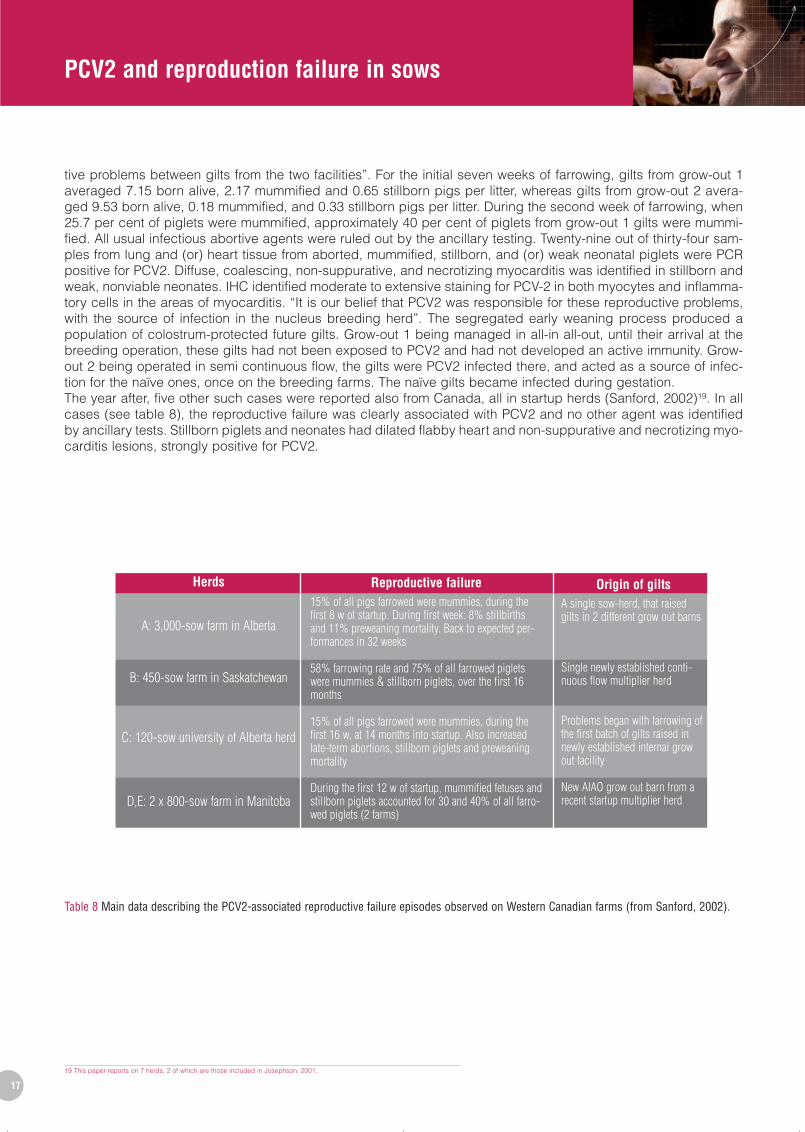
tive problems between gilts from the two facilities”. For the initial seven weeks of farrowing, gilts from grow-out 1averaged 7.15 born alive, 2.17 mummified and 0.65 stillborn pigs per litter, whereas gilts from grow-out 2 avera-ged 9.53 born alive, 0.18 mummified, and 0.33 stillborn pigs per litter. During the second week of farrowing, when25.7 per cent of piglets were mummified, approximately 40 per cent of piglets from grow-out 1 gilts were mummi-fied. All usual infectious abortive agents were ruled out by the ancillary testing. Twenty-nine out of thirty-four sam-ples from lung and (or) heart tissue from aborted, mummified, stillborn, and (or) weak neonatal piglets were PCRpositive for PCV2. Diffuse, coalescing, non-suppurative, and necrotizing myocarditis was identified in stillborn andweak, nonviable neonates. IHC identified moderate to extensive staining for PCV-2 in both myocytes and inflamma-tory cells in the areas of myocarditis. “It is our belief that PCV2 was responsible for these reproductive problems,with the source of infection in the nucleus breeding herd”. The segregated early weaning process produced apopulation of colostrum-protected future gilts. Grow-out 1 being managed in all-in all-out, until their arrival at thebreeding operation, these gilts had not been exposed to PCV2 and had not developed an active immunity. Grow-out 2 being operated in semi continuous flow, the gilts were PCV2 infected there, and acted as a source of infec-tion for the naïve ones, once on the breeding farms. The naïve gilts became infected during gestation.The year after, five other such cases were reported also from Canada, all in startup herds (Sanford, 2002)19. In allcases (see table 8), the reproductive failure was clearly associated with PCV2 and no other agent was identifiedby ancillary tests. Stillborn piglets and neonates had dilated flabby heart and non-suppurative and necrotizing myo-carditis lesions, strongly positive for PCV2.
Table 8 Main data describing the PCV2-associated reproductive failure episodes observed on Western Canadian farms (from Sanford, 2002).
PCV2_sept08 1/10/08 14:48 Page 17

18
Year
2000*
2001*
2002*
2003*
2004**
2005**
200625**
Number of abortion cases submitted to ISU-VDL and attributed to PCV2
1
1024
9
3
4
2
5
20 And also from PDNS-affected sows.24 In (Halbur, 2003), the table mentioned zero cases for 2001.25 For the first eight months of 2006.
In a review on emerging PCVDs, Harding (2004) also provided detailed graphs of the reproductive performancesin a Western Canadian herd where a PCV2-related reproductive failure episode occurred. However, the detailsregarding the farm structure were too scarce to allow identification with any of the cases detailed by Sanford in2002.
In the USA, the first referenced report of a PCV2-associated reproductive failure is in Iowa (Janke, 2000). It occur-red in a closed farm, where both gilts and multiparous sows were affected, with signs “very suggestive of a parvo-virus infection, but laboratory results indicated this virus was not likely the cause”. The problem began with a far-rowing at 116 days of gestation providing two liveborn piglets and eight mummies (6 to 26 cm in size). The secondfarrowing provided two liveborn, two stillborn piglets and 11 mummies (12 cm to full-size), that were collected andsent to the diagnostic laboratory. Histology detected “diffuse non-suppurative myocarditis”, without any grosslesion. No usual abortive agent was identified. The problem lasted for three weeks, at which term another litter wassubmitted to the laboratory, with the same results. “IHC revealed extensive PCV antigen throughout the myocar-dium” and PCV2 was isolated from foetal tissues. Retrospective serology testing indicated the virus had been pre-sent in that herd for several years, and that titres had remained fairly stable over the previous 12-18 months. Thereproductive problem lasted for “approximately three months, after which time sows returned to delivering largenormal litters”. Sorden (Iowa State University) reported in 2001 “we have had several cases in which PCV2 isdemonstrated in aborted porcine foetuses. Typical histories include gilts delivering mummified fetuses. Myocarditiswas the most common lesion, with abundant intralesional PCV2 antigen, and thoracic fluid was PCR positive forPCV2 [DNA]. Similar cases have been reported in the literature” (Sorden, 2001). That same year, PCV2 isolationfrom aborted fetuses was also reported (Meehan et al, 2001)20. In 2003, Halbur et al (Iowa State University, VDL)mentioned that “PCV2-associated reproductive failure is uncommonly diagnosed. When confirmed it is typicallycharacterized by foetal lymphohistiocytic myocarditis and/or myocardial fibrosis and confirmed by demonstrationof abundant amounts of PCV2 antigen associated with the myocardial lesions. PCV2-associated abortions are spo-radic in occurrence and characterized by increased numbers of mummified fetuses” (Halbur et al, 2003). In the fol-lowing years, the same team reported the number of field cases of PCV2-associated diseases among the fieldcases submitted to the ISU-VDL on several occasions (Halbur and Opriessnig, 2004; Opriessnig and Halbur,2006a) (see table 9).
Table 9 Number of field cases of PCV2-associated diseases reported among the field cases submitted to the ISU-VDL over the years (from Halburand Opriessnig, 2004* and Opriessnig and Halbur, 2006a**).
PCV2_sept08 1/10/08 14:48 Page 18

PCV2 and reproduction failure in sows
19
Another interesting report from ISU mentioned pigs presenting with myocardial PCV2-associated lesions at a laterage (Opriessnig et al, 2006b). A first case was a 4-week old pig found dead although previously healthy. Heart tis-sue was strongly positive for PPV and PCV2; the cause of death was heart failure. In two other herds with a historyof piglets found dead without prodromic signs, two piglets (one from each farm, dead at 5 and 7-week old) hadsevere transmural cardiac haemorrhages and the lungs were inflammed and oedematous; nutritional myocardio-pathy was initially suspected, but ruled out by liver and heart vitamin E dosage. Microscopic examination revealedsevere hepatic and pulmonary congestion and severe multifocal myocardial haemorrhage with swelling and necro-sis of myofibres in both pigs. PCV2-IHC staining was strong in the heart and the Küpfer cells of both piglets. Twoother cases are also reported23, with similar age. “The possibility that PCV2 may have predisposed the pigs to heartfailure should be borne in mind”, the authors conclude. But they could not indicate whether these piglets wereinfected in utero by PCV2 and subsequently developed a heart pathology, or whether PCV2 was a post-natal oppor-tunist invader of damaged myocardial cells (or both). Another similar case has been recently reported in a 4,800-sow farrow-to-wean production system with a newly added on-site gilt-development facility (Pittman, 2008).Because of changes in herd organization, internally multiplied gilts were produced. A short time after the first onesof them had farrowed, the herd experienced an increase in incidence of gilt abortions and mummified fetuses thatlasted for eight weeks. Replacement gilts that had entered the herd from an external source remained unaffected.During the outbreak, average mummified fetuses per litter ranged from 0.1 to 0.7 per litter per week, while the ave-rage over the 12 preceding months was <0.1. Although abortion rate was 12 per cent among gilts from externalsource, this rate reached 69 per cent at the peak of clinical disease. Laboratory testing demonstrated numerouscardiac lesions, and PCV2-IHC was positive in all litters from which samples were submitted (heart tissues andother organs). “Based on clinical findings and results of diagnostic tests, the diagnosis was PCV2-associated foe-tal death and mummification with myocardial necrosis or dysplasia”.
Only one documented report has been published from South America (Bermúdez et al, 2008), from two PRRSV-negative farms in Venezuela. The farms (one with 3,000 sows, the other with 2,500 sows) were located in differentregions, but had been experiencing “abortions, postweaning emaciation syndrome, PDNS at different ages andsystemic adenomegaly”. Furthermore, peripartum losses increased from 3.6 to 7 per cent on the first farm and from4.8 to 10 per cent on the second one, between 2006 and 2007. PCV2 was the only agent evidenced from foetalfluids and placenta, and the authors conclude it is the causative agent of the reproductive failure, as well as thePMWS and a number of enteritis cases also observed on those farms.
In 2000, the results of “epidemiological studies in Western European pig producing herds” mentioned that “PCV2 wasshown to produce intra uterine infections between days 32 and 114 of gestation. These infections could lead to 0.5per cent mummified foetuses and to abortions at different stages of gestation with increased frequencies of weak-and stillborn piglets” (Ohlinger et al, 2000). Nevertheless, no details were provided regarding the epidemiologicalprotocol. Furthermore, the same paper reported that “some piglets that were born viremic did not develop anti-PCV2 antibodies”. This has never been reported later, and might be linked to the poor quality of the IF assay usedby the authors. In 2001, a case of transplacental infection has been demonstrated in a gilt in Denmark (Ladekjaer-Mikkelsen et al, 2001). The litter originated from the SPF herd of the Danish Veterinary Institute and comprised 10dead piglets: one mummified fetus, eight late-term dead fetuses and one stillborn pig. That herd was free from PCV(and other agents). The gilt had farrowed 121 days after service, with no obvious clinical sign. Viral isolation in cellculture from dead piglet tissues proved positive for PCV2 only. IHC gave the same result, and IPMA revealed thepresence of anti-PCV2 antibodies in the pleural fluid of piglets “further confirming that piglets had become trans-placentally infected with PCV2”. Q-PCR estimated the viral load over 1011 copies of PCV2 DNA/g tissue. The herdhad remained PCV2 seronegative over 1999 and 2000. By the end of January 2001, the whole herd was seroposi-tive with high titres: “it appears that the herd had an acute outbreak of PCV2 infection in January”. Further investi-gation did not identify other cases of vertical transmission of PCV2 in that herd. A more recent report from Norwayalso documented a case of foetal death and high mortality in piglets associated with PCV2 in a newly establishedherd (Brunborg et al, 2007). The 36 gilts originated from four different multiplier herds and farrowed one month afterarrival (over a 2-week period). Of the 15 gilts with the same origin, 12 gave birth to mummified fetuses or stillbornpiglets (40/192: 20.8%) and nearly 8 per cent of liveborn piglets had congenital tremor or ataxia. One fifth (21%) ofthe piglets born to the 15 gilts died or were killed before weaning. The remaining litters had 0.1 per cent mummiesand stillbirths and 0.3 per cent preweaning mortality. Piglets were submitted to postmortem diagnostic, and pigletsfrom a farm experiencing reproductive failure attributed to mycotoxins were taken as positive controls. Several foe-tuses and weakborn piglets had heart lesions (dilation, mottled myocardium…) with multifocal fibronecrosis and
23 Three older piglets (13 and 14 weeks of age) with a similar pathology are also reported in this paper, but they are from two different PCV2 inoculation experiments, not piglets from conventional farms.
PCV2_sept08 1/10/08 14:48 Page 19

PCV2 and reproduction failure in sows
20
Sow inventory
Number of gilts with PDNS signs
Number of abortions
Number of dead gilts
Unit 1
431
23
4
4
Unit 2
260
2
-
-
Unit 3
340
4
4
-
Unit 4
362
2
-
-
21 PCV1 and PCV2.22 Coalescent red to black macules and crusty areas on the perineal and hind limb skin. Other clinical signs were: fever, anorexia, depression, conjunctivitis, diarrhea, stiff gait, reluctance to move (arthralgia, edema of the lowerlimbs) and labored breathing. At necropsy, kidneys were enlarged with edema, small petechial hemorrhages (0.5-1mm) and multifocal kidney lesions. Focal kidney lesions were found throughout the renal cortex. Peritoneal fluidaccumulation were present. Enlarged hemorrhagic inguinal, subiliacal, popliteal and mesenteric lymph nodes were found. CSF and ASF were ruled out.
infiltration by mononuclear cells. IHC staining of this tissue was strongly positive. High amounts of PCV2 DNA werefound in all stillborn piglets examined. Viral loads were highest in liver from mummified and stillborn piglets (up to1012 copies DNA per 500 ng DNA), as compared to liveborn piglets with myocarditis, and to liveborn piglets withoutmyocarditis. Taken together, these results “further corroborate the theory that myocardium is a target tissue forPCV2 in fetuses and young piglets”. In Eastern and Central Europe, numerous PCV2-associated reproductive failure problems have occurred (Bilkei G,personal communication), but only a few reports have been published. The first one is from a Croatian 600-sownucleus herd (Mauch and Bilkei, 2004) which delivered 94 PCV-seronegative22 middle pregnancy replacement giltsto four PCV-seropositive breeding herds (see table 10). Thirty-one of them developed, within two weeks to twomonths post-entry, skin lesions strongly evocative of PDNS21. Eight of them aborted (4 in units 1 and 3), four of themdied (all in unit 1).
Table 10 Characteristics observed in a Croatian farm with PCV2-associated reproductive failure (from Mauch and Bilkei, 2004).
The condition spread rapidly within a week in unit 1, while it occurred over a time period of 46 days in the otherunits. All four necropsied gilts had hyaline degeneration and necrotizing vasculitis of small and medium sized arte-ries of the skin, renal cortex (with necrotizing to suppurative glomerulonephritis), wall of the uterus, lymph nodes,spleen, and ileum. All 27 gilts that survived PDNS proved seropositive to PCV-1 and PCV-2, four month after entryinto the breeding unit. “Having diagnosed uterine changes, it is reasonable to suggest that the abortion might havebeen the result of PCV2 infection, and that the animals could have been infected with the virus at their entry in ourbreeding units”. Unfortunately, the authors did not collect fetuses for further diagnosis. The second report is alsofrom a herd in Croatia (Ellis and Bilkei, 2004) where an episode of late abortion occurred in gilts in a 600-sow bree-ding, further to the introduction of 60 gilts in their third month of gestation. The herd of origin was PCV2-seronega-tive; the breeding herd was PCV2-positive in serology. Eighteen out of 23 aborted gilts presented with skin grosslesions suggestive of PDNS; necropsy also revealed enlarged hemorrhagic inguinal and mesenteric lymph nodes.Histopathological examination of sections of skin, kidney and uterus stained with haematoxylin and eosin showedhyaline degeneration and necrotizing vasculitis of small and medium-sized arteries. Histology of lymph nodesrevealed presence of large cells with grapes of inclusion bodies. Bacteriology revealed a heavy growth ofStaphylococcus hyicus in samples from renal pelvis, uteri and skin. Post-mortem examination of aborted fetusesrevealed oedema and haemorrhages in the alimentary tract, petechiae on the abdominal serosae, and enlargedlymph nodes. S. hyicus was also cultured from fetus kidney. The authors conclude that this outbreak “appears tohave been the result of concurrent infections of S. hyicus and PCV2”. One of the most recent reports linking PCV2infection to reproductive failure come from the Czech Republic (Zizlavsky et al, 2008). A total of 232 aborted fetusesand stillborn pigs submitted to the diagnostic laboratory between 2005 and 2007 were examined for individualinfectious agents known to cause reproductive disorders with clinical signs including abortion, preterm parturitionand nonviable neonatal pigs variability. Incidence of PPV in fetuses kept decreasing significantly with time (30.4%in 2005; 3.5% in 2007), and the same significant drop was observed for PRRSV (52.2% in 2005; 34.6% in 2006; 5%in 2007). On the contrary, PCV2 incidence kept growing with time (21.7% in 2005; 54.1% in 2007). The most fre-quent cause of reproductive disorders of sows in the Czech Republic is today PCV2, these authors conclude.
PCV2_sept08 1/10/08 14:48 Page 20

PCV2 and reproduction failure in sows
21
Number of cases
35
90
12
9
2
2
Early
4
12
0
2
0
0
Mid
8
23
3
2
0
0
Late
12
32
7
3
1
2
Premature
5
13
1
1
1
0
Stage of gestation
Stillborn
6
10
1
1
0
0
Virus
PCV2
PPV
PRRSV
PCV2 + PPV
PCV2 + PRRSV
PPV + PRRSV
In South-East Asia, a field investigation from South Korea found 13.1 per cent of aborted fetuses and stillborn pigletsto be positive for PCV2 by PCR (Kim et al, 2004). A total of 350 aborted fetuses and stillborn piglets, submitted from321 pig farms from all over the country between October 2000 and September 2002 for the diagnosis of reproduc-tive problems, were screened for the presence of PCV2, PPV and PRRSV. None of the farms which submitted abor-ted fetuses and stillborn piglets was infected with PMWS or PDNS. A total of 150 (43%) of the 350 aborted fetusesand stillborn piglets were positive for at least one of the viruses. Of the single infections, 35 cases were positive forPCV2, but mixed infections were also prevalent (see table 11). Among the 35 single infections with PCV2, four weredetected in fetuses aborted in early gestation, eight in foetuses aborted in mid-gestation, 12 in fetuses aborted inlate gestation, five in premature piglets and six in stillborn piglets. These results “suggest that PCV2 may be asso-ciated with reproductive failure at all stages of gestation”.
Table 11 Stage of gestation of the aborted foetuses and stillborn piglets from which PCV2, PPV and PRRSV were isolated (from Kim et al, 2004).
The authors also suggest that the consequences of maternal infection at different stages of gestation may be rela-ted to the relative or absolute ability of the conceptus to support PCV2 replication (see Sanchez et al, 2001), ratherthan to the effectiveness of the so-called placental barrier. “It therefore seems possible that the placental barriermay not play an important role in determining the reproductive consequences of maternal infection with PCV2 atdifferent stages of gestation”. In Japan PCV2 was identified in a weak-born 8-day old piglet from a litter born duringa reproductive failure episode (Mikami et al, 2005). The herd was a 177-sow farrow-to-finish operation where mum-mified fetuses ad late-term fetuses occurred from June to August 2001. PMWS and PDNS were not in the farm his-tory. One gilt farrowed seven dead foetuses and a weakborn piglet, that died at eight days of age. He was necrop-sied; tonsil, liver and brain were sampled for viral isolation - which provided only PCV2. Focal to extensive lesionswere evidenced in the myocardium by pathology (non-suppurative myocarditis with fibrosis). A lymphocytic deple-tion was obvious in all secondary lymphoid organs. The presence of fibrosis, a sign of ancient infection, togetherwith the presence of myocardial lesions in stillborn fetuses from other sows allows the authors to suggest this pigletwas infected in utero and that the reproductive failure on this farm may be attributed to PCV2.
PCV2_sept08 1/10/08 14:48 Page 21

PCV2 and reproduction failure in sows
22
5. Anecdotal reports
In an article reporting on the identification of PMWS in Brazil, the authors mentioned that among the clinical sam-pled that were obtained between 2000 and 2002, there were “mummified fetuses from sows with reproductive fai-lure” (Castro et al, 2004). Pooled abdominal organs from 297 mummified fetuses provided two PCR positive resultsfor PCV1 and two positive results for PCV2. In the USA, a review on sow mortality observed that sows with more stillbirths are more likely to die subsequent tothe stillbirths. The chance of mortality increases by 24 per cent when one or more stillbirth occurred (Deen and Xue,1999).In a report on PMWS from Europe, it is mentioned that farmers that are PMWS-positive have started taking replace-ments from PMWS-positive farms. There are no technical papers demonstrating its benefits, but field experienceindicates it to be of some help (Donadeau et al, 2003). There are no negative effects reported. Regarding serotherapy, a manager in a Chinese farm reported that the use of sow serum helped more than that ofthe finisher pig serum (Soo Joo, 2003).An American veterinarian reporting on an “UK tour” regarding PMWS occurrence signals that « it was reported tous that piglets born to older parity sows are at higher risk for experiencing PMWS. This theory was reported to usanecdotally by several sources during the week. The two small datasets we were able to see did not support thisinformation » (Engle, 2003). Also for the UK, Stand Done reported “observers of epidemiological effects” of PMWS,among which “a role for PCV2 in congenital tremors and reproductive failure (pre-natal infection). Experimentally,there is doubt as to whether PCV2 crosses the placenta or not, or are they just sow effects. Experimentally, somepiglets may be affected if sows are infected in the second half of pregnancy. There may be litter effects” (Done,2001).In 2007, a veterinarian from Murphy Brown, reporting on North Carolina’s experience with PCVD, stresses that“although the various commercial PCV2 vaccines have helped reduce mortality rates to a more acceptable level,PCVD continues to be diagnosed in NC. At this time, a large part of the US swine industry is dependant on vac-cine manufacturers to supply a consistent volume of quality product. Many producers are now evaluating theimpact of PCVD on their nursery and sow performance” (Battrell, 2007).
PCV2_sept08 1/10/08 14:48 Page 22

PCV2 and reproduction failure in sows
23
6. Conclusion
A large body of evidence, resulting from both field reports and experimental inoculation of gilts and sows concurdraw a causative link between PCV2 and reproductive failure in sows and gilts. Field reports show unequivocallythat gestating naive females introduced in an infected environment experience reproductive failure at primo PCV2-infection. In vitro and transmission studies confirm that PCV2 is able to infect pig embryos at the blastocyst stageand later, and cause early pregnancy losses. Other transmission studies also demonstrate that PCV2 is able to pro-duce transplacental infection at any time during gestation, and that pig fetuses are susceptible to PCV2 in uteroinfection whatever their development stage. This will result in reproductive failure only when a strong viremia hap-pens during gestation, whatever the PCV2 serological status of the dam. Taken together, these data aim at demons-trating that Koch’s postulates are fulfilled by PCV2 as far as reproductive failure in naïve gestating females isconcerned. Nevertheless, this does not correspond to the usual field situation where most breeding animals are seropositive toPCV2, with a limited (if existing) viremia. In healthy PCV2-seropositive sow/gilt, the reproductive tract shows evi-dence of PCV2 infection without any obvious impact on the outcome of future pregnancies. However, transferringPCV2-exposed embryos to PCV2-seropositive recipient sows affects pregnancy outcome. This suggests that PCV2impact on reproduction may be subtler than the reproductive failure described above. Field studies comparing per-formances of several hundreds of farrow-to-finish vaccinated farms demonstrate a significant improvement of thebreeding performances after vaccination as compared to either the national/regional average performances, or theperiod before vaccination. This is also in favour of an impact of PCV2 subclinical infection of gilts/sows on theircareer. The impact of the “natural” reproductive tract infection on reproductive performances of sows at the herdlevel has not been assessed – apart from the papers regarding the effect of Circovac® vaccination on reproductiveperformances, but these trials deal mostly with PCVD-affected breeding farms, rather than with the subclinicalinfection per se. In the only paper describing the latter situation, vaccination allowed to bring the rate of stillbirthinto normal range in this herd.
PCV2_sept08 1/10/08 14:48 Page 23

PCV2 and reproduction failure in sows
24
References
Battrell M. “PCVAD transmission trials”. In Proceedings of the 15th annual Swine disease Conference for Swinepractitioners, ISU, Ames, 2007, p. 54-55.
Bech A. B., Kunstmann L. “Effect of sow vaccination with Circovac® on the performances of 3 Danish herds”. 20thIPVS Durban (RSA) 22-26 June 2008, vol. 2, p. 109.
Bermúdez V, Tonelli T, Escalona M, Morales A, Moreno J, Kwiecien E. “PCV2-associated reproductive failures in twofarms PRRS- in Central Venezuela. First documented report”. 20th IPVS Durban (RSA) 22-26 June 2008, vol. 1, p.61.
Bielanski A, Larochelle R, Algire J, Magar R. “Distribution of PCV-2 DNA in the reproductive tract, oocytes andembryos of PCV-2 antibody-positive pigs”. Vet. Rec., 2004, vol. 155, n° 19, p. 597-598.
Bogdan J, West K, Clark E, Konoby C, Haines D, Allan G, McNeilly F, Meehan B, Krakowka S, Ellis JA. “Associationof porcine circovirus 2 with reproductive failure in pigs: a retrospective study, 1995-1998”. Can. Vet. J., 2001, vol.42, n° 7, p. 548–550. Available online: http://www.pubmedcentral.nih.gov/tocrender.fcgi?iid=131622
Brunborg IM, Jonassen CM, Moldal T, Bratberg B, Lium B, Koenen F, Schönheit J. “Association of myocarditis withhigh viral load of porcine circovirus type 2 in several tissues in cases of fetal death and high mortality in piglets. Acase study”. J. Vet. Diagn. Invest., 2007, vol. 19, n° 4, p. 368-375.
Calsamiglia M, Fraile L, Espinal A, Cuxart A, Seminati C, Martín M, Mateu E, Domingo M, Segalés J. “Sow porcinecircovirus type 2 (PCV2) status effect on litter mortality in postweaning multisystemic wasting syndrome (PMWS)”.Res. Vet. Sci., 2007, vol. 82, n° 3, p. 299-304.
Cariolet R, Blanchard P, Le Dimna M, Mahé D, Jolly JP, de Boisséson C, Truong C, Ecobichon P, Madec F, JestinA. “Experimental infection of pregnant SPF sows with PCV2 through tracheal and muscular routes”. In Proceedingsof Congress on ssDNA viruses of plants, birds, pigs and primates, Saint-Malo (France), Sept. 24-27, 2001a, p. 128.
Cariolet R, Blanchard P, Le Dimna M, Mahé D, Keranflec’h A, Julou P, Beaurepaire B, de Boisséson C, Truong C,Jestin A. “Consequences of PCV2 experimental infection of non-imune SPF sows using the intra-uterine route”. InProceedings of Congress on ssDNA viruses of plants, birds, pigs and primates, Saint-Malo (France), Sept. 24-27,2001b, p. 129.
Castro AM, Cortez A, Ruiz VL, Leomil H, Moreno AM, Doto DS, Richtzenhain LJ. “Detection and differentiation ofporcine circoviruses in Brazilian pigs”. Vet. Rec., 2004, vol. 154, n° 23, p. 728-729.
Deen J, Xue J. “Sow mortality in the US : an industry-wide perspective”. Allen D. Leman Swine Conference, 17-21Sept., 1999, p. 91-94.
Donadeu M, Waddilove J, Marco E. “European management strategies to control PMWS”. Allen D. Leman SwineConference, 2003, vol. 30, p. 136-142.
Done S. “Current European Situation”. In Proceedings of the 9th annual Swine disease Conference for Swine prac-titioners, ISU, Ames, 2001, p. 142-155.
Ebbesen T., Kunstmann L. “Effect of sow vaccination with Circovac® on stillborn piglets”. 20th IPVS Durban (RSA)22-26 June 2008, vol. 2, p. 81.
Ellis CP, Bilkei, G. “Late abortion in gilts caused by concurrent infection with Staphylococcus hyicus and Porcinecircovirus type 2”. Folia Veterinaria, 2004, vol. 48, n° 4, p. 198-201.
PCV2_sept08 1/10/08 14:48 Page 24

PCV2 and reproduction failure in sows
25
Engle MJ. “UK PMWS study tour: lessons for US animal agriculture”. Allen D. Leman Swine Conference, 2003, vol.30, p. 127-132.
Farnham MW, Choi YK, Goyal SM, Joo HS. “Isolation and characterization of porcine circovirus type-2 from sera ofstillborn fetuses”. Can. J. Vet. Res., 2003, vol. 67, n° 2, p. 108-113. Available at: http://www.pubmedcentral.nih.gov/article-render.fcgi?tool=pubmed&pubmedid=12760475 (accessed on 21/04/2008)
Gillespie T. “PCVD: when immunology goes wrong, life on the farm becomes very expensive”. in Proceedings ofthe 48th annual George A. Young Swine Health and management conference, August 16, 2007, University ofNebraska-Lincoln, p. 1-6.
Halbur P, Opriessnig T, Hoogland M, Thacker E, Yu S. “PCV2-associated diseases: research and diagnosticupdate”. In Proceedings of the 11th annual Swine disease Conference for Swine practitioners, ISU, Ames, 2003, p.152-160.
Halbur P, Opriessnig T. “Update on PCV2-associated diseases”. In Proceedings of the 12th annual Swine diseaseConference for Swine practitioners, ISU, Ames, 2004, p. 12-23.
Harding JC. “The clinical expression and emergence of porcine circovirus 2”. Vet. Microbiol., 2004, vol. 98, n° 2,p. 131-135.
Harms P. “Hepatopathology associated with spontaneous type-2 porcine circovirus infection in caesarianderived/colostrum deprived pigs”. In Proceedings of the American Association of Veterinary LaboratoyDiagnosticians, 1999, vol. 42, p. 4.
Janke B. “Case report: porcine circovirus as a cause of reproductive problems”. in Proceeding of the 118th annualmeeting of the Iowa Veterinary Medical Association, 2000, n° 118, p. 101.
Johnson C., Soo Joo H., Direksin C., Hyung Lee W. “ Infection of late-term swine fetuses following in utero inocula-tion with porcine circovirus types –1 and –2”. Allen D. Leman Swine Conference, 17-21 Sept., 1999, p. 250-251.Also published in: Johnson CS, Joo HS, Direksin K, Yoon KJ, Choi YK. « Experimental in utero inoculation of late-term swine fetuses with porcine circovirus type 2 ». J. Vet. Diagn. Invest., 2002, vol. 14, n° 6, p. 507-512.
Joisel F., Brune A., Schade A., Longo S., Charreyre C. (2008) Improvement of reproduction performance inducedby PCV2 vaccination of sows and gilts with Circovac® in 277 German sow farms. 20th IPVS Durban (RSA) 22-26June 2008, vol. 2, p. 72.
Jolie R, Runnels P, McGavin D. “PMWS in a group of cesaerian-derived colostrum-deprived piglets”. InProceedings of the 16th IPVS Congress, Melbourne, Australia, 17-20 Sept. 2000, p. 639.
Josephson G, Charbonneau G. “What’s your interpretation? Case report of reproductive problem in a new startupoperation”. JSHAP, 2001, vol. 9, n° 5, p. 258-259. (non refereed) http://www.aasv.org/shap/issues/v9n5/v9n5wyi.html(accessed 04/19/2008)
Jung K, Kim J, Ha Y, Choi C, Chae C. “The effects of transplacental porcine circovirus type 2 infection on porcineepidemic diarrhoea virus-induced enteritis in preweaning piglets”. Vet. J., 2006, vol. 171, n° 3, p. 445-450.
Kunstmann L., Lau L. (2008) Effect of sow vaccination with Circovac® on the performances of 34 Danish herds.20th IPVS Durban (RSA), 22-26 June 2008, vol. 2, p. 75.
Ladekjaer-Mikkelsen AS, Nielsen J, Storgaard T, Bøtner A, Allan G, McNeilly F. “Transplacental infection with PCV-2 associated with reproductive failure in a gilt”. Vet. Rec., 2001, vol. 148, n° 24, p. 759-760.
Lefebvre DJ, van Reeth K, Vangroenweghe F, Castryck F, Maes D, Laitat M, Nauwynck HJ. “Serosurvey for virusesassociated with reproductive failure in newly introduced gilts and in multiparous sows in belgian sow herds”. 20thIPVS Durban (RSA), 22-26 June 2008, vol. 1, p. 93.
PCV2_sept08 1/10/08 14:48 Page 25

PCV2 and reproduction failure in sows
Madec F, Eveno E, Morvan P, Hamon L, Morvan H, Albina E, Truong C, Hutet E, Cariolet R, Arnaud C, Jestin A. “LaMaladie de l’Amaigrissement du Porcelet (MAP). 1. Aspects descriptifs, impact en élevage”. In Actes des 31èmesJournées de la Recherche Porcine, Paris 2-4 Février 1999, p. 347-354. Available online at : http://www.journees-recherche-porcine.com/texte/index.htm (accessed on 24/04/2008).
Maldonado J, Segalés J, Martínez-Puig D, Calsamiglia M, Riera P, Domingo M, Artigas C. “Identification of viralpathogens in aborted fetuses and stillborn piglets from cases of swine reproductive failure in Spain”. Vet. J., 2005,vol. 169, n° 3, p. 454-6.
Mateusen B . “Interaction of viruses with porcine embryos”. PhD dissertation, FVM Gent, 2007, 198 p. Also in:Mateusen B, Maes DG, Van Soom A, Lefebvre D, Nauwynck HJ. “Effect of a porcine circovirus type 2 infection onembryos during early pregnancy”. Theriogenology, 2007, vol. 68, n° 6, p. 896-901.
Mauch CP, Bilkei G. “Porcine circovirus (PCV) caused losses in pregnant gilts”. Pig J., 2004, vol. 53, p. 69-74.
Meehan BM, McNeilly F, McNair I, Walker I, Ellis JA, Krakowka S, Allan GM. “Isolation and characterization of por-cine circovirus 2 from cases of sow abortion and porcine dermatitis and nephropathy syndrome”. Arch. Virol., 2001,vol. 146, n° 4, p. 835-842.
Mikami O, Nakajima H, Kawashima K, Yoshii M, Nakajima Y. “Nonsuppurative myocarditis caused by porcine cir-covirus type 2 in a weak-born piglet”. J. Vet. Med. Sci., 2005, vol. 67, n° 7, p. 735-738. Available oline at:http://www.jstage.jst.go.jp/article/jvms/67/7/67_735/_article (accessed on 21/04/2008)
Nielsen EO, Enøe C, Jorsal SE, Barfod K, Svensmark B, Bille-Hansen V, Vigre H, Bøtner A, Baekbo P. “Postweaningmultisystemic wasting syndrome in Danish pig herds: productivity, clinical signs and pathology”. Vet. Rec., 2008,vol. 162, p. 505-508.
O’Connor B, Gauvreau H, West K, Bogdan J, Ayroud M, Clark EG, Konoby C, Allan G, Ellis JA. “Multiple porcinecircovirus 2-associated abortions and reproductive failure in a multisite swine production unit”. Can. Vet. J., 2001,vol. 42, n° 7, p. 551–553. Available online: http://www.pubmedcentral.nih.gov/tocrender.fcgi?iid=131622
Ohlinger V, Schmidt U, Pesch S. “Studies on pathogenic aspects of the PMWS”. In Proceedings of the 16th IPVSCongress, Melbourne, Australia, 17-20 Sept. 2000, p. 577.
Opriessnig T, Halbur P. “Current status of PCVAD: diagnostic and research update”. In Proceedings of the 14thannual Swine disease Conference for Swine practitioners, ISU, Ames, 2006a, p. 105-128.
Opriessnig T, Janke BH, Halbur PG. “Cardiovascular lesions in pigs naturally or experimentally infected with por-cine circovirus type 2”. J. Comp. Pathol., 2006b, vol. 134, n° 1, p. 105-110.
Park JS, Kim J, Ha Y, Jung K, Choi C, Lim JK, Kim SH, Chae C. “Birth abnormalities in pregnant sows infected intra-nasally with porcine circovirus 2”. J. Comp. Pathol., 2005, vol. 132, n°2-3, p. 139-144.
Pittman JS. “Reproductive failure associated with porcine circovirus type 2 in gilts”. J. Swine Health Prod., 2008,vol. 16, n° 3, p. 144–148.
Ritzmann M, Wilhelm S, Zimmermann P, Etschmann B, Bogner KH, Selbitz HJ, Heinritzi K, Truyen U. “Prevalenceand association of porcine circovirus type 2 (PCV2), porcine parvovirus (PPV) and porcine reproductive and respi-ratory syndrome virus (PRRSV) in aborted fetuses, mummified fetuses, stillborn and nonviable neonatal piglets[Article in German]”. Dtsch. Tierarztl. Wochenschr., 2005, vol. 112, n° 9, p. 348-351.
Rose N, Blanchard P, Cariolet R, Grasland B, Amenna N, Oger A, Durand B, Balasch M, Jestin A, Madec F.“Vaccination of porcine circovirus type 2 (PCV2)-infected sows against porcine Parvovirus (PPV) and Erysipelas:effect on post-weaning multisystemic wasting syndrome (PMWS) and on PCV2 genome load in the offspring”. J.Comp. Pathol., 2007, vol. 136, n° 2-3, p. 133-144.
26
PCV2_sept08 1/10/08 14:48 Page 26

PCV2 and reproduction failure in sows
27
Sanchez Jr RE. “Pathogenesis of PCV2 infections in pigs from fœtal to early postnatal life”. PhD thesis, Universityof Ghent (Belgium), 2003a.
Sanchez Jr RE, Meerts P, Nauwynck HJ, Pensaert MB. “Change of porcine circovirus 2 target cells in pigs duringdevelopment from fetal to early postnatal life”. Veterinary Microbiology, 2003b, vol. 95, p. 15–25.
Sanchez Jr RE, Meerts P, Nauwynck HJ, Ellis JA, Pensaert MB. “Characteristics of porcine circovirus-2 replicationin lymphoid organs of pigs inoculated in late gestation or postnatally and possible relation to clinical and patholo-gical outcome of infection”. J. Vet. Diagn. Invest., 2004, vol. 16, n° 3, p. 175-185.
Sanford ES. “PCV-2 related reproductive failure in startup herds”. in Proceedings of the 17th International PigVeterinary Society Congress, 2002, vol. 1, p 171.
Schøning T., Nielsen P., Lau L. “Effect of Circovac® (Merial) on Porcine Circovirus type 2 (PCV2) sow reproductivefailure and mortality: a case report”. 20th IPVS Durban (RSA), 22-26 June 2008, vol. 2, p. 108.
Segales S, Rosell C, Domingo M. “Reproductive failure associated to PCV2 infection in Spain ?”. in Proceedings ofthe 17th International Pig Veterinary Society Congress, 2002, vol. 2, p. 171.
Shibata I, Okuda Y, Kitajima K, Asai T. “Shedding of porcine circovirus into colostrum of sows”. J. Vet. Med. SeriesB, 2006, vol. 53, n° 6, p. 278-280.
Soo Joo H. “PMWS: clinical signs and control measures experienced in China”. Allen D. Leman Swine Conference,2003, vol. 30, p. 133-135.
Sorden S. “PMWS – Current Iowa situation”. In Proceedings of the 9th annual Swine disease Conference for Swinepractitioners, ISU, Ames, 2001, p. 156-161.
West KH, Bystrom JM, Wojnarowicz C, Shantz N, Jacobson M, Allan GM, Haines DM, Clark EG, Krakowka S,McNeilly F, Konoby C, Martin K, Ellis JA. “Myocarditis and abortion associated with intrauterine infection of sowswith porcine circovirus 2”. J. Vet. Diagn. Invest., 1999, vol. 11, n° 6, p. 530-532.
Yoon KJ, Jepsen RJ, Pogranichniy RM, Sorden S, Stammer R, Evans LE. “A novel approach to intrauterine viral ino-culation of swine using PCV type 2 as a model”. Theriogenology, 2004, vol. 61, n° 6, p. 1025-1037.
Zizlavsky M, Czanderlova L, Gampova M, Kellnerova D, Tydlitat D, Drabek J. “Significant role of PCV2 in reproduc-tive disorders of sows”. 20th IPVS Durban (RSA), 22-26 June 2008, vol. 2, p. 110.
PCV2_sept08 1/10/08 14:48 Page 27