PATTERNS OFVISUAL FAILURE WITH PITUITARY...
17
Brit. J. Ophthal. (1968) 52, 94 PATTERNS OF VISUAL FAILURE WITH PITUITARY TUMOURS*t CLINICAL AND RADIOLOGICAL CORRELATIONS BY PETER WILSON AND MURRAY A. FALCONER Guy's-Maudsley Neurosurgical Unit, London. IMPAIRMENT of vision in one or both eyes is a common sequel to a suprasellar extension of a pituitary tumour. It is widely held that this impairment takes the form of a bitemporal hemianopia in which the initial defect is a progressive loss in the periphery of the upper temporal quadrants, and moreover that this defect can be readily detected by perimetry. Yet our experience at a neurosurgical unit has suggested that many patients may seek advice repeatedly over many months or even years, from opticians, ophthalmologists, and neuro- logists, before the correct diagnosis is made. A major reason for this is that often the initial visual defect is not an impairment of the peripheral quadrants but a scotomatous defect in the temporal hemicentral fields. Material In order to ascertain the incidence and significance of the differing patterns of visual field disturbance a retrospective study has been made of fifty patients with chromophobe pituitary adenomata causing visual field defects, who were operated upon at the Guy's-Maudsley Neuro- surgical Unit between 1953 and 1964. An attempt was also made to correlate these patterns with certain clinical features, such as the length of history, the presence or absence of optic atrophy, and the radiological findings. By these means we have sought to determine: (1) The anatomical mechanisms responsible for the various patterns of visual defect; (2) The prognosis of these various patterns after surgical operation; (3) Criteria for earlier diagnosis. Only patients with field defects were included, but two of them had also unilateral paresis of oculomotor muscles. The sex distribution was roughly equal (24 men, 26 women), and the age range at operation was from 24 years to 70 years (average 51). The subjective duration of visual deterioration ranged from less than a week to as long as 20 and 50 years in two patients. The average length of history, when these two last-mentioned patients are excluded, was 2 years. Quantitative perimetric examination was performed in 49 cases before operation. One patient (Case 29) was too ill to co-operate and in fact died in coma shortly after operation, and there was one other post-operative death (Case 35). Both distance (Snellen) and reading (Jaeger) visual acuity was assessed in 43 of these 49 cases, while in the others only the reading acuity was recorded. Of the surviving patients, 45 have undergone at least one quantitative perimetric examination since the operation, whilst in the remaining three the findings on simple clinical confrontation were recorded. Although all patients had pre-operative x-ray examination of the skull, the films were * Received for publication May 3, 1967. t Address for reprints: M. A. Falconer, as above. 94 on 21 June 2018 by guest. Protected by copyright. http://bjo.bmj.com/ Br J Ophthalmol: first published as 10.1136/bjo.52.2.94 on 1 February 1968. Downloaded from
-
Upload
truonghanh -
Category
Documents
-
view
222 -
download
1
Transcript of PATTERNS OFVISUAL FAILURE WITH PITUITARY...
Brit. J. Ophthal. (1968) 52, 94
PATTERNS OF VISUAL FAILURE WITHPITUITARY TUMOURS*t
CLINICAL AND RADIOLOGICAL CORRELATIONS
BY
PETER WILSON AND MURRAY A. FALCONERGuy's-Maudsley Neurosurgical Unit, London.
IMPAIRMENT of vision in one or both eyes is a common sequel to a suprasellar extension of apituitary tumour. It is widely held that this impairment takes the form of a bitemporalhemianopia in which the initial defect is a progressive loss in the periphery of the uppertemporal quadrants, and moreover that this defect can be readily detected by perimetry.Yet our experience at a neurosurgical unit has suggested that many patients may seek advicerepeatedly over many months or even years, from opticians, ophthalmologists, and neuro-logists, before the correct diagnosis is made. A major reason for this is that often theinitial visual defect is not an impairment of the peripheral quadrants but a scotomatousdefect in the temporal hemicentral fields.
MaterialIn order to ascertain the incidence and significance of the differing patterns of visual field
disturbance a retrospective study has been made of fifty patients with chromophobe pituitaryadenomata causing visual field defects, who were operated upon at the Guy's-Maudsley Neuro-surgical Unit between 1953 and 1964. An attempt was also made to correlate these patterns withcertain clinical features, such as the length of history, the presence or absence of optic atrophy, andthe radiological findings. By these means we have sought to determine:
(1) The anatomical mechanisms responsible for the various patterns of visual defect;(2) The prognosis of these various patterns after surgical operation;(3) Criteria for earlier diagnosis.Only patients with field defects were included, but two of them had also unilateral paresis of
oculomotor muscles. The sex distribution was roughly equal (24 men, 26 women), and the agerange at operation was from 24 years to 70 years (average 51). The subjective duration of visualdeterioration ranged from less than a week to as long as 20 and 50 years in two patients. Theaverage length of history, when these two last-mentioned patients are excluded, was 2 years.
Quantitative perimetric examination was performed in 49 cases before operation. One patient(Case 29) was too ill to co-operate and in fact died in coma shortly after operation, and there wasone other post-operative death (Case 35). Both distance (Snellen) and reading (Jaeger) visualacuity was assessed in 43 of these 49 cases, while in the others only the reading acuity was recorded.Of the surviving patients, 45 have undergone at least one quantitative perimetric examination sincethe operation, whilst in the remaining three the findings on simple clinical confrontation wererecorded. Although all patients had pre-operative x-ray examination of the skull, the films were
* Received for publication May 3, 1967.t Address for reprints: M. A. Falconer, as above.
94
on 21 June 2018 by guest. Protected by copyright.
http://bjo.bmj.com
/B
r J Ophthalm
ol: first published as 10.1136/bjo.52.2.94 on 1 February 1968. D
ownloaded from
VISUAL FAILURE WITH PITUITARY TUMOURS
available for study in only forty cases, the remainder having been lost or expatriated. Again,whilst 41 patients underwent pre-operative pneumo-encephalography, the films of only 36 wereavailable for personal scrutiny. All were submitted to subtotal or intracapsular removal of thepituitary tumour via a subfrontal approach to relieve optic pathway compression as completely aspossible. Postoperative radiotherapy was given in 24 cases with the aim of suppressing any residualtumour tissue.The 48 surviving patients have since been followed up for periods of between 1 and 10 years.
In the Tables the postoperative visual result has been taken as the best improvement obtained up to3 years after operation. Five patients subsequently had recurrent visual deterioration some monthsor years after an apparently successful operation, none of them having had postoperative irradia-tion. These five were operated upon a second time and in one instance a third time, all withultimate visual improvement. In all five the tumour was recrudescent. These cases of relapse aredistinguished in certain of the figures by a symbol. Another patient (Case 50), following surgeryand radiotherapy for a very large tumour, with initial dramatic visual restoration, later suffered aslight visual relapse but had no radiographic evidence of any suprasellar recurrence.
Patterns of Visual FailureField defects due to tumorous compression of the optic nerves, chiasm, or tract are dynamic
phenomena. Although it is often possible to classify them with reasonable certainty on a singleperimetric assessment, sometimes one has to take into account the manner in which the field defectsregress after operation. Traquair (1938), for instance, stated that "after a successful operation thefields retrace the stages through which they originally developed", a surmise that Falconer (1949)also confirmed.By adopting this principle, we were able to distinguish in our fifty patients three clear groups of
visual field disturbance:
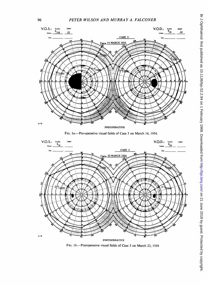
(1) Bitemporal hemicentral scotomatous hemianopia (BHS)24 patients (48 per cent.) showed an essentially scotomatous hemianopia (Figs la and lb, overleaf). Therewere ten men and fourteen women; ages 24 to 67 years (average 52).
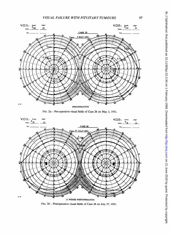
(2) Classical bitemporal hemianopia (BTH)Eighteen patients (36 per cent.) showed a classical peripheral hemianopia (Figs 2a and 2b, overleaf). Therewere eleven men and seven women; ages 24 to 67 years (average 52).
(3) Atypical field defects (AFD)Eight patients (16 per cent.) showed defects not classifiable in the first two groups, viz. homonymoushemianopia (3); unilateral central scotoma (2); complex hemianopic defects with large scotoma (3). Therewere three men and five women; ages 24 to 70 years (average 54).
The correlations which exist between each group and certain clinical and radiologicalcharacteristics will now be described.
Length of History and Mode of Progression of Symptoms
The history of the onset and course of visual failure in optic pathway compression isnotoriously protean (Table I, overleaf). Some patients first notice a gross defect such asuniocular near-blindness suddenly but quite fortuitously, though it must have been presentfor weeks, months, or years beforehand (termed "spurious abrupt onset" in Table I).Others describe an insidious progressive deterioration. There may be intervals in which thevisual decline spontaneously slows or arrests. There may be periods of rapid or urgent pro-gression of visual failure. Sometimes a genuine abrupt onset of visual failure is due to acutechiasmal compression as in so-called "pituitary apoplexy",
95
on 21 June 2018 by guest. Protected by copyright.
http://bjo.bmj.com
/B
r J Ophthalm
ol: first published as 10.1136/bjo.52.2.94 on 1 February 1968. D
ownloaded from
PETER WILSON AND MURRA Y A. FALCONER
V.0. S.: Snellin Jaeger6
Uncor. /18 J2
c. _
V.Q.D.: s-lu Jaegerun,r. /6 J2Cor_
PREOPERATIVE
FIG. la.-Pre-operative visual fields of Case 3 on March 14, 1954.
V.0 S.: Sna.li. Jaeger VO D. Szin Jaeger
UlCO. /6 Une.. /6
CO. CASE 3 C
90 so
_ o90 l _ _
POSTOPERATIVE
FIG. lb.-Postoperative visual fields of Case 3 on March 23, 1954
96
on 21 June 2018 by guest. Protected by copyright.
http://bjo.bmj.com
/B
r J Ophthalm
ol: first published as 10.1136/bjo.52.2.94 on 1 February 1968. D
ownloaded from
VISUAL FAILURE WITH PITUITARY TUMOURS 97
V.0. S.: Snllin Jaeger V.0D. 6"1 JaegerU. 6/36 J2 U /12 JiC_. CASE 26 _Coe
PREOPERATIVE
FIG. 2a.-Pre-operative visual fields of Case 26 on May 3, 1951.
V. .S.: Sn<lln Jaeger V.O.D.: S Jaeger.6/9 J1 Uro. /6A J1
CW. JCASE 26 En Cor_an_an
11 WEEKS POSTOPERATIVE
FIG. 2b.-Postoperative visual fields of Case 26 on July 27, 1951.
on 21 June 2018 by guest. Protected by copyright.
http://bjo.bmj.com
/B
r J Ophthalm
ol: first published as 10.1136/bjo.52.2.94 on 1 February 1968. D
ownloaded from
PETER WILSON AND MURRA Y A. FALCONER
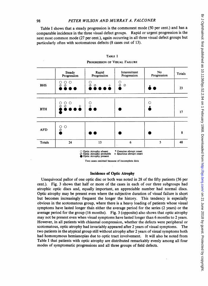
Table I shows that a steady progression is the commonest mode (50 per cent.) and has acomparable incidence in the three visual defect groups. Rapid or urgent progression is thenext most common mode (27 per cent.), again occurring in all three visual defect groups butparticularly often with scotomatous defects (8 cases out of 13).
TABLE I
PROGRESSION OF VISUAL FAILURE
Steady Rapid Intermittent No TProgression Progression Progression Progression Ttl
0 00 0 0BHS 00 00 + 00 0
*000 *0@@ 0 *23
000 0 000 + +
6BTH ||**|*|*|1* ~~~~~~~~~~~~~~~~~~~~17
0 0AFD 0
* @~~~000 8
Totals 24 13 6 5 48
O Optic Atrophy absent * Genuine abrupt onset0 Optic Atrophy probable + Spurious abrupt onset* Optic Atrophy present
Two cases omitted because of incomplete data
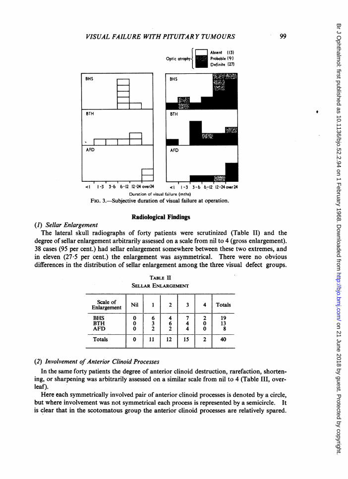
Incidence of Optic AtrophyUnequivocal pallor of one optic disc or both was noted in 28 of the fifty patients (56 per
cent.). Fig. 3 shows that half or more of the cases in each of our three subgroups hadatrophic optic discs and, equally important, an appreciable number had normal discs.Optic atrophy may be present even where the subjective duration of visual failure is shortbut becomes increasingly frequent the longer the history. This tendency is especiallyobvious in the scotomatous group, where there is a heavy loading of patients whose visualsymptoms have lasted longer than either the average period for the series (2 years) or theaverage period for the group (16 months). Fig. 3 (opposite) also shows that optic atrophymay not be present even when visual symptoms have lasted longer than 6 months to 2 years.However, in all patients with chiasmal compression, whether the defects were peripheral orscotomatous, optic atrophy had invariably appeared after 2 years of visual symptoms. Thetwo patients in the atypical group still without atrophy after 2 years of visual symptoms bothhad homonymous hemianopias due to optic tract involvement. It will also be noted fromTable I that patients with optic atrophy are distributed remarkably evenly among all fourmodes of symptomatic progressions and all three groups of field defects.
98
on 21 June 2018 by guest. Protected by copyright.
http://bjo.bmj.com
/B
r J Ophthalm
ol: first published as 10.1136/bjo.52.2.94 on 1 February 1968. D
ownloaded from
VISUAL FAILURE WITH PITUITAR Y TUMOURS
Absent (13)Optic atrophy. Probable (9)
Definite (27)
BHS
<I 1-3 36 b -12 12-24 over24 <I 1-3 3-6 6-12 12-24over24Duration of visual failure (mths)
FIG. 3.-Subjective duration of visual failure at operation.
Radiological Findings(1) Sellar EnlargementThe lateral skull radiographs of forty patients were scrutinized (Table II) and the
degree of sellar enlargement arbitrarily assessed on a scale from nil to 4 (gross enlargement).38 cases (95 per cent.) had sellar enlargement somewhere between these two extremes, andin eleven (27.5 per cent.) the enlargement was asymmetrical. There were no obviousdifferences in the distribution of sellar enlargement among the three visual defect groups.
TABLE IISELLAR ENLARGEMENT
Enlargement Nil 1 2 3 4 Totals
BHS 0 6 4 7 2 19BTH 0 3 6 4 0 13AFD 0 2 2 4 0 8
Totals 0 1 12 15 2 40
(2) Involvement of Anterior Clinoid ProcessesIn the same forty patients the degree of anterior clinoid destruction, rarefaction, shorten-
ing, or sharpening was arbitrarily assessed on a similar scale from nil to 4 (Table III, over-leaf).Here each symmetrically involved pair of anterior clinoid processes is denoted by a circle,
but where involvement was not symmetrical each process is represented by a semicircle. Itis clear that in the scotomatous group the anterior clinoid processes are relatively spared.
99
on 21 June 2018 by guest. Protected by copyright.
http://bjo.bmj.com
/B
r J Ophthalm
ol: first published as 10.1136/bjo.52.2.94 on 1 February 1968. D
ownloaded from
100 PETER WILSON AND MURRA Y A. FALCONER
TABLE IIIDESTRUCTION OF ANTERIOR CLINOID PROCESSES
Nil 1 2 3 4
BHS 00000 00000 00 aca 19OOaDD OD
BTH aUaD oocicDr ooaci OODDD 13DD
AFD |OD oooocI ODa 8
0 Both U Right D Left
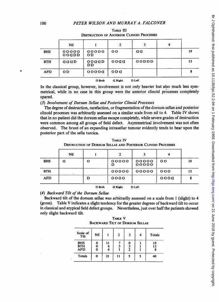
In the classical group, however, involvement is not only heavier but also much less sym-metrical, while in no case in this group were the anterior clinoid processes completelyspared.(3) Involvement of Dorsum Sellae and Posterior Clinoid ProcessesThe degree of destruction, rarefaction, or fragmentation ofthe dorsum sellae and posterior
clinoid processes was arbitrarily assessed on a similar scale from nil to 4. Table IV showsthat in no patient did the dorsum sellae escape completely, while severe grades of destructionwere common among all groups of field defect. Asymmetrical involvement was not oftenobserved. The brunt of an expanding intrasellar tumour evidently tends to bear upon theposterior part of the sella turcica.
TABLE IVDESTRUCTION OF DORSUM SELLAE AND POSTERIOR CLINOID PROCESSES
Nil 2 3 41
BHS a 0 00000 00000 00 19D 00000
BTH 00000 00000 000 13
AFD D 0000 OOO 8
0 Both El Right D Left
(4) Backward Tilt of the Dorsum SellaeBackward tilt of the dorsum sellae was arbitrafily assessed on a scale from 1 (slight) to 4
(gross). Table V indicates a slight tendency for the greater degrees of backward tilt to occurin classical and atypical field defect groups. Nevertheless, just over half the patients showedonly slight backward tilt.
TABLE VBACKWARD TILT OF DORSUM SELLAE
Scale of Nil 1 2 3 4 TotalsTilt
BHS 0 11 7 0 1 19BTH 0 6 3 3 1 13AFD 0 4 1 2 1 8
Totals 0 21 1 5 3 40
9
on 21 June 2018 by guest. Protected by copyright.
http://bjo.bmj.com
/B
r J Ophthalm
ol: first published as 10.1136/bjo.52.2.94 on 1 February 1968. D
ownloaded from
VISUAL FAILURE WITH PITUITAR Y TUMOURS
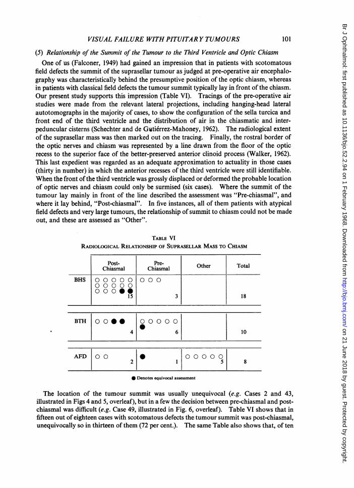
(5) Relationship of the Summit of the Tumour to the Third Ventricle and Optic ChiasmOne of us (Falconer, 1949) had gained an impression that in patients with scotomatous
field defects the summit of the suprasellar tumour as judged at pre-operative air encephalo-graphy was characteristically behind the presumptive position of the optic chiasm, whereasin patients with classical field defects the tumour summit typically lay in front of the chiasm.Our present study supports this impression (Table VI). Tracings of the pre-operative airstudies were made from the relevant lateral projections, including hanging-head lateralautotomographs in the majority of cases, to show the configuration of the sella turcica andfront end of the third ventricle and the distribution of air in the chiasmatic and inter-peduncular cisterns (Schechter and de Gwtierrez-Mahoney, 1962). The radiological extentof the suprasellar mass was then marked out on the tracing. Finally, the rostral border ofthe optic nerves and chiasm was represented by a line drawn from the floor of the opticrecess to the superior face of the better-preserved anterior clinoid process (Walker, 1962).This last expedient was regarded as an adequate approximation to actuality in those cases(thirty in number) in which the anterior recesses of the third ventricle were still identifiable.When the front of the third ventricle was grossly displaced or deformed the probable locationof optic nerves and chiasm could only be surmised (six cases). Where the summit of thetumour lay mainly in front of the line described the assessment was "Pre-chiasmal", andwhere it lay behind, "Post-chiasmal". In five instances, all of them patients with atypicalfield defects and very large tumours, the relationship of summit to chiasm could not be madeout, and these are assessed as "Other".
TABLE VIRADIOLOGICAL RELATIONSHIP OF SUPRASELLAR MASS TO CHIASM
Post- Pre-OteToaChiasmal Chiasmal Other Total
BHS 0 0 000 000O O O O O
15 3 18
BTH 0 000 0000 0
4 6 10
AFD 00 0 000 0 02 1 5 8
0 Denotes equivocal assessment
The location of the tumour summit was usually unequivocal (e.g. Cases 2 and 43,illustrated in Figs 4 and 5, overleaf), but in a few the decision between pre-chiasmal and post-chiasmal was difficult (e.g. Case 49, illustrated in Fig. 6, overleaf). Table VI shows that infifteen out of eighteen cases with scotomatous defects the tumour summit was post-chiasmal,unequivocally so in thirteen of them (72 per cent.). The same Table also shows that, of ten
101
on 21 June 2018 by guest. Protected by copyright.
http://bjo.bmj.com
/B
r J Ophthalm
ol: first published as 10.1136/bjo.52.2.94 on 1 February 1968. D
ownloaded from
102 PETER WILSON AND MURRA Y A. FALCONER
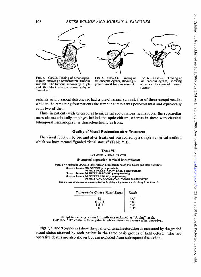
FIG. 4.-Case 2. Tracing of air encepha- FIG. 5.-Case 43. Tracing of FIG. 6.-Case 49. Tracing oflogram, showing a retrochiasmal tumour air encephalogram, showing a air encephalogram, showingsummit. The tumour is shown by stipple pre-chiasmal tumour summit. equivocal location of tumourand the black shadow shows subara- summit.chnoid air.
patients with classical defects, six had a pre-chiasmal summit, five of them unequivocally,while in the remaining four patients the tumour summit was post-chiasmal and equivocallyso in two of them.
Thus, in patients with bitemporal hemicentral scotomatous hemianopia, the suprasellarmass characteristically impinges behind the optic chiasm, whereas in those with classicalbitemporal hemianopia it is characteristically in front.
Quality of Visual Restoration after TreatmentThe visual function before and after treatment was scored by a simple numerical method
which we have termed "graded visual status" (Table VII).
TABLE VIIGRADED VISUAL STATUS
(Numerical expression of visual improvement)Note: Two functions, ACUITY and FIELD, are scored for each eye, before and after operation.
Score 2 denotes NO DEFECT pre-operatively;DEFECT FULLY RECOVERED postoperatively
Score 1 denotes DEFECT IMPROVED postoperatively;Score 0 denotes DEFECT PRESENT pre-operatively;
DEFECT UNCHANGED OR WORSE postoperativelyThe average of the scores is multiplied by 6, giving a figure on a scale rising from 0 to 12.
Postoperative Graded Visual Status Result
12 "A"6-10-5 "B"1 5-6 "C"0 "D"
Complete recovery within 1 month was reckoned as "A plus" result.Category "D" contains three patients whose vision was worse after operation.
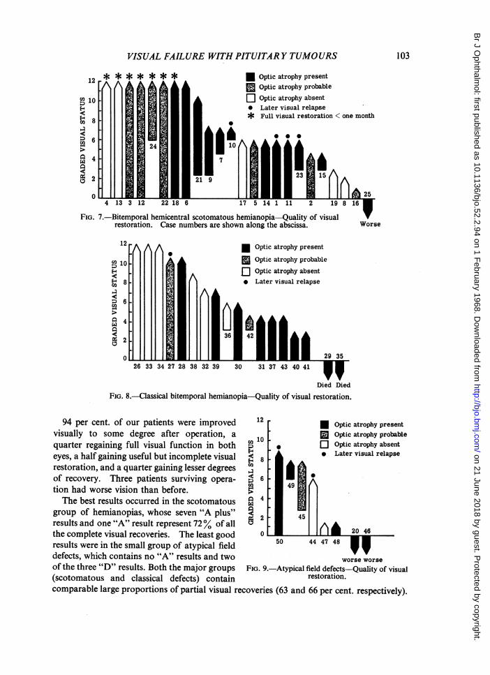
Figs 7, 8, and 9 (opposite) show the quality of visual restoration as measured by the gradedvisual status attained by each patient in the three basic groups of field defect. The twooperative deaths are also shown but are excluded from subsequent discussion.
on 21 June 2018 by guest. Protected by copyright.
http://bjo.bmj.com
/B
r J Ophthalm
ol: first published as 10.1136/bjo.52.2.94 on 1 February 1968. D
ownloaded from
VISUAL FAILURE WITH PITUITARY TUMOURS 103
12 M P qWPvvqbqptic;aTropny prsents11
Optic atrophy probable
10 X Optic atrophy absent* Later visual relapse
D6 I. * ~~~~~~~~~Fullvisual restoration < one month.6 Al
0 ~~~2.0
FIG. 7.-Bitemporal hemicentral scotomatous hemianopia-Quality of visualWrestoration. Case numbers are shown along the abscissa. Worse
12 * Optic atrophy present
D 10 . Optic atrophy probable¢ Optic atrophy absent
E- 8. b Later visual relapseco
02~~ ~ ~ ~~~6 4
0 3
26 33 3427 283832 39 30 31 3743 404
Died Died
FIG. 8.-Classical bitemporal hemianopia-Quality of visual restoration.
94 per cent. of our patients were improved 12 * Optic atrophy presentvisually to some degree after operation, a 10 S Optic atrophy probablequarter regaining full visual function in both 2 * Q Optic atrophy absenteyes, a half gaining useful but incomplete visual ¢ 8 * Later visual relapserestoration, and a quarter gaining lesser degrees EIof recovery. Three patients surviving opera- 6tion had worse vision than before.The best results occurred in the scotomatous 4
group of hemianopias, whose seven "A plus" iresults and one "A" result represent 72% of all 2 45
the complete visual recoveries. The least good 0 * 46
results were in the small group of atypical field 50 44 47 48defects, which contains no "A" results and two worse worseof the three "D" results. Both the major groups FIG. 9.-Atypical field defects-Quality of visual(scotomatous and classical defects) contain restoration.comparable large proportions of partial visual recoveries (63 and 66 per cent. respectively).
M f%,n+i,, a+-rf%r%h%r invacion+
on 21 June 2018 by guest. Protected by copyright.
http://bjo.bmj.com
/B
r J Ophthalm
ol: first published as 10.1136/bjo.52.2.94 on 1 February 1968. D
ownloaded from
PETER WILSON AND MURRA Y A. FALCONER
Discussion(1) Incidence of Bitemporal Hemicentral Scotomatous HemianopiaThe importance or even the occurrence of scotomatous defects as a form of presentation
of chiasmal compression by pituitary tumours receives little emphasis either in under-graduate texts or in publications intended for postgraduate study (Handfield-Jones andPorritt, 1957; Blackburn and Lawrie, 1958; Jennett, 1964; Houston, Joiner, and Trounce,1966).Chamlin, Davidoff, and Feiring (1955) found bitemporal hemianopic defects in 96 per
cent. of 109 cases of proven or presumptive chromophobe adenoma, but while stating thatfield changes occur first in the small central isopters (e.g. 1/2000) concluded that centralhemianopic defects, whether unilateral or bilateral, were very rare except with peripheralhemianopic defects, although the latter might be very inconspicuous. Traquair (1938) hadearlier described the scotomatous type of hemianopia without giving a precise incidence,but indicated that it could merge with the classical type as it progresses. Clarke, Knighton,and Bebin (1963) reviewing 75 cases of chromophobe adenoma found a visual field defect in81 per cent. and remarked that "the characteristic field defect is an asymmetrical bitemporalhemianopsia" without mentioning the occurrence of scotomatous hemianopia. Rucker(1957) found "ordinary bitemporal hemianopsia" the characteristic field defect but alsodescribed patients with bitemporal hemicentral scotomatous defects, bilateral central scoto-mata, and incongruous homonomous hemianopias. Clover and Lyle (1961), reporting 100cases of proven pituitary adenoma (not all of them chromophobe adenomata), found clear-cut bitemporal hemianopic defects in 94, the, peripheral fields being affected in 79 (i.e.classical bitemporal hemianopia) and unaffected in fifteen (i.e. hemicentral scotomatoushemianopia.)Our series contains a high proportion of scotomatous hemianopias (48 per cent.) partly
because in classifying the final preoperative field defect we have also taken into accountthe pattern of postoperative regression of the defect. All our patients had asymmetricalfield defects.
(2) Anatomical ConsiderationsThe interruption of chiasmal function in compression by a tumour is probably due much
more to interference with the blood supply of the chiasm than to mere pressure or displace-ment (Traquair, 1938; Rucker, 1957). Nevertheless Rucker felt that the early visualdisturbance, before mechanical factors had been overshadowed by vascular ones, hadreliable localizing value. He quoted Wilbrand's classical concept (Wilbrand, 1926) of theoptic chiasm as comprising three layers. The upper layer contains mainly fibres from theupper retinal quadrants (i.e. subserving the lower visual quadrants) with non-crossing out-numbering crossing fibres. The middle layer contains crossing fibres from the upper nasalretinal quadrants in roughly equal proportion to non-crossing fibres from the lowertemporal quadrants. The lower layer contains mainly crossing fibres from the lower nasalretinal quadrants (i.e. subserving the upper temporal visual quadrants) which loop for-wards into the opposite optic nerve before turning backwards into the optic tract. Thefunction of these lowest fibres is commonly the first to be interrupted by a tumour compres-sing the antero-inferior face of the chiasm, resulting in a peripheral bitemporal upperquadrantic hemianopia. A few non-crossing fibres from the inferior temporal retinalquadrants also run in the third or lower layer. The decussating central retinal fibres cross
104
on 21 June 2018 by guest. Protected by copyright.
http://bjo.bmj.com
/B
r J Ophthalm
ol: first published as 10.1136/bjo.52.2.94 on 1 February 1968. D
ownloaded from
VISUAL FAILURE WITHPITUITARY TUMOURS
in the posterior part of the chiasm. According to Traquair, scotomatous hemianopia is
due to compression of these decussating fibres of the papillo-macular bundles, which hedescribes as forming the entire substance of the posterior edge of the chiasm. Hughes(1954) has pointed out that the point of chiasmal compression depends not only on theposition of the lesion but also on whether the chiasm is pre-fixed or post-fixed. Our ownmaterial supports these views.
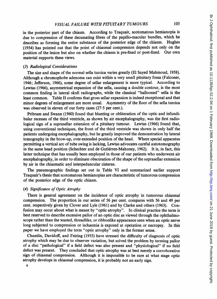
(3) Radiological ConsiderationsThe size and shape of the normal sella turcica varies greatly (El Sayed Mahmoud, 1958).
Although a chromophobe adenoma can exist within a very small pituitary fossa (Falconer,1946; Jefferson, 1966), some degree of sellar enlargement is more typical. According toLewtas (1966), asymmetrical expansion of the sella, causing a double contour, is the mostcommon finding in lateral skull radiographs, while the classical "ballooned" sella is theleast common. Table II confirms that gross sellar expansion is indeed exceptional and thatminor degrees of enlargement are more usual. Asymmetry of the floor of the sella turcicawas observed in eleven of our forty cases (27-5 per cent.).Pribram and Swann (1960) found that blunting or obliteration of the optic and infundi-
bular recesses of the third ventricle, as shown by air encephalography, was the first radio-logical sign of a suprasellar extension of a pituitary tumour. Lewtas (1966) found that,using conventional techniques, the front of the third ventricle was shown in only half thepatients undergoing encephalography, but he greatly improved the demonstration by lateraltomography in the brow-up, over-extended position of the head. Where special apparatuspermitting a vertical arc of tube swing is lacking, Lewtas advocates careful autotomographyin the same head position (Schechter and de Gutierrez-Mahoney, 1962). It is, in fact, thislatter technique that has usually been employed in those of our patients who underwent airencephalography, in order to eliminate obscuration of the shape of the suprasellar extensionby air in the chiasmatic and interpeduncular cisterns.The pneumographic findings set out in Table VI and summarized earlier support
Traquair's thesis that scotomatous hemianopias are characteristic of tumorous compressionof the posterior edge of the optic chiasm.
(4) Significance of Optic AtrophyThere is general agreement on the incidence of optic atrophy in tumorous chiasmal
compression. The proportion in our series of 56 per cent. compares with 56 and 49 percent. respectively given by Clover and Lyle (1961) and by Clarke and others (1963). Con-fusion may occur about what is meant by "optic atrophy". In clinical practice the term isbest reserved to describe excessive pallor of an optic disc as viewed through the ophthalmo-scope rather than the wasted, threadlike, or ribbonlike appearance seen when an optic nervelong subjected to compression or ischaemia is exposed at operation or necropsy. In thispaper we have employed the term "optic atrophy" only in the former sense.
Chamlin, Davidoff, and Feiring (1955) have stressed the difficulty of diagnosis of opticatrophy which may be due to observer variation, but solved the problem by terming pallorof a disc "pathological" if a field defect was also present and "physiological" if no fielddefect was present. They concluded that optic atrophy was at best merely a corroborativesign of chiasmal compression. Although it is impossible to be sure at what stage opticatrophy develops in chiasmal compression, it is probably not an early sign.
9
105
on 21 June 2018 by guest. Protected by copyright.
http://bjo.bmj.com
/B
r J Ophthalm
ol: first published as 10.1136/bjo.52.2.94 on 1 February 1968. D
ownloaded from

PETER WILSON AND MURRA Y A. FALCONER
The prognostic significance of optic atrophy is not as gloomy as is sometimes thought.Rucker (1957) considered that, in cases with slight pallor but relatively great visual loss,improvement after surgery was to be expected, and that even in cases with distinct pallor,improvement might sometimes unexpectedly occur. Our own findings in Figs 7, 8, and 9confirm this. Six of the eleven perfect visual recoveries were seen in patients with definiteor probable optic atrophy, whilst of 21 good quality but incomplete visual recoveries,definite or probable optic atrophy was present in sixteen. In the scotomatous group opticatrophy was present in six of the eight patients who made perfect visual recoveries, whereasin the classical group none of the three patients whose vision returned to normal had opticatrophy. The three worst visual results were seen in patients with definite optic atrophy.Again, the absence of optic atrophy in a particular case does not by itself seem necessarilyto imply a good visual prognosis. In our series three out of sixteen patients with poorquality postoperative visual status had no optic atrophy, although two of these weresuffering from optic tract involvement rather than chiasmal compression.Thus the presence of optic atrophy does not preclude the possibility of full and rapid
recovery of vision in cases where the field defect is a bitemporal hemicentral scotomatoushemianopia. On the other band a perfect recovery is not to be expected if optic atrophy ispresent in cases of classical bitemporal hemianopia, although good partial recovery is stilllikely.
(5) Mode ofProgression of Visual FailureTraquair (1938) stated that the scotomatous type of field defect was characteristic of
activity of the lesion and the non-scotomatous type of a slowly-growing or stationarycondition. Table I indicates that our series of cases broadly supports this opinion. Onlythree out of seventeen cases (17 per cent.) of classical bitemporal hemianopia described arapid or urgent progression of visual failure, whereas eight out of 23 cases (35 per cent.) ofscotomatous hemianopia did so. Conversely, while only eleven of 23 cases (48 per cent.)of scotomatous hemianopia described either a stationary defect or an insidiously progressiveone, thirteen of seventeen cases (76 per cent.) of classical hemianopia did so. However, noclear correlation exists between the mode of progression of visual failure and subsequentvisual improvement.
(6) Subjective Duration of Visual FailureWhen speed and completeness of visual recovery were studied, six of the seven "A plus"
results followed a history of visual failure of between 6 months and 2 years. Two "A"results occurred in patients with histories of less than 1 month. Only one patient (a case ofclassical bitemporal hemianopia) regained full visual function after a history exceeding 2years. All three patients with deterioration of vision after operation had histories greatlyin excess of 2 years.
It would seem therefore that good results can be expected only if subjective visual failurehas been of less than 2 years' duration.
(7) Quality of Diagnosis before Definite ReferralLong delays were often noted between the patient's first seeking advice and his final
definitive referral to a neurosurgeon. These delays were due to failure to detect or torealize the significance of a visual field defect and thus the time was often taken up withmultiple refractions or other ocular treatments.
106
on 21 June 2018 by guest. Protected by copyright.
http://bjo.bmj.com
/B
r J Ophthalm
ol: first published as 10.1136/bjo.52.2.94 on 1 February 1968. D
ownloaded from
VISUAL FAILURE WITH PITUITAR Y TUMOURS
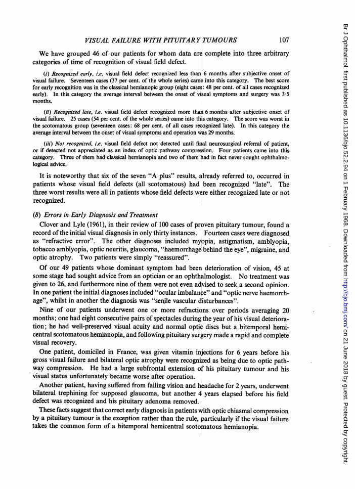
We have grouped 46 of our patients for whom data are complete into three arbitrarycategories of time of recognition of visual field defect.
(i) Recognized early, i.e. visual field defect recognized less than 6 months after subjective onset ofvisual failure. Seventeen cases (37 per cent. of the whole series) came into this category. The best scorefor early recognition was in the classical hemianopic group (eight cases: 48 per cent. of all cases recognizedearly). In this category the average interval between the onset of visual symptoms and surgery was 3-5months.
(ii) Recognized late, i.e. visual field defect recognized more than 6 months after subjective onset ofvisual failure. 25 cases (54 per cent. of the whole series) came into this category. The score was worst inthe scotomatous group (seventeen cases: 68 per cent. of all cases recognized late). In this category theaverage interval between the onset of visual symptoms and operation was 29 months.
(iii) Not recognized, i.e. visual field defect not detected until finXal neurosurgical referral of patient,or if detected not appreciated as an index of optic pathway compression. Four patients came into thiscategory. Three of them had classical hemianopia and two of them had in fact never sought ophthalmo-logical advice.
It is noteworthy that six of the seven "A plus" results, already referred to, occurred inpatients whose visual field defects (all scotomatous) had been recognized "late". Thethree worst results were all in patients whose field defects were either recognized late or notrecognized.
(8) Errors in Early Diagnosis and TreatmentClover and Lyle (1961), in their review of 100 cases of proven pituitary tumour, found a
record of the initial visual diagnosis in only thirty instances. Fourteen cases were diagnosedas "refractive error". The other diagnoses included myopia, astigmatism, amblyopia,tobacco amblyopia, optic neuritis, glaucoma, "haemorrhage behind the eye", migraine, andoptic atrophy. Two patients were simply "reassured".Of our 49 patients whose dominant symptom had been deterioration of vision, 45 at
some stage had sought advice from an optician or an ophthalmologist. No treatment wasgiven to 26, and furthermore nine of them were not even advised to seek a second opinion.In one patient the initial diagnoses included "ocular imbalance" and "optic nerve haemorrh-age", whilst in another the diagnosis was "senile vascular disturbances".Nine of our patients underwent one or more refractions over periods averaging 20
months; one had eight consecutive pairs of spectacles during the year of his visual deteriora-tion; he had well-preserved visual acuity and normal optic discs but a bitemporal hemi-central scotomatous hemianopia, and following pituitary surgery made a rapid and completevisual recovery.One patient, domiciled in France, was given vitamin injections for 6 years before his
gross visual failure and bilateral optic atrophy were recognized as being due to optic path-way compression. He had a large subfrontal extension of his pituitary tumour and hisvisual status unfortunately became worse after operation.Another patient, having suffered from failing vision and headache for 2 years, underwent
bilateral trephining for supposed glaucoma, but another 4 years elapsed before his fielddefect was recognized and his pituitary adenoma removed.These facts suggest that correct early diagnosis in patients with optic chiasmal compression
by a pituitary tumour is the exception rather than the rule, particularly if the visual failuretakes the common form of a bitemporal hemicentral scotomatous hemianopia.
107
on 21 June 2018 by guest. Protected by copyright.
http://bjo.bmj.com
/B
r J Ophthalm
ol: first published as 10.1136/bjo.52.2.94 on 1 February 1968. D
ownloaded from
PETER WILSON AND MURRA Y A. FALCONER
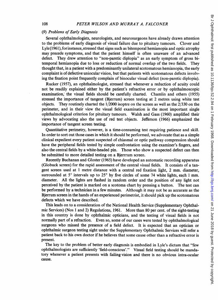
(9) Problems of Early DiagnosisSeveral ophthalmologists, neurologists, and neurosurgeons have already drawn attention
to the problems of early diagnosis of visual failure due to pituitary tumours. Clover andLyle(1961), forinstance, stressed that signs such as bitemporal hemianopia and optic atrophymay precede symptoms, and that the patient himself is often unaware of an advanceddefect. They drew attention to "non-paretic diplopia" as an early symptom of gross bi-temporal hemianopia due to loss or reduction of normal overlap of the two fields. Theythought that, in a patient with a predominantly unilateral scotomatous hemianopia, the earlycomplaint is of defective uniocular vision, but that patients with scotomatous defects involv-ing the fixation point frequently complain of binocular visual defect (non-paretic diplopia).Rucker (1957), an ophthalmologist, stressed that whenever a reduction of acuity could
not be readily explained either by the patient's refractive error or by ophthalmoscopicexamination, the visual fields should be carefully charted. Chamlin and others (1955)stressed the importance of tangent (Bjerrum) screen testing at 2 metres using white testobjects. They routinely charted the 1/2000 isoptre on the screen as well as the 2/330 on theperimeter, and in their view the visual field examination is the most important singleophthalmological criterion for pituitary tumours. Walsh and Gass (1960) amplified theirviews by advocating also the use of red test objects. Jefferson (1966) emphasized theimportance of tangent screen testing.
Quantitative perimetry, however, is a time-consuming test requiring patience and skill.In order to sort out those cases in which it should be performed, we advocate that as a simpleclinical expedient every patient suspected of chiasmal or optic pathway compression shouldhave the peripheral fields tested by simple confrontation using the examiner's fingers, andalso the central fields by a white-headed pin. Those who show a suspected defect can thenbe submitted to more detailed testing on a Bjerrrum screen.
Recently Buchanan and Gloster (1965) have developed an automatic recording apparatus(Globuck screen) for the rapid assessment of the central visual fields. It consists of a tan-gent screen used at 1 metre distance with a central red fixation light, 2 mm. diameter,surrounded at 50 intervals up to 25° by five circles of some 74 white lights, each 1 mm.diameter. All the lights are flashed in random order and the position of any light notperceived by the patient is marked on a scotoma chart by pressing a button. The test canbe performed by a technician in a few minutes. Although it may not be as accurate as theBjerrum screen in the hands of an experienced perimetrist, it should pick up the scotomatousdefects which we have described.
This leads on to a consideration of the National Health Service (Supplementary Ophthal-mic Services) (Nos 1 and 2) Regulations, 1961. More than 80 per cent. of the sight-testingin this country is done by ophthalmic opticians, and the testing of visual fields is notnormally part of a refraction. Even so, some of our cases were tested by ophthalmologicalsurgeons who missed the presence of a field defect. It is expected that an optician orophthalmic surgeon testing sight under the Supplementary Ophthalmic Services will refer apatient back to his own doctor if he believes that some cause other than a refractive error ispresent.The key to the problem of better early diagnosis is embodied in Lyle's dictum that "few
ophthalmologists are sufficiently 'field-conscious'." Visual field testing should be manda-tory whenever a patient presents with failing vision and there is no obvious intra-ocularcause.
108
on 21 June 2018 by guest. Protected by copyright.
http://bjo.bmj.com
/B
r J Ophthalm
ol: first published as 10.1136/bjo.52.2.94 on 1 February 1968. D
ownloaded from
VISUAL FAILURE WITH PITUITAR Y TUMOURS
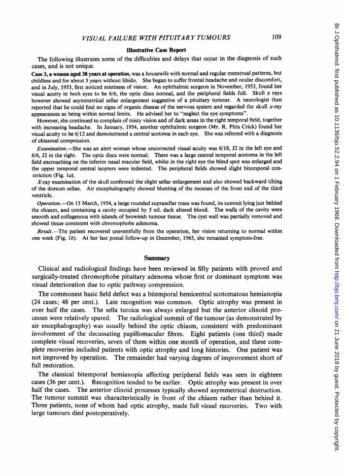
Illustrative Case ReportThe following illustrates some of the difficulties and delays that occur in the diagnosis of such
cases, and is not unique.Case 3, a woman aged 38 years at operation, was a housewife with normal and regular menstrual patterns, butchildless and for about 5 years without libido. She began to suffer frontal headache and ocular discomfort,and in July, 1953, first noticed mistiness of vision. An ophthalmic surgeon in November, 1953, found hervisual acuity in both eyes to be 6/6, the optic discs normal, and the peripheral fields full. Skull x rayshowever showed asymmetrical sellar enlargement suggestive of a pituitary tumour. A neurologist thenreported that he could find no signs of organic disease of the nervous system and regarded the skull x-rayappearances as being within normal limits. He advised her to "neglect the eye symptoms".However, she continued to complain of misty vision and of dark areas in the right temporal field, together
with increasing headache. In January, 1954, another ophthalmic surgeon (Mr. R. Pitts Crick) found hervisual acuity to be 6/12 and demonstrated a central scotoma in each eye. She was referred with a diagnosisof chiasmal compression.Examination.-She was an alert woman whose uncorrected visual acuity was 6/18, J2 in the left eye and
6/6, J2 in the right. The optic discs were normal. There was a large central temporal scotoma in the leftfield encroaching on the inferior nasal macular field, whilst in the right eye the blind spot was enlarged andthe upper temporal central isopters were indented. The peripheral fields showed slight bitemporal con-striction (Fig. la).X-ray examination of the skull confirmed the slight sellar enlargement and also showed backward tilting
of the dorsum sellae. Air encephalography showed blunting of the recesses of the front end of the thirdventricle.Operation.-On 15 March, 1954, a large rounded suprasellar mass was found, its summit lying just behind
the chiasm, and containing a cavity occupied by 5 ml. dark altered blood. The walls of the cavity weresmooth and collagenous with islands of brownish tumour tissue. The cyst wall was partially removed andshowed tissue consistent with chromophobe adenoma.
Result.-The patient recovered uneventfully from the operation, her vision returning to normal withinone week (Fig. lb). At her last postal follow-up in December, 1965, she remained symptom-free.
Summary
Clinical and radiological findings have been reviewed in fifty patients with proved andsurgically-treated chromophobe pituitary adenoma whose first or dominant symptom wasvisual deterioration due to optic pathway compression.The commonest basic field defect was a bitemporal hemicentral scotomatous hemianopia
(24 cases; 48 per cent.). Late recognition was common. Optic atrophy was present inover half the cases. The sella turcica was always enlarged but the anterior clinoid pro-cesses were relatively spared. The radiological summit of the tumour (as demonstrated byair encephalography) was usually behind the optic chiasm, consistent with predominantinvolvement of the decussating papillomacular fibres. Eight patients (one third) madecomplete visual recoveries, seven of them within one month of operation, and these com-plete recoveries included patients with optic atrophy and long histories. One patient wasnot improved by operation. The remainder had varying degrees of improvement short offull restoration.The classical bitemporal hemianopia affecting peripheral fields was seen in eighteen
cases (36 per cent.). Recognition tended to be earlier. Optic atrophy was present in overhalf the cases. The anterior clinoid processes typically showed asymmetrical destruction.The tumour summit was characteristically in front of the chiasm rather than behind it.Three patients, none of whom had optic atrophy, made full visual recoveries, Two withlarge tumours died postoperatively.
109
on 21 June 2018 by guest. Protected by copyright.
http://bjo.bmj.com
/B
r J Ophthalm
ol: first published as 10.1136/bjo.52.2.94 on 1 February 1968. D
ownloaded from
PETER WILSON AND MURRA Y A. FALCONER
The remaining eight patients had miscellaneous field defects indicating optic nerve, optictract, or composite optic pathway involvement. Optic atrophy was present in five cases.The tumour was often very large and bore no characteristic relation to the optic chiasm.No patient in this group regained full visual function, and two were worse after operation.An important factor in the frequent failure of recognition of chiasmal compression in its
early stages is that the field defect more commonly evolves as an asymmetrical bitemporalhemicentral scotomatous hemianopia which is overlooked if only the peripheral fields areexamined. Patients with visual failure not obviously due to a local ocular cause shouldhave detailed quantitative perimetric assessment. A quick preliminary expedient for"screening" the central fields of such patients is simple confrontation with a white-headedpin.
REFERENCESBLACKBURN, G., and LAWRIE, R. (1958). "Textbook of Surgery". Blackwell, Oxford.BUCHANAN, W. M., and GLOSTER, J. (1965). Brit. J. Ophthal., 49, 57.CHAmUN, M., DAVIDOFF, L. M., and FEIRING, E. H. (1955). Amer. J. Ophthal., 40, 353.CLARKE, H. A., KNIGHTON, R. S., and BEBIN, J. (1963). J. Mich. med. Soc., 62, 1183.CLovR, P., and LY, T. KErm (1961). Proc. roy. Soc. Med., 54, 611.EL SAYED MAHMoUD, M. (1958) "The Sella in Health and Disease". Brit. J. Radiol., SuppI. 8.FALCONER, M. A. (1946). N.Z med. J., 45, 343.
(1949). Ibid., 48 (October Suppl.), 8.HANDFIELD-JONES, R. M., and PoRRr, A. E. (1957). "Essentials of Modern Surgery", 5th ed. Living-
stone, Edinburgh.HousToN, J. C., JOINER, C. L., and TROUNCE, J. R. (1966). "A Short Textbook of Medicine", 2nd ed.
English Universities Press, London.HUGHES, B. (1954). "The Visual Fields". Blackwell, Oxford.JEFFERSON, A. (1966). Clin. Radiol., 17, 141.JENNETr, W. B. (1964). "An Introduction to Neurosurgery". Heinemann, London.LEWTAS, N. A. (1966). Clin. Radiol., 17, 149.PRIBRAM, H. F. W., and SWANN, G. F. (1960). Radiology, 75, 877.RUCKER, C. W. (1957). Clin. Neurosurg., 4, 34.SCHECHTER, M. M., and DE GuTLiRREz-MAHONEY, C. G. (1962). Brit. J. Radiol., 35, 438.TRAQUAIR, H. M. (1938). "Introduction to Clinical Perimetry", 3rd ed. Kimpton, London.WALKER, A. E. (1962). Amer. J. Ophthal., 54, 563.WALSH, F. B., and GASS, J. D. (1960). Ibid., 50, 1031.WILBRAND, H. (1926). Z. Augenheilk., 59, 135.
110
on 21 June 2018 by guest. Protected by copyright.
http://bjo.bmj.com
/B
r J Ophthalm
ol: first published as 10.1136/bjo.52.2.94 on 1 February 1968. D
ownloaded from