Patient Setup for Precision Proton Therapy
48
Patient Setup for Precision Proton Therapy Hsiao-Ming Lu, Ph.D.
Transcript of Patient Setup for Precision Proton Therapy

Patient Setup for Precision Proton Therapy
Hsiao-Ming Lu, Ph.D.

• What’s so special about setup for PT?• Anything different in immobilization?• Imaging systems• Organ motion issues• Future tools
Content

• Clinical data show significant decrease in tumor control (≥ 5%) when tumor dose decreased by 4 - 5 %
• Therefore, “significant”portion of CTV cannot fall outside high dose region more than once in fractionated course of radiation L. Vehey UCSF
What Precision Is Needed?

What Precision Is Needed?
• The uncertainty of the target position is more important than the uncertainty of the dosimetry– Daily dose errors of ≥ 20 % possible with
positioning error– Dose errors due to errors in dose
calculation probably not more than 5 - 10%
L. Vehey UCSF

Proton Dose Distribution Extra SensitiveVery Sensitive to WED Variations
Over or Undershoot!
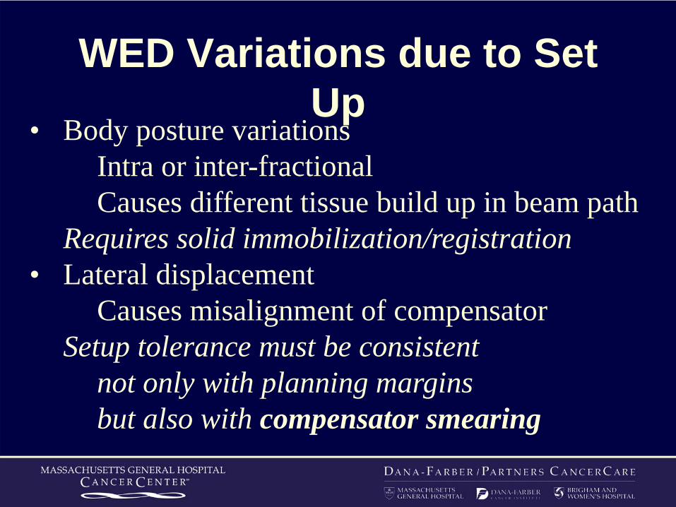
• Body posture variationsIntra or inter-fractionalCauses different tissue build up in beam path
Requires solid immobilization/registration• Lateral displacement
Causes misalignment of compensatorSetup tolerance must be consistent
not only with planning marginsbut also with compensator smearing
WED Variations due to Set Up

Inter-fraction Set Up Variation
Head and Neck
Pelvis
Rosenthal 2004

Hair
• Style & Placement• Clips (even plastic)• Gel & Water
Potential Range Perturbation
Rosenthal 2004

• Generic requirementsSmall inter- and intra-fractional variationUncertainty well-understoodPatient comfortableFixed to treatment table, if possiblePractical
Immobilization
• Proton specificNo potential range perturbations
e.g., passing part of the frameHardware clearance
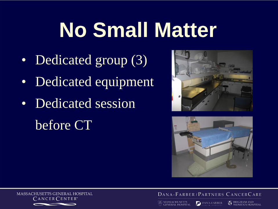
• Dedicated group (3)• Dedicated equipment• Dedicated session
before CT
No Small Matter

Immobilization devices
• Alpha Cradle• Bean Bag• Duncan Head Rest• Leg Abductor• Wing Board
• Base of Skull *• IC-Mask (reinforced)• Modified GTC• Neck Cast *• Para-Nasal Sinus *
Non-rigid DevicesRigid Devices
Engelsman 2005

Immobilization For Proton Therapy
Allow treatment snout proximity to cervical/thoracic spine
Prevent Articulation of head/neck
Base of Skull (BOS)Contoured Carbon Base (0.3 - 0.5 cm)Mask w. Low Profile Pin Arms
Rosenthal 2004

Immobilization For Proton TherapyIntra-Cranial (IC)
Curved Wafer Filled Carbon BaseDense Foam PadReinforced Mask
Rosenthal 2004

Immobilization For Proton TherapyParanasal Sinus (PNS) Frame
Sturdy Lucite Base and ArchDense Foam Head PadRigid Bite Mold - Prosthedontal
Table 1A: Measured motion between set-up and post treatment film pairs using the PNS immobilization system (N=20).
(Mean time between films = 16 ± 4 minutes (SD)) Average Standard
Deviation A-P 0.3 mm 0.4 mm S-I 0.0 mm 0.5 mm Lat. -0.4 mm 0.5 mm 3-D Displacement 0.9 mm 0.4 mm 2-D Displacement 0.7 mm 0.4 mm Roll 0.07 degrees 0.45 degreesPitch 0.03 degrees 0.42 degreesTurntable -0.14 degrees 0.67 degrees
Rosenthal 2004

In-House DevicesRadionics Gill-Thomas-Cosman (GTC) Frame
Occipital Cup with Heavy-Body Reprosil ®Velcro Head RestraintBite Mold
Bussiere 2006

GTC ModificationsReplace High Density Occipital Cup with MoldcareTM Cushion
Bussiere 2006

GTC ModificationsCushion
Bussiere 2006

Immobilization For Proton Therapy
Medulloblastoma FrameFull Body Prone MoldMask, Head/Body Articulation Minimal
Rosenthal 2004

CSI (Cranio-Spinal Immobilization)
Rosenthal 2004
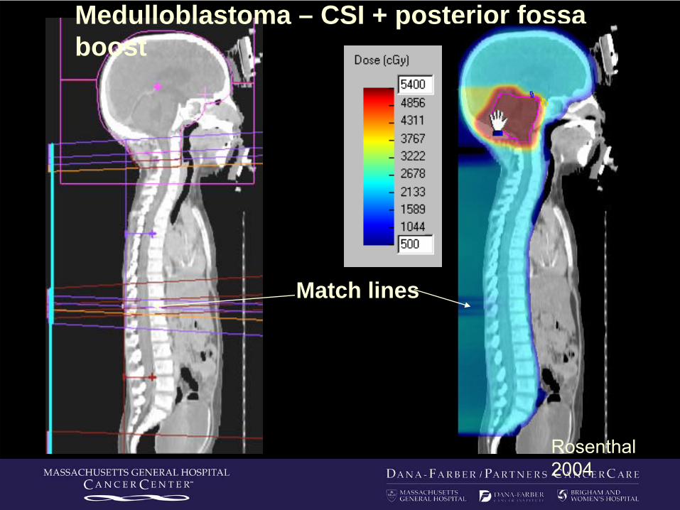
Medulloblastoma – CSI + posterior fossa boost
Match lines
Rosenthal 2004

2 Spine fields matching at anterior cord surface
Dose %11010510210090805010
Protons Photons
Non-aligned divergence
Rosenthal 2004

Current• Orthogonal X-ray images (DIPS)
Digital image panelsIntegrated with TPSLandmark matching to DRR(bony anatomy or implants) 6D correction
• Ultrasound (prostate only)
Imaging System

Radiographic Set Up w.Diagnostic X-rays DRR From CT
1993 Implanted Fiducials Digitized from Film 3D - High Accuracy - 1/2 mm (Gall, Verhey ‘93)
1996 Digital Radiography – Filmless CCD Camera
Flat Panel Imager Receptors at NPTCSmaller Lighter Device, Accurate Couch, High Resolution
1999
1980
History proton X-ray positioning
Digirad – Application Integrates Planning and Set Up2001
2003/4DIPSTM2 – Dicom Database, TCS Integration Ready, Feature Matching Automation Rosenthal
2004

Repositioning For Proton Therapy
X-rays ( anterior-posterior )
Orthogonal X-ray Tubes
Simultaneous Lateral and AP Radiographs
Rosenthal 2004

Repositioning For Proton Therapy
X-Ray Tubes
Nozzle Snout (withaperture & compensator)
6-axis patient positioner
Rosenthal 2004

Cage Source
Cage Detector
Nozzle Detector
Nozzle Source
Hitachi gantry with orthogonal X-ray system
Zhu, 2007

Repositioning For Proton Therapy
Amorphous Silicon Flat Panel Imagers
Rosenthal 2004

Repositioning For Proton Therapy
DIPS Radiographic Set Up Software
Gold Seeds in Prostate
Rosenthal 2004

Diagnostic x-ray positioningDIPSTM Relies on Identifying Landmarks
Rosenthal 2004

1) Patient immobilization2) Initial positioning (lasers and skin marks)3) Take AP/LAT orthogonal image pair
General position adjustment(translation and rotation)
4) Take beam-line image (BEV)Small adjustment (translation only)(Tolerance must agree with smearing)
Setup Process

Set Up Uncertainty - Summary
Intercranial tissues movement
• 1 mm invasive skull fixation
• 2-3 mm high quality head mask
• 3 mm neck articulation
Associated with breathing and cardiac motion:
• Lung tumors• Diaphragm• Liver• Kidneys• Pancreas
3.9 mm12-17 mm10-25 mm11-19 mm20 mm
K. Langen NAC Capetown SA
Digestive and Muscular Variation:
• Prostate• Bladder• Rectum• Abdomen
Highly Unpredictable
Daily Verification or Tracking Required
Rosenthal 2004

Sonarray System3D ultrasound to CT contours
Ultrasound for Prostate

Workstation
CT-Sim Room
U/S-SIM
Ultrasound to Ultrasound The RESTITU Platform
Treatment Room
U/S-GUIDE

STAR (Stereotactic Alignment radiosurgery)Fixed Beam SRS/SRT
Bussiere 2006

Imaging/Alignment for STARFilm (3D) → Digital (2+D) → Digital 3D
Varian PaxScan 4030: 127 μm pixel
(Gadolinium Oxysulfide Scintillator)
Bussiere 2006

Ocular Treatment
Bussiere 2006

Imaging uncertainty due to respiration
Organ Motion
• Instant X-ray image
• Averaged DRR from 3D CT

Example: LiverRL fieldUse same range, mod, compensator for 50% and 0% phases 50% 0% (50% field)
Proton Range Variation
In many cases, gating is the only option

• Gated imaging• Use bony landmarks
for pitch, roll, and rotation
• Use seed implants for translation
Patient Set Up for Gating

• Verify patient posture• Verify treatment
position correction• Monitor intra-
fractional motion• Respiratory gating by
tracking multiple points
Future – Surface Imaging
Bert, C. 2004

• Fast digital fluoroscopy panels• Organ motion management• Direct target volume tracking• Respiratory gating by target position
Future – Fluoroscopy

Fluoroscopic Tracking - Liver
Visualize Clips Computer Tracks Clips
Rosenthal 2004

• 3D target verification
• Adaptive therapy
• On-line range verification
Future – Cone Beam CT/CT

The ultimate proof!But, post-analysis only, non-interactive
In Vivo Range VerificationActivated PET imaging, in-beam, or offline

Detectors: MOSFET, TLD, DiodesSurface, cavity or implant
What not Point Dose Measurement
Widely practiced in photon/electron therapy
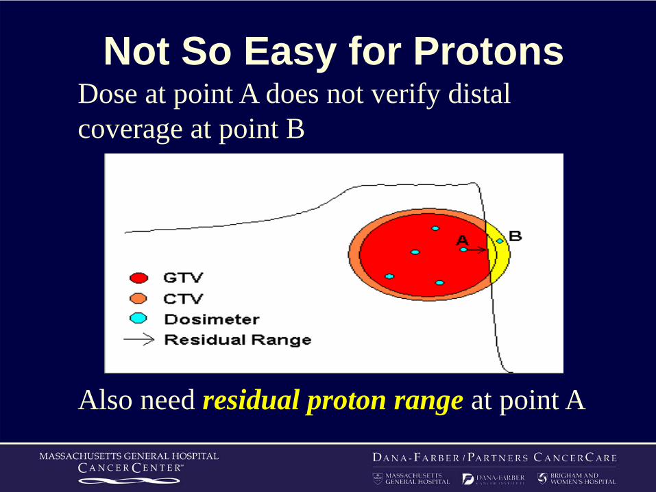
Dose at point A does not verify distal coverage at point B
Not So Easy for Protons
Also need residual proton range at point A
Stanley RosenthalMarc BussiereMartijn EngelsmanKatia ParodiAlejandro MazalRonald Zhu……
Acknowledgment

Thank You