Patient packet - Headaches and Migraine - NeuroSensory Center
14
Copyright - 2007 NeuroSensory Centers of America NeuroSensory Center of Eastern Pennsylvania 250 Pierce Street, Suite 317 Kingston, PA 18704 (570) 763-0054 Headaches and Migraine
Transcript of Patient packet - Headaches and Migraine - NeuroSensory Center

Copyright - 2007
NeuroSensory Centers of America
NeuroSensory Center of Eastern Pennsylvania 250 Pierce Street, Suite 317
Kingston, PA 18704 (570) 763-0054
Headaches
and
Migraine
Copyright - 2007
NeuroSensory Centers of America
HHeeaaddaacchheess aanndd MMiiggrraaiinnee It has been well documented that there is now an epidemic increase in the symptomatic syndromes the medical
community recognizes as chronic “symptomatic” syndromes that include recurrent headaches and migraines. This
disturbing rise in prevalence also accompanies a generalized increase in the number of neurological disorders in the
general population. Unfortunately, as with many neurological diseases of unknown origin, the environment of poorly
coordinated care between medical professionals have left patients in the state of relative confusion and frustration.
WWhhaatt aarree NNeerrvvee HHyyppeerrsseennssiittiivviittyy SSyynnddrroommeess?? It is quite difficult for the medical community to recognize, much less, categorize nerve based syndromes due to the
inability to visualize a “symptom”. In fact, there are two major hurdles a patient must overcome with their doctors:
1. Having a physician believe the patient is telling the truth regarding their symptoms
2. Having objective or measurable indicators of their symptoms
These two facts leave most physicians in a state of major denial as to the existence of these debilitating processes and
invariability leaves the patient traveling from doctor to doctor looking for help.
Numerous studies in the past have attempted to implicate the source of these conditions in a tissue or organ system only
to find no apparent abnormality at the source of the pain or symptom. Simply put, these syndromes represent
“symptoms” that are out of proportion when compared to a normal state of nervous system. In fact, these syndromes
simply represent an abnormal state of inflammation in the nervous system resulting in an inability to send electrical
information in an accurate manner.
After treating thousands of patients with these conditions, there is no doubt that these conditions exist, but new insights
in medical diagnostics have given us the ability to objectively measure some of the indicators of these complicated
disease processes.
Where is the problem in these disorders? In order to begin to define the problem, it is important to realize where the problem actually exists. One commonality of these
patients is that they almost always experience significant fluctuations in their symptoms on a day to day basis. In general, this
means that the condition has “good” days and “bad” days. Only after a reasonable period of time do the symptoms become
chronic or constant. This simple realization has enormous implications.
If the brains perception of pain was abnormal, meaning it represented an abnormal psychiatric state associated with conscious or
unconscious secondary gain, then the symptoms should have a relatively little variability. The hallmark of a primary brain
abnormality is that it stays consistent because the brain is damaged or altered and cannot change on a daily basis. However, in
the “Nerve Hypersensitivity” Disorders, these patients have notable fluctuations, sometimes in the same day. This fluctuation
can only be accomplished by changes in the information feeding the brain and causing an abnormal response. This simple fact
may indeed show that we have been focusing our study in the wrong place. These patients do not appear to have a problem in
the brain; they have a disorder in feeding the brain the proper electrical information.
Copyright - 2007
NeuroSensory Centers of America
NNoorrmmaall SSeennssoorryy DDeevveellooppmmeenntt In order to understand the abnormal situation that relates to these patient groups, you must first understand normal function of the
sensory systems. The sensory nervous system can be divided into subgroups according to:
1. What sense they transmit
2. How fast they carry electrical information
For example, touch nerves are relatively slow in speed compared to other nerves and visual nerves are ultra-fast, meaning they
carry information at a very high rate. The anatomical differences between these nerves relates to the amount of myelin necessary to
insulate them and the size of the nerve fibers.
HHooww ddoo wwee sseeee tthhee pprroobblleemm wwiitthh tthhee sseennssoorryy ssyysstteemmss??
Beginning in 1998, nerve specialists began using a clinical method of combining known FDA approved diagnostic tests to
visualize the entire array of sensory systems feeding the brain information. Using this diagnostic method, these physicians were
able to “visualize” sensory disorders and provide greatly improved therapeutic outcomes in patients with a myriad of nervous
system sensory abnormalities. This system, the Sensory-View, is now in use in the NeuroSensory Centers of America.
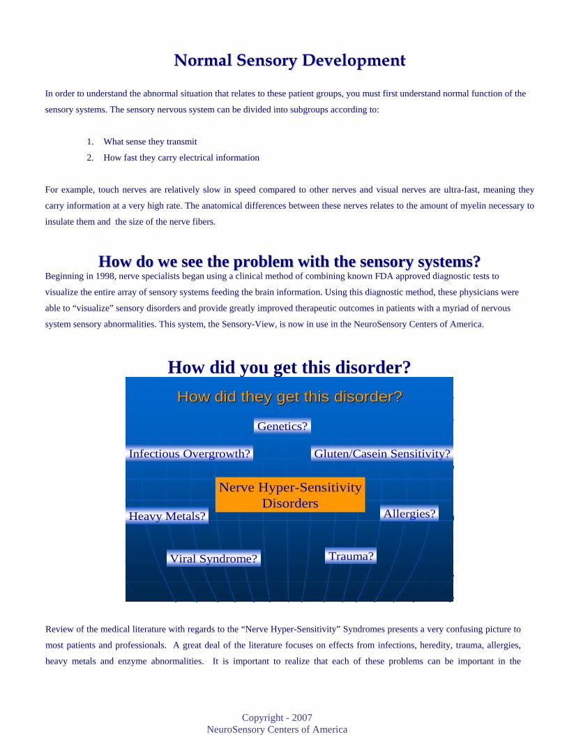
How did you get this disorder?
How did they get this disorder?How did they get this disorder?
Nerve Hyper-SensitivityDisorders
Heavy Metals?
Viral Syndrome?
Allergies?
Infectious Overgrowth?
Genetics?
Gluten/Casein Sensitivity?
Trauma?
Review of the medical literature with regards to the “Nerve Hyper-Sensitivity” Syndromes presents a very confusing picture to
most patients and professionals. A great deal of the literature focuses on effects from infections, heredity, trauma, allergies,
heavy metals and enzyme abnormalities. It is important to realize that each of these problems can be important in the

Copyright - 2007
NeuroSensory Centers of America
individual patient and must be considered in each patient’s unique situation. It is equally important to realize that these
disorders are nerve based problems that may have a multi-factorial origin and each patient must have a customized treatment
plan unique to their needs.
Our clinical methods are based on a model that assumes latent viral activation initiated inflammatory flux in the sensory nerves.
The latent viral activity was initiated by hormonal depletion due to decrease production, increased utilization or hormonal
interruption. Multiple states, including high heavy metal concentrations, infectious overgrowth and/or poor sleep patterns,
create an environment of IGF-1 depletion that appears to trigger continued activations of neurotropic (nerve infecting) viral
agents causing fluctuating inflammation in the sensory nerves. The variability in the sensory system function does not allow
the brain to integrate the different sensory systems and leads to a dynamic and confusing clinical state.
One could assume that this simple concept of nervous system disease should have been seen previously by experienced
physicians, however, there is only one problem with proving this concept; the offending agents are difficult or impossible to
quantify by blood or urine testing. Most neurotropic viral groups (Herpes for instance) have no good method of quantification.
Additionally, heavy metals are very difficult to quantify (measure) due to their ability to collect in “fatty” tissues. Much debate
has centered on the best way to determine if heavy metal exposure or metabolism is an inherent problem. These facts leave
many physicians questioning the validity of these disease processes.
How did they get these disorders?How did they get these disorders?A complex web developsA complex web develops
Infectious overgrowthInflammatory States
Growth
IGF-1 deficiency(Fatty Acid/Amino Acid delivery)
Heavy Metal Elevations Sleep pattern abnormalities
Poor nutritional support of myelinand nerve development
Poor Sensory Development and AccuracyImmune Hyper-stimulation
Deficiency of Neurotrophic Factors
Herpetic Inflammation of MyelinYeast/Bacterial Activation
Fluctuant functionStatic dysfunction
Interruption of IGF function
Decreased IGF Production
Increased IGF requirements
Copyright - 2007
NeuroSensory Centers of America
Is there a way to reverse these disorders? After many years of clinical experience, it is quite apparent that there is the possibility of reversing adult chronic inflammatory or
demyelinating syndromes. In short, with our current state of understanding, creating a “healthier”, less inflammatory environment
in the body will produce less nerve inflammation and allow re-myelination of nerves in many individuals. Successful healing or
reversal appears to require four necessary clinical steps:
Four necessary elements
1. Evaluate and reduce any heavy metal issue to erase toxic environment
2. Reduce herpetic load to eliminate inflammation of myelin
3. Reduce fungal/bacterial/allergic immune hyper-stimulation
4. Maximize hormonal and nutritional status to improve the repair of myelin
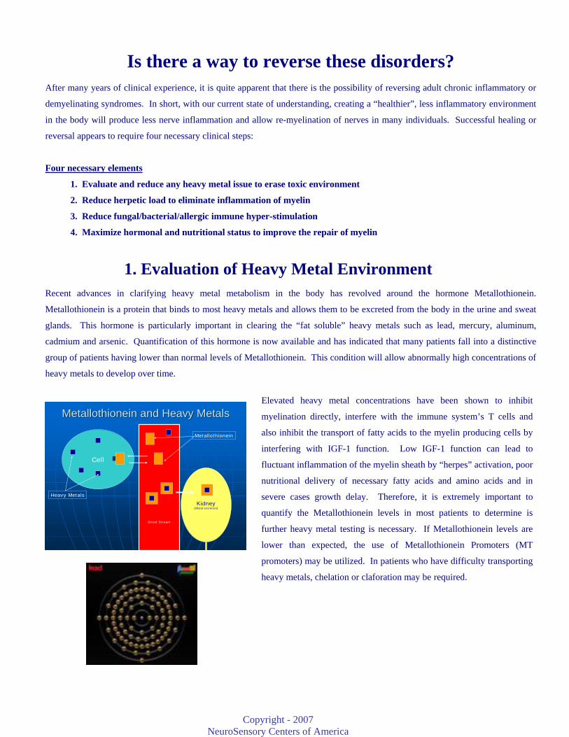
1. Evaluation of Heavy Metal Environment Recent advances in clarifying heavy metal metabolism in the body has revolved around the hormone Metallothionein.
Metallothionein is a protein that binds to most heavy metals and allows them to be excreted from the body in the urine and sweat
glands. This hormone is particularly important in clearing the “fat soluble” heavy metals such as lead, mercury, aluminum,
cadmium and arsenic. Quantification of this hormone is now available and has indicated that many patients fall into a distinctive
group of patients having lower than normal levels of Metallothionein. This condition will allow abnormally high concentrations of
heavy metals to develop over time.
MetallothioneinMetallothionein and Heavy Metalsand Heavy Metals
Cell
Kidney(Metal excretion)
Heavy Metals
Metallothionein
Blood Stream
Elevated heavy metal concentrations have been shown to inhibit
myelination directly, interfere with the immune system’s T cells and
also inhibit the transport of fatty acids to the myelin producing cells by
interfering with IGF-1 function. Low IGF-1 function can lead to
fluctuant inflammation of the myelin sheath by “herpes” activation, poor
nutritional delivery of necessary fatty acids and amino acids and in
severe cases growth delay. Therefore, it is extremely important to
quantify the Metallothionein levels in most patients to determine is
further heavy metal testing is necessary. If Metallothionein levels are
lower than expected, the use of Metallothionein Promoters (MT
promoters) may be utilized. In patients who have difficulty transporting
heavy metals, chelation or claforation may be required.

Copyright - 2007
NeuroSensory Centers of America
2 Reduce the viral load to decrease myelin inflammation
Is there a way to reverse these disorders?Is there a way to reverse these disorders?2. Reduce viral load to reduce continuous myelin inflammation2. Reduce viral load to reduce continuous myelin inflammation
Nerve Body
Myelin Layer
Myelin Layer
Anti-Herpetic
Immune Control Immune Destruction
Herpes family virus
33.. RReedduuccee ffuunnggaall//bbaacctteerriiaall// iimmmmuunnee hhyyppeerrssttiimmuullaattiioonn It is well documented that many types of opportunistic infections can be active in the
patient with Autistic Spectrum and Sensory Integration Disorders. These “opportunists”
can be represented by fungal, bacterial or viral agents. Common “opportunists” include
Candida, Mycoplasma, Clostridium, E. coli, measles, human papilloma virus and many
others. It is essential for your physician to quantify these agents if possible and reduce the
amount of immune stimulation caused by activation or overgrowth. This helps to create the
“ideal” environment necessary to recover the sensory nerve deficit.
4. Maximize the hormonal and nutritional status Nerves, in general, are very difficult to repair or develop. Nerves typically require the “healing” hormones (thyroid, cortisol,
insulin and IGF-1) to have levels that approach the middle of the normal range for that patient’s age. These hormone levels will
generally be evaluated by blood testing and adjusted in necessary.
Many physicians falsely believe that it is impossible to “kill” the herpes virus because we do not have a medication that
“kills” herpes directly. This statement is short-sighted because the immune system is perfectly capable of destroying a
herpes family virus. It is only essential to provide exposure of the virus to the immune system. Immune exposure is
accomplished by anti-viral medications that arrest the viral division while it is outside the nerve body. This allows the
immune system to have adequate time to kill the exposed virus and gradually reduce the amount of viral load in the nervous
system.

Copyright - 2007
NeuroSensory Centers of America
WWhhyy iiss sslleeeepp ssoo iimmppoorrttaanntt?? NECA’s clinical research has been able to show a correlation between sleep abnormalities and nervous system abnormalities. This
correlation seems to be related to an absence of Stage IV sleep, commonly known as “deep sleep”. During Stage IV sleep the body
produces a group of hormones called Insulin Dependent Growth Factors (IGF I-IV), substances that are related to growth hormone.
The IGF hormones are probably misnamed and should be called “Human Repair Hormones” because they are important throughout
life. These factors are required for the transport of proteins, fats and cholesterols into cells.
All patients that have difficulty entering Stage IV sleep and will typically have low normal or abnormal levels of IGF. In adults, a
deficiency of these hormones causes only one symptom; fatigue. Fatigue is almost a universal complaint of all patients with
nervous system abnormalities. Many times patients cannot explain the feeling of fatigue. Deficiency of this hormone will be seen
clinically as an increase in body fat, growth delay, poor muscle tone or weakness, and an increase in cholesterol and triglyceride
levels in the blood stream.
FFrreeqquueennttllyy AAsskkeedd QQuueessttiioonnss
IIss tthhiiss aa nneeww eexxppeerriimmeennttaall ttrreeaattmmeenntt?? The treatment protocol utilized by the NeuroSensory Centers of America is not new or experimental. Specialist physicians have
been utilizing this therapeutic protocol for over 7 years and have treated thousands adults and children with sensory abnormalities
and/or cognitive and emotional syndromes. The current protocols are the result of extensive clinical research experience in
diagnostic methods and treatment outcomes.
HHooww lloonngg uunnttiill mmyy ssyymmppttoommss iimmpprroovvee??
Many symptoms can improve as early as 2-3 months, however, resolution of secondary symptoms may take up to 3-24 months to
improve. There are 3 main phases of treatment in these patients. First, decrease the viral load will take approximately 6 months.
Please remember that the inner ear is dynamic and symptoms can fluctuate from day to day and fluctuation is particularly
common during the first 6-8 weeks. Each patient is treated individually according to his/her problem, and improvement may
vary from patient to patient.
HHooww oofftteenn wwiillll II bbee eexxppeecctteedd ttoo ffoollllooww uupp wwiitthh rree--tteessttiinngg?? The doctor would like to see re-testing done every 3 months. This re-testing provides the healthcare team with specific
information to customize your plan of care and accurately follow your progress. This method is unique and above all provides
the most successful approach to evaluating and treating the inner ear problem.
WWiillll mmyy iinnssuurraannccee ccoovveerr tthhee ttrreeaattmmeenntt?? Insurance is a contract between you and your insurance carrier. All of our testing is approved by Medicare and most insurance
carriers. Platform Posturography is not reimbursed by Medicare in the State of Texas, but is reimbursable in other states, and
may be denied by other insurance carriers. It is essential for objective verification of improvement and modification of your care
plan. No testing is performed in our office without prior research indicating its’ necessity and benefit to your care plan.
Copyright - 2007
NeuroSensory Centers of America
Recent Articles of Interest Clin Endocrinol (Oxf). 2003 Jul;59(1):56-61. Diagnostic reliability of a single IGF-I measurement in 237 adults with total anterior hypopituitarism and severe GH deficiency. Aimaretti G, Corneli G, Baldelli R, Di Somma C, Gasco V, Durante C, Ausiello L, Rovere S, Grottoli S, Tamburrano G, Ghigo E. Division of Endocrinology and Metabolism, Department of Internal Medicine, University of Turin, Italy. OBJECTIVE: Within an appropriate clinical context, GH deficiency (GHD) in adults must be demonstrated biochemically by a single provocative test. Insulin-induced hypoglycaemia (ITT) and GH-releasing hormone (GHRH) + arginine (ARG) are indicated as the tests of choice, provided that appropriate cut-off limits are defined. Although IGF-I is the best marker of GH secretory status, its measurement is not considered a reliable diagnostic tool. In fact, considerable overlap between GHD and normal subjects is present, at least when patients with suspected GHD are considered independently of the existence of other anterior pituitary defects. Considering the time and cost associated with provocative testing procedures, we aimed to re-evaluate the diagnostic power of IGF-I measurement. DESIGN: To this goal, in a large population [n = 237, 139 men, 98 women, age range 20-80 years, body mass index (BMI) range 26.4 +/- 4.3 kg/m2] of well-nourished adults with total anterior pituitary deficit including severe GHD (as shown by a GH peak below the 1st centile limit of normal response to GHRH + ARG tests and/or ITT) we evaluated the diagnostic value of a single total IGF-I measurement. IGF-I levels in hypopituitary patients were evaluated based on age-related normative values in a large population of normal subjects (423 ns, 144 men and 279 women, age range 20-80 years, BMI range 18.2-24.9 kg/m2). RESULTS: Mean IGF-I levels in GHD were lower than those in normal subjects in each decade, but not the oldest one (74.4 +/- 48.9 vs. 243.9 +/- 86.7 micro g/l for 20-30 years; 81.8 +/- 46.5 vs. 217.2 +/- 56.9 micro g/l for 31-40 years; 85.8 +/- 42.1 vs. 168.5 +/- 69.9 micro g/l for 41-50 years; 82.3 +/- 39.3 vs. 164.3 +/- 60.3 micro g/l for 51-60 years; 67.5 +/- 31.8 vs. 123.9 +/- 50.0 micro g/l for 61-70 years; P < 0.0001; 54.3 +/- 33.6 vs. 91.6 +/- 53.5 micro g/l for 71-80 years, P = ns). Individual IGF-I levels in GHD were below the age-related 3rd and 25th centile limits in 70.6% and 97.63% of patients below 40 years and in 34.9% and 77.8% of the remaining patients up to the 8th decade, respectively. CONCLUSIONS: Total IGF-I levels are often normal even in patients with total anterior hypopituitarism but this does not rule out severe GHD that therefore ought to be verified by provocative testing of GH secretion. However, despite the low diagnostic sensitivity of this parameter, very low levels of total IGF-I can be considered definitive evidence of severe GHD in a remarkable percentage of total anterior hypopituitary patients who could therefore skip provocative testing of GH secretion. Horm Metab Res. 2003 Feb;35(2):114-9. Impact of growth hormone on central nervous activity, vigilance, and tiredness after short-term therapy in growth hormone-deficient adults. Pavel ME, Lohmann T, Hahn EG, Hoffmann M. Division of Endocrinology, Department of Internal Medicine I, University of Erlangen-Nurnberg, Germany. [email protected] Impairment of well-being and cognitive function has been reported in growth hormone-deficient adults, as well as an improvement of these parameters after GH substitution, albeit inconsistently. The effect of growth hormone on central nervous activity, vigilance and sleepiness was studied prospectively in 16 growth hormone-deficient adults (7 females, 9 males, mean age: 36.8 yrs) with multiple pituitary hormone deficiencies before and 3 months after the start of growth hormone substitution using two objective methods of measurement, pupillographic sleepiness test and a choice reaction time test. Significant differences were found for neither pupillary unrest index nor for reaction time, false or missing reactions in 12 evaluable patients (7 females, 5 males, mean age 37.8 years). Because of the known interrelationships between growth hormone, sleep and mood, the visual analogue scale for tiredness and standardized retrospective questionnaires regarding sleep and mood (Pittsburgh sleep quality index, Epworth sleepiness scale, Depression scale) were used as additional methods. After GH substitution, there was no difference in sleep efficiency and daytime sleepiness, but some of the subjective sleep parameters (sleep quality and sleep latency) improved significantly. There was a tendency for mood improvement, too. Although results must be interpreted cautiously due to the small sample size, we conclude that the improved sleep and mood parameters might be caused by other indices of general well-being in our study. J Endocrinol Invest. 2003 Jun;26(6):588-94.
Copyright - 2007
NeuroSensory Centers of America
Circulating free insulin-like growth-factor-I (IGF-I) levels should also be measured to estimate the IGF-I bioactivity. Janssen JA, van der Lely AJ, Lamberts SW. Department of Internal Medicine, Erasmus MC, Rotterdam, The Netherlands. [email protected] Free IGF-I by analogy with sex and adrenal steroids and thyroid hormones, may be the major biologically active hormonal form of IGF-I. Because of methodological difficulties in measuring the free IGF-I the measurement of total IGF-I in blood is often used to assess the activity of the endocrine GH-IGF-I axis in clinical studies. However, there is currently no reliable standard reference method for circulating total IGF-I against which individual samples can be calibrated. In addition, in many of the common methods used to measure circulating total IGF-I levels, remaining insulin-like growth factor, binding proteins (IGFBPs) or binding protein fragments after sample extraction, may still interfere and produce falsely increased or decreased circulating total IGF-I levels. This latter phenomenon occurs especially under pathologic conditions. In addition, it has also been suggested that altered post-sampling integrity of IGF-I in vitro might contribute to the reported inconsistencies in circulating total IGF-I levels in literature. Although at the moment there is also no "golden standard" for the measurement of circulating free IGF-I levels, we discuss some studies in this paper that in our opinion, have demonstrated conclusively that circulating free IGF-I levels in several conditions reflect the IGF-I bioactivity better than circulating total IGF-I levels. Therefore, when evaluating the IGF-I bioactivity in health and disease, we recommend measuring also circulating free IGF-I. J Neurosci Res. 2003 Nov 15;74(4):512-23. Systemic insulin-like growth factor-I administration prevents cognitive impairment in diabetic rats, and brain IGF regulates learning/memory in normal adult rats. Lupien SB, Bluhm EJ, Ishii DN. Department of Biomedical Sciences, Colorado State University, Fort Collins, CO 80523, USA. Diabetic patients have impaired learning/memory, brain atrophy, and two-fold increased risk of dementia. The cause of cognitive disturbances that progress to dementia is unknown. Because neurotrophic insulin-like growth factor (IGF) levels are reduced in diabetic patients and rodents, and IGF can cross the blood-central nervous system barrier (B-CNS-B), the hypothesis was tested that IGF administered systemically can prevent cognitive disturbances, independently of hyperglycemia and a generalized catabolic state. Latency to escape to a hidden platform in the Morris Water Maze is used widely to test spatial memory, a hippocampus-dependent task. Adult rats were rendered diabetic with streptozotocin and implanted 4 weeks later with subcutaneous pumps that released either vehicle (D + Veh) or 20 microg/day IGF-I (D + IGF). Latency to escape to the hidden platform was prolonged in (D + Veh) versus non-diabetic rats (P < 0.003) 10.5 weeks after the onset of diabetes. Such prolongation was prevented in (D + IGF) versus (D + Veh) rats (P < 0.03). The data show that IGF-I can act across the B-CNS-B to prevent loss of cognition-related performance in the water maze independently of ongoing hyperglycemia and reduction in brain (P < 0.001) and whole body weight (P < 0.001) in diabetic rats. The hypothesis that brain IGF contributes to learning/memory was tested. An anti-IGF antibody, or preimmune serum, was infused into the lateral ventricles in non-diabetic rats. Learning in a passive avoidance task was impaired significantly in the IGF antibody versus preimmune serum-treated groups on test Days 1, 2, and 3 (P = 0.04, 0.02 and 0.004, respectively). The data together are consistent with a model in which brain IGF is essential for learning/memory, and a loss of IGF activity due to diabetes may contribute to cognitive disturbances. Mol Psychiatry. 2004 Jan 27 Activation of methionine synthase by insulin-like growth factor-1 and dopamine: a target for neurodevelopmental toxins and thimerosal. Waly M, Olteanu H, Banerjee R, Choi SW, Mason JB, Parker BS, Sukumar S, Shim S, Sharma A, Benzecry JM, Power-Charnitsky VA, Deth RC. 1Department of Pharmaceutical Sciences, Northeastern University, Boston, MA 02115, USA. Methylation events play a critical role in the ability of growth factors to promote normal development. Neurodevelopmental toxins, such as ethanol and heavy metals, interrupt growth factor signaling, raising the possibility that they might exert adverse effects on methylation. We found that insulin-like growth factor-1 (IGF-1)- and dopamine-stimulated methionine synthase (MS) activity and folate-dependent methylation of phospholipids in SH-SY5Y human neuroblastoma cells, via a PI3-kinase- and
Copyright - 2007
NeuroSensory Centers of America
MAP-kinase-dependent mechanism. The stimulation of this pathway increased DNA methylation, while its inhibition increased methylation-sensitive gene expression. Ethanol potently interfered with IGF-1 activation of MS and blocked its effect on DNA methylation, whereas it did not inhibit the effects of dopamine. Metal ions potently affected IGF-1 and dopamine-stimulated MS activity, as well as folate-dependent phospholipid methylation: Cu(2+) promoted enzyme activity and methylation, while Cu(+), Pb(2+), Hg(2+) and Al(3+) were inhibitory. The ethylmercury-containing preservative thimerosal inhibited both IGF-1- and dopamine-stimulated methylation with an IC(50) of 1 nM and eliminated MS activity. Our findings outline a novel growth factor signaling pathway that regulates MS activity and thereby modulates methylation reactions, including DNA methylation. The potent inhibition of this pathway by ethanol, lead, mercury, aluminum and thimerosal suggests that it may be an important target of neurodevelopmental toxins.Molecular Psychiatry advance online publication, 27 January 2004; doi:10.1038/sj.mp.4001476 Exp Cell Res. 2003 Dec 10;291(2):289-98. Intracellular zinc fluctuations modulate protein tyrosine phosphatase activity in insulin/insulin-like growth factor-1 signaling. Haase H, Maret W. Center for Biochemical and Biophysical Sciences and Medicine, Harvard Medical School, One Kendall Square, Bldg. 600, 3rd Floor, Cambridge, MA 02139, USA. Zinc is an effector of insulin/IGF-1 signaling and has insulinomimetic effects, the molecular basis of which is not understood. The present study establishes the capacity of zinc to inhibit protein tyrosine phosphatases (PTPs) as a cause for these effects and, moreover, demonstrates modulation of the insulin response by changes in intracellular zinc. The inhibition of PTPs by zinc occurs at significantly lower concentrations than previously reported. In vitro, zinc inhibits PTPs 1B and SHP-1 with IC(50) values of 17 and 93 nM, respectively. A fluorescent probe with a similar binding constant [FluoZin-3, K(D)(Zn) = 15 nM] detects corresponding concentrations of zinc within cells. Increase of cellular zinc after incubation with both zinc and the ionophore pyrithione augments protein tyrosine phosphorylation, and in particular the phosphorylation of three activating tyrosine residues of the insulin/IGF-1 receptor. Vice versa, specific chelation of cellular zinc with the membrane-permeable N,N,N',N'-tetrakis(2-pyridylmethyl)ethylenediamine suppresses insulin- and IGF-1-stimulated phosphorylation. In the context of the emerging concept that intracellular zinc is tightly regulated and fluctuates dynamically, these results suggest that a pool of cellular zinc modulates phosphorylation signaling. Neurotoxicology. 2005 Jan;26(1):1-8. Thimerosal Neurotoxicity is Associated with Glutathione Depletion: Protection with Glutathione Precursors. James SJ, Slikker W 3rd, Melnyk S, New E, Pogribna M, Jernigan S. Department of Pediatrics, University of Arkansas for Medical Sciences and Arkansas Children's Hospital Research Institute, Little Rock, AR 72202, USA. Thimerosol is an antiseptic containing 49.5% ethyl mercury that has been used for years as a preservative in many infant vaccines and in flu vaccines. Environmental methyl mercury has been shown to be highly neurotoxic, especially to the developing brain. Because mercury has a high affinity for thiol (sulfhydryl (SH)) groups, the thiol-containing antioxidant, glutathione (GSH), provides the major intracellular defense against mercury-induced neurotoxicity. Cultured neuroblastoma cells were found to have lower levels of GSH and increased sensitivity to thimerosol toxicity compared to glioblastoma cells that have higher basal levels of intracellular GSH. Thimerosal-induced cytotoxicity was associated with depletion of intracellular GSH in both cell lines. Pretreatment with 100muM glutathione ethyl ester or N-acetylcysteine (NAC), but not methionine, resulted in a significant increase in intracellular GSH in both cell types. Further, pretreatment of the cells with glutathione ethyl ester or NAC prevented cytotoxicity with exposure to 15muM Thimerosal. Although Thimerosal has been recently removed from most children's vaccines, it is still present in flu vaccines given to pregnant women, the elderly, and to children in developing countries. The potential protective effect of GSH or NAC against mercury toxicity warrants further research as possible adjunct therapy to individuals still receiving Thimerosal-containing vaccinations.
Mech Ageing Dev. 2003 Apr;124(4):371-8Interrelationships among brain, endocrine and immune response in ageing and successful ageing: role of metallothionein III isoform. Giacconi R, Cipriano C, Muzzioli M, Gasparini N, Orlando F, Mocchegiani E.
Copyright - 2007
NeuroSensory Centers of America
Metallothionein-III (MT-III) a brain-specific member of metallothionein family contributes to zinc neuronal homeostasis, and zinc is an important regulator of many brain functions, including the activity of hormone realising factors by hippocampus. Among them, somatostatin is pivotal because affecting thyroid hormones turnover and consequently thymic and peripheral immune efficiency (Natural Killer, NK) cell activity. Somatostatin is in turn affected by somatomedin-C, which is also zinc-dependent. Therefore, somatomedin-C may be a marker of somatostatin status in the hippocampus. MTs sequester and release zinc in transient stress, as it may occur in young age, to protect cells by reactive oxygen species. In order to accomplish this task, MTs are induced by IL-6 for a prompt immune and anti-inflammatory response. During ageing, MTs are high with a role of sequester of zinc, but with very limited role in zinc release because stress-like condition and inflammation is persistent. Therefore, high MTs may become to protective in young age to harmful during ageing leading to low zinc ion bioavailability for many body homeostatic mechanisms, including brain function. As a consequence, an altered physiological cascade from the brain (upstream) to endocrine and immune system (downstream) may occur. The aim of this work is to study the role of MT-III in the interrelationships among brain-endocrine-immune response in ageing and successful ageing. The main results are: (1) MT-III and IL-6 gene expressions increase in the hippocampus from old mice, in comparison with young and very old mice. (2) Somatomedin-C plasma levels decrease in old mice in comparison with young and very old mice. (3) Low zinc ion bioavailability (tested by the ratio total thymulin/active thymulin) is coupled with altered thyroid hormone turnover and depressed IL-2 in old mice in comparison with young and very old mice. (4) 'In vitro' experiments display more increments on NK cells activity by adding zinc-bound active thymulin than T3 alone. In conclusion, low MT-III in the hippocampus from young and very old mice leads to good zinc ion bioavailability that it is upstream coupled with normal hippocampal function affecting downstream normal thyroid hormones turnover and satisfactory NK cell activity, via complete saturation of zinc-bound active thymulin molecules. Therefore, a correct MTs homeostasis is pivotal for brain-endocrine-immune response in order to reach successful ageing. Eur Arch Otorhinolaryngol. 1996;253(4-5):264-7. Detection of viral antigen in the endolymphatic sac. Kumagami H. Department of Otolaryngology, School of Medicine, Nagasaki University, Japan. A study was devised to determine whether or not any immune defense mechanism is present when a virus invades the human endolymphatic sac (ES). The ES was removed from 14 fresh autopsy cases having no known pre-mortem diseases in the middle and inner ears. Specimens were then examined for viral antigens including herpes simplex (HSV) type 1 and 2, mumps and cytomegalovirus using immunohistochemical methods. DNA examination by in situ hybridization was also performed for HSV. HSV antigen and DNA were observed in 9 of the 14 cases studied. These findings suggest that the virus invades the ES but is impeded by an immune defense mechanism under normal conditions. Since disease may alter host defenses, further studies are warranted to study the relationship between HSV and patients with Meniere's disease. Acta Otolaryngol Suppl. 1993;503:85-9.
Latent herpes simplex virus type 1 in human vestibular ganglia. Furuta Y, Takasu T, Fukuda S, Inuyama Y, Sato KC, Nagashima K. Department of Pathology, Hokkaido University School of Medicine, Sapporo, Japan. Viral infection has been considered to be a possible pathogenesis of vestibular neuronitis, and reactivation of the herpes simplex virus (HSV) is one of the most likely causes. However, it remains unknown whether the human vestibular ganglia contain latent HSV. We examined 26 vestibular ganglia from autopsied adults in search of HSV type 1 (HSV-1). To detect HSV-1, we used polymerase chain reaction (PCR), in situ hybridization and immunohistochemical staining. HSV DNA was detected in 6 of 10 vestibular ganglia using the PCR method. However, the latency-associated transcript (LAT) of HSV-1 was negative in all of the 16 vestibular ganglia examined. No HSV antigen was detected in any of the ganglia. These results indicate that HSV-1 is latently infected in the human vestibular ganglia, and that LAT is transcribed weakly or not at all. Pediatr Nurs. 2001 Mar-Apr;27(2):125-6, 129-30. Lead poisoning: a summary of treatment and prevention. Cohen SM. Nursing Faculty, College of Health Sciences, Roanoke, VA, USA.
Copyright - 2007
NeuroSensory Centers of America
Lead poisoning affects an estimated 890,000 young children in the United States annually (American Academy of Pediatrics [AAP], 1998). Extremely high levels in the child can cause mental retardation, coma, seizures, and death. Chronic low level exposure is more commonly seen with multiple effects, including learning disabilities, impaired growth, and hearing loss. Lead poisoning prevention efforts have significantly reduced the number of children affected by this serious health hazard. Health care providers need to continue their vigilant efforts to educate families living in older homes about the risks, screening, and treatment. Neurotoxicol Teratol. 2003 Jan-Feb;25(1):69-76. Neurotoxic effects of mercury on auditory cortex networks growing on microelectrode arrays: a preliminary analysis. Gopal KV. Department of Speech and Hearing Sciences and Center for Network Neuroscience, University of North Texas, PO Box 305010, Denton, TX 76203, USA. [email protected] Mercury is known to cause sensorineural hearing loss and impaired speech perception. However, there is still a lack of a quantitative description of mercury toxicity on central auditory structures. This is a preliminary study using the novel technique of microelectrode array (MEA) recordings to evaluate acute and chronic neurotoxic effects of mercury on auditory cortex networks (ACNs) in vitro. Morphological and electrophysiological effects of mercuric chloride (HgCl(2)) were studied.Neurons dissociated from auditory cortices of 14-day-old mouse embryos were grown on photoetched MEAs containing 64 transparent indium-tin oxide (ITO) electrodes. For acute electrophysiological experiments, the spontaneous spiking and bursting activity from ACNs were compared before and after application of HgCl(2). For chronic electrophysiological experiments, auditory cortex cultures were treated with various concentrations of HgCl(2) from the day of seeding, and were tested 4 weeks later for the presence of spontaneous activity. Morphological analysis was conducted on 8-day-old ACNs treated with HgCl(2) for 3 days. Results of acute experiments indicated that <75 mM of HgCl(2) had an excitatory effect of variable magnitude on the spontaneous activity of ACNs; however, concentrations above 100 microM completely and irreversibly inhibited spike and burst activity. Chronic exposure of ACNs to 10 microM HgCl(2) completely blocked the spontaneous activity. Morphological analysis indicated that 10 microM HgCl(2) caused neuronal cell death in 3 days. It is concluded that HgCl(2) has a more toxic effect on auditory networks when exposed chronically, and the levels of mercury showing toxic effects on ACNs are within the dose range shown to cause neurologic symptoms in humans. Biol Trace Elem Res. 2002 Aug;88(2):153-63.
• Effect of zinc ion on cadmium-induced auditory changes. Agirdir BV, Bilgen I, Dinc O, Ozcaglar HU, Fisenk F, Turhan M, Oner G. Department of Otorhinolaryngology, Faculty of Medicine, Akdeniz University, Antalya, Turkey. Cadmium, which has adverse effects on many physiological systems, is an important environmental pollutant. Our previous experimental study showed that cadmium also has a dose-dependent deleterious effect on the auditory system in rats. Because zinc reverses cadmium cytotoxicity in many systems, we investigated the possible preventive effect of a zinc-enriched diet given isochronally on cadmium-induced hearing loss in rats. Fifty-four male rats were divided into three equal groups. Control rats were fed normal rat food and tap water, whereas the cadmium group was subjected to 15 ppm cadmium-containing water as CdCl2. The third group received 15 ppm CdCl2 and food enriched with 200 ppm zinc as ZnSO4 for 30 d. On d 30, eight animals from each group were used for the measurement of kidney functions. In the remaining animals, hearing functions were measured by auditory brainstem response and distortion product otoacoustic emission. Blood cadmium increased from 1.87+/-1.69 to 6.08+/-2.62 microg/dL and elevated cadmium contents of ear ossicles and kidney cortex were associated with a decreased glomerular filtration rate in rats subjected to high cadmium. A zinc-enriched diet obviously reduced cadmium accumulation in the kidney and prevented the nephrotoxicity. Our data indicated that cadmium-induced ototoxicity seems to be partially zinc preventable and zinc addition to diet without altering cadmium content in ear ossicles may help to prevent cadmium-induced hearing loss.
J Occup Environ Med. 2002 Jan;44(1):30-8Neuro-ototoxicity in andean adults with chronic lead and noise exposure.
Copyright - 2007
NeuroSensory Centers of America
Counter SA, Buchanan LH. Department of Neurology, Massachusetts General Hospital, Harvard Medical School, Biological Laboratories, 16 Divinity Avenue, Cambridge, MA 02138, USA. [email protected] Brainstem auditory evoked responses and audiological thresholds were used as biomarkers for neuro-ototoxicity in adults with chronic lead (Pb) intoxication from long-term Pb exposure in ceramic-glazing work. Venous blood samples collected from 30 adults (15 men and 15 women) indicated a mean blood Pb level of 45.1 micrograms/dL (SD, 19.5; range, 11.2 to 80.0 micrograms/dL) and in excess of the World Health Organization health-based biological limits (men, 46.2 micrograms/dL; SD, 19.6; range, 18.3 to 80.0 micrograms/dL; women, 44.0 micrograms/dL; SD, 20.1; range, 11.2 to 74.2 micrograms/dL). Mean auditory thresholds at frequencies susceptible to ototoxicity (2.0, 3.0, 4.0, 6.0, and 8.0 kHz) revealed sensory-neural hearing loss in men, which may be attributable to occupational noise exposure in combination with Pb intoxication. Bilateral brainstem auditory evoked response tests on participants with elevated blood Pb levels (mean, 47.0 micrograms/dL) showed delayed wave latencies consistent with sensory-neural hearing impairment. The results suggest that environmental noise exposure must be considered an important factor in determining sensory-neural hearing status in occupationally Pb-exposed adults. Toxicology. 2001 Apr 12;162(1):11-22. Abnormal auditory brainstem responses for mice treated with mercurial compounds: involvement of excessive nitric oxide. Chuu JJ, Hsu CJ, Lin-Shiau SY. Institutes of Toxicology, College of Medicine, National Taiwan University, No. 1, Section 1, Jen-Ai Road, Taipei 10043, Taiwan. In this paper, we attempted to construct an animal (mouse) model for monitoring the oto-neurotoxicity of mercuric sulfide, comparing its toxicity with the well-known (organic) mercury compound methyl-mercury. Mice were treated with either mercuric sulfide (HgS, 0.1 and 1.0 g/kg per day) or methyl-mercury (MeHg, 0.2, 2.0 and 10 mg/kg per day) by gastric gavage for 7 consecutive days. Analysis of auditory brainstem response (ABR) indicated that significant elevation of the physiological hearing threshold as well as significant prolongation of interwave latency I-V was observed for MeHg -- (2.0 and 0.2 mg/kg per day) or HgS -- (1.0 g/kg per day, but not 0.1 g/kg per day) treated mice. Further, both MeHg- and HgS-treated animals demonstrated a significant prolongation of interwave latency I-V that increased with an increasing mean blood-Hg level. The oto-neurotoxicity of MeHg (2.0 mg/kg per day) persisted to at least 11 weeks subsequent to the cessation of its administration. The toxic effect of HgS, however, disappeared completely 5 weeks subsequent to the cessation of its administration. These results suggest a correlation between the Hg-elicited hearing dysfunction and the availability of mercury in brain tissue. Both inhibition of Na(+)/K(+)-ATPase activity and overproduction of nitric oxide in the brainstem are consistent with an analysis of the physiological hearing threshold and latencies of ABR waveform at all time points throughout the experimental process. Thus, it is proposed that high-dose HgS or MeHg intoxication is associated with a decrease in functional Na(+)/K(+)-ATPase activity in the brainstem of affected animals, this presumably arising via excessive nitric oxide production, and suggesting that brainstem damage may play a role in mercury-induced hearing loss. Environ Res. 1999 Jan;80(1):1-8. Lead exposure and hearing effects in children in Katowice, Poland. Osman K, Pawlas K, Schutz A, Gazdzik M, Sokal JA, Vahter M. Institute of Environmental Medicine, Karolinska Institutet, Stockholm, S-171 77, Sweden. The objective of the study was to investigate the relationship between lead exposure and hearing in children in the Katowice region, an industrial area in Poland. Blood lead was determined using inductively coupled plasma mass spectrometry, with appropriate quality control. The concentrations of lead in blood (B-Pb) in 155 children, aged 4-14, ranged from 19 to 281 microg/L (0.09 to 1.4 micromol/L), with a median of 72 microg/L (0.34 micromol/L). The hearing thresholds increased significantly with increasing blood lead levels at all investigated frequencies (0.5, 1, 2, 4, 6, and 8 kHz). The relationship also remained significant for B-Pb below 100 microg/L (0.48 micomol/L; n=107). The brainstem auditory evoked potential latency of wave I was significantly increased (also after adjustment for age) in the group of children with the highest blood lead levels (B-Pb above 100 microg/L, 0.48 micromol/L; n=51), compared to the group with the lowest ones (B-Pb below 46 microg/L, 0.22
Copyright - 2007
NeuroSensory Centers of America
micromol/L; n=51). The audiometric results clearly indicate that auditory function in children is impaired at a blood lead concentration even below 100 microg/L (0.5 micromol/L). Copyright 1999 Academic Press. J Occup Environ Med. 1997 Jul;39(7):658-60. Occupational lead exposure and hearing loss. Forst LS, Freels S, Persky V. Department of Emergency Medicine, College of Medicine, University of Illinois at Chicago 60612, USA. Studies of adults, children, and laboratory animals suggest an association between lead exposure and hearing loss. A causal relationship might direct mandated medical surveillance of lead-exposed workers to include audiometric testing. A cross-sectional, computerized dataset was obtained from a private occupational health screening company to examine the relationship between blood lead level and hearing loss. Audiometry and blood lead results were available for 183 workers. A statistically significant correlation was found between blood lead level and an elevated hearing threshold at 400 Hz (P = 0.03); no other frequencies showed such a correlation. This finding suggests either an interaction between nose exposure and lead, interaction of other exposure factors (such as cigarette smoking), or that factors other than biomechanical ones render the organ of Corti more susceptible at 4000 Hz. Further evaluation of these questions should be undertaken. Computerized databases created for worker surveillance may be a source for data useful for examining other causal connections in occupational settings.