Patient Educational Files Kaiser Permanente Northern ......5 Intrathecal Baclofen (ITB) Therapy...
132
1 Neurosurgical Programs Patient Educational Files Kaiser Permanente Northern California
Transcript of Patient Educational Files Kaiser Permanente Northern ......5 Intrathecal Baclofen (ITB) Therapy...

1
Neurosurgical Programs
Patient Educational Files
Kaiser Permanente Northern California

2
Table of Contents
Chapter 1: Kaiser Permanente – Northern California Intrathecal
Baclofen (ITB) Management and Surgical Centers: Pages 3-6
Chapter 2: Comprehensive NeuroVascular Program: Pages 7-
21
Chapter 3: Comprehensive Hydrocephalus Programs: Pages 22-
33
Chapter 4: Comprehensive Spine Surgery Program: Pages 34-
51
Chapter 5: Comprehensive Brain Tumor Program: Pages 52-82
Chapter 6: Comprehensive Level 4 Epilepsy Surgery Program:
Pages 83-97
Chapter 7: Deep Brain Stimulation Program – Kaiser
Permanente Redwood City Parkinson’s Disease: Pages 99-110
Chapter 6: Deep Brain Stimulation Program – Kaiser
Permanente Redwood City: Essential Tremor: Pages 111-122
Chapter 7: Comprehensive Level 4 Interventional Pain Program
Kaiser Permanente Redwood City: Pages 124-134

3
Kaiser Permanente – Northern California
Intrathecal Baclofen (ITB) Management and
Surgical Centers
Surgical Implantation Sites
Redwood City (650) 299 - 2290:
Ramon Quesada, MD: Chief of Physical Medicine and Rehabilitation
Elizabeth Heilman, MD: Physical Medicine and Rehabilitation
Taissa Cherry, MD: Anesthesiologist, Interventional Pain
Mark Sedrak, MD: Director of Functional Neurosurgery, Northern California Kaiser Permanente
Patrick Pezeshkian, MD: Functional Neurosurgeon
Diana Bruce, PA: Physician Assistant in Functional Neurosurgery
Ivan Bernstein, PA: Physician Assistant in Functional Neurosurgery, Surgery Scheduling
Supervisor
Julie Perez: Medical Assistant to Functional Neurosurgery
Sacramento (916) 973 - 5490:
Conrad Papas, MD, Phd: Functional Neurosurgeon

4
Medical Management and Refill Sites
South Sacramento (916) 688-2005 Susan Scholey, MD Redwood City (650) 299-4741 Ramon Quesada, MD Elizabeth Heilman, MD Santa Rosa (707) 571-3086 Andrea Rubinstein, MD Tabitha Washington, MD Stockton (209) 476-2080 Michael Rehbein, MD Sacramento (916) 973-7481 Marian TeSelle, MD South San Francisco (650) 742-7226 Dennis Nakamura, MD San Jose (408) 972-3033 Wayne Smith, MD Santa Clara (408) 554-9810 Benjamen Mandac, MD Union City (510) 675-3070 Eunice Lau, MD San Rafael (415) 444-2000 Vincenzo Vitto, MD Fresno/Clovis (559) 324-5035 Muhammad Akbar, MD Oakland (510) 752-2679 Joshua Rittenberg, MD Martinez (925) 313-4740 Eric Alexander, MD Vallejo (707) 651-1044 Lynn Kostecki-Csanyi, MD San Francisco (415) 833-4342 Xiao-Ping Cheng, MD

5
Intrathecal Baclofen (ITB) Therapy
Intrathecal baclofen therapy (ITB) is a system that utilizes baclofen for infusion directly around the
nerves of spine for the treatment of spasticity. The advantage of ITB is that it can afford a continuous
infusion of medicine and one can achieve much higher doses than what can be delivered with pills, often
times with lower side-effects. The disadvantage of the therapy is that it required physical filling of the
pump with a needle procedure and carries more long-term risk, especially of withdrawal problems
which can be lethal. Also, the amount of spasticity relief varies for each person.
Trial Period
While ITP can be helpful in a number of people, it does not work in everybody. To help decide in
whom this may or may not work, almost all patients have a trial to test the effectiveness. There
are two forms of trials: bolus or catheter. A bolus trial involves placement of the medication via
a needle stick (lumbar puncture) in the back and then assessing the spasticity over the
subsequent hours in the hospital then at home in the following days. A catheter trial requires
actual placement of a catheter in the spine and then infusing the medication over the following
week in the hospital. Typically for this week long trial period, we are looking for a substantial
amount of ongoing pain relief, typically more than 50%.
Permanent Implant
For those who have a successful trial, a permanent full implantation may be the next step. This
surgery is typically a much bigger procedure than the trial, involving larger incisions. Patients
may also be in the hospital at least overnight after a permanent implant. You may have the full
implant surgery as early as three weeks after your catheter trial, but sometimes a bit longer.
The surgery will go much the same as the trial with an incision in the back and in the abdomen
on the left or right side. You will have discussed this with your surgeon in advance. The battery
in the device lasts about 7 years and will need future replacement around that time.
Your infusion will initially be set at a very low dose to avoid over-infusion problems. Over the
next several months and years, the device can be programmed to optimally achieve therapy.
You will receive wound care instructions before you go home.
Programming/Adjustment/Refills
Shortly after your surgery you may return for programming and adjustments. You will have
post-operative care and can use those opportunities to have programming done by making
arrangements with the care management team. You can request additional sessions when you
think you’re not exactly where you need to be for spasticity control. It is notable that the
infusion rate, and therefore dose, can be adjusted non-invasively with the device. Because this
is an infusion system, refills are necessary and typically occur between 6 weeks-3month
intervals.

6
Appendix
Figure 1: Intrathecal Pump, Medtronic Synchromed II, there are 20cc and 40cc options
Figure 2: Lumbar Puncture. A procedure where a needle is inserted in the spine to gain access to the
cerebrospinal fluid (CSF). For intrathecal baclofen, the medication is injected in this CSF space.

7
Comprehensive NeuroVascular Program
Kaiser Permanente Redwood City
Redwood City Neurosurgeons (650) 299 - 2290:
William Sheridan, MD: Chief of Neurosurgery
Allen Efron, MD: Assistant Chief of Neurosurgery, Skull Base Specialist
John Duncan, MD, Phd: Chief of Pediatric Neurosurgery Kaiser Santa Clara, Director of Epilepsy
Surgery program
Nicole Moayeri, MD: Assistant Physician in Chief (APIC) of Surgical Services, Vascular
specialist
Cornelia Von Koch, MD, Phd: Neurosurgeon, Spine specialist
Aleksandyr Lavery, MD: Neurosurgeon, brain tumor/vascular and spine specialist
Lyndell Wang, MD: Neurosurgeon, brain tumor specialist
Lewis Hou, MD: Neurosurgeon, skull base and vascular specialist
Victor Tse, MD, Phd: Neurosurgeon, director of radiosurgery program, brain tumor and
peripheral nerve specialist
Prasad Reddy, MD: Neurosurgeon, Vascular and Endovascular specialist
Mark Sedrak, MD: Neurosurgeon, Director of Functional Neurosurgery, brain tumor specialist
Patrick Pezeshkian, MD: Functional Neurosurgeon and peripheral nerve specialist
Kevin Chao, MD: Neurosurgeon, Pediatric and Adult specialist
Redwood City EndoVascular Team (650) 299 - 2290:
Sean Cullen, MD: Director of NeuroInterventional Radiology
Daniel Hsu, MD: NeuroInterventional Radiologist
Prasad Reddy, MD: Neurosurgeon and Endovascular Specialist
Redwood City NeuroCritical Care Team (650) 299 - 2290:
Vivek Rao, MD: Chief of Neurology, Neurocritical care and stroke physician
Alexander Flint, MD: Neurocritical care and stroke physician
Sheila Chan, MD: Neurocritical care and stroke physician

8
Redwood City Physical Medicine and Rehabilitation Care Team (650) 299 - 2290:
William Firtch, MD: Physician in Chief (PIC) of Kaiser Redwood City, Physical Medicine and
Rehabilitation physician
Ramon Quesada, MD: Chief of Physical Medicine and Rehabilitation
Elizabeth Heilman, MD: Physician Medicine and Rehabilitation physician
Raymond Lai, DO: Physical Medicine and Rehabilitation physician
Sacramento Neurosurgeons (916) 973 – 5490:
Amit Banerjee, MD: Chief of Neurosurgery, Skull Base and Complex Spine Surgery
Mark Hawk, MD: Assistant Physician in Chief, Complex Spine surgery and Vascular
Kaveh Barami, MD: Neurosurgeon
Kamran Sahrakar, MD: Neurosurgeon, Neuro-Oncology and Radiosurgery
James Silverthorn, DO: Neurosurgeon
Abraham Bosckovitz, MD: Neurosurgeon
Indro Chakrabarti, MD: Neurosurgeon, Neuro-Oncology and Vascular
Huy Duong, MD: Neurosurgeon
Adam Griffith, MD: Neurosurgeon
Kern Guppy, MD: Neurosurgeon, Complex Spine
Brian Jian, MD: Neurosurgeon
David Moller, MD: Neurosurgeon
Conrad Pappas, MD, Phd: Functional Neurosurgeon
Alan Williams, MD: Neurosurgeon, Vascular
Sean McNatt, MD: Neurosurgeon Adult and Pediatric
Sacramento EndoVascular Team (916) 973 – 5490:
Jonathan Hartman, MD: Neurointerventional radiologist
Sacramento Neuro-Critital Care Team (916) 973 – 5490:
Paul Akins, MD: NeuroCritical Care Specialist
9
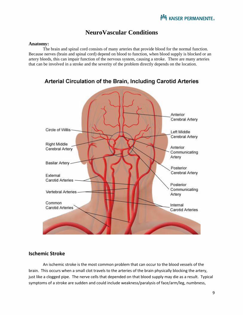
NeuroVascular Conditions
Anatomy: The brain and spinal cord consists of many arteries that provide blood for the normal function.
Because nerves (brain and spinal cord) depend on blood to function, when blood supply is blocked or an
artery bleeds, this can impair function of the nervous system, causing a stroke. There are many arteries
that can be involved in a stroke and the severity of the problem directly depends on the location.
Ischemic Stroke
An ischemic stroke is the most common problem that can occur to the blood vessels of the
brain. This occurs when a small clot travels to the arteries of the brain physically blocking the artery,
just like a clogged pipe. The nerve cells that depended on that blood supply may die as a result. Typical
symptoms of a stroke are sudden and could include weakness/paralysis of face/arm/leg, numbness,

10
blindness and should be treated as an emergency. Occasionally, if a patient presents to a hospital with a
major stroke within hours of the onset, they can be treated with a clot buster (ivTPA) and occasionally
could be a candidate for direct clot retrieval. These treatments are very time sensitive, making the
detection extremely important to occur in a timely fashion. There are many causes of ischemic strokes,
but most common origins of these clots include disease to the carotid arteries (atherosclerosis) and
atrial fibrillation. In younger patients, a possible cause is something called a dissection.
Hemorrhagic Stroke
Another type of stroke that can occur is when a blood vessel actually breaks or pops, causing
blood to leak out around or into the brain itself. Because arteries are under pressure from the beating
of the heart, the pressure can be high and cause damage to the brain by shearing through the tissue.
There are several causes of a hemorrhagic stroke, but the most common is because of a
weakened/diseased vessel which can occur from age, diabetes, or high blood pressure. Unfortunately,
there is no direct treatment for this type of weakened vessel which are typically very small except for
blood pressure control, control of blood lipids. Please consult your primary care physician for further
questions or concerns. Other important causes of a hemorrhagic stroke include aneurysms and
arteriovenous malformation.

11
Cerebral Aneurysms: A brain/cerebral aneurysm is an outpouching of a blood vessel in the brain. This outpouching
signified a weakened wall in that region, which can increase the chances of bleeding called a
“subarachnoid hemorrhage”. Subarachnoid hemorrhages are an immediate life-threatening disease
and about 50% of patients will not even make it to the hospital because of the high pressure transmitted
to the brain, preventing blood flow throughout. However, many patients can present with severe
sudden onset lightning bolt headache. When this happens, diagnosis and treatment is needed urgently.
There are several steps in the treatment of ruptured aneurysms that needs to be noted, which include
direct treatment of the aneurysm with “clipping” or “coiling”, monitoring in the hospital for
“hydrocephalus” or backing up of fluid in the brain, and monitoring for delayed “ischemic” strokes from
a problem called vasospasm. Many of these issues can become quite problematic even weeks after the
subarachnoid hemorrhage (SAH), requiring patients to stay in the hospital for weeks of monitoring.
“Clipping” versus “Coiling”
Much progress has been made in the neurosurgical/neurointerventional community geared
towards treating and understanding aneurysms. Because of this collaborative effort, we’ve developed
certain criteria which dictate the need for surgical clipping or coiling, which are primarily based off of the
configuration of the aneurysm. In general, if the aneurysm neck is very wide, clipping is needed, and if
the neck is narrow, coiling can be performed.

12
Clipping of an aneurysm refers to a procedure called a “craniotomy”, where the skull is opened
and surgeon with use of a microscope identities the “neck” of the aneurysm and places a clip to prevent
he aneurysm from rebleeding. This procedure is performed by neurosurgeons.

13
Coiling of an aneurysm is procedure where a small puncture in the artery of the groin is
performed, followed by placing a catheter in the arteries of the body all the way up to the arteries of the
brain, then placing small metallic coils in the aneurysm dome. The process of placing this catheter from
the groin to the brain with injection of contrast is called an “angiogram”. This procedure is minimally
invasive and doesn’t require opening of the skull. These coils cause a clot to form which then prevents
blood flow into the aneurysm and preventing it from re-bleeding.
Hydrocephalus
Hydrocephalus is a problem that occurs about 20% of the time after a subarachnoid hemorrhage
(SAH). Normally, the fluid spaces of the brain have normal circulation making about ½ liter of
cerebrospinal fluid (CSF) that becomes absorbed into the veins. When a SAH occurs, the normal
channels for CSF flow are thought to become clogged, causing backing up of CSF like a dam.
Hydrocephalus symptoms can include cognitive dysfunction, walking problems and urinary incontinence.
Sometimes, this backing up of fluid, called hydrocephalus, needs to drained with a catheter, which can
be external (called a ventriculostomy) or internal (called a ventriculoperitoneal shunt).

14
Vasospasm
Lastly, following an SAH, some patients can developed delayed ischemic strokes, which can
occur even up to several weeks after the hemorrhage. Nobody knows the exact reason for this problem,
but is likely related to blood irritation on the arteries of the brain. When vessels go into spasm, patients
can develop stroke like symptoms. They are often then treated with elevation of blood pressure and
fluids, but can be treated with specific angiographic interventions such as intra-arterial verapamil or
angioplasty. This condition is a reasons for ongoing hospitalization after a SAH.
Arteriovenous Malformations (AVM):
An AVM is a condition where an artery (high flow) is abnormally connected to a vein (low flow).
This creates a condition where high flow and elevated pressures are introduced into a vein, rather than
being filtered through a series of capillaries or tiny blood vessels (which act like resistors to reduce
pressure and flow). Because of this problem, occasionally the blood vessels may bleed into the brain
tissue and can occasionally cause seizure disorders. However, we know now that many of these AVMs
do not bleed. Typically, when an AVM has bled, it needs to be treated. Treatment of AVM’s can include
surgical removal (a craniotomy), endovascular treatment (embolization of gluing), or radiosurgery
treatment.

15

16
Procedures
Cerebral/Brain Angiogram:
An angiogram is a procedure performed by interventional neuro-radiologists, where a catheter
is inserted into the groin and tunneled all the way up into the arteries of the brain. Once in position,
contrast is injected into the arteries which can be seen with special Xray imaging. It is important to not
move during these XRays so as to prevent a fuzzy picture and needing to repeat the injections.
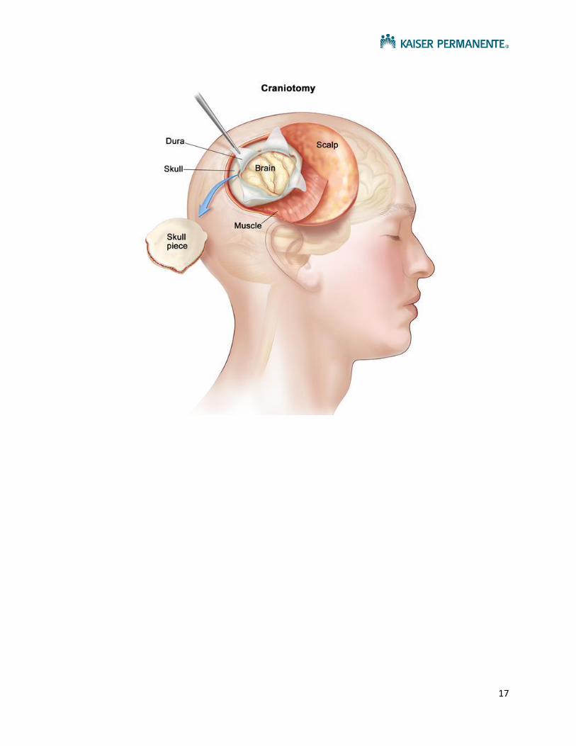
Craniotomy:
Craniotomies (entering the brain for surgery) can be done with great success. A craniotomy is
often the procedure needed access the brain to remove an AVM or clip an aneurysm. The actual
location of the surgery would depend on the location of the vascular. There are several stages to a
typical procedure. Most often, patients are completely under general anesthesia. The head is put into a
surgical clamp to make sure no movement can occur during the procedure. Depending on the type of
operation of and location of the vascular abnormality, neuro-monitoring can also be used to
physiologically map important parts of the brain, such as the motor regions to prevent major paralysis.
After the surgery is complete, the bone is replaced using metallic plates and screws that are MRI safe to
bridge the flap to the remainder of the skull, which are permanent implants.
17

18
Appendix
Figure 1: Symptoms of a Stroke is an emergency and need to act FAST.
Figure 2: Two major types of a stroke, ischemic (blocked /clogged artery) and hemorrhagic (bleeding
artery).

19
Figure 3: Arterial Dissection. This occurs when the wall of an artery physically tears, causing blocking of
the artery and sometimes clots that can dislodge and cause strokes.
Figure 4: Atrial Fibrillation. This is a condition of the heart which causes the heart to beat abnormally,
causing clots within the heart. Typically, patients need to be on blood thinners to prevent these clots
from forming and moving into the brain, producing strokes.
20
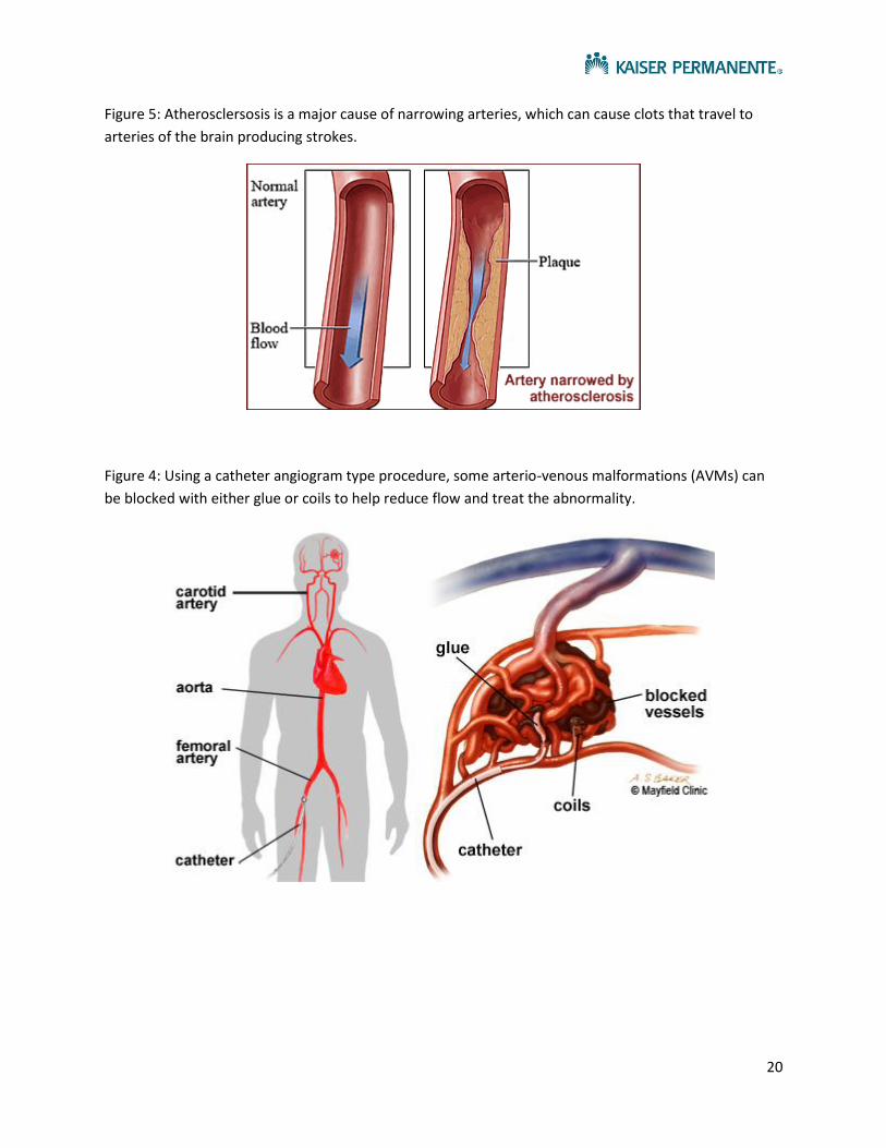
Figure 5: Atherosclersosis is a major cause of narrowing arteries, which can cause clots that travel to
arteries of the brain producing strokes.
Figure 4: Using a catheter angiogram type procedure, some arterio-venous malformations (AVMs) can
be blocked with either glue or coils to help reduce flow and treat the abnormality.

21
Figure 5: Stereotactic Radiosurgery (SRS) is a procedure where focused radiation is targeted on an AVM.
Typically, the radiation effects take between 3 months to 3 years to take effect, causing clogging of the
arteries in that area. Each beam of radiation is notably minimal, but when all the beams converge there
is a high dose at that target site.

22
Comprehensive Hydrocephalus Programs
Kaiser Permanente Redwood City
Redwood City Neurosurgeons (650) 299 - 2290:
William Sheridan, MD: Chief of Neurosurgery
Allen Efron, MD: Assistant Chief of Neurosurgery, Skull Base Specialist
John Duncan, MD, Phd: Chief of Pediatric Neurosurgery Kaiser Santa Clara, Director of Epilepsy
Surgery program
Nicole Moayeri, MD: Assistant Physician in Chief (APIC) of Surgical Services, Vascular
specialist
Cornelia Von Koch, MD, Phd: Neurosurgeon, Spine specialist
Aleksandyr Lavery, MD: Neurosurgeon, brain tumor/vascular and spine specialist
Lyndell Wang, MD: Neurosurgeon, brain tumor specialist
Lewis Hou, MD: Neurosurgeon, skull base and vascular specialist
Victor Tse, MD, Phd: Neurosurgeon, director of radiosurgery program, brain tumor and
peripheral nerve specialist
Prasad Reddy, MD: Neurosurgeon, Vascular and Endovascular specialist
Mark Sedrak, MD: Neurosurgeon, Director of Functional Neurosurgery, brain tumor specialist
Patrick Pezeshkian, MD: Functional Neurosurgeon and peripheral nerve specialist
Kevin Chao, MD: Neurosurgeon, Pediatric and Adult specialist

23
Sacramento Neurosurgeons (916) 973 – 5490:
Amit Banerjee, MD: Chief of Neurosurgery, Skull Base and Complex Spine Surgery
Mark Hawk, MD: Assistant Physician in Chief, Complex Spine surgery and Vascular
Kaveh Barami, MD: Neurosurgeon
Kamran Sahrakar, MD: Neurosurgeon, Neuro-Oncology and Radiosurgery
James Silverthorn, DO: Neurosurgeon
Abraham Bosckovitz, MD: Neurosurgeon
Indro Chakrabarti, MD: Neurosurgeon, Neuro-Oncology and Vascular
Huy Duong, MD: Neurosurgeon
Adam Griffith, MD: Neurosurgeon
Kern Guppy, MD: Neurosurgeon, Complex Spine
Brian Jian, MD: Neurosurgeon
David Moller, MD: Neurosurgeon
Conrad Pappas, MD, Phd: Functional Neurosurgeon
Alan Williams, MD: Neurosurgeon, Vascular
Sean McNatt, MD: Neurosurgeon Adult and Pediatric
Santa Clara Neurosurgical Group (408) 851 – 1240:
John Duncan, MD, Phd: Chief of Pediatric Neurosurgery Kaiser Santa Clara, Director of Epilepsy
Surgery program
Kevin Chao, MD: Pediatric Neurosurgeon
Roseville Pediatric Neurosurgery Group (916) 474 – 2600:
Sean McNatt, MD: Pediatric Neurosurgeon
Oakland Pediatric Neurosurgery Group (510) 752 – 1749:
Dachling Pang, MD: Chief of Pediatric Neurosurgery
John Zovickian, MD
Fresno Neurosurgery Group (559) 448 – 4437:
Steven Hysell, MD
Donald Myers, MD

24
Hydrocephalus
Anatomy: The brain consists of fluid spaces and a circulation system, similar to that of the heart and blood
vessels. The brain produces about 500 mL of cerebrospinal fluid (csf) each day that circulates and
becomes reabsorbed into the blood stream. The fluid spaces of the brain are called “ventricles” and they
are interconnected with small channels.
Hydrocephalus:
Hydrocephalus is a problem that occurs when there is reduced absorption somewhere in the
system, possibly an obstruction, or rarely very increased production. We distinguish two main types of
hydrocephalus as communicating and non-communicating (obstructive). Non-communicating
hydrocephalus is a more serious condition that when left untreated can become lethal.
25
Communicating Hydrocephalus
Communicating hydrocephalus refers to that type of hydrocephalus that does not have a
specific block in any of the csf channels (ie, foramen of Monro or cerebral aqueduct). Typically, there is
not an elevated pressure in the brain as a result causing headaches, but more frequently there can be
more subtle symptoms. The classic triad of symptoms related to hydrocephalus include: “wet”,
“wacky”, and “wobbly”, which is to say patients may have urinary incontinence, cognitive dysfunction,
and difficulty walking. One of the common types of communicating hydrocephalus is “normal pressure
hydrocephalus”, which usually occurs in older age, and the exact reason this problem occurs is largely
unknown. Other causes of communicating hydrocephalus include subarachnoid hemorrhage (from
aneurysm), traumatic brain injury, perinatal hemorrhages, and after some tumor operations.
In children with perinatal hemorrhages (intraventricular hemorrhage), a baby may be monitored
for signs of enlarging head or pressure (bulging soft spot or fontanelle). Sometimes, when the baby is
too small to accommodate a shunt or major surgery, a catheter may be placed in those fluid spaces with
a reservoir that can be accesses through the skin of the scalp. Eventually, these babies may need to
have a full shunt implanted to divert the csf indefinitely.
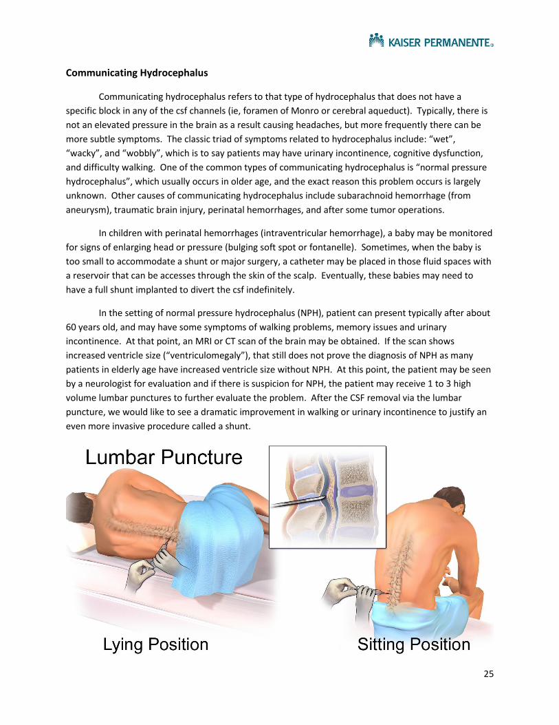
In the setting of normal pressure hydrocephalus (NPH), patient can present typically after about
60 years old, and may have some symptoms of walking problems, memory issues and urinary
incontinence. At that point, an MRI or CT scan of the brain may be obtained. If the scan shows
increased ventricle size (“ventriculomegaly”), that still does not prove the diagnosis of NPH as many
patients in elderly age have increased ventricle size without NPH. At this point, the patient may be seen
by a neurologist for evaluation and if there is suspicion for NPH, the patient may receive 1 to 3 high
volume lumbar punctures to further evaluate the problem. After the CSF removal via the lumbar
puncture, we would like to see a dramatic improvement in walking or urinary incontinence to justify an
even more invasive procedure called a shunt.

26
Non-communicating “Obstructive” Hydrocephalus
Obstructive hydrocephalus refers to the type of hydrocephalus in which there in fact is a specific
block in the channel of flow of csf. Typically this can occur at the level of the aqueduct (called
aqueductal stenosis) or from a mass/tumor (such as a colloid cyst which blocks the third ventricle).
Because there is a specific block in the fluid flow, the CSF may build up like a dam causing elevated
pressures in the brain. Depending on the cause of the obstruction, treatments may vary but include
“bypass” type procedures such as what’s called an endoscopic third venticulosotomy (ETV) or removal of
the tumor/mass. If these are not options, performing a shunting procedure may be the best option.
Example of Aqueductal Stenosis
Other Conditions that May Require Shunting:
Pseudotumor Cerebri
Another type of problem that may require a neurosurgical procedure is called pesudotumor
cerebri. Typically, the fluid spaces in the brain are small, rather than being enlarged with this condition.
This is a condition where pressures in the brain are increased and can cause a problem called
papilledema, which may cause vision changes and blindness if untreated. Nobody knows the exact
cause of this syndrome, but it typically occurs in overweight young females around the age of 40. A cure
for this problem could include significant weight loss notably. Also, we typically would want to exclude
other problems, such as a vein issue called sinus thrombosis, which can mimic the syndrome. Many of
these patients can be treated medically with a medication called Diamox, which reduces CSF production,
repeat lumbar punctures, and significant weight loss or even gastric surgery. If significant visual
impairment occurs, the treatment need is more urgent which may include optic sheath fenestration,
shunting or ICP monitoring.

27
Surgical Procedures
There are many types of surgical procedures geared towards treating hydrocephalus. The
mainstay of treatments include implanting tubing that redirects CSF to a different location in the body to
become reabsorbed, which are called shunts. Occasionally only in specific situations, a different type of
procedure can be performed, which does not involve implanting any tubing material, called an
endoscopic third ventriculostomy (ETV).
Types of Shunts:
There are several types of shunts that involve implanting tubing with a valve. The most
common of these is called a VentriculoPeritoneal Shunt (VPS). In these shunts, a catheter is placed from
the brain fluid spaces (ventricle) and tunneled underneath the skin all the way into the abdomen.
Occasionally, the abdomen may not be a good place for CSF to be reabsorbed because of prior surgery,
so other locations that can be used include the chest/lung space called the “pleural”or the main vein
which goes into the heart called “atrial”. These two are called ventriculopleural and ventriculoatrial
shunts, respectively. In addition, sometimes it’s unnecessary to place the main catheter in the brain
and one can place that main catheter in the back (“lumbar”), which is then usually tunneled into the
abdomen (lumboperitoneal shunt).
Endoscopic Third Venticulosotomy (ETV)
An endoscopic third ventriculostomy is a procedure that performed using an endoscope camera,
which is placed into the brain fluid spaces (ventricles) and a physical hole is made at the base of the

28
brain (technically called the tuber cinerium). That region that’s punctured is a very thin membrane
which does not have any physical function and is usually thinned as a result of the hydrocephalus. This
procedure effectively re-routes the CSF past the blocked channel into a new direction which allows for
more normal flow. An ETV may not work for long-standing hydrocephalus or in patients who have had
prior catheter shunts and typically can only work with a new diagnosis of obstructive hydrocephalus.
Types of Shunt Implants
There are two main types of shunt implants, programmable and non-programmable.
Programmable shunt allow for pressure adjustments non-invasively, which can then be tailored to an
individual patient. The disadvantage of these programmable shunts is that they are often affected by
strong magnetic fields, such as which is present in an MRI, which means the shunt needs to be reset
after each MRI scan. Some valves may be MRI resistant notably.
Non-programmable valves are valves with a set pressure. These valves do not require as much
“maintenance” and are implanted and essentially left alone. These non-programmable valves are not
affected by magnetic fields and so patients do not need shunt adjustment or assessment after an MRI.
Lastly, there is often an option of placing an extra in the shunt tubing called an “antisiphon
device”. This antisiphon device prevents overshunt as a result of gravity as the tip of the tube is often
below the head. Siphoning is akin to placing a water hose in a pool and placing the other end
underneath the pool, which results in water flowing through the hose “downhill”. The antisiphon
device prevents this problem. However, sometimes siphoning can be used to the advantage of a patient
to promote treatment of the hydrocephalus.
29
Appendix
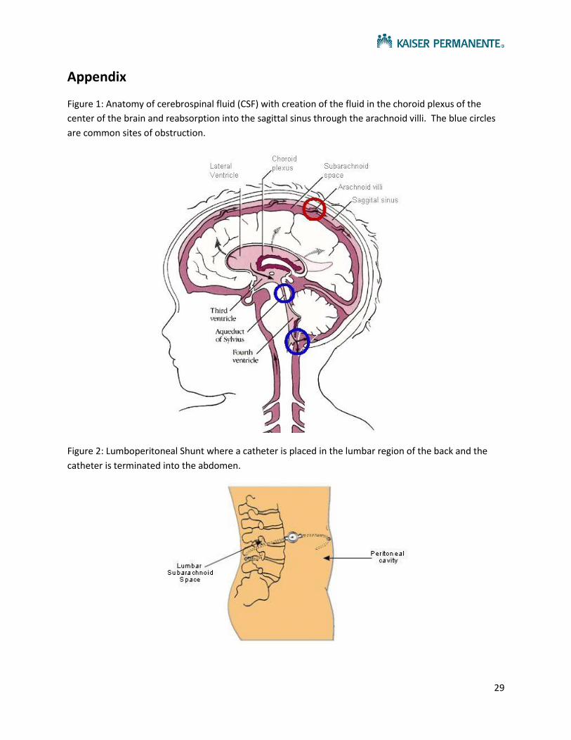
Figure 1: Anatomy of cerebrospinal fluid (CSF) with creation of the fluid in the choroid plexus of the
center of the brain and reabsorption into the sagittal sinus through the arachnoid villi. The blue circles
are common sites of obstruction.
Figure 2: Lumboperitoneal Shunt where a catheter is placed in the lumbar region of the back and the
catheter is terminated into the abdomen.

30
Figure 3: Ventriculopleural shunt where there is a catheter inserted into the brain and the terminus is in
the chest cavity around the lungs.
Figure 4: Ventriculoatrial shunt where there is a catheter inserted into the brain and the terminus is in
the major veins leading into the heart.

31
Figure 5: Strata Valve Adjustable valve. NSC valve does not have an antisiphon device, whereas the II
does have that additional component. These valves need adjustment after an MRI. The pressures listed
are the opening pressures for the Strata II

32
Figure 6: Codman CERTAS Programmable Valve: MRI Compatible

33
Figure 7: Codman-Hakim Programmable Valve. Valve needs adjustment after exposure to MRI.

34
Comprehensive Spine Surgery Program
Kaiser Permanente Redwood City
Redwood City Neurosurgeons (650) 299 - 2290:
William Sheridan, MD: Chief of Neurosurgery
Allen Efron, MD: Assistant Chief of Neurosurgery, Skull Base Specialist
John Duncan, MD, Phd: Chief of Pediatric Neurosurgery Kaiser Santa Clara, Director of Epilepsy
Surgery program
Nicole Moayeri, MD: Assistant Physician in Chief (APIC) of Surgical Services, Vascular
specialist
Cornelia Von Koch, MD, Phd: Neurosurgeon, Spine specialist
Aleksandyr Lavery, MD: Neurosurgeon, brain tumor/vascular and spine specialist
Lyndell Wang, MD: Neurosurgeon, brain tumor specialist
Lewis Hou, MD: Neurosurgeon, skull base and vascular specialist
Victor Tse, MD, Phd: Neurosurgeon, director of radiosurgery program, brain tumor and
peripheral nerve specialist
Prasad Reddy, MD: Neurosurgeon, Vascular and Endovascular specialist
Mark Sedrak, MD: Neurosurgeon, Director of Functional Neurosurgery, brain tumor specialist
Patrick Pezeshkian, MD: Functional Neurosurgeon and peripheral nerve specialist
Kevin Chao, MD: Neurosurgeon, Pediatric and Adult specialist
35
Sacramento Neurosurgeons (916) 973 – 5490:
Amit Banerjee, MD: Chief of Neurosurgery, Skull Base and Complex Spine Surgery
Mark Hawk, MD: Assistant Physician in Chief, Complex Spine surgery and Vascular
Kaveh Barami, MD: Neurosurgeon
Kamran Sahrakar, MD: Neurosurgeon, Neuro-Oncology and Radiosurgery
James Silverthorn, DO: Neurosurgeon
Abraham Bosckovitz, MD: Neurosurgeon
Indro Chakrabarti, MD: Neurosurgeon, Neuro-Oncology and Vascular
Huy Duong, MD: Neurosurgeon
Adam Griffith, MD: Neurosurgeon
Kern Guppy, MD: Neurosurgeon, Complex Spine
Brian Jian, MD: Neurosurgeon
David Moller, MD: Neurosurgeon
Conrad Pappas, MD, Phd: Functional Neurosurgeon
Alan Williams, MD: Neurosurgeon, Vascular
Sean McNatt, MD: Neurosurgeon Adult and Pediatric
Santa Clara Neurosurgical Group (408) 851 – 1240:
John Duncan, MD, Phd: Chief of Pediatric Neurosurgery Kaiser Santa Clara, Director of Epilepsy
Surgery program
Kevin Chao, MD: Pediatric Neurosurgeon
Roseville Pediatric Neurosurgery Group (916) 474 – 2600:
Sean McNatt, MD: Pediatric Neurosurgeon
Oakland Pediatric Neurosurgery Group (510) 752 – 1749:
Dachling Pang, MD: Chief of Pediatric Neurosurgery
John Zovickian, MD
Fresno Neurosurgery Group (559) 448 – 4437:
Steven Hysell, MD
Donald Myers, MD
Oakland Orthopedic Spine Group (559) 448 – 4437:
Ravinder-Raj Bains, MD
Andrew Slucky, MD
Josef Gorek, MD
Todd Lincoln, MD
San Jose Orthopedic Spine Group (408) 972 – 6100:
Anthony Matan, MD
Joseph Matthews, MD
Elliot Carlisle, MD
Steven Spisak, MD

36
Kaiser Permanente – Northern California Non-Surgical Spine/Pain
Service Areas
DIABLO Service Area (WCR, MTZ, ANT) 925-372-1741
DIABLO 1 ANS Epidural Clinic
Dr Angela Chiang Dr Cynthia Rahn
Dr Mark Rahn Dr Sabrina Martinez
Dr Prakash Raygor Dr Mark Moore
DIABLO 2 ANS Interventional Pain Clinic 925-372-1283
Dr Andrew Maher
Dr James Mura
Dr Darko Vodopich
Dr. Norm Aleks
DIABLO 3 PMR/SPINE CLINIC (925) 313-4740
Dr. Eric Alexander
Fresno (559) 448-4555
Dr. Eugene Huang (ANS)
Dr. Jonathan Grossman (PMR/SPINE CLINIC)
Dr. Rupinder Singh (ER)
Dr. Marta Bator (ANS - Pool)
Modesto & Stockton Chronic Pain & Interventional Pain 209-735-3255
Dr Tanja Frey - ans
Dr Brandon Valine

37
Dr. Raul Calderon
Petaluma Intervention Pain Clinic PMR/SPINE CLINIC 415-444-2988
Dr Diane Murphy
Dr Vitto
Dr Tabitha Washington
Redwood City 1 of 2– ANS/NSG 650-299-5454
Dr. Taissa Cherry (ANS, Dept of Neurosurg)
Dr. Mark Sedrak (NSG)
Dr. Patrick Pezeshkian (NSG)
PA Ivan Bernstein
PA Diana Bruce
Redwood City 2 of 2 – PMR/SPINE CLINIC 650-299-4741
Dr Firtch Dr Ray Lai
Dr Maratukulam Dr Nguyen
Dr Treinen
Richmond PMR/SPINE CLINIC 510-307-1661
Dr John H Lim PMR/SPINE CLINIC
Dr Angelita Balbas PMR/SPINE CLINIC
Roseville PMR/SPINE CLINIC 916-771-6611
Dr William Fenton - ANS
Dr Mark Tyburski
Dr Ryan Carver

38
Sacramento (916) 973-5490
Conrad Pappas, MD (Neurosurgery)
San Francisco Pain Interventional Center- ANS 415-833-0095
Dr Justin McKendry ANS
Dr Taissa Cherry ANS
Dr Salman Dasti
San Leandro (Union City) books appts/epidurals are done in Fremont PMR/SPINE
CLINIC (510) 675-3070
Dr Eunice Lau (chief) - PMR/SPINE CLINIC
Dr Sergiy Rybiy
Dr Raghu Katragadda ANS
San Jose (Santa Theresa) Interventional Pain ANS 408-972-6283
Dr Darshan Patel Dr Samuel Park
Dr Shabeen Tharani Dr Prasad Movva
Dr Laurence Won Dr Vital Kandula
San Rafael PMR/SPINE CLINIC 415-444-2988
Dr Murphy
Dr Vitto
Santa Rosa 1 of 2 – PMR/SPINE CLINIC 707-566-5557
Dr David Vidaurri
Drs. Donald F Green

39
Hari Lakshmanan
Kirk Pappas
Tracey Jones
Todd Weitzenberg
Santa Rosa 2 of 2– Chronic/Interventional Pain ANS 707-571-3921
Dr Andrea Rubinstein (ANS)
Dr. Tabitha Washington (ANS)
South Sacramento Pain Clinic (ANS) (916) 688-6353
Dr Mike Bicocca
Dr Rod Youssefi
Dr Kegang Hu
Dr Sabina Tahera
South San Francisco ANS Pain Services 650-742-2395
Dr Eddie Busracamwongs ANS
Dr. Hamid Motamed
Dr. Tin-Na Kan
Dr. Ali Abdollahi-Fard
Santa Clara PMR/SPINE CLINIC 408-851-9200
Dr Richard Kim
Dr Kevin Wang
Dr Dhiruj Kirpalani
Vallejo – PMR/SPINE CLINIC (707) 651-1025

40
Dr. Linda Cho (PMR/SPINE CLINIC)
Conditions of the Spine
Anatomy: The spine consists of bone, discs, muscles, ligaments and nerves. Each one of these can cause
conditions that produce pain or other problems. The most common reason for a patient to see a spine
surgeon, however, is related to problems of the bone, nerves and/or discs. Nerves, both the nerve roots
and spinal cord, pass through small tunnels throughout the spine. When these tunnels become narrowed,
such as from a herniated disc, they can compress the nerve causing pain, numbness, weakness. It’s
notable that many patients who have no symptoms can actually have what appears to be compression of a
nerve on an MRI, making correlation between symptoms and a scan of utmost importance. In addition,
may patients may have pain from muscles, ligaments, or even nerves without compression, making direct
spine operations not a reasonable option.
Disc Herniation
The disc of the spine is located in between each level of bone or vertebra, which is why we call it
the intervertebral disc named after the surrounding bones. For example, in between L4 and L5 is the
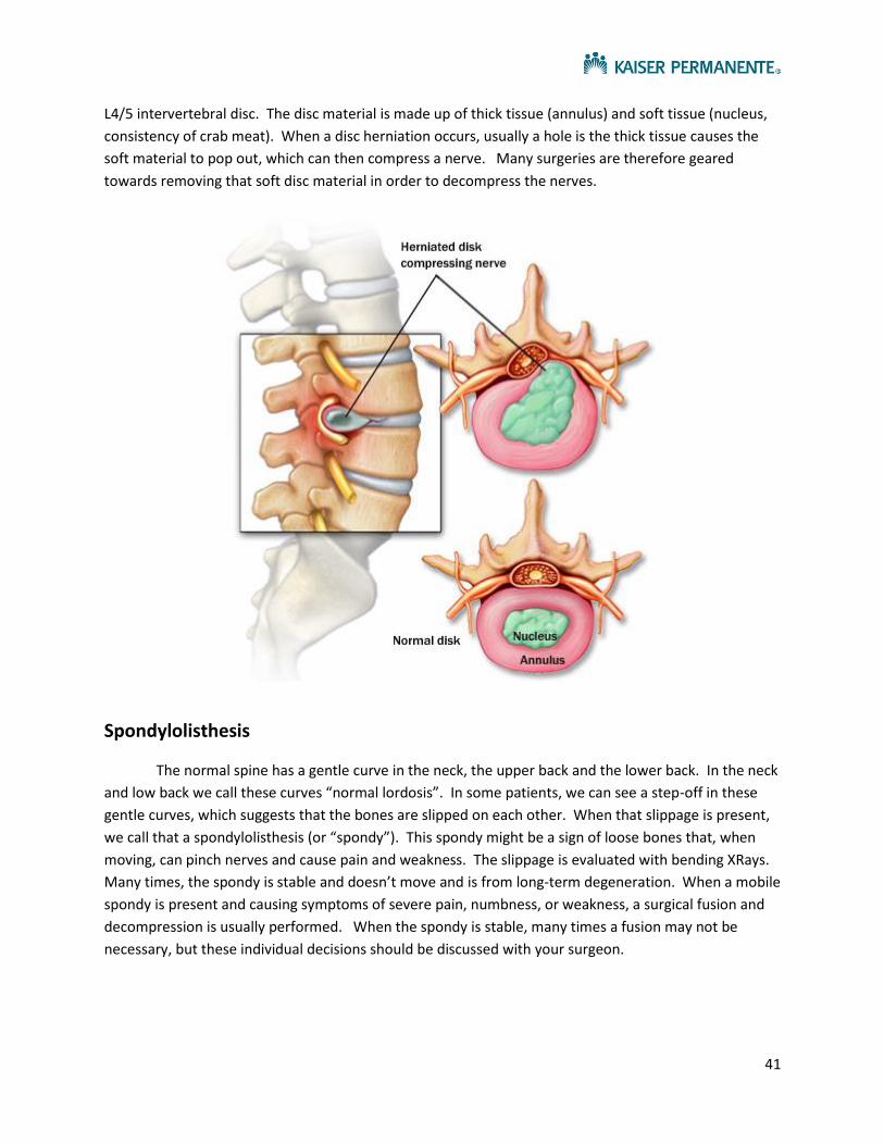
41
L4/5 intervertebral disc. The disc material is made up of thick tissue (annulus) and soft tissue (nucleus,
consistency of crab meat). When a disc herniation occurs, usually a hole is the thick tissue causes the
soft material to pop out, which can then compress a nerve. Many surgeries are therefore geared
towards removing that soft disc material in order to decompress the nerves.
Spondylolisthesis
The normal spine has a gentle curve in the neck, the upper back and the lower back. In the neck
and low back we call these curves “normal lordosis”. In some patients, we can see a step-off in these
gentle curves, which suggests that the bones are slipped on each other. When that slippage is present,
we call that a spondylolisthesis (or “spondy”). This spondy might be a sign of loose bones that, when
moving, can pinch nerves and cause pain and weakness. The slippage is evaluated with bending XRays.
Many times, the spondy is stable and doesn’t move and is from long-term degeneration. When a mobile
spondy is present and causing symptoms of severe pain, numbness, or weakness, a surgical fusion and
decompression is usually performed. When the spondy is stable, many times a fusion may not be
necessary, but these individual decisions should be discussed with your surgeon.

42
Stenosis
Stenosis refers to narrowing of the canal that the nerves are passing through. There are many
passage ways that can have stenosis, but generally speaking we refer to the “central canal” and the
“neural foramen”. The central canal is the canal that the spinal cord and the clump of nerve roots
travel. The neural foramen is where the nerve root exit the main spinal canal before going to our arms
or legs. Stenosis can be caused by disc herniations, bone spurs from arthritis, ligaments growing
(“hypertrophy”). The main goal of surgeries aimed at stenosis is to open the canal and provide room for
the nerves passing through their canal.

43
Types of Spinal Surgery
There are many types of spinal operations, but the main two are those of decompression and
those of fusion. Many times, surgery would include both a decompression and fusion.
Decompression:
A decompressive operation is an operation geared towards opening a canal where nerves are
passing. If the central canal has stenosis, typically we’d perform a procedure called a “laminectomy”.
This is akin to removing the soft side of a lobster tail, which then opens one side of the canal and
provides room for the nerves. Sometimes, the canal that needs decompression is the neural foramen
and the procedure performed can include a “foraminotomy”. Many times, both a laminectomy and
foraminotomy can be performed.
In the neck in particular, it’s notable that there is a procedure that can provide more space for
the spinal cord without the need for fusion, which is called a laminoplasty. In this case, the lamina of
the spine is elevated, allowing it to be hinged in a way that increases the amount of room for the spinal
cord.

44
Fusion:
A fusion procedure is a procedure performed to stabilize the bones and prevent future
movement. There are numerous types of spinal fusion procedures that vary based off of the location in
the body.
Cervical Region (Neck):
In the neck, there are two main fusion procedures, one from the front (ACDF) and one from the
back. The surgery from the front goes through the neck to the spine and is technically called an
“anterior cervical discectomy and fusion”. This procedure allows excellent removal of the disc space and
the ability to fuse the bones well. Most common issues that can occur with this procedure include
temporary swallowing and a hoarse voice, which usually are transient problems. This procedure
typically carries the least pain afterwards compared to other spine procedures and is one of the most
common procedures of the spine performed.
The other option is a fusion procedure from the back of the neck. Typically, that procedure is
combined with a laminectomy as well. In this instance, screws are placed at the desired levels and are
connected with rods. The bone is usually prepared in a way that allows ingrowth of bone cells over the
next several months, creating a solid bone mass.
45
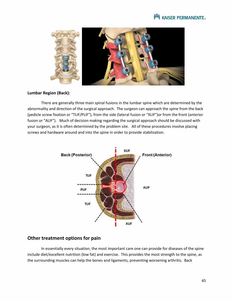
Lumbar Region (Back):
There are generally three main spinal fusions in the lumbar spine which are determined by the
abnormality and direction of the surgical approach. The surgeon can approach the spine from the back
(pedicle screw fixation or “TLIF/PLIF”), from the side (lateral fusion or “XLIF”)or from the front (anterior
fusion or “ALIF”). Much of decision making regarding the surgical approach should be discussed with
your surgeon, as it is often determined by the problem site. All of these procedures involve placing
screws and hardware around and into the spine in order to provide stabilization.
Other treatment options for pain
In essentially every situation, the most important care one can provide for diseases of the spine
include diet/excellent nutrition (low fat) and exercise. This provides the most strength to the spine, as
the surrounding muscles can help the bones and ligaments, preventing worsening arthritis. Back

46
strengthening exercises are very useful, including swimming/aquatherapy, pilates/yoga and physical
therapy.
Further, in many instances no surgical options to decompress or fuse the spine are reasonable.
In these situations, besides medications and epidural steroid injections (ESI), other treatments could
include spinal cord stimulation, intrathecal pump therapy, ketamine infusions or other functional
neurosurgical procedures. Typically, a patient would need to have stable and refractory pain for years
before undergoing these advanced procedures. Please consult your chronic pain physician if you feel
like you may be at the point of needing these types of procedures.

47
Other Non-Compressive Spine Related Pain Syndromes There are many syndromes which give rise to pain that is related to the spine. It’s notable that many pain syndromes are not caused by nerve compression in the spine, but rather of an origin that’s likely an abnormal reflex or circuit involving either the spinal cord itself or the dorsal root ganglion. So often times, an MRI or CT scan show no obvious problem to explain the origin of pain. Chronic Regional Pain Syndrome (CRPS) CRPS is a chronic pain condition most often affected the arms, legs, hands or feet and most typically after a trauma (herniated disc or surgery). A portion of the nervous system involved with this pain syndrome is called the autonomic nervous system, rather than the peripheral nervous system or central nervous system. Typically patients can develop sensitivity to touch (called allodynia) and pain can spread beyond a single site, there can be changes in color (blotchy/purple/red), swelling, joint stiffness, hair loss. Since MRI’s/CT’s are often negative, there is no good diagnostic test to confirm this disorder, but rather it’s a clinical diagnosis and a diagnosis where other problems are excluded on imaging. Typically, treatment includes: rehabilitation therapy, psychotherapy, medications, sympathetic nerve blocks, ketamine infusion, and spinal cord stimulation. It’s important to both recognize and treat this condition as soon as possible to prevent it from worsening or becoming a chronic condition.
Failed back surgery syndrome (FBSS) FBSS is a group of issues related to the spine that result in persistent back pain with a prior history of spine surgery. Patients can often have back pain or shooting pain that persists without any evidence of continued pinched nerves or unstable joints. It’s important to know that the spine is made up of not only nerves and discs, but has a constellation of joints, muscles and ligaments, which may all contribute to pain in addition to nerves that are not compressed. Patients with FBSS are typically

48
treated with physical therapy, medical management, epidural steroid injections and can also be candidates for spinal cord stimulation, dorsal root ganglion stimulation, intrathecal pump therapy.

49
Appendix
Figure 1: Types of Epidural Steroid Injections (ESI) including: Translaminar epidural injection and
transforaminal epidural injection
Figure 2: Dermatomal map, which helps to correlate a nerve compression problem

50
Figure 3: TLIF Procedure from back
Figure 4: XLIF/DLIF procedure or extreme lateral from side

51
Figure 5: ALIF procedure, anterior or from front
Figure 6: Example of Spinal cord stimulation to help chronic pain conditions
Spinal cord Stimulator as implanted X-ray of a single lead in place

52
Comprehensive Brain Tumor Program
Kaiser Permanente
Redwood City Neurosurgeons (650) 299 - 2290:
William Sheridan, MD: Chief of Neurosurgery
Allen Efron, MD: Assistant Chief of Neurosurgery, Skull Base Specialist
John Duncan, MD, Phd: Chief of Pediatric Neurosurgery Kaiser Santa Clara, Director of Epilepsy
Surgery program
Nicole Moayeri, MD: Assistant Physician in Chief (APIC) of Surgical Services, Vascular
specialist
Cornelia Von Koch, MD, Phd: Neurosurgeon, Spine specialist
Aleksandyr Lavery, MD: Neurosurgeon, brain tumor/vascular and spine specialist
Lyndell Wang, MD: Neurosurgeon, brain tumor specialist
Lewis Hou, MD: Neurosurgeon, skull base and vascular specialist
Victor Tse, MD, Phd: Neurosurgeon, director of radiosurgery program, brain tumor and
peripheral nerve specialist
Prasad Reddy, MD: Neurosurgeon, Vascular and Endovascular specialist
Mark Sedrak, MD: Neurosurgeon, Director of Functional Neurosurgery, brain tumor specialist
Patrick Pezeshkian, MD: Functional Neurosurgeon and peripheral nerve specialist
Kevin Chao, MD: Neurosurgeon, Pediatric and Adult specialist
Redwood City Neuro-Oncology Group (650) 299 – 2290:
Scott Peak, MD: Neurooncologist
Victor Levin, MD: Neurooncologist
Piia Thomas, MD: Neurooncologist
Nisha Hazari, NP: Neurooncology Nurse Practitioner

53
Sacramento Neurosurgeons (916) 973 – 5490:
Amit Banerjee, MD: Chief of Neurosurgery, Skull Base and Complex Spine Surgery
Mark Hawk, MD: Assistant Physician in Chief, Complex Spine surgery and Vascular
Kaveh Barami, MD: Neurosurgeon
Kamran Sahrakar, MD: Neurosurgeon, Neuro-Oncology and Radiosurgery
James Silverthorn, DO: Neurosurgeon
Abraham Bosckovitz, MD: Neurosurgeon
Indro Chakrabarti, MD: Neurosurgeon, Neuro-Oncology and Vascular
Huy Duong, MD: Neurosurgeon
Adam Griffith, MD: Neurosurgeon
Kern Guppy, MD: Neurosurgeon, Complex Spine
Brian Jian, MD: Neurosurgeon
David Moller, MD: Neurosurgeon
Conrad Pappas, MD, Phd: Functional Neurosurgeon
Alan Williams, MD: Neurosurgeon, Vascular
Sean McNatt, MD: Neurosurgeon Adult and Pediatric
Sacramento Neuro-Oncology Group (916) 973 – 5490:
Enrico Lallana, MD
Santa Clara Neurosurgical Group (408) 851 – 1240:
John Duncan, MD, Phd: Chief of Pediatric Neurosurgery Kaiser Santa Clara, Director of Epilepsy
Surgery program
Kevin Chao, MD: Pediatric Neurosurgeon
Roseville Pediatric Neurosurgery Group (916) 474 – 2600:
Sean McNatt, MD: Pediatric Neurosurgeon
Oakland Pediatric Neurosurgery Group (510) 752 – 1749:
Dachling Pang, MD: Chief of Pediatric Neurosurgery
John Zovickian, MD
Fresno Neurosurgery Group (559) 448 – 4437:
Steven Hysell, MD
Donald Myers, MD

54
GENERAL INFORMATION ABOUT BRAIN TUMORS
Tumors can arise from essentially any living cell in the body. With respect to the brain, tumors
often arise from the meninges (meningioma), brain cells (glioma’s), pituitary gland (pituitary
adenoma’s), surrounding skull/bone (chordoma’s), white blood cells (lymphoma). Depending on the
type of tumor, different treatment strategies can be planned which may include surgery, chemotherapy,
and or radiation. Oftentimes, the diagnosis is the first step in the treatment plan, which typically
involves surgery to obtain a piece or remove the tumor/mass.

55
Craniotomy or Tumor Biopsy:
Craniotomies (entering the brain for surgery) can be done with great success. A craniotomy is
often the procedure needed to remove a brain tumor. Depending on the tumor site, tumor removal
may be attempted or possibly a biopsy. There are several stages to a typical procedure. Most often,
patients are completely under general anesthesia. However, on occasion, it may be necessary to
perform the surgery awake with mapping of speech centers. Once in position, the surgeon will often
utilize a clamp that will both hold the head and be used to register your head to a special MRI that was
obtained prior to surgery, called a “navigation” scan. This “navigation” scan is used during surgery, like
GPS to help locate and pinpoint the tumor location. Depending on the location of the tumor, neuro-
monitoring can also be used to physiologically map important parts of the brain, such as the motor
regions to prevent major paralysis. After the surgery is complete, the bone is replaced using metallic
plates and screws that are MRI safe to bridge the flap to the remainder of the skull, which are
permanent implants.

56
Typically, after a craniotomy procedure, patients will be in the hospital on average 3 days, but
that time-frame may vary based on patient’s status and tumor type/location. A bandage will be present
on the incision that will be kept clean and dray for approximately 3 days, then removed and left open to
air. Staples or stitches will be used on the skin surface, which can be removed at the home Kaiser facility
in 2-3 weeks after surgery. Once the bandage is removed in 3 days, it would be okay to shower using
baby shampoo with brief washing and without aggressive combing or brushing of the hair. We
recommend avoiding direct sunlight on the incision or heavy physical activity for about 6 weeks. If a hat
or beanie are worn, make sure they are loose fitting, cleanly washed daily, and that there is no
significant pressure or sweating on the incision site. Total healing time from any operation is about 6
weeks to 3 months. Patients may often need chemotherapy or radiation after surgery, which typically
are not started for approximately 4 weeks after surgery.
Pathology:
Within about 3-7 days after surgery, the pathology results may become finalized. This tells your
doctors what kind of tumor was present and will help to dictate subsequent treatment. The reason it

57
takes several days is that the tissue must be processed with special stains and microscopy. Sometimes
after first processing rounds, further processing and stains are needed to refine the diagnosis which may
extend the length of time to have the results. On occasion for difficult tumors, the pathologists may also
collaborate with other neuropathologists to decide on the diagnosis.
Chemotherapy:
The type of chemotherapy recommended depends on the tumor type. For many high grade
gliomas, Temodar (Temozolomide) is utilized. Temodar is often delivered along with radiation, which
may work synergistically to fight the tumor. Other kinds of chemotherapy include Avastin
(bevacizumab), which is often used as a backup for recurrent glioblastomas or radiation necrosis.
Radiation:
There are many types of radiation. Typically, two forms of radiation exist to treat brain tumors:
stereotactic radiosurgery (SRS) and IMRT/EBRT. With stereotactic radiosurgery, radiation beams are
focused on the tumor, which receives a high dose whereas all the surrounding tissues receive very little.
SRS is often used for meningiomas and schwannomas where the focus is most easily identified. For
other tumors, such as high grade gliomas, the margins of the tumor are not clearly defined and a more
broad form of radiation is delivered over many fractions or doses, to reduce the risk of collateral
damage to important portions of the brain. This type of radiation, which can treat a wider field for
invasive or large tumors, is often referred to as IMRT (Intensity-modulated radiation therapy) and
maybe delivered over the course of a month.

58
TUMOR TYPES
http://www.abta.org/brain-tumor-information/
Meningiomas:
Description
Meningiomas usually grow inward, causing pressure on the brain or spinal cord. They also can grow
outward toward the skull, causing it to thicken. Most meningiomas are noncancerous, slow-growing
tumors. Some contain sacs of fluid (cysts), mineral deposits (calcifications), or tightly packed bunches of
blood vessels.
Symptoms
Meningiomas usually grow slowly, and may reach a large size before interfering with the normal functions
of the brain. The resulting symptoms depend on the location of the tumor within the brain. Headache and
weakness in an arm or leg are the most common symptoms. However, seizures, personality changes,
and/or visual problems may also occur.
Incidence
Meningiomas account for about 36.1% of all primary brain tumors, which are tumors that form in the brain
or its coverings. They are most likely to be found in adults older than 60; the incidence appears to
increase with age. Rarely are meningiomas found in children. They occur about twice as often in women
as in men.
Cause
Researchers are studying meningiomas carefully to find out what causes them. Between 40% and 80% of
meningiomas contain an abnormal chromosome 22. The cause of this abnormality is not known. We do
know, however, that this chromosome is normally involved in suppressing tumor growth. Meningiomas
also frequently have extra copies of the platelet-derived growth factor (PDGFR) and epidermal growth
factor receptors (EGFR), which may contribute to the growth of these tumors.
Previous radiation to the head, a history of breast cancer, or neurofibromatosis type 2 may be risk factors
for developing meningioma. Multiple meningiomas occur in 5% to 15% of patients, particularly those with
neurofibromatosis type 2.
Some meningiomas have receptors that interact with hormones, including progesterone, androgen, and
less commonly, estrogen. Although the exact role of hormones in the growth of meningiomas has not
been determined, researchers have observed that meningiomas occasionally grow faster during
pregnancy.
Treatment

59
Surgery and radiation are the most common forms of treatment for meningioma. Surgery is the primary
treatment for meningiomas, although some tumors may not be removed this way. Radiation therapy may
be used to tumors that cannot be removed with surgery, tumors that are not completely removed in
surgery, malignant/anaplastic tumors, or recurrent tumors.
Lymphoma
Lymphoma is a cancer that arises from the cells of the lymphatic system. In the brain, this type of cancer
is called Primary CNS Lymphoma (PCNSL).
Location
Lymphoma occurs most often in the cerebral hemisphere, but may also involve the cerebrospinal fluid,
the eyes, or the spinal cord. In addition, some people may have evidence of lymphoma elsewhere in the
body. It is not unusual for this tumor to be found in multiple areas of the cerebral hemisphere, as it can
spread throughout the central nervous system.
Description
Lymphoma is a cancer that starts in the cells of the lymphatic system.
Symptoms

60
The most common symptoms of CNS lymphoma include personality and behavioral changes, confusion,
symptoms associated with increased pressure within the brain (eg, headache, nausea, vomiting,
drowsiness), weakness on one side of the body, and seizures. Problems with eyesight may also occur.
Incidence
This disease affects people with healthy immune systems, as well as those whose immune systems are
not functioning properly, for example organ transplant recipients, patients with autoimmune disease or
people who are HIV positive.
The incidence of CNS lymphoma has been increasing over the past 20 years; it now represents between
2% and 3% of all primary brain tumors.
Cause
CNS lymphoma usually originates from B lymphocytes and is classified as non-Hodgkin’s (meaning it is
different from Hodgkin’s disease).
Treatment
Once a diagnosis is confirmed, steroids are used to control brain swelling; this may result in the
immediate disappearance of the tumor on a later scan. Chemotherapy and radiation, or chemotherapy
alone may then be used the primary treatment. Surgery is not usually an option because lymphomas tend
to occur deep within the brain and the risk of surgical complications is too high.
Glioma:
“Glioma” is a general term used to describe any tumor that arises from the supportive (“gluey”) tissue of
the brain. This tissue, called “glia,” helps to keep the neurons in place and functioning well.
There are three types of normal glial cells that can produce tumors. An astrocyte will produce
astrocytomas (including glioblastomas), an oligodendrocyte will produce oligodendrogliomas, and

61
ependymomas come from ependymal cells. Tumors that display a mixture of these different cells are
called mixed gliomas.
Tumors such as “optic nerve glioma” and “brain stem glioma” are named for their locations, not the tissue
type from which they originate.
Location
The location of the tumor depends on the type of cells from which it originates.
Description
Three types of normal glial cells can produce tumors—astrocytes, oligodendrocytes, and ependymal
cells. Tumors that display a mixture of these cells are called mixed gliomas.
Astrocytoma: Click here to learn more about astrocytomas, including juvenile pilocytic astrocytoma, low
grade astrocytoma, anaplastic astrocytoma, or glioblastoma.
Ependymoma: Click here to learn more about ependymoma.
Mixed Glioma (also called Oligoastrocytoma): These tumors usually contain a high proportion of more
than one type of cell, most often astrocytes and oligodendrocytes. Occasionally, ependymal cells are also
found. The behavior of a mixed glioma appears to depend on the grade of the tumor. It is less clear
whether their behavior is based on that of the most abundant cell type.
Oligodendroglioma: Click here to learn more about oligodendroglioma.
Optic Glioma: These tumors may involve any part of the optic pathway, and they have the potential to
spread along these pathways. Most of these tumors occur in children under the age of 10. Grade I
pilocytic astrocytoma and grade II fibrillary astrocytoma are the most common tumors affecting these
structures. Higher-grade tumors may also arise in this location. Twenty percent of children with
neurofibromatosis (NF-1) will develop an optic glioma. These gliomas are typically grade I, pilocytic
astrocytomas. Children with optic glioma are usually screened for NF-1 for this reason. Adults with NF-1
typically do not develop optic gliomas.
Gliomatosis Cerebri: This is an uncommon brain tumor that features widespread glial tumor cells in the
brain. This tumor is different from other gliomas because it is scattered and widespread, typically involving
two or more lobes of the brain. It could be considered a “widespread low-grade glioma” because it does
not have the malignant features seen in high-grade tumors.
The widespread nature of gliomatosis cerebri causes enlargement of any part of the brain it involves. This
may include the cerebral hemispheres, or less often, the cerebellum or brain stem.
Symptoms
Symptoms vary based on tumor type:
Astrocytoma:Click here to learn more about astrocytoma symptoms.
Ependymoma: Click here to learn more about ependymoma symptoms.
Mixed Glioma (also called Oligoastrocytoma): The initial symptoms, including headache and nausea,
usually are the result of increased pressure inside the brain. Vision problems, as well as changes in
behavior and personality, are also fairly common in mixed glioma patients.
Oligodendroglioma: Click here to learn more about oligodendroglioma symptoms.
Optic Glioma: These tumors may cause few or no symptoms. Their placement along the optic nerve,
however, can cause vision loss (depending on the location of the tumor) or strabismus (“crossed eyes”).

62
Hormonal disturbance might also occur, causing developmental delay(s), early puberty, and other
symptoms.
Gliomatosis Cerebri: Symptoms are often nonspecific and can include personality and behavioral
changes, memory disturbance, increased intracranial pressure with headache and sometimes seizures.
Incidence
The incidence of this tumor varies by type.
Cause
Like many tumor types, the exact cause of glioma is not known.
Treatment
Treatment is based on tumor type:
Astrocytoma: Click here to learn more about treatment for astrocytoma.
Ependymoma: Click here to learn more about treatment for ependymoma.
Mixed Glioma (also called Oligoastrocytoma): Treatment may include surgery followed by radiation
therapy, particularly if the tumor is high-grade. Chemotherapy will also generally be used in high-grade
tumors.
Oliogdendroglioma: Click here to learn more about treatment for oligodendroglioma.
Optic Glioma: Careful observation may be an option for patients with stable or slow-growing tumors.
Surgery might be recommended for a growing tumor which involves only the optic nerve. Radiation might
be used for a tumor of the chiasm or other pathways. Local radiation and chemotherapy with radiation
therapy are used for recurrent tumors. Patients with primary and/or recurrent tumors may wish to take
part in a clinical trial.
Gliomatosis Cerebri: Treatment is less well defined because this tumor is so rare. Surgical removal is
generally not attempted, because it is so widespread. Radiation and chemotherapy may be considered.
Astrocytoma:
Astrocytomas are tumors that arise from astrocytes—star-shaped cells that make up the “glue-like” or
supportive tissue of the brain.
These tumors are “graded” on a scale from I to IV based on how normal or abnormal the cells look. There
are low-grade astrocytomas and high-grade astrocytomas. Low-grade astrocytomas are usually localized
and grow slowly. High-grade astrocytomas grow at a rapid pace and require a different course of
treatment. Most astrocytoma tumors in children are low grade. In adults, the majority are high grade.

63
Below are descriptions of the various grades of these tumors:
Pilocytic Astrocytoma (also called Juvenile Pilocytic Astrocytoma)—These grade I astrocytomas
typically stay in the area where they started and do not spread. They are considered the “most benign”
(noncancerous) of all the astrocytomas. Two other, less well known grade I astrocytomas are cerebellar
astrocytoma and desmoplastic infantile astrocytoma.
Diffuse Astrocytoma (also called Low-Grade or Astrocytoma Grade II) Types: Fibrillary, Gemistocytic,
Protoplasmic Astrocytoma—These grade II astrocytomas tend to invade surrounding tissue and grow at a
relatively slow pace.
Anaplastic Astrocytoma—An anaplastic astrocytoma is a grade III tumor. These rare tumors require
more aggressive treatment than benign pilocytic astrocytoma.
Astrocytoma Grade IV (also called Glioblastoma, previously named “Glioblastoma Multiforme,” “Grade
IV Glioblastoma,” and “GBM”)— There are two types of astrocytoma grade IV—primary, or de novo, and
secondary. Primary tumors are very aggressive and the most common form of astrocytoma grade IV. The
secondary tumors are those which originate as a lower-grade tumor and evolve into a grade IV tumor.
Subependymal Giant Cell Astrocytoma—Subependymal giant cell astrocytomas are ventricular tumors
associated with tuberous sclerosis.
Location
Astrocytomas can appear in various parts of the brain and nervous system, including the cerebellum, the
cerebrum, the central areas of the brain, the brainstem, and the spinal cord.
Description
Pilocytic Astrocytomas generally form sacs of fluid (cysts), or may be enclosed within a cyst. Although
they are usually slow-growing, these tumors can become very large.
Diffuse Astrocytomas tend to contain microcysts and mucous-like fluid. They are grouped by the
appearance and behavior of the cells for which they are named.
Anaplastic Astrocytomas tend to have tentacle-like projections that grow into surrounding tissue,
making them difficult to completely remove during surgery.
Astrocytoma Grade IV (glioblastoma) may contain cystic material, calcium deposits, blood vessels,
and/or a mixed grade of cells.
Symptoms
Headaches, seizures, memory loss, and changes in behavior are the most common early symptoms of
astrocytoma. Other symptoms may occur depending on the size and location of the tumor.

64
Incidence
Pilocytic astrocytomas are typically seen in children and young adults. The other types tend to occur in
males more often than females, and most often in people age 45 and over.
Cause
Like many tumor types, the exact cause of astrocytoma is not known.
Treatment
Treatment options depend on the type, size, and location of the tumor, if and how far it has spread,
previous treatment received, and the patient’s overall health. Treatment methods for the various types of
astrocytomas are briefly explained below.
Pilocytic Astrocytoma: These tumors are often removed by surgery alone. In adults and older children,
radiation may follow surgery if the tumor cannot be completely removed. Or, the patient may be watched
carefully for signs that the tumor has returned.
Diffuse Astrocytoma: If the tumor is accessible and can be completely removed, the only additional care
required is follow-up scans. In adults and older children, radiation may be suggested in addition to
surgery. Radiation may also be used to treat an unremovable low-grade astrocytoma. The role of
chemotherapy in treating these tumors is being investigated.
Anaplastic Astrocytoma: The first step in treatment of anaplastic astrocytoma is surgery. Radiation is
then used to treat the remaining tumor. Chemotherapy may be recommended immediately after radiation
or when and if the tumor recurs.
Astrocytoma Grade IV: The first treatment step is surgery to remove as much tumor as possible.
Surgery is almost always followed by radiation. Chemotherapy is often given at the same time as
radiation and may be used to delay radiation in young children.
Glioblastoma:
Glioblastomas (GBM) are tumors that arise from astrocytes—the star-shaped cells that make up the
“glue-like,” or supportive tissue of the brain. These tumors are usually highly malignant (cancerous)
because the cells reproduce quickly and they are supported by a large network of blood vessels.
Location
Glioblastomas are generally found in the cerebral hemispheres of the brain, but can be found anywhere in
the brain or spinal cord.
Description
Glioblastomas usually contain a mix of cell types. It is not unusual for these tumors to contain cystic
mineral, calcium deposits, blood vessels, or a mixed grade of cells.
Glioblastomas are usually highly malignant—a large number of tumor cells are reproducing at any given
time, and they are nourished by an ample blood supply. Dead cells may also be seen, especially toward
the center of the tumor. Because these tumors come from normal brain cells, it is easy for them to invade
and live within normal brain tissue. However, glioblastoma rarely spreads elsewhere in the body.

65
There are two types of glioblastomas:
Primary, or de novo: These tumors tend to form and make their presence known quickly. This is the
most common form of glioblastoma; it is very aggressive.
Secondary: These tumors have a longer, somewhat slower growth history, but still are very aggressive.
They may begin as lower-grade tumors which eventually become higher grade. They tend to be found in
people 45 and younger, and represent about 10% of glioblastomas.
Symptoms
Because glioblastomas can grow rapidly, the most common symptoms are usually caused by increased
pressure in the brain. These symptoms can include headache, nausea, vomiting, and drowsiness.
Depending on the location of the tumor, patients can develop a variety of other symptoms such as
weakness on one side of the body, memory and/or speech difficulties, and visual changes.
Incidence
This tumor represents about 15.4% of all primary brain tumors and about 60-75% of all astrocytomas.
They increase in frequency with age, and affect more men than women. Only three percent of childhood
brain tumors are glioblastomas.
Cause
Like many tumor types, the exact cause of glioblastoma is not known.
Treatment
Glioblastoma can be difficult to treat because the tumors contain so many different types of cells. Some
cells may respond well to certain therapies, while others may not be affected at all. This is why the
treatment plan for glioblastoma may combine several approaches.
The first step in treating glioblastoma is a procedure to make a diagnosis, relieve pressure on the brain,
and safely remove as much tumor as possible through surgery. Because gliblastomas have finger-like
tentacles, they are very difficult to completely remove. This is particularly true when they are growing near
the parts of the brain that control important functions such as language and coordination.
Radiation and chemotherapy may be used to slow the growth of tumors that cannot be removed with
surgery. Chemotherapy may also be used to delay the need for radiation in young children.
Learn more about different treatment options for brain tumors on our Treatment page.
Some glioblastoma treatments are available through research studies called clinical trials. Click here to
access Trial ConnectTM, the ABTA's clinical trial matching service.
Prognosis
Prognosis is usually reported in years of "median survival." Median survival is the time at which an equal
number of patients do better and an equal number of patients do worse. With standard treatment, median
survival for adults with an anaplastic astrocytoma is about two to three years. For adults with more
aggressive glioblastoma, treated with concurrent temozolamide and radiation therapy, median survival is
about 14.6 months and two-year survival is 30%. However, a 2009 study reported that almost 10% of
patients with glioblastoma may live five years or longer.

66
Children with high-grade tumors (grades III and IV) tend to do better than adults; five-year survival for
children is about 25%.
In addition, glioblastoma patients who have had their MGMT gene shut off by a process called
methylation also have prolonged survival rates. The MGMT gene is thought to be a significant predictor of
response.
However, not all glioblastomas have the same biologic abnormalities. This may be the reason different
patients respond differently to the same treatment and why different patients with the same tumor have
different outcomes. Researchers continue to study the common characteristics of long-term brain tumor
survivors, and how personalized and targeted treatments may be optimally used to treat brain tumor
patients.
Emerging Biomarkers in Glioblastoma
There are a number of biomarkers, or molecular signatures, which have the potential to contribute
to diagnosis, prognosis and prediction of response to therapy in glioblastoma.
Schwannoma:
Schwannoma is a benign tumor of the nerve of hearing (the 8th cranial nerve, also known as the acoustic
or vestibulocochlear nerve).
Location

67
Schwannomas are usually located in the angle between the cerebellum and the pons, in the back of the
skull (the posterior fossa).
Description
Schwannomas are usually very slow-growing.
Symptoms
Common symptoms of Schwannoma are one-sided hearing loss and buzzing or ringing in the ear.
Dizziness may also occur, although it is less common. If the tumor affects the facial nerve (the 7th cranial
nerve, which is located next to the 8th cranial nerve), facial paralysis may occur. Other symptoms include
difficulty in swallowing, impaired eye movement, taste disturbances, and unsteadiness.
Incidence
Schwannomas account for about 8% of all primary brain tumors. They typically occur in middle-aged
adults, and females are twice as likely as males to have this tumor.
Cause
Like many tumor types, the exact cause of Schwannoma is not known. However, it is believed to occur
when there is a defect in a gene that normally prevents tumors from forming.
Treatment
Total removal using microsurgical techniques is often possible. Stereotactic radiosurgery may be used as
an alternative to surgery in some patients.
Neurofibromas:
Neurofibromas are tumors of the nerve fibers. The term neurofibromatosis refers to two different genetic
diseases characterized by skin abnormalities and nervous system tumors:
Neurofibromatosis type 1: Also called NF-1 or Von Recklinghausen’s disease.
Neurofibromatosis type 2: Also known as NF-2.
Location
Tumor location depends on the type of neurofibromatosis present.
NF-1 can cause neurofibromas to appear throughout the body. These tumors often are visible underneath
the skin. It can also cause skin discolorations called “café-au-lait” spots, as well as freckles in the armpits
and groin.
Other nervous system tumors can be associated with NF-1; these occur in approximately 10% of patients
and include optic pathway gliomas (usually pilocytic astrocytoma), cerebral hemisphere, posterior fossa
(brain stem and cerebellum), and low-grade astrocytomas in the spinal cord.
NF-2 Nervous system tumors associated with this type may include tumors of the hearing nerve (acoustic
neuromas or vestibular Schwannoma), typically on both sides, meningiomas, schwannomas or the spinal
root of nerves, and ependymomas (spinal cord or brain).
Description

68
A neurofibroma is a tumor or growth located along a nerve or nervous tissue. Neurofibromatosis refers to
two different genetic diseases characterized by skin abnormalities and nervous system tumors.
Symptoms
The symptoms of NF-1 and NF-2 present themselves in very different ways. One of the most common
early signs of NF-1 are the skin discolorations referred to as “café-au-lait” spots. Because NF-2 tumors
may involve the hearing nerve, unexplained hearing loss or ringing in the ears might be an early
symptom.
Incidence
NF-1 is the more common form of neurofibroma, appearing in one of every 3000 or 4000 people
worldwide. NF-2 is a little more rare, occurring in one of every 35,000-40,000.
Cause
The predisposition to tumor formation in both disorders is related to genetic abnormalities which interfere
with a protein that normal regulates and prevents tumor formation.
Treatment
Surgery is the most common form of treatment for neurofibroma. However, removal of the tumor can be
tricky because these growths are often woven through the nerve structure.
Pituitary Tumors:
The pituitary gland is involved in the production of several essential hormones.

69
Tumors arising from the pituitary gland itself are called adenomas or carcinomas. Pituitary adenomas are
benign, slow-growing masses that represent about 10% of primary brain tumors. Pituitary carcinoma is
the rare malignant form of pituitary adenoma.
Location
Most pituitary adenomas grow in the front two-thirds of the pituitary gland. These tumors are classified as
“secreting” and “non-secreting.” A “secreting tumor” produces excessive amounts of hormones. Most
pituitary tumors fall into this category; they are further classified by the type(s) of hormone they produce.
Description
Pituitary adenomas are benign, slow-growing tumors. Pituitary carcinoma is the rare malignant form of
pituitary adenoma. It is diagnosed only when there is proven spread (metastases) inside or outside the
nervous system.
Symptoms

70
Symptoms are caused when the growing tumor pushes on surrounding structures. This pressure can
result in headache, visual impairment, and behavioral changes.
Tumors can also either produce excessive amounts of hormone or limit how much hormone is produced.
The hormones most commonly affected include: growth hormone (regulates body height and structure),
prolactin (controls lactation, or milk production), sex hormones (control the menstrual cycle and other
sexual functions), thyroid gland hormones (control the thyroid gland), adrenal gland hormones, and
vasopressin (a hormone involved in water and electrolyte balance).
Symptoms of pituitary adenoma and pituitary carcinoma are identical.
Incidence
Pituitary tumors account for 9% to 12% of all primary brain tumors. They can occur at any age, but they
are more common in older people. Women are more affected than men, particularly during the
childbearing years.
Cause
Like many tumor types, the exact cause of pituitary tumors is not known.
Treatment
Because the pituitary gland impacts so many of the body’s functions, a multi-disciplinary approach to
tumor treatment is needed to ensure the best possible outcome.
Treatment of pituitary adenoma or carcinoma usually includes surgery to remove it. In some cases,
however drug therapy may be used to reduce the size of the tumor without surgery. Radiation can be
used to treat a persistent and/or recurring tumor that does not respond to medication, as long as the
tumor is secreting hormone. For tumors that do not secrete hormone, radiation may be used following
partial removal, or if the tumor was invasive. Replacement hormone therapy is often prescribed following
surgery and/or radiation.
Craniopharyngioma:
A craniopharyngioma is a benign (noncancerous) tumor arising from small nests of cells near the pituitary
stalk.

71
Adamantinomatous (ordinary) craniopharyngioma occurs in children and tends to be less solid than
papillary craniopharyngioma. Papillary craniopharyngioma occurs in adults and is a more solid tumor.
Location
Craniopharyngiomas occur in the sellar region of the brain, near the pituitary gland. They often involve the
third ventricle, optic nerve, and pituitary gland.
Description
Crangiopharyngiomas are localized tumors and become large before they are diagnosed. How malignant
they are and how quickly they are likely to spread are unknown.
Symptoms
Increased pressure within the brain causes many of the symptoms associated with this tumor. Other
symptoms result from pressure on the optic tract and pituitary gland. Obesity, delayed development,
impaired vision, and a swollen optic nerve are common.
Incidence
Craniopharyngiomas represent 2-5% of all primary brain tumors, and 5-10% of all childhood brain tumors.
This tumor tends to be found in two age groups—patients up to age 14 and patients over age 45. They
are more common in African-American patients.
Cause
Like many tumor types, the exact cause of craniopharyngioma is not known.
Treatment
Surgery to remove the tumor is usually the first step in treatment. If hydrocephalus (excess water in the
brain) is present, a shunt (drainage system) may be placed during surgery. The shunt will help remove
excess cerebrospinal fluid from the brain and ease the pressure.
Radiation therapy may be suggested if all visible tumor cannot be removed. In children younger than 3,
radiation may be delayed by the use of surgery or hormone therapies.

72
This tumor tends to be located close to the pituitary gland, which controls hormone balance in the body.
To ensure the best outcome, an endocrinologist (a doctor trained to treat hormone imbalances) may work
with the treatment team to develop a long-term care plan.
Pineal Tumors:
These tumors originate from normal cells in the pineal gland. The pineal gland is located in the center of
the brain and is involved in the secretion of specific hormones.
Tumor types occurring in the pineal region may or may not involve the pineal gland. True pineal cell
tumors—pineocytoma, pineoblastoma, and mixed pineal tumors—are covered on this page.
Tumors that may occur in this region but are not necessarily pineal tumors include: germinoma, non-
germinoma (eg, teratoma, endodermal sinus tumor, embryonal cell tumor, choriocarcinoma, and mixed
tumors), meningioma, astrocytoma, ganglioglioma, and dermoid cysts. Information on these particular
tumors can be found elsewhere on this site.
Location
The pineal gland is located at the rear of the third ventricle, which is one of the fluid-filled cavities of the
brain. Pineal tumors come from the normal cells of the pineal gland.
Description
There are three types of pineal tumors:
Pineocytoma: Slow-growing, grade II tumor.
Pineoblastoma: More aggressive, grade IV, malignant tumor. A grade III intermediate form has also
been described.
Mixed Pineal Tumor: Contains a combination of cell types.
Symptoms
Symptoms are most often caused by blockage of the cerebrospinal fluid flow and problems with the eye
movement pathways. Headache, nausea and vomiting, and double vision are common.
Incidence
Pineal region tumors represent less than 1% of all primary brain tumors; however, 3% to 8% of childhood
brain tumors occur in this area. These tumors tend to occur in young adults between 20 and 40 years old.
About 10% to 20% of the tumors, particularly pineoblastoma, have the potential to spread through the
cerebrospinal fluid. This usually occurs late in the disease. The tumors, however, rarely spread elsewhere
in the body.
Cause
Like many tumor types, the exact cause of pineal cell tumors is not known. However, scientists have
identified chromosomal abnormalities which may play a role in the development of these tumors.
Treatment

73
Standard treatment for these kinds of tumors is radiation therapy. Radiation of the entire brain and spinal
cord is recommended in patients with pineoblastoma. Chemotherapy may also be considered, particularly
if the tumor has spread or if it regrows.
Surgery may be possible in some individuals to determine the tumor type and remove part of the tumor.
In some cases, placement of a shunt (similar to a drain) is used to relieve pressure caused by buildup of
cerebrospinal fluid.
Metastatic Cancer:
A metastatic, or secondary, brain tumor is formed by cancer cells from a primary cancer elsewhere in the
body that have spread to the brain.
Location
The locations of metastatic brain tumors varies.
Description
Cancers that frequently spread to the brain include lung cancer, breast cancer, melanoma (a malignant
form of skin cancer), colon cancer, and kidney cancer.
Symptoms
Symptoms depend on the size and location of the tumor.
Incidence
People are surviving cancer longer than ever before. As a result, it is likely that the incidence of
metastatic brain tumors will rise in the years to come.
Cause
Metastatic brain tumors are caused by cancer that has spread from another part of the body.
Treatment
Treatment consists of radiation and/or chemotherapy.
Medulloblastoma:
Medulloblastoma is a fast-growing, high-grade tumor. The various types of medulloblastoma include:
classic medulloblastoma
desmoplastic nodular medulloblastoma
large-cell or anaplastic medulloblastoma
medulloblastoma with neuroblastic or neuronal differentiation
medulloblastoma with glial differentiation

74
medullomyoblastoma
melanotic medulloblastoma
Each type of medulloblastoma is described in our Medulloblastoma publication, which can be downloaded
at the bottom of the page.
Location
Medulloblastoma is always located in the cerebellum—the lower, rear portion of the brain. It is unusual for
medulloblastomas to spread outside the brain and spinal cord.
Description
Medulloblastoma is a fast-growing, high-grade tumor. It is the most common of the embryonal tumors—
tumors that arise from “emybryonal” or “immature” cells at the earliest stage of their development.
Symptoms
The most common symptoms of medulloblastoma include behavioral changes, changes in appetite,
symptoms of increased pressure on the brain (eg, headache, nausea, vomiting, and drowsiness, as well
as problems with coordination). Unusual eye movements may also occur.
Incidence

75
Medulloblastoma is relatively rare, accounting for less than 2% of all primary brain tumors and 18% of all
pediatric brain tumors. More than 70% of all pediatric medulloblastomas are diagnosed in children under
age 10. Very few occur in children up to age 1.
Medulloblastoma in adults is less common, but it does occur. About one-third of all medulloblastomas
diagnosed in the United States are found in adults between the ages of 20-44. The incidence in adults
sharply decreases in frequency after age 45, with very few older adults having this tumor.
Medulloblastoma occurs more often in men than in women.
Cause
Like many tumor types, the exact cause of medulloblastoma is not known. However, scientists are making
significant strides in understanding its biology. Changes have been identified in genes and chromosomes
(the cell’s DNA blueprints) that may play a role in the development of this tumor. There are also a few
rare, genetic health syndromes that are associated with increased risk for developing this tumor.
Treatment
Treatment consists of surgical removal of as much tumor as possible, radiation, and then chemotherapy
(in older children and adults). Learn more about different treatment options for brain tumors on
our Treatment page.
New approaches to treatment are currently in development. These new therapies are offered in organized
research studies called clinical trials. Click here to access TrialConnect®, the ABTA's clinical
trial match service.
Prognosis
How well a patient responds to treatment is affected by the age they are at the time of diagnosis, the size
and extent of the tumor, the amount of mass that can be removed safely, and the level of metastatic
disease.
Overall, the Central Brain Tumor Registry of the United States reports about 57%-60% of adults (age
20+) with medulloblastoma are alive at five years following diagnosis, and 44% at 10 years. It is important
to realize these statistics do not reflect the differences in outcome between low risk and high risk groups
(since high risk groups may not do as well), differences in patient characteristics, nor differences in
patient responses to treatment. And "10 year survival" means the patients were followed for only 10
years; we do not know how well they did beyond that length of time.
With current therapies, 70% - 80% of children with average-risk medulloblastoma can be expected to be
alive and free of disease five years from diagnosis. Even in those children with high-risk disease, effective
therapy is possible and results in long-term disease control in as high as 60% - 65% of patients. Outcome
for infants is poorer, but for those infants with localized disease at the time of diagnosis, survival rates in
the 30% - 50% range are being seen.
Ependymoma:

76
Ependymomas arise from the ependymal cells that line the ventricles of the brain and the center of the
spinal cord.
These tumors are divided into four major types:
Subependymomas (grade I): Typically slow-growing tumors.
Myxopapillary ependymomas (grade I): Typically slow-growing tumors.
Ependymomas (grade II): The most common of the ependymal tumors. This type can be further divided
into the following subtypes, including cellular ependymomas, papillary ependymomas, clear cell
ependymomas, and tancytic ependymomas.
Anaplastic ependymomas (grade III): Typically faster-growing tumors.
Location
The various types of ependymomas appear in different locations within the brain and spinal column.
Subependymomas usually appear near a ventricle. Myxopapillary ependymomas tend to occur in the
lower part of the spinal column. Ependymomas are usually located along, within, or next to the ventricular
system. Anaplastic ependymomas are most commonly found in the brain in adults and in the lower back
part of the skull (posterior fossa) in children. They are rarely found in the spinal cord.
Description
Ependymomas are soft, grayish, or red tumors which may contain cysts or mineral calcifications.
Symptoms
Symptoms of an ependymoma are related to the location and size of the tumor. In babies, increased head
size may be one of the first symptoms. Irritability, sleeplessness, and vomiting may develop as the tumor
grows. In older children and adults, nausea, vomiting, and headache are the most common symptoms.
Incidence
Ependymomas are relatively rare tumors in adults, accounting for 2-3% of primary brain tumors. However,
they are the sixth most common brain tumor in children. About 30% of pediatric ependymomas are
diagnosed in children younger than 3 years of age.

77
Cause
Like many tumor types, the exact cause of ependymomas is not known.
Treatment
The first step of ependymoma treatment is to remove as much of the tumor as possible. Radiation is
usually recommended for older children and adults following surgery, in some cases even if the tumor
was completely removed.
The role of chemotherapy in treating newly diagnosed ependymomas is not clear. However, it may be
used to treat tumors that have grown back after radiation therapy, or to delay radiation in infants and very
young children.
Learn more about different treatment options for brain tumors on our Treatment page.
New approaches to treatment are currently in development. These new therapies are offered in organized
research studies called clinical trials.
78
Tumor Grading:
Medical professionals assign tumors “grades” to help your healthcare team communicate better, plan
treatment, and predict outcomes. The grades (1-4) assess how cancerous the tumor cells are. A grade 1
tumor is the slowest growing and easiest to treat. A grade 4 tumor is the most cancerous and can be
difficult to treat.
Grade 1 tumors are the least cancerous and are usually associated with long-term survival. The tumors
grow slowly and have an almost normal cellular appearance when viewed through a microscope. Surgery
alone might be an effective treatment for this grade of tumor. Pilocytic astrocytoma is an example of a
grade 1 tumor.
Grade 2 tumors are relatively slow-growing and have a slightly abnormal cellular microscopic
appearance. Some can spread into nearby normal tissue and reproduce themselves, and can become a
higher grade tumor. Examples are grade 2 oligodendroglioma and grade 2 astrocytoma.
Grade 3 tumors are by definition malignant (cancerous.) The cells of a grade 3 tumor are actively
reproducing abnormal cells which grow into nearby normal brain tissue. These tumors tend to recur, or
reproduce themselves, and may recur as a grade 3 or change to a grade 4. A cancer recurrence is
defined as a return of cancer after treatment and after a period of time during which the cancer cannot be
detected.
Grade 4 are the most cancerous brain tumors. They reproduce rapidly, can have a bizarre cellular
appearance when viewed under the microscope and easily grow into surrounding normal brain tissue.
These tumors form new blood vessels so they can maintain their rapid growth. They also have areas of
dead cells in their center. Glioblastoma is the most common example of a grade 4 tumor.
A single tumor may contain several grades of cells. The highest or most cancerous grade of cell
determines the tumor grade, even if most of the cells are a lower grade. Some tumors undergo changes.
A lower-grade tumor might recur as a higher-grade tumor. Your doctor can tell you if your tumor might
have this potential.

79
APPENDIX:
Figure 1: Examples of Locations and different types of brain tumors

80
Figure 2: Neuronavigation system: GPS like system used during surgery to guide the surgeon to the
target and avoid important structures.

81
Figure 3: Eloquent Areas of the Brain on the Dominant Hemisphere (the side that controls speech which
is typically the left). These are areas of the brain of critical importance, involving motor function, speech
and sight. Motor function and vision are also present on the non-dominant hemisphere (typically the
right).

82
Figure 4: World Health Organization Grading of Tumors

83
Comprehensive Level 4 Epilepsy Surgery Program
Kaiser Permanente Northern California
Kaiser Permanente – Redwood City [RWC] (650) 299 - 2290
John Duncan, MD, Phd: Neurosurgeon, Chief of Pediatric Neurosurgery, Kaiser Santa Clara,
Director of Epilepsy surgery, Northern California Kaiser Permanente (Pediatric clinic and
pediatric surgeries SCH, adult clinic and adult surgeries RWC)
Everett Austin, MD: Neurologist, epileptologist (Clinic in RWC and San Rafael)
Josiah Ambrose, MD: Neurologist, epileptologist
Mark Sedrak, MD: Neurosurgeon, Director of Functional Neurosurgery, Northern California
Kaiser Permanente
Patrick Pezeshkian, MD: Neurosurgeon, Functional Neurosurgeon
Cam Tran, MD: Neuroradiologist
Tina Nguyen, MD: Neurologist, Neuromonitoring specialist
Dorris Luong, NP: Nurse Practitioner for Epilepsy Program
Diana Bruce, PA: Physician Assistant in Functional Neurosurgery
Ivan Bernstein, PA: Physician Assistant in Functional Neurosurgery
Eric Sabelman, Phd: Bioengineer in Functional Neurosurgery
Siddharth Srivastava, Phd: Bioengineer in Functional Neurosurgery
Brenda Melconian, RN, Redwood City Program Coordinator/Scheduler
Nancy Mendelssohn: Neuroscience Research coordinator

84
Kaiser Permanente – San Francisco [SFO] (415) 833 - 4625
Sharon McDaniel, MD: Pediatric Neurologist, epileptologist (Clinic/EMU in SFO, Surgery SCH)
John Kuratani, MD: Pediatric Neurologist, epileptologist (Clinic in SFO/SCH, EMU SFO/SCH,
Surgery SCH)
Wendy Lewis, Psy.D: neuropsychologist
Kaiser Permanente – Santa Clara [SCH] (408) 851 - 1240
John Duncan, MD, Phd: Neurosurgeon, Chief of Pediatric Neurosurgery, Kaiser Santa Clara,
Director of Epilepsy surgery, Northern California Kaiser Permanente (Pediatric clinic and
pediatric surgeries SCH, adult clinic and adult surgeries RWC)
Sharon McDaniel, MD: Pediatric Neurologist, epileptologist (Clinic/EMU in SFO, Surgery SCH)
John Kuratani, MD: Pediatric Neurologist, epileptologist (Clinic in SFO/SCH, EMU SFO/SCH,
Surgery SCH)
Kevin Chao, MD: Pediatric Neurosurgeon
Kaiser Permanente – Sacramento [SAC] (916) 973 – 5175
Leo Chen, MD: Neurologist, epileptologist
Ning Zhong, MD: Neurologist, epileptologist
Lupe Kimble, NP: Nurse Practitioner for Epilepsy program

85
(Epilepsy: A disorder in which nerve cell activity in the brain is disturbed, causing seizures)
What is Epilepsy?
Epilepsy is a chronic disorder characterized by recurrent, unprovoked seizures. These seizures
occur as a result of activation of clusters of abnormal neurons/cells in the brain. When these clusters of
abnormal neurons become activated, many problems can occur which is often related to the actual
location of that cluster and its spread to other parts of the brain. It’s notable that there are many types of
seizures, which depend on various factors, and only some of these are amendable to surgery.
How To Become Eligible for Epilepsy Surgery?
Most patients are initially managed by their local neurologist for their epilepsy. If a patient
becomes refractory, or the case becomes complicated, a referral can be made to an epileptologist, who is a
specialty neurologist located at several sites across Northern California Kaiser Permanente. The
epileptologist will evaluate the patient, decide on possibly further advanced medical management
strategies, which, if the patient’s symptoms are refractory, may be considered for a more in depth
evaluation. It’s notable that there are many seizure types that are not amendable to epilepsy surgery,
please consult your neurologist for further information. This evaluation typically includes an MRI and
long-term surface EEG (Phase I study), but may also include: WADA testing, Neuropsychological
testing, specialized MRI imaging (e.g., functional MRI, DTI), PET imaging, MEG imaging, and Ictal
SPECT imaging. If the site of the seizures are still unknown, the patient may be considered for invasive
EEG. Invasive EEG is a surgical procedure where electrodes are inserted into the brain to better evaluate
the source of the seizures.
WADA testing is often an important component in the workup of epilepsy. This procedure
involves a procedure called an angiogram, where a catheter is inserted through an artery in the groin, the
catheter is brought all the way up to the carotid artery and a medication (amytal) is injected which shuts
down that half of the brain. During that time, the patient is unable to move that whole side of the body

86
and the patient undergoes testing of speech and memory. The procedure is then repeated on the opposite
side. Based off of this information, we are able to identify the brain hemisphere where speech is located
and derive some information as to where memory is encoded. This helps in planning for a surgical
procedure.
What is Epilepsy Surgery?
When a patient is deemed to have refractory epilepsy despite at least 2-3 medication trials,
appears to have focal epilepsy, that patient may be presented at a regional epilepsy board for next possible
steps. If a patient is approved for surgery there are two main categories of epilepsy surgery, those
addressing the location of the seizures and those used to treat the seizures.
Lateralization/Localization Surgery (Phase II):
Often times, all the non-invasive methods to evaluate the actual location of the seizures do not
give enough information to identify the seizure focus. Because of this, patients often undergo placement
of invasive EEG to better identify the focus. There are many types of electrode implants, the main:
subdural strips (A), grids (B), and depth electrodes (C). The type of electrodes inserted would depend on
the seizure type, the information gathered from the surface EEG, MRI and other studies.
These invasive EEG electrodes are temporarily implanted as a part of a “Phase II” study, typically
about 1 week long, but will depend on the findings. Patients are in the hospital after these implants in the
Epilepsy Monitoring Unit (EMU), while the team awaits seizures. Oftentimes, medications are reduced
and occasionally stopped, VNS machines may be turned down or off, patients are also often sleep
deprived and whatever triggers the seizures can be encouraged. The team often wants to have
approximately 3 or more seizures of the type the patient normally would experience. Once enough
information has been acquired, the leads can be removed and, possibly, a second operation planned.
Depth electrodes can often be removed at the bedside, but the other electrodes require removal in the
operating room.

87
Treatment Surgery:
After there is enough information gathered regarding the epilepsy location, treatment surgeries
can then be discussed with the comprehensive epilepsy group, led by your epileptologist(s) and
surgeon(s). There are many types of epilepsy surgery, which are in two broad categories:
excision/ablation (includes “topectomy”) or device implantation.
Excision/Ablation:
The most common adult type of surgical epilepsy involves a region of the brain called the
temporal lobe. In particular, a region of the brain called the hippocampus is often the cause of the
refractory seizures. There are two main types of excisional of temporal lobe operations: selective
amygdalohippocampectomy (A+H) and complete anterior temporal lobectomy (ATL). In essence, a
selective procedure is geared towards removing a smaller portion of brain tissue called the amygdala and
hippocampus. The selective procedure is often the procedure of choice on the dominant (speech) side of
the brain when the disease is thought to be “mesial”, or on that inner side of the temporal lobe. The ATL
procedure is a more complete excision of the region of interest in the temporal lobe. Depending on the
prior information gathered from a patient’s workup, speech center, and anatomy, the A+H or ATL
procedure can be decided. It’s notable that temporal lobe operations, overall, have the highest chance of
developing seizure freedom.

88
There is another form of treatment used for mesial temporal lobe epilepsy (MTE) called Laser-
Induced Thermal Therapy (LITT). This procedure involves implanting a laser applicator probe in the area
causing the seizures, while patients are asleep under anesthesia. This is often performed using what’s
called stereotaxis, which is a procedure that helps pinpoint targets within the brain. A small incision is
placed at the entry point, then after the probe is inserted, the patient is taken to the MRI where laser
energy is delivered, causing the tissue to heat up, thereby rendering that calculated region of brain to be
dead or non-functional.
Topectomy:
Many times, seizures are found outside of the temporal lobe. These are called “extratemporal
neocortical”. When there is no obvious focus on the MRI, the case is deemed “non-lesional”, meaning
that there is no clear anatomical case of the seizures. There are often microscopic causes, however, such
as a malformation called cortical dysplasia, of which there are varying degrees. Sometimes, the MRI
does show an abnormality and we call this “lesional”. Often times, the lesion, or abnormality is a
malformation/dysplasia or a tumor. A similar procedure, called a craniotomy, is performed where a
window of bone is opened over that region of interest. The area is mapped and removed if surgically
accessible and safe. Oftentimes, a seizure focus may be close to or involve important areas of the brain
(called “eloquent” regions). These eloquent regions include speech, motor, and visual zones. If the
seizure focus is near or potentially involving these eloquent zones, the surgery may need to be performed

89
awake, or with neuro-monitoring (a specialty service in the operating room), or perhaps the zone may
need to able to be addressed by surgical excision. When the later occurs, other surgical options can be
considered that involve implanted devices, such as neuropace or vagal nerve stimulation.
In addition to topectomies, pediatric epilepsy procedures include corpus callosotomy as well as
hemispherectomy, both anatomic and functional. Like other epilepsy operations, these procedures depend
on the clinical problem and findings during the evaluation.
Device Implantation:
There are now several devices that can be utilized for the treatment of refractory epilepsy. These
include: neuropace, vagal nerve stimulation, and deep brain stimulation. Typically with these treatments,
seizures can be successfully treated, but only rarely cured. Also, the benefit from these treatments are
often slow, and start to accrue months after implantation and may even continue 5 years or more
afterwards.
Neuropace
Neuropace, also called Responsive Neuro-Stimulation (RNS), is a device that’s implanted in a
region of the brain geared towards treatment refractory seizures. This type of implant is often used when
a patient has multifocal epilepsy (multiple areas causing the seizures) that cannot be removed, or when
the seizure focus is located in or too close to an eloquent/important structure within the brain. This device
is similar to invasive EEG, in that both depth and strip electrodes can be utilized. However, those
electrodes are in this case implanted in or around the brain, a small portion of bone is removed and a
battery is implanted in the skull which is connected to those electrodes. The device can then both monitor
for seizures, but it can also deliver electricity to the brain in response to the detection of a seizure.
Typically, after the device is implanted, a patient would undergo monitoring with the device for about 1
month, before the device is programmed to deliver electricity.

90
Vagal Nerve Stimulation:
Vagal Nerve Stimulation (VNS) is one of the most common implants used to treat epilepsy and is
the broadest reaching for seizure type. VNS involves placement of wires on the vagal nerve in the neck,
which is then connected to a batter in the chest below the collar bone. Electrical impulses are delivered
using the device to the vagal nerve, which then is relayed to the brain.
Deep Brain Stimulation:
Deep Brain Stimulation (DBS) is a procedure where electrodes are implanted in the brain and
connected to a battery in the chest, below the collar bone. Unlike VNS, DBS electrodes are implanted
physically in the brain tissue at target areas. Further, unlike RNS, DBS involves stimulation that is non-
responsive, utilizing higher energy and typically physically located in different strategic targets. DBS can
be utilized for both multifocal epilepsy or generalized epilepsy.

91
Multimodality Team Approach:
The Kaiser Permanent Epilepsy Center team consists of outstanding physicians who were trained at the
country's top medical schools and are driven to have the best clinical results. The clinicians are outcome
driven and actively involved in multiple research studies. Our approach in Northern California Kaiser
Permanente has been to take advantage of the system and integrate a full team approach for treating our
epilepsy patients. A comprehensive team of epilepsy neurologists, epilepsy neurosurgeons,
neuroradiologists, neuropsychologists, dietitians, nurse practitioners, physician assistants review all
patient data and formulate your comprehensive epilepsy treatment plan. This group decides who will
most likely have a significant benefit from surgery and who may not, who may be high risk and who
should be able to tolerate the surgery without a problem. In addition, we have a group in the operating
room of anesthesiologists, nurses, operating room technicians, and circulators all specifically dedicated to
epilepsy surgeries. This team approach adds another layer of clinical excellence to the overall program.

92

93
Appendix:
Figure 1: Example of Surface EEG and the Epilepsy Monitoring Unit (EMU). The EMU is a hospital bed
where patients have long-term EEG and video recordings to capture seizure activity. This helps your
doctors understand the location of the seizures, clinical manifestation (what it looks like or “semiology”),
and the seizure type (e.g., focal or generalized).

94
Figure 2: Depiction of WADA testing, used to evaluate the side of the brain that controls speech (most
often left) and memory encoding. A catheter is inserted through an artery in the groin and brought all the
way up to the carotid artery, where amytal is injected (a). Testing is then performed for including verbal
responses to test speech (b), and to test object recognition and memory (c).

95
Figure 3: Diffusion Tensor Imaging. This type of imaging allows us to image the physical tracts within
the brain, which give us an understanding of the connections
Figure 4: PET imaging. This type of imaging involves using tagged glucose (fluorine-
18/fluorodesoxyglucose) to evaluate changes in metabolism in the brain. Often times we try to observe a
reduced area of metabolism suggestive of the seizure focus, such as that seen in figures C and D (red
arrows)

96
Figure 5: Invasive EEG. These are electrode systems implanted in or around the brain to evaluate the
physical location of the seizures.
Figure 6: Eloquent Areas of the Brain on the Dominant Hemisphere (the side that controls speech which
is typically the left). These are areas of the brain of critical importance, involving motor function, speech
and sight. Motor function and vision are also present on the non-dominant hemisphere (typically the
right).

97
Figure 7: Hemispherectomy; Anatomic versus Functional, which describe degree of abnormal brain
removed, which depend on clinical condition.
Figure 8: Corpus Callostomy: a procedure where the corpus callosum, the portion of the brain that
connects the two hemisphere’s, is sectioned, typically to reduce “drop attacks” related to epilepsy.

98
Deep Brain Stimulation Program – Kaiser Permanente Redwood City
Parkinson’s Disease
Redwood City Personnel (650) 299 - 2290:
Mark Sedrak, MD: Director of Functional Neurosurgery, Northern California Kaiser Permanente*
Patrick Pezeshkian, MD: Functional Neurosurgeon
Ramon Quesada, MD: Physical Medicine and Rehabilitation (PMR) Physician
William T Wong, MD: Interventional Psychiatrist
Diana Bruce, PA: Physician Assistant in Functional Neurosurgery*
Ivan Bernstein, PA: Physician Assistant in Functional Neurosurgery, Surgery Scheduling
Supervisor
Eric Sabelman, Phd: Senior Bioengineer in Functional Neurosurgery
Siddharth Srivastava, Phd: Bioengineer in Functional Neurosurgery
Julie Perez: Medical Assistant to Functional Neurosurgery
Redwood City Associated Sites:
Kaiser Permanente – San Leandro (510) 454-3180
Han Lee, MD: Movement Disorder Neurologist
Carol Evans, NP: Movement Disorder Nurse Practitioner
Kaiser Permanente – San Francisco (415) 833 - 4555
Rima Ash, MD: Movement Disorder Neurologist*
Kaiser Permanente – Walnut Creek (925) 295 - 6953
Jeff Klingman, MD: Movement Disorder Neurologist, Chief of Neurology in Northern California
Kaiser Permanente
Heidi Shale, MD: Movement Disorder Neurologist

99
Robin Bon, NP: Movement Disorder Nurse Practitioner
Stacey Rohrer, Phd: Neuropsychologist
Maia del Fierro-Lemon, MHCA: Consultant
Susan Bodell, RN: Regional Program Coordinator for the Movement Disorders Deep Brain
Stimulation Program
Other Regional Site:
Kaiser Permanente – Sacramento (925) 295 - 7848
Conrad Pappas, MD, Phd: Functional Neurosurgeon
Suketu Khandhar, MD: Movement Disorder Neurologist, Director of Movement Disorders
Program*
Dawn Levine, Phd: Neuropsychologists
Steven Jinks, Phd: Clinical Neuroscientist/Neurophysiologist
May J Kim, Phd: Neuropsychologist
Catherine Nitifan-Young, FNP: Movement Disorders Nurse Practitioner
*Involved with INTREPID Boston Scientific Study

100
How To Become Eligible for Deep Brain Stimulation?
Most patients are initially managed by their local neurologist for their movement disorder
(Parkinson’s Disease, Dystonia, or Essential Tremor). If a patient becomes refractory, or the case
becomes complicated, a referral can be made to the movement disorder specialist, who is a Neurologist at
4 sites across Northern California Kaiser Permanente. The movement disorder specialist will evaluate
the patient, decide on possibly further advanced medical management strategies, which, if the patient’s
symptoms are refractory, may be considered for deep brain stimulation. There are complicated and strict
criteria that need to be met for a patient to become eligible. The first depends on a patients symptoms,
which for Parkinson’s Disease, only some components are treatable with DBS (including motor
fluctuations/ON/OFF swings, dystonia, tremor, rigidity). Certain symptoms with Parkinson’s Disease are
less likely to respond, including gait dysfunction (walking difficulty). Medication response with PD is
important and may predict DBS outcome. Lastly, cognitive dysfunction or significant psychiatric disease
may contraindicate the surgery. These are typically formally assessed using neuropsychological
evaluation (s). A screening MRI is typically performed at the home facility to identify any obvious
structural abnormalities or issues that may make surgery difficult or dangerous.
If there are no clear contraindications to surgery, the case file is brought to the Movement
Disorders Board, which is a panel that meet approximately every 6 weeks. During this meeting, the entire
case is reviewed, history, medication trials, videos of patient (ON/OFF for PD), neuropsychological
evaluation, and MRI/Imaging. A group consensus needs to be made for patient to continue on meeting
the surgical team. Also, during this meeting, a decision about the target is typically made. For essential
tremor, the target is the thalamus (Ventral Intermediate Nucleus of the Thalamus). For dystonia, the
target is typically the globus pallidus interna (GPi). For Parkinson’s disease, there are multiple possible
targets, but most commonly subthalamic nucleus (STN) and globus pallidus interna (GPi). In PD, STN
may be favored by our group if patient doesn’t have significant psychiatric comorbidities, impulse control
problems, if there is tremor dominance, and to maximize medication reduction. The literature is
essentially split between these two targets, but STN is a smaller target that requires higher precision than
GPi and STN has a slightly faster response time (figure 1). Lastly, there is usually a decision about
staging an operation (2 surgeries instead of 1) and also about different battery types (Activa PC, dual
channel non-rechargeable; Activa SC, single channel non-rechargeable, or Activa RC, dual channel
rechargeable). The advantage of the dual channel devices, is that the patient would only require 1 general

101
anesthesia event (during the time of tunneling to the chest). However, in comparing the SC to the PC, the
SC has about 30% more battery life. SC’s and PC’s typically last about 3-6 years and require
replacement surgery which is an outpatient local anesthetic procedure. The RC requires charging,
typically about 20-30minutes every 1-2 days, but if well maintained will last 9 total years. For surgeries
that require high output (GPi), the RC may be advantageous to avoid frequent battery replacement
operations.
If a patient is approved for surgery they will meet with the surgical team either in Redwood City
or Sacramento typically after a phone appointment with a member of the team. During that time, the
surgery will be discussed in greater detail and a decision to proceed or not can be made at that time.
Typically, if a patient agrees to surgery, they will come back within 1 month of the operation and have a
specific meeting with one of the PA’s for a History and Physical, the Pre-operative Medical Clinic
(POM), a special MRI will be obtained and specific instructions around the time of surgery will be
discussed (including stopping Parkinson’s medications 48hours prior to each operation.)
How we do it at Kaiser Permanente-Redwood City
Deep brain stimulation (DBS) is a high tech surgical procedure, geared towards improving symptoms of
neurological origin by manipulating circuits within the brain. This technology progressed after historical
destructive procedures were fraught with complications and high frequency stimulation was found to
produce similar clinical features to those destructive procedures with the added features of being
reversible and adjustable.
There are many ways these surgeries can be performed and you will see many advertisements with
various techniques. The “gold” standard operation contains three key elements: image guidance,
microelectrode recordings, and intraoperative testing. I’m proud to say that we, at Redwood City Kaiser,
utilize all three of these elements to provide the best possible patient outcome every time.
Image Guidance:
We all may know that CT and MRI scans give us images containing anatomical information about the
internal structure of our body. These images acquired are in 2-dimensional slices. When aimed at the
brain, these techniques provide images of the various structures in the brain, including the targets of
interest for DBS surgeries. There is much discussion in the world of functional neurosurgery attempting
to understand where each and every target is physically located not only in 2D space, but more
importantly in 3D. How do we exactly know where the targets are? Do MRI’s actually show the
subthalamic nucleus (or other targets) and surrounding structures in enough detail? Although special MRI
sequences can demonstrate where various structures seem to be located, these targets used for movement
disorder surgeries are tiny and ambiguous even with the best of systems.
We do two special things in our program with regards to image guidance: specialized MRI diffusion
tensor imaging/colored fractional anisotropy (DTI/FA), susceptibility weighted imaging (SWI), and a
calibrated XR technique for real-time imaging in the operating room. The DTI/FA method, the technique
on which we have published, gives us patient specific information about the structural organization of
fiber/axonal tracts in the brain, which are pathways for the diffusion of water (i.e, high FA) in the brain,
and which also connects various brain structures together. Hence, in addition to providing anatomical
information, DTI/FA also provide information about the relative connectivity of brain areas. . Using
computational techniques, the direction of water diffusion, and the amount of diffusion in that direction is
combined in a spectacular color map. When used in combination with the classical MRI, it provides an
enhanced information about the anatomical localization of the small DBS targets also in relation to the

102
fiber tracts, and the pattern of water diffusion in their proximity. Secondly, we use an amazing technique
developed at Stanford that utilizes XR machines set up in the operating room, calibrated to a very specific
fixated point, which gives our bioengineer the ability to calculate positions in the brain in real-time. We
uniquely have the ability to calculate even the position of the microelectrode recordings in addition to the
DBS electrode. These can be calculated relative to each other or relative to referential structures in the
brain, the actual landmarks used for MRI/CT guidance. In addition, this method is the most accurate
method of calculating positions that is known in the world (accurate to 0.6mm or better)! It is more
accurate than intraoperative MRI, intraoperative CT, fluoroscopy, or frameless systems. In addition, it is
notable that MRI and CT systems have a theoretical problem of depth/angular measurement error as a
result of software “smoothing” called interpolation. The XR system we use, does not have this problem.
So at the end of our surgeries, we are quite certain about our stereotactic location.
Microelectrode Recordings:
It is amazing to think that we now have the ability to measure electrical activity (action potentials) created
by single neurons and a small cluster of neurons in the brain. This ability to visualize brain activity at the
level of a single neuron has led to the discovery of many structures within the brain, hinting at their role
in understanding the functional organization of this extremely complex organ. This method has become a
very useful tool for neurosurgeons to identify many of the key targets during deep brain stimulation.
During the time we measure these actual “action potentials” (actual neurons firing!), we can induce
activity by manipulating the arm, the leg, or sometimes by flashing light in the eyes and many other
maneuvers. The target areas of the brain become very active when patients are awake and off of their
medications (typically 1-2 days). This is a second layer of assurance that have identified the proper
position in the brain. We perform an XR when we identify the target and ensure that the DBS electrode
eventually will make it to the same exact spot we want, with the help of our calibrated XR technique.
In addition to these MER’s, we’ve recently started utilizing what are called “local field potential”
recordings. This method is similar to what many know as EEG/ECOG, where pools of neurons are
sampled for their electrical activity. However, we are measuring these fields of neurons deep within the
brain rather than on the surface. It turns out that many of the LFP signals may in fact help us understand
the overall activity in a certain portion of the brain. This information is typically not utilized during the
procedure, but can be analyzed when recorded afterwards.
Intraoperative Testing:
The last and final test is one of the most important. “What happens when we introduce electricity in the
brain at a specific target?”. This is a very critical step during the surgery. Patients do receive much local
anesthesia at incision site (like numbing medication at dentist) in addition to having an anesthesiologist
who administers some meds that won’t interfere with surgery, allowing some sedation (some patients
even sleep). The awake testing can yield very critical information to the surgeon. For example,
occasionally we can map the area we think is the best target utilizing MER, have our image guidance to
get us to where we want to be, and we find that with stimulation at that precise target, we induce effects
that suggest we are too close to surrounding structures. When this occurs, our knowledge of the mapped
area and our image guidance help steer us away from these surrounding areas, giving our patients the best
possible outcome and allowing our programmers the greatest degree of flexibility.

103
Staging Each Side of the surgery:
We stage each side of the surgery for essentially 3 reasons. First, we have found that when both sides of
the brain are exposed, patient tend to have more post-operative confusion. When each side is done
individually, patients typically only require one night hospital stay. Second, we have noticed increased
inaccuracy when both sides are done simultaneously, and our technique has demonstrated increased brain
shift when this is done. Third, the physiology in the target site may actually be affected by the first site of
implant. There is evidence that these regions are interconnected, both right and left. This interference
seems to dissipate in a few weeks. We typically implant a dual channel implantable pulse generator (IPG;
Activa PC or Activa RC) at the end of the second surgery under general anesthesia. If we implant single
channel IPG’s (Activa SC), we typically implant each generator at the end of each individual surgery. It is
notable that we can do the surgeries in this format, which is our preference, because we are not under
reimbursement practices as a result of Medicare and other insurers.
Activation of Device and Titration of Medications
Typically after the entire system is implanted surgically, the device is off. Some patients notably have a
“microlesioning” effect, where symptoms may improve slightly over hours, days, or weeks and then
return to baseline. We allow this effect and any small amount of swelling in the brain to improve before
turning on the device. This programming is typically done by the referring specialized movement disorder
neurologist or associated personnel, about 2-4 weeks after surgery completion. Final effects from the DBS
system, which has thousands of settings, may not be present for 6-12 months.
Team Approach:
It is important to realize that the physicians of Kaiser are truly outstanding individuals, many of which are
excellent at what they do, graduating top of their respective classes, and from top universities around the
country. A major benefit of being a part of Kaiser, is that these physicians are truly driven to have the best
clinical results. The clinicians are outcome driven, not income driven. Our approach in Northern
California Kaiser, has been to take advantage of the system and integrate a full team approach for treating
our movement disorders patients. With this in mind, a board was created, incorporating: functional
neurosurgeons, movement disorder neurologists, neuropsychologists, bioengineers, physician assistants,
nurse practitioners, nurses, and administrators who all take part in creating a consensus decision for each
patient. This is an organized meeting where patients are formally presented, they are videotaped in “On”
and “Off” medication states, formal UPDRS-III (Parkinson’s Scale) scores are tabulated, MRI’s are
reviewed, neuro-psychological evaluations are discussed. With all this information in mind, the board
decides who will most likely have a significant benefit and who may not, who may be high risk and who

104
should be able to tolerate the surgery without a problem. This type of board meeting is very unique. Most
institutions simply work using “hallway conversations” or simple consultations/referrals for this type of
advanced surgery. In addition, we have a dedicated group in the operating room, anesthesiologists, nurses,
operating room technicians, circulators, all specifically dedicated to DBS surgeries. This team approach
adds another layer of clinical excellence to our overall program.
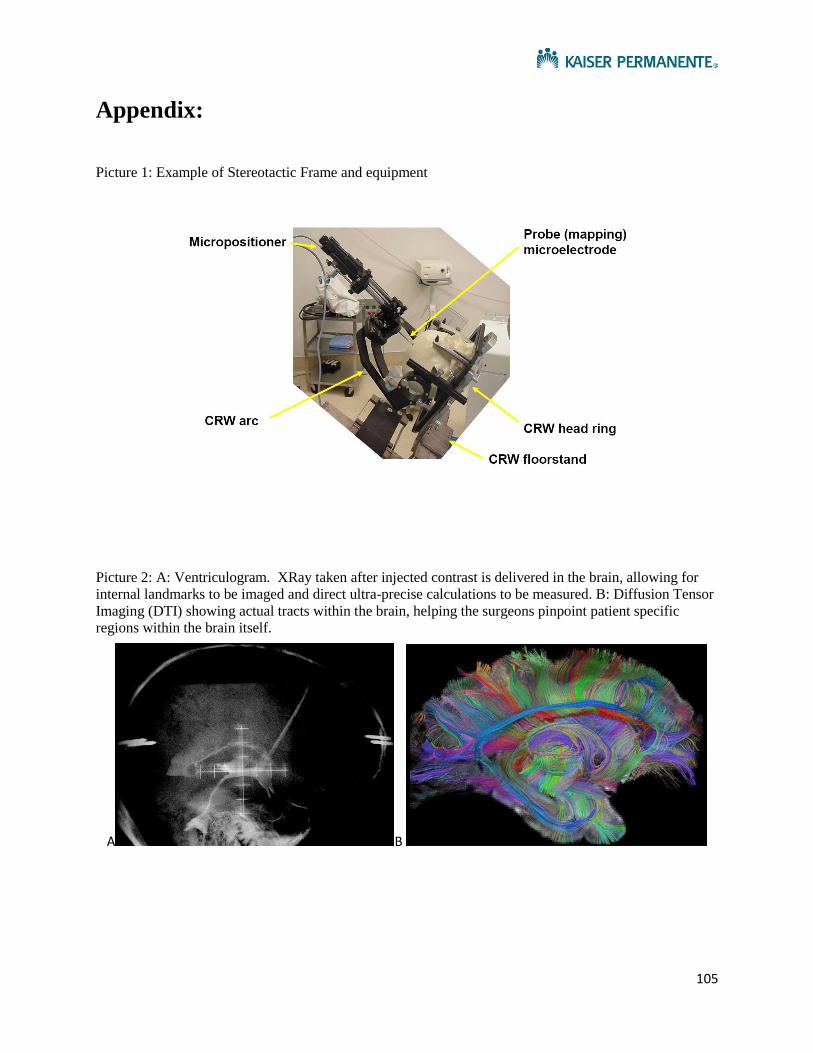
105
Appendix:
Picture 1: Example of Stereotactic Frame and equipment
Picture 2: A: Ventriculogram. XRay taken after injected contrast is delivered in the brain, allowing for
internal landmarks to be imaged and direct ultra-precise calculations to be measured. B: Diffusion Tensor
Imaging (DTI) showing actual tracts within the brain, helping the surgeons pinpoint patient specific
regions within the brain itself.
A B

106
Picture 3: X-Ray setup, with to both AP and Lateral Image Sets
Picture 4: Demonstration of “curving” of a lead in the operating room. Despite this movement, we are
able to calculate a very precise location.

107
Figure 5: Microelectrode recordings are performed, measuring single cell and multiunit activity in the
brain.
Figure 6: Microelectrode recordings demonstrate key findings in different parts of the brain. Image on
left is for STN, right is GPi.

108
Figure 7: Navigus cap, which mounts the electrode lead to the skull
Figure 8: Shows the gradual improvement of the UPDRS III scale in Parkinson’s patients. Note that the
improvement takes about 6 months and is slightly faster with STN stimulation than GPi, but the net
outcome is the same.

109
Figure 9: The narrowed therapeutic window in Parkinson’s Disease is a disabling feature, causing motor
fluctuations to ON/OFF throughout a given day. After DBS, these fluctuations are typically less
prominent and the PD symptoms are overall under better control.
Figure 10: Outcomes from DBS Surgery, Bilateral STN, Score is UPDRS-III

110
Deep Brain Stimulation Program – Kaiser Permanente Redwood City
Essential Tremor
Redwood City Personnel (650) 299 - 2290:
Mark Sedrak, MD: Director of Functional Neurosurgery, Northern California Kaiser Permanente
Patrick Pezeshkian, MD: Functional Neurosurgeon
Ramon Quesada, MD: Physical Medicine and Rehabilitation (PMR) Physician
William T Wong, MD: Interventional Psychiatrist
Diana Bruce, PA: Physician Assistant in Functional Neurosurgery
Ivan Bernstein, PA: Physician Assistant in Functional Neurosurgery, Surgery Scheduling
Supervisor
Eric Sabelman, Phd: Senior Bioengineer in Functional Neurosurgery
Siddharth Srivastava, Phd: Bioengineer in Functional Neurosurgery
Julie Perez: Medical Assistant to Functional Neurosurgery
Redwood City Associated Sites:
Kaiser Permanente – San Leandro (510) 454-3180
Han Lee, MD: Movement Disorder Neurologist
Carol Evans, NP: Movement Disorder Nurse Practitioner
Kaiser Permanente – San Francisco (415) 833 - 4555
Rima Ash, MD: Movement Disorder Neurologist
Kaiser Permanente – Walnut Creek (925) 295 - 6953
Jeff Klingman, MD: Movement Disorder Neurologist, Chief of Neurology in Northern California
Kaiser Permanente
Heidi Shale, MD: Movement Disorder Neurologist

111
Robin Bon, NP: Movement Disorder Nurse Practitioner
Stacey Rohrer, Phd: Neuropsychologist
Maia del Fierro-Lemon, MHCA: Consultant
Susan Bodell, RN: Regional Program Coordinator for the Movement Disorders Deep Brain
Stimulation Program
Other Regional Site:
Kaiser Permanente – Sacramento (925) 295 - 7848
Conrad Pappas, MD, Phd: Functional Neurosurgeon
Suketu Khandhar, MD: Movement Disorder Neurologist, Director of Movement Disorders
Program
Dawn Levine, Phd: Neuropsychologists
Steven Jinks, Phd: Clinical Neuroscientist/Neurophysiologist
May J Kim, Phd: Neuropsychologist
Catherine Nitifan-Young, FNP: Movement Disorders Nurse Practitioner

112
(Ventral Intermediate Nucleus (VIM) of the Thalamus)
How To Become Eligible for Deep Brain Stimulation?
Most patients are initially managed by their local neurologist for their movement disorder
(Parkinson’s Disease, Dystonia, or Essential Tremor). If a patient becomes refractory, or the case
becomes complicated, a referral can be made to the movement disorder specialist, who is a Neurologist at
4 sites across Northern California Kaiser Permanente. The movement disorder specialist will evaluate
the patient, decide on possibly further advanced medical management strategies, which, if the patient’s
symptoms are refractory, may be considered for deep brain stimulation. There are complicated and strict
criteria that need to be met for a patient to become eligible. The first depends on a patients symptoms,
which for Essential Tremor, extremity symptoms are more treatable with DBS. Voice and head
symptoms may be more difficult to control, especially with a single electrode. Lastly, cognitive
dysfunction or significant psychiatric disease may contraindicate the surgery. These are typically
formally assessed using neuropsychological evaluation (s). A screening MRI is typically performed at
the home facility to identify any obvious structural abnormalities or issues that may make surgery
difficult or dangerous.
If there are no clear contraindications to surgery, the case file is brought to the Movement
Disorders Board, which is a panel that meet approximately every 6 weeks. During this meeting, the entire
case is reviewed, history, medication trials, videos of patient, neuropsychological evaluation, and
MRI/Imaging. A group consensus needs to be made for patient to continue on meeting the surgical team.
Also, during this meeting, a decision about the target is typically made. For essential tremor, the target is
the thalamus (Ventral Intermediate Nucleus of the Thalamus). For dystonia, the target is typically the
globus pallidus interna (GPi). For Parkinson’s disease, there are multiple possible targets, but most
commonly subthalamic nucleus (STN) and globus pallidus interna (GPi). In PD, STN may be favored by
our group if patient doesn’t have significant psychiatric comorbidities, impulse control problems, if there
is tremor dominance, and to maximize medication reduction. The literature is essentially split between
these two targets, but STN is a smaller target that requires higher precision than GPi and STN has a
slightly faster response time (figure 1). STN can also be used in difficult/complex essential tremor
patients as a secondary resort. Lastly, there is usually a decision about staging an operation (2 surgeries
instead of 1) and also about different battery types (Activa PC, dual channel non-rechargeable; Activa SC,
single channel non-rechargeable, or Activa RC, dual channel rechargeable). The advantage of the dual
channel devices, is that the patient would only require 1 general anesthesia event (during the time of
tunneling to the chest). However, in comparing the SC to the PC, the SC has about 30% more battery
life. SC’s and PC’s typically last about 3-6 years and require replacement surgery which is an outpatient
local anesthetic procedure. The RC requires charging, typically about 20-30minutes every 1-2 days, but if
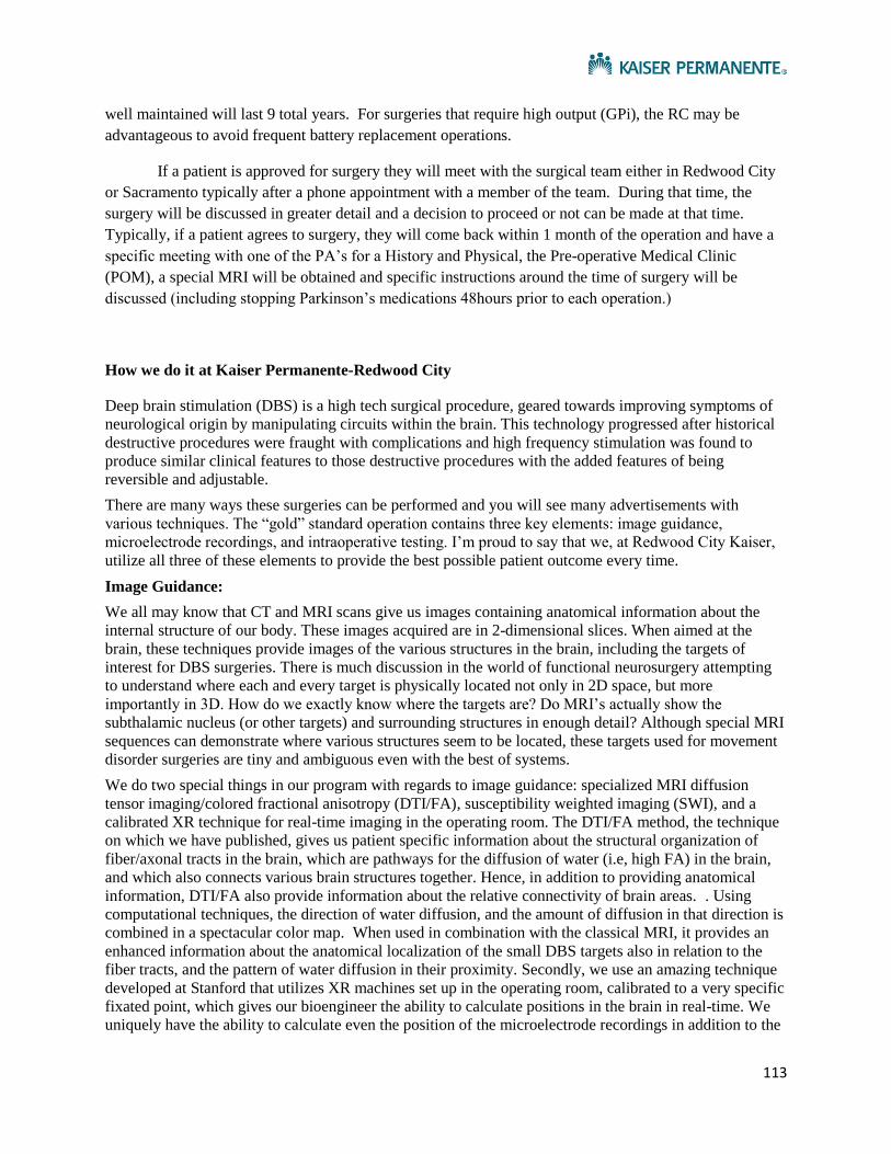
113
well maintained will last 9 total years. For surgeries that require high output (GPi), the RC may be
advantageous to avoid frequent battery replacement operations.
If a patient is approved for surgery they will meet with the surgical team either in Redwood City
or Sacramento typically after a phone appointment with a member of the team. During that time, the
surgery will be discussed in greater detail and a decision to proceed or not can be made at that time.
Typically, if a patient agrees to surgery, they will come back within 1 month of the operation and have a
specific meeting with one of the PA’s for a History and Physical, the Pre-operative Medical Clinic
(POM), a special MRI will be obtained and specific instructions around the time of surgery will be
discussed (including stopping Parkinson’s medications 48hours prior to each operation.)
How we do it at Kaiser Permanente-Redwood City
Deep brain stimulation (DBS) is a high tech surgical procedure, geared towards improving symptoms of
neurological origin by manipulating circuits within the brain. This technology progressed after historical
destructive procedures were fraught with complications and high frequency stimulation was found to
produce similar clinical features to those destructive procedures with the added features of being
reversible and adjustable.
There are many ways these surgeries can be performed and you will see many advertisements with
various techniques. The “gold” standard operation contains three key elements: image guidance,
microelectrode recordings, and intraoperative testing. I’m proud to say that we, at Redwood City Kaiser,
utilize all three of these elements to provide the best possible patient outcome every time.
Image Guidance:
We all may know that CT and MRI scans give us images containing anatomical information about the
internal structure of our body. These images acquired are in 2-dimensional slices. When aimed at the
brain, these techniques provide images of the various structures in the brain, including the targets of
interest for DBS surgeries. There is much discussion in the world of functional neurosurgery attempting
to understand where each and every target is physically located not only in 2D space, but more
importantly in 3D. How do we exactly know where the targets are? Do MRI’s actually show the
subthalamic nucleus (or other targets) and surrounding structures in enough detail? Although special MRI
sequences can demonstrate where various structures seem to be located, these targets used for movement
disorder surgeries are tiny and ambiguous even with the best of systems.
We do two special things in our program with regards to image guidance: specialized MRI diffusion
tensor imaging/colored fractional anisotropy (DTI/FA), susceptibility weighted imaging (SWI), and a
calibrated XR technique for real-time imaging in the operating room. The DTI/FA method, the technique
on which we have published, gives us patient specific information about the structural organization of
fiber/axonal tracts in the brain, which are pathways for the diffusion of water (i.e, high FA) in the brain,
and which also connects various brain structures together. Hence, in addition to providing anatomical
information, DTI/FA also provide information about the relative connectivity of brain areas. . Using
computational techniques, the direction of water diffusion, and the amount of diffusion in that direction is
combined in a spectacular color map. When used in combination with the classical MRI, it provides an
enhanced information about the anatomical localization of the small DBS targets also in relation to the
fiber tracts, and the pattern of water diffusion in their proximity. Secondly, we use an amazing technique
developed at Stanford that utilizes XR machines set up in the operating room, calibrated to a very specific
fixated point, which gives our bioengineer the ability to calculate positions in the brain in real-time. We
uniquely have the ability to calculate even the position of the microelectrode recordings in addition to the

114
DBS electrode. These can be calculated relative to each other or relative to referential structures in the
brain, the actual landmarks used for MRI/CT guidance. In addition, this method is the most accurate
method of calculating positions that is known in the world (accurate to 0.6mm or better)! It is more
accurate than intraoperative MRI, intraoperative CT, fluoroscopy, or frameless systems. In addition, it is
notable that MRI and CT systems have a theoretical problem of depth/angular measurement error as a
result of software “smoothing” called interpolation. The XR system we use, does not have this problem.
So at the end of our surgeries, we are quite certain about our stereotactic location.
Microelectrode Recordings:
It is amazing to think that we now have the ability to measure electrical activity (action potentials) created
by single neurons and a small cluster of neurons in the brain. This ability to visualize brain activity at the
level of a single neuron has led to the discovery of many structures within the brain, hinting at their role
in understanding the functional organization of this extremely complex organ. This method has become a
very useful tool for neurosurgeons to identify many of the key targets during deep brain stimulation.
During the time we measure these actual “action potentials” (actual neurons firing!), we can induce
activity by manipulating the arm, the leg, or sometimes by flashing light in the eyes and many other
maneuvers. The target areas of the brain become very active when patients are awake and off of their
medications (typically 1-2 days). This is a second layer of assurance that have identified the proper
position in the brain. We perform an XR when we identify the target and ensure that the DBS electrode
eventually will make it to the same exact spot we want, with the help of our calibrated XR technique.
In addition to these MER’s, we’ve recently started utilizing what are called “local field potential”
recordings. This method is similar to what many know as EEG/ECOG, where pools of neurons are
sampled for their electrical activity. However, we are measuring these fields of neurons deep within the
brain rather than on the surface. It turns out that many of the LFP signals may in fact help us understand
the overall activity in a certain portion of the brain. This information is typically not utilized during the
procedure, but can be analyzed when recorded afterwards.
Intraoperative Testing:
The last and final test is one of the most important. “What happens when we introduce electricity in the
brain at a specific target?”. This is a very critical step during the surgery. Patients do receive much local
anesthesia at incision site (like numbing medication at dentist) in addition to having an anesthesiologist
who administers some meds that won’t interfere with surgery, allowing some sedation (some patients
even sleep). The awake testing can yield very critical information to the surgeon. For example,
occasionally we can map the area we think is the best target utilizing MER, have our image guidance to
get us to where we want to be, and we find that with stimulation at that precise target, we induce effects
that suggest we are too close to surrounding structures. When this occurs, our knowledge of the mapped
area and our image guidance help steer us away from these surrounding areas, giving our patients the best
possible outcome and allowing our programmers the greatest degree of flexibility.

115
Staging Each Side of the surgery:
We stage each side of the surgery for essentially 3 reasons. First, we have found that when both sides of
the brain are exposed, patient tend to have more post-operative confusion. When each side is done
individually, patients typically only require one night hospital stay. Second, we have noticed increased
inaccuracy when both sides are done simultaneously, and our technique has demonstrated increased brain
shift when this is done. Third, the physiology in the target site may actually be affected by the first site of
implant. There is evidence that these regions are interconnected, both right and left. This interference
seems to dissipate in a few weeks. We typically implant a dual channel implantable pulse generator (IPG;
Activa PC or Activa RC) at the end of the second surgery under general anesthesia. If we implant single
channel IPG’s (Activa SC), we typically implant each generator at the end of each individual surgery. It is
notable that we can do the surgeries in this format, which is our preference, because we are not under
reimbursement practices as a result of Medicare and other insurers.
Activation of Device and Titration of Medications
Typically after the entire system is implanted surgically, the device is off. Some patients notably have a
“microlesioning” effect, where symptoms may improve slightly over hours, days, or weeks and then
return to baseline. We allow this effect and any small amount of swelling in the brain to improve before
turning on the device. This programming is typically done by the referring specialized movement disorder
neurologist or associated personnel, about 2-4 weeks after surgery completion. Final effects from the DBS
system, which has thousands of settings, may not be present for 6-12 months.
Team Approach:
It is important to realize that the physicians of Kaiser are truly outstanding individuals, many of which are
excellent at what they do, graduating top of their respective classes, and from top universities around the
country. A major benefit of being a part of Kaiser, is that these physicians are truly driven to have the best
clinical results. The clinicians are outcome driven, not income driven. Our approach in Northern
California Kaiser, has been to take advantage of the system and integrate a full team approach for treating
our movement disorders patients. With this in mind, a board was created, incorporating: functional
neurosurgeons, movement disorder neurologists, neuropsychologists, bioengineers, physician assistants,
nurse practitioners, nurses, and administrators who all take part in creating a consensus decision for each
patient. This is an organized meeting where patients are formally presented, they are videotaped in “On”
and “Off” medication states, formal UPDRS-III (Parkinson’s Scale) scores are tabulated, MRI’s are
reviewed, neuro-psychological evaluations are discussed. With all this information in mind, the board
decides who will most likely have a significant benefit and who may not, who may be high risk and who

116
should be able to tolerate the surgery without a problem. This type of board meeting is very unique. Most
institutions simply work using “hallway conversations” or simple consultations/referrals for this type of
advanced surgery. In addition, we have a dedicated group in the operating room, anesthesiologists, nurses,
operating room technicians, circulators, all specifically dedicated to DBS surgeries. This team approach
adds another layer of clinical excellence to our overall program.

117
Appendix:
Picture 1: Example of Stereotactic Frame and equipment
Picture 2: A: Ventriculogram. XRay taken after injected contrast is delivered in the brain, allowing for
internal landmarks to be imaged and direct ultra-precise calculations to be measured. B: Diffusion Tensor
Imaging (DTI) showing actual tracts within the brain, helping the surgeons pinpoint patient specific
regions within the brain itself.
A B
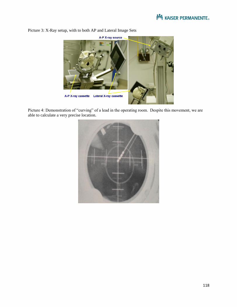
118
Picture 3: X-Ray setup, with to both AP and Lateral Image Sets
Picture 4: Demonstration of “curving” of a lead in the operating room. Despite this movement, we are
able to calculate a very precise location.

119
Figure 5: Microelectrode recordings are performed, measuring single cell and multiunit activity in the
brain.
Figure 6: Microelectrode recordings demonstrate key findings in different parts of the brain

120
Figure 7: Navigus cap, which mounts the electrode lead to the skull
Figure 8: Depiction of preoperative tremor (A) and post-operative with DBS on (B)
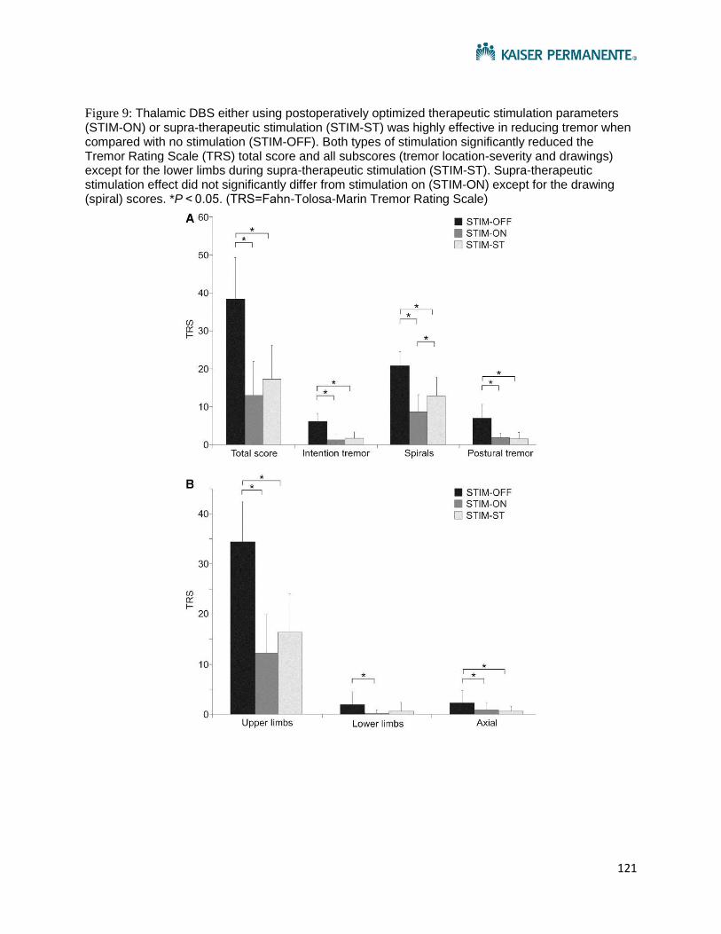
121
Figure 9: Thalamic DBS either using postoperatively optimized therapeutic stimulation parameters (STIM-ON) or supra-therapeutic stimulation (STIM-ST) was highly effective in reducing tremor when compared with no stimulation (STIM-OFF). Both types of stimulation significantly reduced the Tremor Rating Scale (TRS) total score and all subscores (tremor location-severity and drawings) except for the lower limbs during supra-therapeutic stimulation (STIM-ST). Supra-therapeutic stimulation effect did not significantly differ from stimulation on (STIM-ON) except for the drawing (spiral) scores. *P < 0.05. (TRS=Fahn-Tolosa-Marin Tremor Rating Scale)
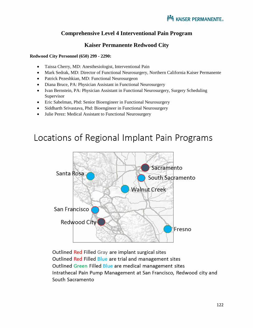
122
Comprehensive Level 4 Interventional Pain Program
Kaiser Permanente Redwood City
Redwood City Personnel (650) 299 - 2290:
Taissa Cherry, MD: Anesthesiologist, Interventional Pain
Mark Sedrak, MD: Director of Functional Neurosurgery, Northern California Kaiser Permanente
Patrick Pezeshkian, MD: Functional Neurosurgeon
Diana Bruce, PA: Physician Assistant in Functional Neurosurgery
Ivan Bernstein, PA: Physician Assistant in Functional Neurosurgery, Surgery Scheduling
Supervisor
Eric Sabelman, Phd: Senior Bioengineer in Functional Neurosurgery
Siddharth Srivastava, Phd: Bioengineer in Functional Neurosurgery
Julie Perez: Medical Assistant to Functional Neurosurgery

123
Kaiser Permanente – Northern California Service Areas
DIABLO Service Area (WCR, MTZ, ANT) 925-372-1741
DIABLO 1 ANS Epidural Clinic
Dr Angela Chiang Dr Cynthia Rahn
Dr Mark Rahn Dr Sabrina Martinez
Dr Prakash Raygor Dr Mark Moore
DIABLO 2 ANS Interventional Pain Clinic 925-372-1283
Dr Andrew Maher
Dr James Mura
Dr Darko Vodopich
Dr. Norm Aleks
DIABLO 3 PMR/SPINE CLINIC (925) 313-4740
Dr. Eric Alexander
Fresno (559) 448-4555
Dr. Eugene Huang (ANS)
Dr. Jonathan Grossman (PMR/SPINE CLINIC)
Dr. Rupinder Singh (ER)
Dr. Marta Bator (ANS - Pool)
Modesto & Stockton Chronic Pain & Interventional Pain 209-735-3255
Dr Tanja Frey - ans
Dr Brandon Valine
Dr. Raul Calderon

124
Petaluma Intervention Pain Clinic PMR/SPINE CLINIC 415-444-2988
Dr Diane Murphy
Dr Vitto
Dr Tabitha Washington
Redwood City 1 of 2– ANS/NSG 650-299-5454
Dr. Taissa Cherry (ANS, Dept of Neurosurg)
Dr. Mark Sedrak (NSG)
Dr. Patrick Pezeshkian (NSG)
PA Ivan Bernstein
PA Diana Bruce
Redwood City 2 of 2 – PMR/SPINE CLINIC 650-299-4741
Dr Firtch Dr Ray Lai
Dr Maratukulam Dr Nguyen
Dr Treinen
Richmond PMR/SPINE CLINIC 510-307-1661
Dr John H Lim PMR/SPINE CLINIC
Dr Angelita Balbas PMR/SPINE CLINIC
Roseville PMR/SPINE CLINIC 916-771-6611
Dr William Fenton - ANS
Dr Mark Tyburski
Dr Ryan Carver
Sacramento (916) 973-5490

125
Conrad Pappas, MD (Neurosurgery)
San Francisco Pain Interventional Center- ANS 415-833-0095
Dr Justin McKendry ANS
Dr Taissa Cherry ANS
Dr Salman Dasti
San Leandro (Union City) books appts/epidurals are done in Fremont PMR/SPINE
CLINIC (510) 675-3070
Dr Eunice Lau (chief) - PMR/SPINE CLINIC
Dr Sergiy Rybiy
Dr Raghu Katragadda ANS
San Jose (Santa Theresa) Interventional Pain ANS 408-972-6283
Dr Darshan Patel Dr Samuel Park
Dr Shabeen Tharani Dr Prasad Movva
Dr Laurence Won Dr Vital Kandula
San Rafael PMR/SPINE CLINIC 415-444-2988
Dr Murphy
Dr Vitto
Santa Rosa 1 of 2 – PMR/SPINE CLINIC 707-566-5557
Dr David Vidaurri
Drs. Donald F Green
Hari Lakshmanan
Kirk Pappas

126
Tracey Jones
Todd Weitzenberg
Santa Rosa 2 of 2– Chronic/Interventional Pain ANS 707-571-3921
Dr Andrea Rubinstein (ANS)
Dr. Tabitha Washington (ANS)
South Sacramento Pain Clinic (ANS) (916) 688-6353
Dr Mike Bicocca
Dr Rod Youssefi
Dr Kegang Hu
Dr Sabina Tahera
South San Francisco ANS Pain Services 650-742-2395
Dr Eddie Busracamwongs ANS
Dr. Hamid Motamed
Dr. Tin-Na Kan
Dr. Ali Abdollahi-Fard
Santa Clara PMR/SPINE CLINIC 408-851-9200
Dr Richard Kim
Dr Kevin Wang
Dr Dhiruj Kirpalani
Vallejo – PMR/SPINE CLINIC (707) 651-1025
Dr. Linda Cho (PMR/SPINE CLINIC)

127
SPINAL CORD STIMULATION, PERIPHERAL NERVE STIMULATION
Spinal Cord Stimulation (SCS) or Dorsal Column Stimulation (DCS) is a treatment for primarily for limb
pain, chronic regional pain syndrome (CRPS). This technology uses a low- voltage current to block nerve
impulses in the spinal cord traveling from a troubled area to the brain. Peripheral Nerve Stimulation
(PNS) uses the same technology, with targeted nerves and nerve bundles. PNS is useful in some
headaches, facial pain, pelvic and groin pain. These technologies are not cures for these various pain
syndromes, but sometimes can alleviate pain. Stimulation typically induces a smooth tingling sensation
in the area of interest. The amount of pain relief varies for each person.
Trial Period
While SCS and PNS can be helpful in a number of people, it does not work in everybody. To help
decide in whom this may or may not work, almost all patients have a trial implant to test the
effectiveness. Typically for this week long trial period, we are looking for a substantial amount
of ongoing pain relief, for specific areas of the stimulators which are contributing to pain relief,
and in the end or a significant functional improvement in a patient’s level of function and daily
living. Some patients have some pain relief, but not enough to qualify for implant.
Furthermore, some patients have no pain relief and likewise do not qualify.
The SCS trial begins with the surgical implant of the leads, usually done via a needle puncture,
where the leads are placed along the back portion of your spinal cord. In the case of PNS, the
leads are placed via needle puncture along the area in question as determined by the surgeon
based on your description of location.
Little numbing medication is used during the insertion of the trial stimulator, in order to be able
to test proper placement during the surgical procedure. You will be made comfortable for the
surgery by the anesthesiologist (“twilight anesthesia”), but will remain awake so that the
surgeon can determine if the lead placement is correct for your symptoms. Your feedback is
important as you know where your pain is. We are looking for tingling in the area of the pain.
We want to know during the procedure if we are inducing tingling in the area of interest and if
we are covering it completely or if we are missing a particular area.
The tails leads are external, sutured and taped in place, and you will go home with the leads
connected to a special box that will behave just as the implanted device would.
In the recovery room, and/or post-operative period, a representative from the company your
doctor has chosen will do some programming and teach you how to use the patient remote
control. Most people leave the hospital on the day of surgery. You may not shower for the
entire period of the trial.
You will return on several occasions during the 7-10 period to have programming, working
closely with the rep to achieve the best coverage and pain reduction possible. It is critically
important during this trial period to keep a meticulous LOG of your pain levels, the program that

128
you are using, and to try different programs and turning device on and off to make sure it is
functioning well for you.
The trial will end either when it is determined it is a successful trial or maximum on day 10. The
leads are then pulled in the clinic at the bedside. This is a relatively painless procedure and you
may shower the next day.
Occasionally, the trial period is performed using a full implanted electrode, which is buried
underneath the skin like a permanent lead. This permanent lead is connected to temporary
extensions and brought externally for a trial period. In this type of situation, the end of the trial
results in either a full implant with a battery, or removal of the buried leads. Either way, this
requires open surgery.
Permanent Implant
For those who have a successful trial, a permanent full implantation may be the next step. This
surgery is typically a much bigger procedure than the trial, involving larger incisions. Patients
may also be in the hospital at least overnight after a permanent implant. You may have the full
implant surgery as early as three weeks after your lead pull, but sometimes a bit longer.
The surgery will go much the same as the trial with the addition of the IPG or generator
placement. Most people choose this to be at the upper buttocks/flank, abdomen, or upper
chest and you will have discussed this with your surgeon in advance.
You will have programming done similarly to the trial period. You will receive wound care
instructions before you go home.
Programming/Adjustment
About 1- 2 weeks after your surgery you may return for programming. You will have post-
operative care and can use those opportunities to have programming done by making
arrangements both with the company’s representative at the clinic where you are being seen.
You can request additional sessions when you think your stimulation is not exactly where you
need it. This can be done at any Kaiser facility, but must be under the supervision of a medical
doctor, and must be coordinated with the provider and the company representative.

129
Intrathecal Pump Therapy
Intrathecal pump therapy (ITP) is a pump device with a catheter that utilizes pain medications for
infusion. ITP are useful for low back pain and cancer pain and are often used as a second line to
stimulation therapy. The advantage of ITP is that it can afford a continuous infusion of pain medicine
and can work for axial pain and cancer pain. The disadvantage of the therapy is that it required physical
filling of the pump with a needle procedure every 6 weeks to 3 months and carries more long-term risk,
especially of withdrawal problems. The amount of pain relief varies for each person.
Trial Period
While ITP can be helpful in a number of people, it does not work in everybody. To help decide in
whom this may or may not work, almost all patients have a trial to test the effectiveness.
Typically for this week long trial period, we are looking for a substantial amount of ongoing pain
relief, typically more than 50%.
Permanent Implant
For those who have a successful trial, a permanent full implantation may be the next step. This
surgery is typically a much bigger procedure than the trial, involving larger incisions. Patients
may also be in the hospital at least overnight after a permanent implant. You may have the full
implant surgery as early as three weeks after your catheter trial, but sometimes a bit longer.
The surgery will go much the same as the trial with an incision in the back and in the abdomen
on the left or right side. You will have discussed this with your surgeon in advance.
Your infusion will initially be set at a very low dose to avoid over-infusion problems. Over the
next several months and years, the device can be programmed to optimally achieve therapy.
You will receive wound care instructions before you go home.
Programming/Adjustment
Shortly after your surgery you may return for programming. You will have post-operative care
and can use those opportunities to have programming done by making arrangements with the
care management team, typically at either San Francisco, Redwood City, or South Sacramento
facilities. You can request additional sessions when you think you’re not exactly where you need
to be for pain control. It is notable that not only can the infusion rate be adjusted with the
device, but the actual formulation of the medications can be changed during each refill period,
offering nearly innumerable possibilities.

130
Appendix
Figure 1: Spinal cord stimulation
Spinal cord Stimulator as implanted X-ray of a single lead in place
Figure 2: Peripheral Nerve stimulation, occipital and supraorbital locations
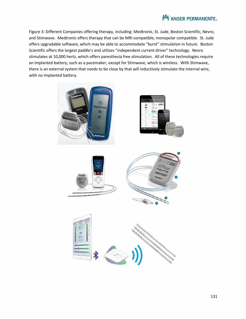
131
Figure 3: Different Companies offering therapy, including: Medtronic, St. Jude, Boston Scientific, Nevro,
and Stimwave. Medtronic offers therapy that can be MRI compatible, monopolar compatible. St. Jude
offers upgradable software, which may be able to accommodate “burst” stimulation in future. Boston
Scientific offers the largest paddle’s and utilizes “independent current driver” technology. Nevro
stimulates at 10,000 hertz, which offers paresthesia free stimulation. All of these technologies require
an implanted battery, such as a pacemaker, except for Stimwave, which is wireless. With Stimwave,
there is an external system that needs to be close by that will inductively stimulate the internal wire,
with no implanted battery.

132
Figure 4: Differences between percutaneous and paddle leads, percutaneous are wire and cylindrical,
whereas paddles are flat like a spatula
Figure 5: Intrathecal Pump, Medtronic Synchromed II, there are 20cc and 40cc options