Pathophysiology of the respiratory system II - common diseases
55
Pathophysiology of the respiratory system II - common diseases Respiratory insufficiency Classification of respiratory diseases - ventilation disease - diffusion diseases - perfusion diseases Chronic obstructive pulmonary disease (COPD) Bronchial asthma
Transcript of Pathophysiology of the respiratory system II - common diseases

Pathophysiology of the respiratory system II - common diseases
Respiratory insufficiencyClassification of respiratory diseases- ventilation disease- diffusion diseases- perfusion diseasesChronic obstructive pulmonary disease (COPD)Bronchial asthma
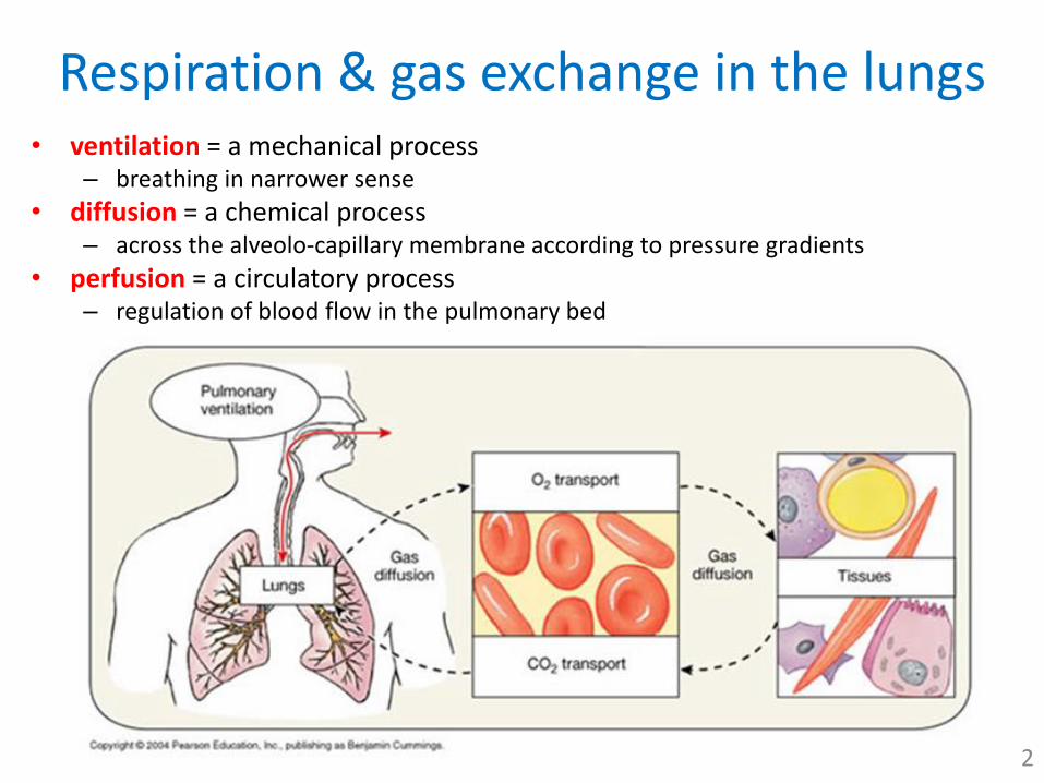
Respiration & gas exchange in the lungs• ventilation = a mechanical process
– breathing in narrower sense
• diffusion = a chemical process– across the alveolo-capillary membrane according to pressure gradients
• perfusion = a circulatory process– regulation of blood flow in the pulmonary bed
2
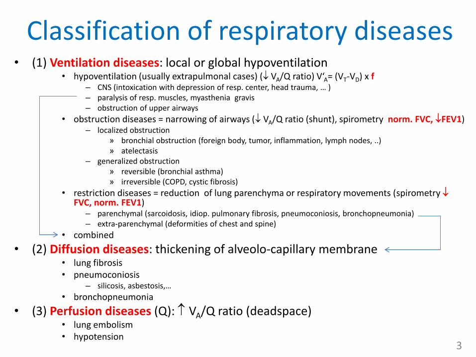
Classification of respiratory diseases• (1) Ventilation diseases: local or global hypoventilation
• hypoventilation (usually extrapulmonal cases) ( VA/Q ratio) V‘A= (VT-VD) x f– CNS (intoxication with depression of resp. center, head trauma, … )– paralysis of resp. muscles, myasthenia gravis– obstruction of upper airways
• obstruction diseases = narrowing of airways ( VA/Q ratio (shunt), spirometry norm. FVC, FEV1)– localized obstruction
» bronchial obstruction (foreign body, tumor, inflammation, lymph nodes, ..)» atelectasis
– generalized obstruction» reversible (bronchial asthma)» irreversible (COPD, cystic fibrosis)
• restriction diseases = reduction of lung parenchyma or respiratory movements (spirometry FVC, norm. FEV1)
– parenchymal (sarcoidosis, idiop. pulmonary fibrosis, pneumoconiosis, bronchopneumonia)– extra-parenchymal (deformities of chest and spine)
• combined
• (2) Diffusion diseases: thickening of alveolo-capillary membrane• lung fibrosis• pneumoconiosis
– silicosis, asbestosis,…
• bronchopneumonia
• (3) Perfusion diseases (Q): VA/Q ratio (deadspace)• lung embolism• hypotension
3
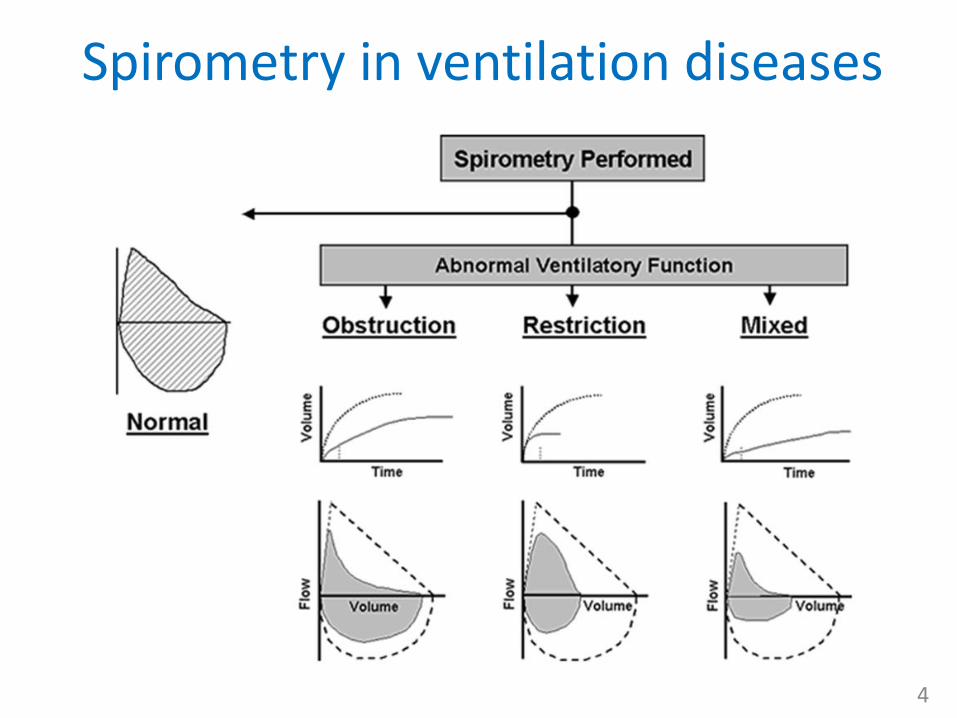
Spirometry in ventilation diseases
4
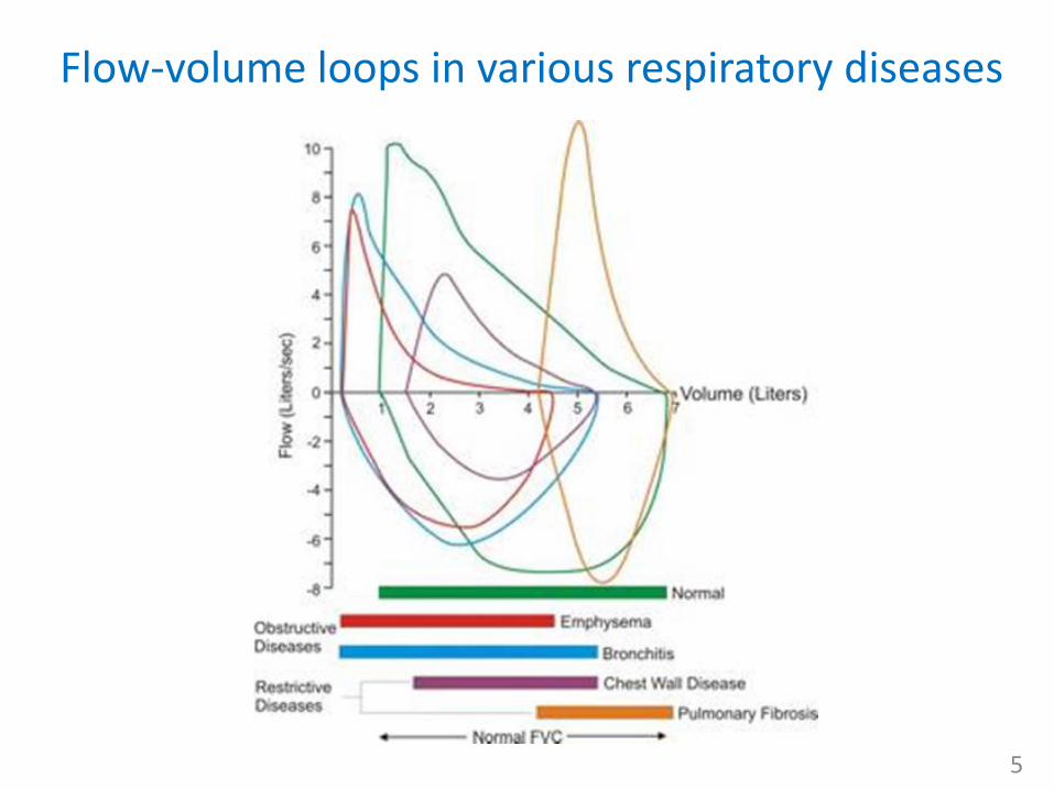
Flow-volume loops in various respiratory diseases
5
RESPIRATORY INSUFFICIENCY
6
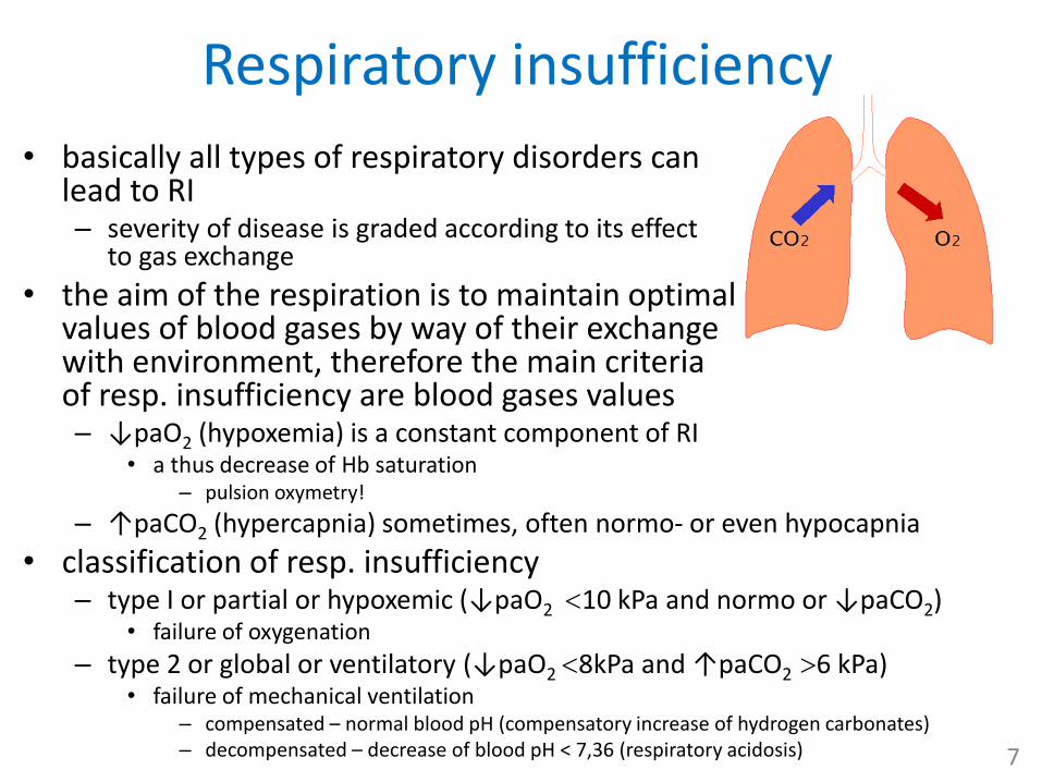
Respiratory insufficiency
• basically all types of respiratory disorders can lead to RI– severity of disease is graded according to its effect
to gas exchange
• the aim of the respiration is to maintain optimal values of blood gases by way of their exchange with environment, therefore the main criteria of resp. insufficiency are blood gases values– ↓paO2 (hypoxemia) is a constant component of RI
• a thus decrease of Hb saturation – pulsion oxymetry!
– ↑paCO2 (hypercapnia) sometimes, often normo- or even hypocapnia
• classification of resp. insufficiency– type I or partial or hypoxemic (↓paO2 10 kPa and normo or ↓paCO2)
• failure of oxygenation
– type 2 or global or ventilatory (↓paO2 8kPa and ↑paCO2 6 kPa)• failure of mechanical ventilation
– compensated – normal blood pH (compensatory increase of hydrogen carbonates) – decompensated – decrease of blood pH < 7,36 (respiratory acidosis) 7
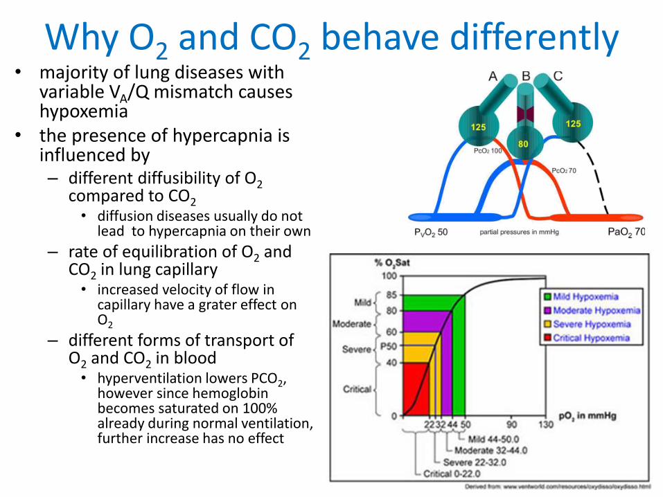
Why O2 and CO2 behave differently• majority of lung diseases with
variable VA/Q mismatch causes hypoxemia
• the presence of hypercapnia is influenced by– different diffusibility of O2
compared to CO2• diffusion diseases usually do not
lead to hypercapnia on their own
– rate of equilibration of O2 and CO2 in lung capillary• increased velocity of flow in
capillary have a grater effect on O2
– different forms of transport of O2 and CO2 in blood • hyperventilation lowers PCO2,
however since hemoglobin becomes saturated on 100%already during normal ventilation, further increase has no effect
8
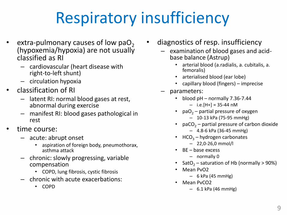
Respiratory insufficiency• extra-pulmonary causes of low paO2
(hypoxemia/hypoxia) are not usually classified as RI– cardiovascular (heart disease with
right-to-left shunt)– circulation hypoxia
• classification of RI– latent RI: normal blood gases at rest,
abnormal during exercise – manifest RI: blood gases pathological in
rest
• time course: – acute: abrupt onset
• aspiration of foreign body, pneumothorax, asthma attack
– chronic: slowly progressing, variable compensation • COPD, lung fibrosis, cystic fibrosis
– chronic with acute exacerbations:• COPD
• diagnostics of resp. insufficiency– examination of blood gases and acid-
base balance (Astrup) • arterial blood (a.radialis, a. cubitalis, a.
femoralis) • arterialised blood (ear lobe) • capillary blood (fingers) – imprecise
– parameters: • blood pH – normally 7.36-7.44
– i.e.[H+] = 35-44 nM
• paO2 – partial pressure of oxygen – 10-13 kPa (75-95 mmHg)
• paCO2 – partial pressure of carbon dioxide – 4.8-6 kPa (36-45 mmHg)
• HCO3 – hydrogen carbonates – 22,0-26,0 mmol/l
• BE – base excess– normally 0
• SatO2 – saturation of Hb (normally > 90%) • Mean PvO2
– 6 kPa (45 mmHg)
• Mean PvCO2– 6.1 kPa (46 mmHg)
9

Generalized hypoxia• = deficiency of O2 in the organism (paO2 10kPa/75mm Hg)• types:
– (1) hypoxemic hypoxia = arterial PO2 - leads to central cyanosis• causes of hypoxemia
– PO2 in inspired air (PO2 (high altitude, low FiO2)– hypoventilation due to damage of respiration center– diffusion impairment (fibrosis, emphysema)– anatomical shunting of non-oxygenated blood (heart)– ventilation-perfusion mismatch
– (2) anemic hypoxia = normal arterial PO2• concentration of hemoglobin
– anemia, leukemias
• abnormal hemoglobin with low ability to bind oxygen– carbonylhemoglogin (COHb )– methemoglobin
– - (3) circulatory hypoxia = normal arterial PO2 – leads to peripheral cyanosis• decreased cardiac output• decreased of systemic blood pressure• (local tissue ischemia) • microcirculation defects
– - (4) histotoxic hypoxia – normal arterial PO2 , venous PO2• Intoxication with cyanides, cobalt, …)
10

pO2
kidney
erythropoetin
Blood marrow
erytrop
oesis
Sugar
Lipids
Proteins
cell
+ O2 CO2
CO2
H2O
mitochondria
mitochondria
Histotoxic hypoxia
Hypoxic hypoxia
O2
Circulation hypoxia (ischemic)
Anemic hypoxia

Intermittent, chronic intermittent and chronic hypoxia
• Intermittent hypoxia – an effective stimulus for evoking the respiratory, cardiovascular, and
metabolic to some extent beneficial • they may provide protection against disease as well as improve exercise performance
in athletes
• Long-term consequences of chronic intermittent hypoxia (such as OSA) may have detrimental effects– hypertension, cerebral and coronary vascular
problems– right ventricular heart mass, pulmonary
vascular remodeling and pulmonary hypertension– developmental and neurocognitive deficits and
neurodegeneration
• Chronic hypoxia induces proliferation of the vasculature due to angiogenesis (up-regulation of VEGF) but can also change the integrity of vessels, leading to changes in vascular permeability (e.g. contribution to acute mountain sickness)
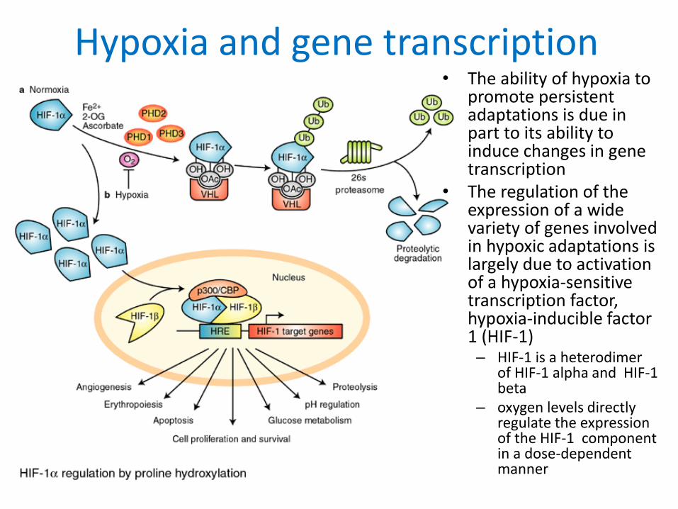
Hypoxia and gene transcription• The ability of hypoxia to
promote persistent adaptations is due in part to its ability to induce changes in gene transcription
• The regulation of the expression of a wide variety of genes involved in hypoxic adaptations is largely due to activation of a hypoxia-sensitive transcription factor, hypoxia-inducible factor 1 (HIF-1)– HIF-1 is a heterodimer
of HIF-1 alpha and HIF-1 beta
– oxygen levels directly regulate the expression of the HIF-1 component in a dose-dependent manner
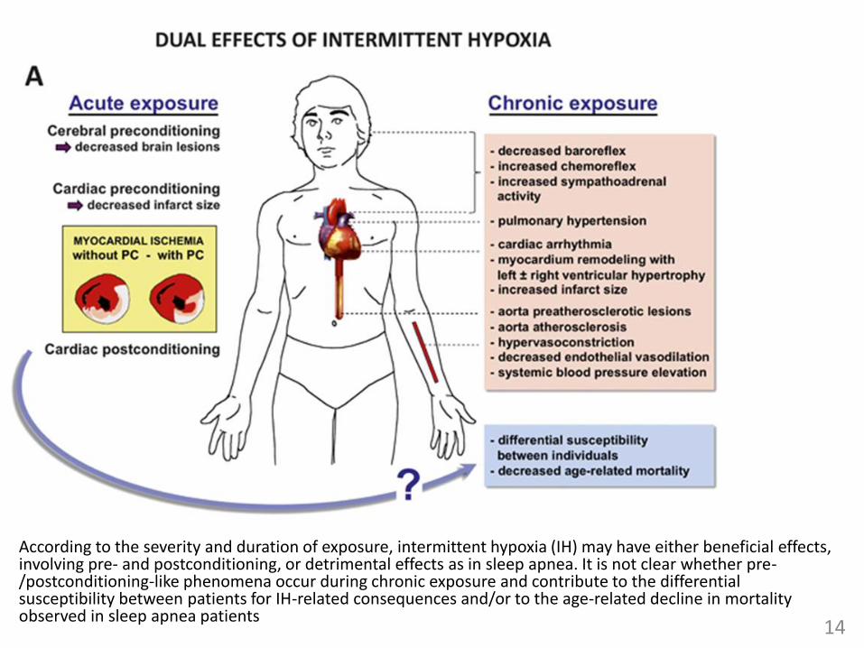
According to the severity and duration of exposure, intermittent hypoxia (IH) may have either beneficial effects, involving pre- and postconditioning, or detrimental effects as in sleep apnea. It is not clear whether pre-/postconditioning-like phenomena occur during chronic exposure and contribute to the differential susceptibility between patients for IH-related consequences and/or to the age-related decline in mortality observed in sleep apnea patients
14

MOST COMMON OBSTRUCTION DISEASES
15

Breathing = periodical changes in preassures and resistances
16

Ventilation disorders due to bronchial obstruction –basic pathophysiological characteristics
17
• obstruction in airways massively increases their resistance (dynamic resistance)– Hagen-Poiseuille law
• since inspirium is an active process (muscles and negative alv. and transthoracic pressure overcome resistances) but expirium passive one, obstruction leads to an impairment of expiration
• participation of auxiliary respiratory muscles leads to – dynamic compression, air trapping
and hyperinflation of the lungs• ↑ residual volume (FRC, RV, TLC)
– ↑ breathing effort and thus dyspnea
• ↓dynamic ventilatory parameters (spirometry)– more time needed to exhale FVC
(↓FEV1)
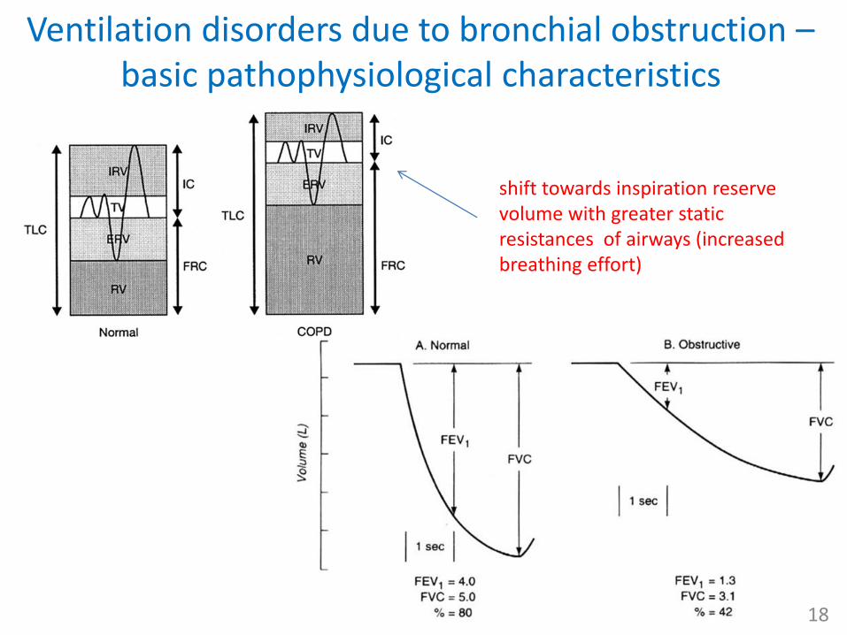
Ventilation disorders due to bronchial obstruction –basic pathophysiological characteristics
18
shift towards inspiration reserve volume with greater static resistances of airways (increased breathing effort)

Ventilation disorders due to bronchial obstruction –basic pathophysiological characteristics
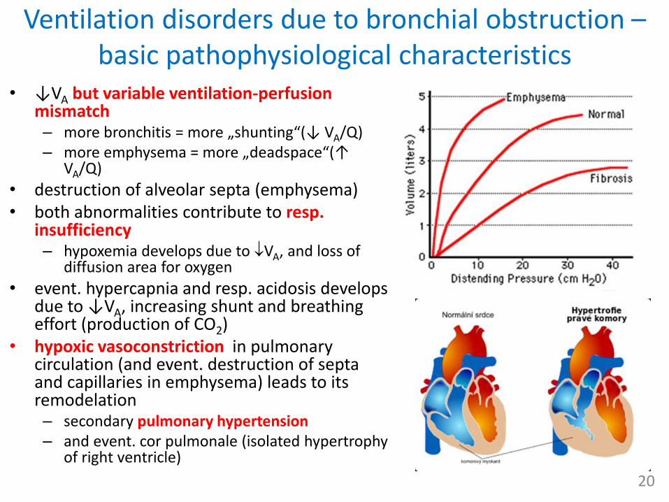
• ↓VA but variable ventilation-perfusion mismatch– more bronchitis = more „shunting“(↓ VA/Q)– more emphysema = more „deadspace“(↑
VA/Q)
• destruction of alveolar septa (emphysema)• both abnormalities contribute to resp.
insufficiency– hypoxemia develops due to VA, and loss of
diffusion area for oxygen
• event. hypercapnia and resp. acidosis develops due to ↓VA, increasing shunt and breathingeffort (production of CO2)
• hypoxic vasoconstriction in pulmonary circulation (and event. destruction of septa and capillaries in emphysema) leads to its remodelation– secondary pulmonary hypertension– and event. cor pulmonale (isolated hypertrophy
of right ventricle)
20
Chronic obstructive pulmonary disease (COPD)
21
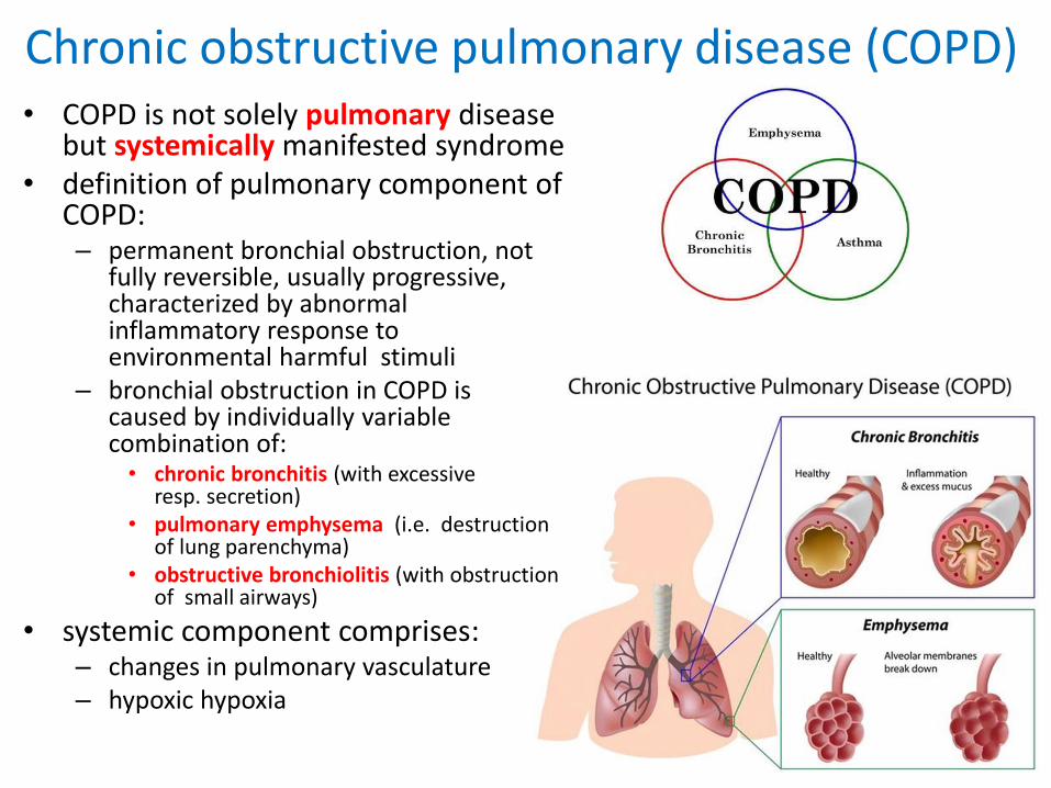
• COPD is not solely pulmonary disease but systemically manifested syndrome
• definition of pulmonary component of COPD: – permanent bronchial obstruction, not
fully reversible, usually progressive, characterized by abnormal inflammatory response to environmental harmful stimuli
– bronchial obstruction in COPD is caused by individually variable combination of: • chronic bronchitis (with excessive
resp. secretion) • pulmonary emphysema (i.e. destruction
of lung parenchyma)• obstructive bronchiolitis (with obstruction
of small airways)
• systemic component comprises:– changes in pulmonary vasculature– hypoxic hypoxia

COPD
23
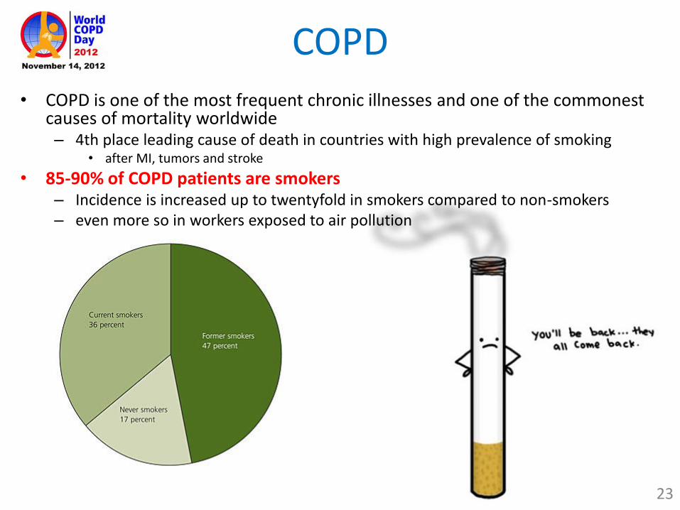
• COPD is one of the most frequent chronic illnesses and one of the commonest causes of mortality worldwide– 4th place leading cause of death in countries with high prevalence of smoking
• after MI, tumors and stroke
• 85-90% of COPD patients are smokers– Incidence is increased up to twentyfold in smokers compared to non-smokers– even more so in workers exposed to air pollution

Chronic bronchitis ( 2mm) and bronchiolitis ( 2mm)
24
• symptomatic definition – hypersecretion of mucus and chronic productive
cough that continues for at least 3 months of years for at least 2 consecutive years
• however patients typically suffer from chronic bronchitis without obstruction for a long time and only then develop bronchial obstruction (i.e. COPD)– there are of course patients with COPD without
clinical signs of chronic bronchitis– some chronic bronchitis cases never progress to
COPD
• in manifest COPD presence of chronic bronchiolitis is obligatory dominantly responsible (together with pulmonary emphysema) for obstruction– chronic persistent inflammation of small airways
( ≤ 2 mm)– the ratio between chronic bronchiolitis and
pulmonary emphysema is entirely individual
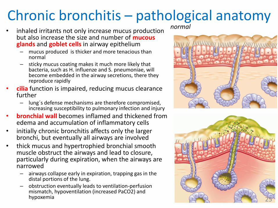
Chronic bronchitis – pathological anatomy• inhaled irritants not only increase mucus production
but also increase the size and number of mucous glands and goblet cells in airway epithelium– mucus produced is thicker and more tenacious than
normal– sticky mucus coating makes it much more likely that
bacteria, such as H. influenze and S. pneumoniae, will become embedded in the airway secretions, there they reproduce rapidly
• cilia function is impaired, reducing mucus clearance further– lung´s defense mechanisms are therefore compromised,
increasing susceptibility to pulmonary infection and injury
• bronchial wall becomes inflamed and thickened from edema and accumulation of inflammatory cells
• initially chronic bronchitis affects only the larger bronchi, but eventually all airways are involved
• thick mucus and hypertrophied bronchial smooth muscle obstruct the airways and lead to closure, particularly during expiration, when the airways are narrowed– airways collapse early in expiration, trapping gas in the
distal portions of the lung.– obstruction eventually leads to ventilation-perfusion
mismatch, hypoventilation (increased PaCO2) and hypoxemia 25
normal

Lung emphysema• abnormal permanent enlargement of
gas-exchange airways = acini (i.e distally from terminal bronchioles) accompanied by destruction of alveolar walls and without obvious fibrosis– obstruction results from changes in lung
tissues, rather than mucus production and inflammation, as in chronic bronchitis
• functional consequence:– major mechanism of airflow limitation is
loss of elastic recoil leading to the collapse of small airways during expiration
– expiration becomes difficult because loss of elastic recoil reduces the volume of air that can be expired passively,
– combination of increased RV (and FRC) in the alveoli and diminished caliber of the bronchioles causes part of each inspiration to be trapped in the acinus
– hyperinflation of alveoli causes large air spaces (bullae) and air spaces adjacent to pleura (blebs) to develop 26
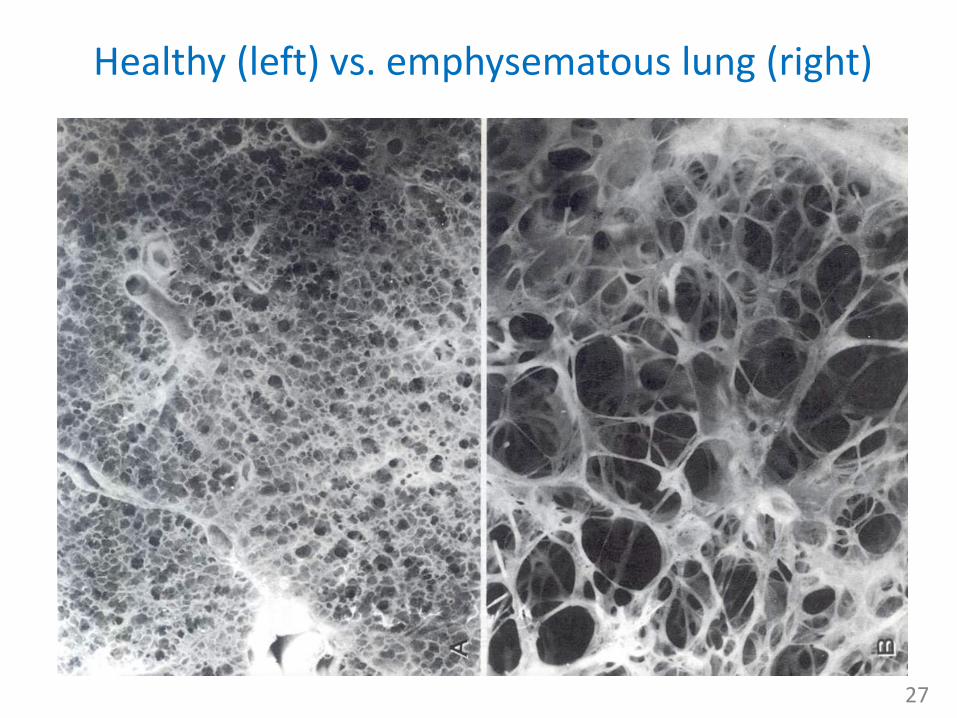
Healthy (left) vs. emphysematous lung (right)
27
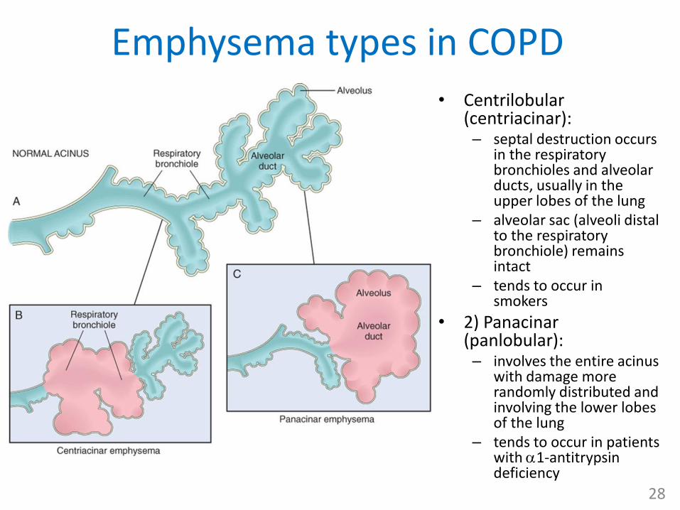
Emphysema types in COPD• Centrilobular
(centriacinar):– septal destruction occurs
in the respiratory bronchioles and alveolar ducts, usually in the upper lobes of the lung
– alveolar sac (alveoli distal to the respiratory bronchiole) remains intact
– tends to occur in smokers
• 2) Panacinar (panlobular): – involves the entire acinus
with damage more randomly distributed and involving the lower lobes of the lung
– tends to occur in patients with 1-antitrypsin deficiency
28


Variable overlap in COPD


Etiology of COPD - multifactorial • smoking
– cigarette smoke and air pollution, tip the normal balance of elastases (proteolytic enzymes) and antielastases(such as 1-antitrypsin) so that elastin is destroyed at an increased rate
– number of neutrophil granulocytes in inflammed airways• source of elastases and proteases favoring emphysema development
– tissue injury due to reactive oxygen and nitrogen species• healing with the participation of macrophages (source of matrix metaloprotinases)
– hypertrophy of mucus glands and thus CHB– impairment of surfactant
• airway hyper-reactivity• genetics (= variable consequencies in two persons with equal „smoking“ history)
– α1-antitrypsin deficiency • α1-antitrypsin inhibits neutrophil elastase which has the ability to destruct lung tissue• identified more than 75 alleles in the gene for α1-antitrypsin
– other genes • pro-inflammatory cytokines, growth factors, protease/antiprotease balance, antioxidants etc.
• exposure to other air pollutants (dust, smoke, professional exposure, car traffic fumes, biomass burning etc.)– the most risky are small particles ≤ 2.5 μm
• recurrent lower airways and lung infections 32

33
Effect of smoking

Pulmonary vessels in COPD
• remodelation (i.e. wall thickening, lumen narrowing and increased resistance) present very early in the time course of COPD– endothelial dysfunction
• due to oxidative stress
– hyperplasia of tunica intima• cells (inflammatory cells and SMCs)
as well as ECM
– hypertrophy of tunica media
• gradually hypoxia and loss of capillaries (emphysema) contributes to remodelation as well– vasoconstriction– later pre-capillary form of
secondary pulmonary hypertension
• cor pulmonale
34
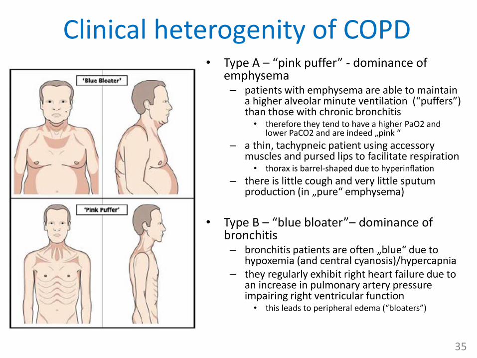
Clinical heterogenity of COPD• Type A – “pink puffer” - dominance of
emphysema– patients with emphysema are able to maintain
a higher alveolar minute ventilation (“puffers”) than those with chronic bronchitis• therefore they tend to have a higher PaO2 and
lower PaCO2 and are indeed „pink “
– a thin, tachypneic patient using accessory muscles and pursed lips to facilitate respiration• thorax is barrel-shaped due to hyperinflation
– there is little cough and very little sputum production (in „pure“ emphysema)
• Type B – “blue bloater”– dominance of bronchitis– bronchitis patients are often „blue“ due to
hypoxemia (and central cyanosis)/hypercapnia – they regularly exhibit right heart failure due to
an increase in pulmonary artery pressure impairing right ventricular function • this leads to peripheral edema (“bloaters”)
35


COPD stages
37
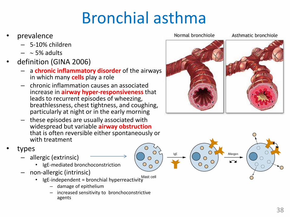
Bronchial asthma• prevalence
– 5-10% children – 5% adults
• definition (GINA 2006)– a chronic inflammatory disorder of the airways
in which many cells play a role – chronic inflammation causes an associated
increase in airway hyper-responsiveness that leads to recurrent episodes of wheezing, breathlessness, chest tightness, and coughing, particularly at night or in the early morning
– these episodes are usually associated with widespread but variable airway obstruction that is often reversible either spontaneously or with treatment
• types– allergic (extrinsic)
• IgE-mediated bronchoconstriction
– non-allergic (intrinsic)• IgE-independent = bronchial hyperreactivity
– damage of epithelium– increased sensitivity to bronchoconstrictive
agents
38
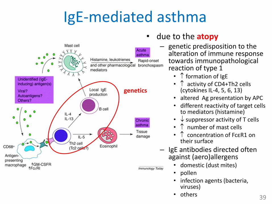
IgE-mediated asthma• due to the atopy
– genetic predisposition to the alteration of immune response towards immunopathologicalreaction of type 1• formation of IgE• activity of CD4+Th2 cells
(cytokines IL-4, 5, 6, 13)• altered Ag presentation by APC• different reactivity of target cells
to mediators (histamine)• suppressor activity of T cells• number of mast cells• concentration of FcεR1 on
their surface
– IgE antibodies directed often against (aero)allergens • domestic (dust mites) • pollen• infection agents (bacteria,
viruses)• others 39
genetics

Polygenic nature of asthma

Sensibilisation phase in atopic subjects
41
Proportions of asthmatic children sensitized to common allergens

Pathogenesis of allergic asthma
Inhaled antigen is processed by dendritic cells and presented to Th2 CD4+ T cells. B cells are stimulated to produce IgE, which binds to mast cells. Inhaled antigen binds to IgE, stimulating the mast cell to degranulate, which in turn leads to the release of mediators of the immediate response and the late response. Histamine and the leukotrienes produce bronchospasm and airway edema. Released chemotactic factors, along with factors from the Th2 CD4+ T cells, facilitate eosinophil traffic from the bone marrow to the airway walls. These late responses are proposed to lead to excessive mucus production, airway wall inflammation, injury, and hyperresponsiveness. (GM-CSF—granulocyte-macrophage colony-stimulating factor; IFN-y—interferon gamma; IL—interleukin) 42
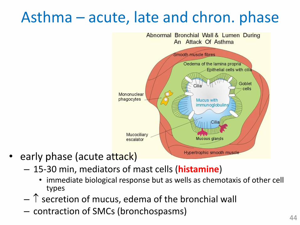
Asthma – acute, late and chron. phase
• early phase (acute attack)– 15-30 min, mediators of mast cells (histamine)
• immediate biological response but as wells as chemotaxis of other cell types
– secretion of mucus, edema of the bronchial wall– contraction of SMCs (bronchospasms)
44
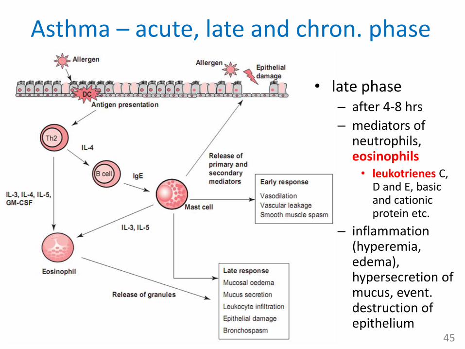
Asthma – acute, late and chron. phase
• late phase– after 4-8 hrs
– mediators of neutrophils, eosinophils• leukotrienes C,
D and E, basic and cationic protein etc.
– inflammation (hyperemia, edema), hypersecretion of mucus, event. destruction of epithelium
45
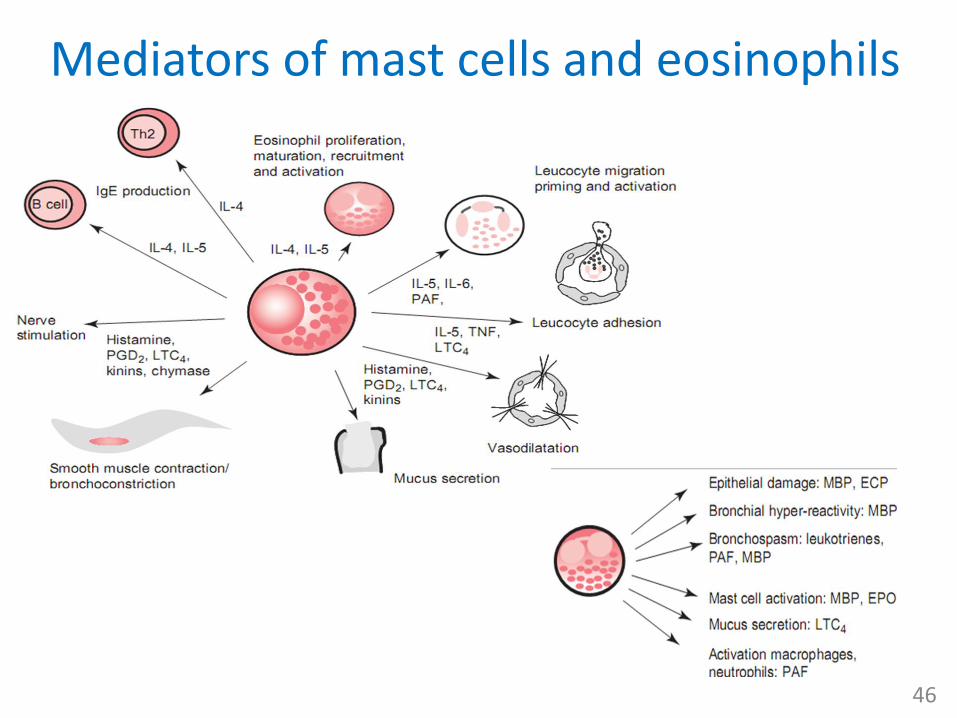
Mediators of mast cells and eosinophils
46

Asthma – acute, late and chron. phase• chronic phase
– chronic inflammation + repair processes lead to irreversible structural (remodelation) and functional (hyper-reactivity) changes of airways constituting a vicious cycle
– epithelium• cilia, desquamation
• hypertrophy of mucus glands and hyperplasia of goblet cells
– basal membrane• fibrotisation in
subepithelialspace (collagen)
– muscle layer • hypertrophy and
hyperplasia of SMCs 47
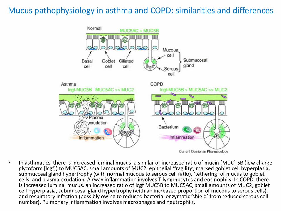
Mucus pathophysiology in asthma and COPD: similarities and differences
• In asthmatics, there is increased luminal mucus, a similar or increased ratio of mucin (MUC) 5B (low charge glycoform [lcgf]) to MUC5AC, small amounts of MUC2, epithelial ‘fragility’, marked goblet cell hyperplasia, submucosal gland hypertrophy (with normal mucous to serous cell ratio), ‘tethering’ of mucus to goblet cells, and plasma exudation. Airway inflammation involves T lymphocytes and eosinophils. In COPD, there is increased luminal mucus, an increased ratio of lcgf MUC5B to MUC5AC, small amounts of MUC2, goblet cell hyperplasia, submucosal gland hypertrophy (with an increased proportion of mucous to serous cells), and respiratory infection (possibly owing to reduced bacterial enzymatic ‘shield’ from reduced serous cell number). Pulmonary inflammation involves macrophages and neutrophils.
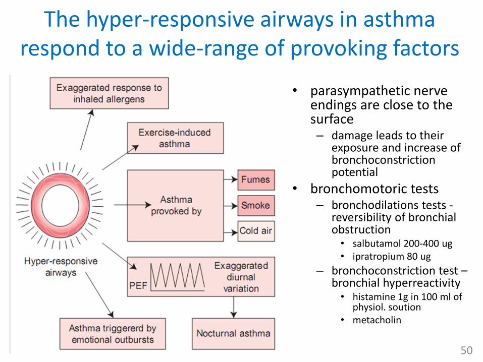
The hyper-responsive airways in asthma respond to a wide-range of provoking factors
• parasympathetic nerve endings are close to the surface – damage leads to their
exposure and increase of bronchoconstriction potential
• bronchomotoric tests– bronchodilations tests -
reversibility of bronchial obstruction• salbutamol 200-400 ug• ipratropium 80 ug
– bronchoconstriction test –bronchial hyperreactivity• histamine 1g in 100 ml of
physiol. soution• metacholin
50

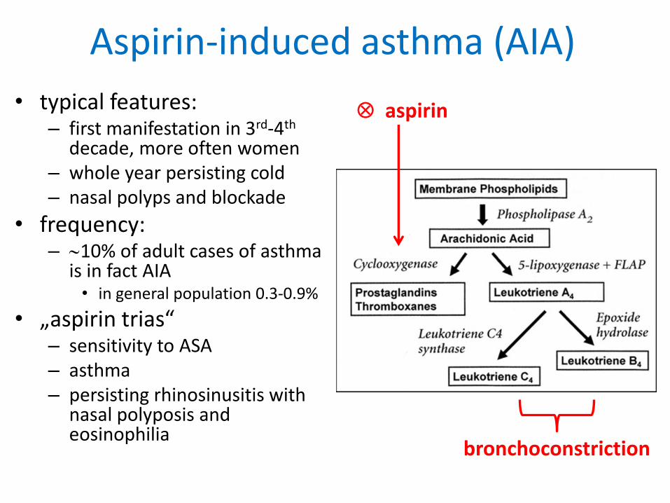
Aspirin-induced asthma (AIA)
• typical features:– first manifestation in 3rd-4th
decade, more often women – whole year persisting cold – nasal polyps and blockade
• frequency: – 10% of adult cases of asthma
is in fact AIA• in general population 0.3-0.9%
• „aspirin trias“– sensitivity to ASA– asthma– persisting rhinosinusitis with
nasal polyposis and eosinophilia
aspirin
bronchoconstriction

Pathogenesis of virus-induced asthma
Inhaled virus infects epithelial cells and leads to apoptosis of some of them. The release of chemotactic factors promotes the recruitment of macrophages into the lung parenchyma, where they ingest the dead epithelium. An acute response consisting of bronchospasm occurs at this time. Similar to allergic asthma, the inhaled virus is processed by dendritic cells and presented to Th2 CD8+ T cells. These cells produce copious amounts of IFN-y. Perforin released from the T cells leads to apoptosis of infected cells. B cells produce IgG, which is capable of neutralizing the virus. These events are thought to be related to the chronic response, which consists of airway inflammation, goblet cell hyperplasia, and airway hyperresponsiveness. (IFN-Y—interferon gamma; IL—interleukin; CCL—chemokine ligand) 53
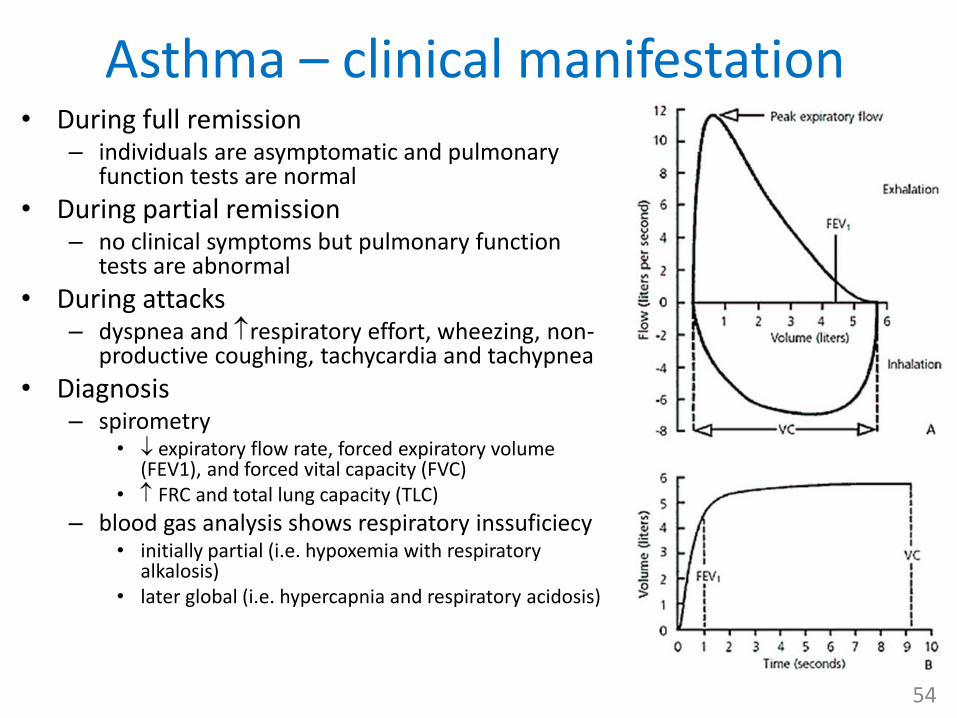
Asthma – clinical manifestation• During full remission
– individuals are asymptomatic and pulmonary function tests are normal
• During partial remission – no clinical symptoms but pulmonary function
tests are abnormal
• During attacks– dyspnea and respiratory effort, wheezing, non-
productive coughing, tachycardia and tachypnea
• Diagnosis– spirometry
• expiratory flow rate, forced expiratory volume (FEV1), and forced vital capacity (FVC)
• FRC and total lung capacity (TLC)
– blood gas analysis shows respiratory inssuficiecy• initially partial (i.e. hypoxemia with respiratory
alkalosis)• later global (i.e. hypercapnia and respiratory acidosis)
54