Phagocyte-specific S100 proteins in the local response to ...
Shannon Martinson, September 2015
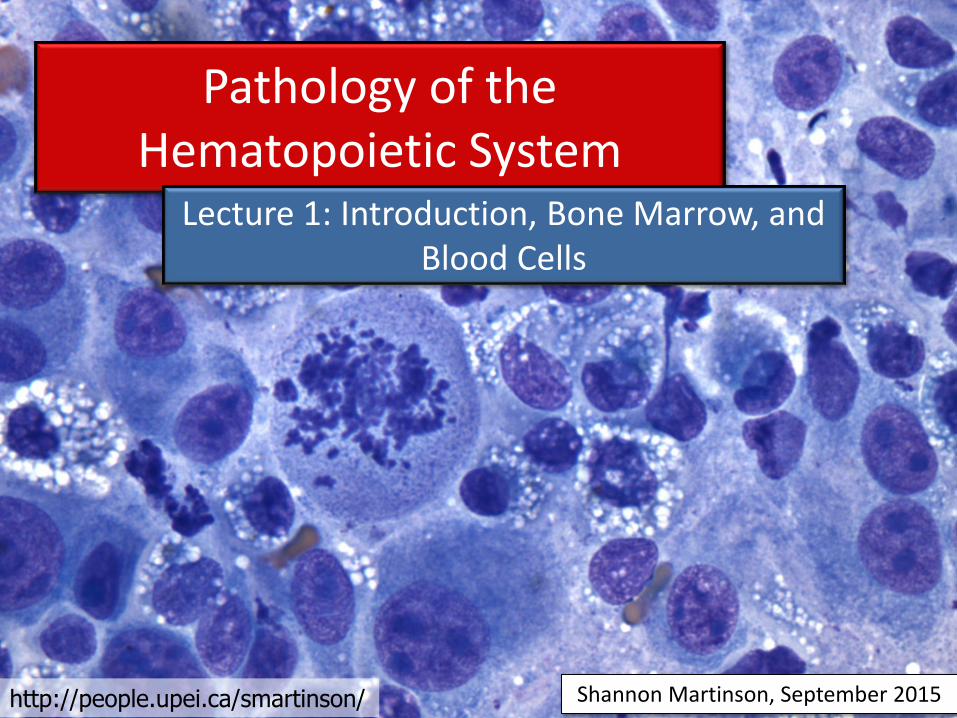
Pathology of the Hematopoietic System
Lecture 1: Introduction, Bone Marrow, and Blood Cells
http://people.upei.ca/smartinson/

Hematopoietic system - Introduction
Myeloid Tissue
• Bone marrow
• Blood cells
• Mononuclear-phagocyte system
Lymphoid Tissue
• Lymph nodes
• Spleen
• Thymus
• Accessory lymphoid tissue
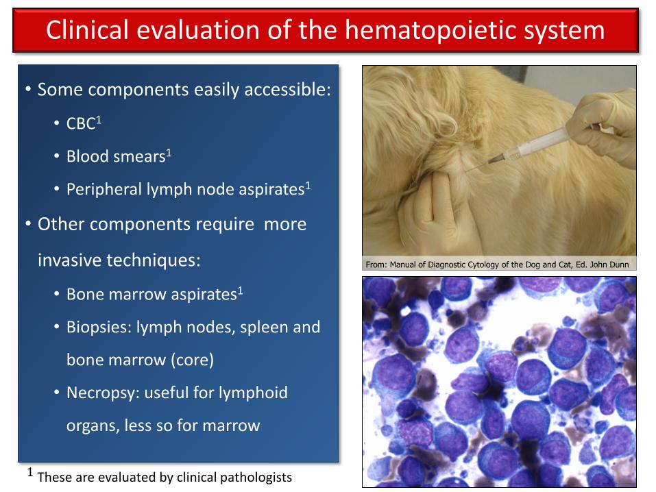
• Some components easily accessible:
• CBC1
• Blood smears1
• Peripheral lymph node aspirates1
• Other components require more
invasive techniques:
• Bone marrow aspirates1
• Biopsies: lymph nodes, spleen and
bone marrow (core)
• Necropsy: useful for lymphoid
organs, less so for marrow
Clinical evaluation of the hematopoietic system
1 These are evaluated by clinical pathologists
From: Manual of Diagnostic Cytology of the Dog and Cat, Ed. John Dunn

Blood cells are made in the following sites:
• Embryo: yolk sac*
• Fetus: liver, spleen, thymus, lymph node & bone marrow
• Neonates: mostly bone marrow (long & flat bones)
• Adults: bone marrow in all regions of flat bones & extremities of long bones
• Elsewhere depending on need = Extramedullary hematopoiesis (EMH)
*
*
• The process through which blood cells are made Hematopoiesis
Myeloid system: Bone marrow and blood cell development
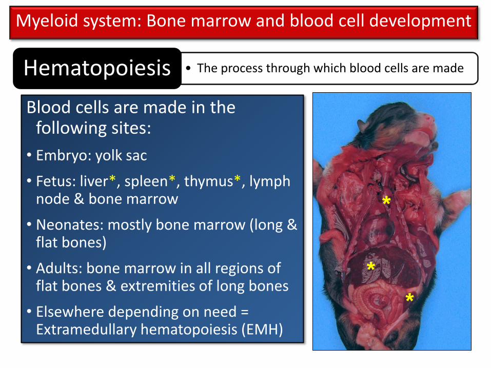
Blood cells are made in the following sites:
• Embryo: yolk sac
• Fetus: liver*, spleen*, thymus*, lymph node & bone marrow
• Neonates: mostly bone marrow (long & flat bones)
• Adults: bone marrow in all regions of flat bones & extremities of long bones
• Elsewhere depending on need = Extramedullary hematopoiesis (EMH)
*
• The process through which blood cells are made Hematopoiesis
Myeloid system: Bone marrow and blood cell development
*
*

Blood cells are made in the following sites:
• Embryo: yolk sac
• Fetus: liver, spleen, thymus, lymph node & bone marrow
• Neonates: mostly bone marrow (long & flat bones) *
• Adults: bone marrow in all regions of flat bones & extremities of long bones*
• Elsewhere depending on need = Extramedullary hematopoiesis (EMH)
*
• The process through which blood cells are made Hematopoiesis
Myeloid system: Bone marrow and blood cell development
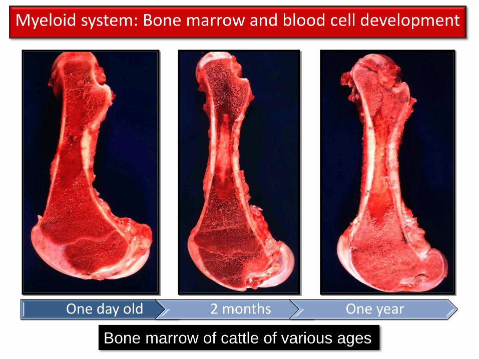
Bone marrow of cattle of various ages
Myeloid system: Bone marrow and blood cell development
One day old 2 months One year
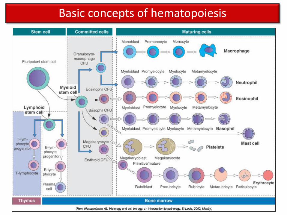
Basic concepts of hematopoiesis

Basic concepts of hematopoiesis
• Hematopoietic tissue is highly prolific
• All blood cells are derived from a common pluripotential stem cell
• Pluripotential stem cells are capable of self renewal and further differentiation
• Pluripotent stem cell ➝ committed cells ➝ maturing cells ➝ mature cells
• Mature cells have a limited lifespan
• Production and turnover of blood cells are balanced in health
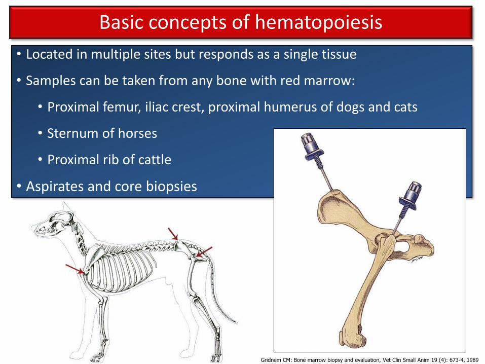
• Located in multiple sites but responds as a single tissue
• Samples can be taken from any bone with red marrow:
• Proximal femur, iliac crest, proximal humerus of dogs and cats
• Sternum of horses
• Proximal rib of cattle
• Aspirates and core biopsies
http://www.vetmed.wsu.edu/resources/Techniques/images/bm_core.jpg
Basic concepts of hematopoiesis
Gridnem CM: Bone marrow biopsy and evaluation, Vet Clin Small Anim 19 (4): 673-4, 1989

• Located in multiple sites but responds as a single tissue
• Samples can be taken from any bone with red marrow:
• Proximal femur, iliac crest, proximal humerus of dogs and cats
• Sternum of horses
• Proximal rib of cattle
• Aspirates and core biopsies
http://www.vetmed.wsu.edu/resources/Techniques/images/bm_core.jpg
Basic concepts of hematopoiesis

• Indicated when abnormalities are identified on hematology:
• Unexplained cytopenias
• Maturation or morphological defects (atypical cells in circulation)
• Suspected myeloproliferative diseases
• Potential malignancies metastatic to marrow
http://www.vetmed.wsu.edu/resources/Techniques/images/bm_core.jpg
Basic concepts of hematopoiesis

Bone marrow aspirate/smears:
Interpreted by clinical pathologists
Important for:
• Cellular morphology
• Erythroid to myeloid ratio
• Primary or metastatic neoplasia
Bone marrow core biopsy: Interpreted by morphologic pathologists
Important for:
• Ratio of fat to hematopoietic cells
• Myelofibrosis • Primary or
metastatic neoplasia
**Should be interpreted in conjunction with a CBC!**
Bone marrow: Microscopic evaluation
Courtesy of Dr Noel Clancey

End result depends on the type of cell damaged
• Pluripotent stem cells = multiple cell lines affected
• Committed stem cells = one or more lines affected
• Differentiated cells = one cell type affected
Alterations are reflected in the peripheral blood
• Decreases in cell lines = cytopenias, anemia
• Increases in cell lines = ‘cytoses and ‘philias
Alterations are reflected in the bone marrow
• Increased or decreased cellularity
• Changes in the proportion of hematopoietic tissue (red marrow) to adipose tissue (yellow marrow)
Pathology of the Bone Marrow and Blood Cells
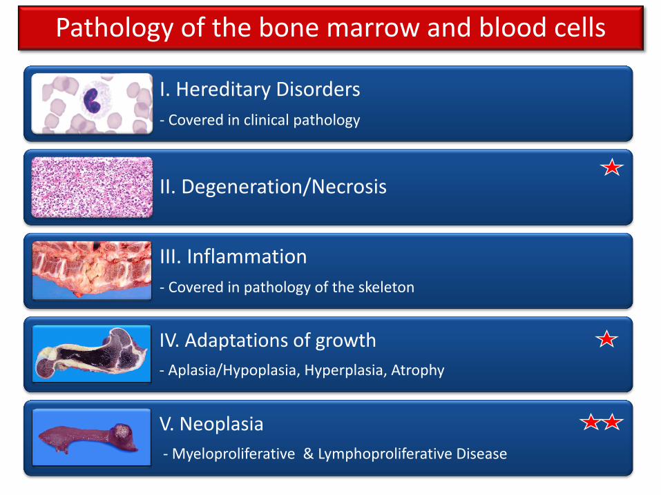
I. Hereditary Disorders
- Covered in clinical pathology
II. Degeneration/Necrosis
III. Inflammation
- Covered in pathology of the skeleton
IV. Adaptations of growth
- Aplasia/Hypoplasia, Hyperplasia, Atrophy
V. Neoplasia
- Myeloproliferative & Lymphoproliferative Disease
Pathology of the bone marrow and blood cells

Hematopoietic tissue is highly active ➝ susceptible to insults
Bone marrow and blood cells: Degeneration and necrosis
• Radiation
• Toxins/Drugs • Antineoplastic / immunosuppressive
drugs
• Idiosyncratic drug reactions
• Toxic chemicals
• Viral agents • Feline and canine parvovirus
• Feline Leukemia Virus
• Feline Immunodeficiency Virus
• Equine Infectious Anemia
• Immune-mediated destruction • Systemic Lupus Erythematosus
• Idiopathic Bone marrow degeneration: canine parvovirus infection

Hematopoietic tissue is highly active ➝ susceptible to insults
Bone marrow and blood cells: Degeneration and necrosis
• Radiation
• Toxins/Drugs • Antineoplastic / immunosuppressive
drugs
• Idiosyncratic drug reactions
• Toxic chemicals
• Viral agents • Feline and canine parvovirus
• Feline Leukemia Virus
• Feline Immunodeficiency Virus
• Equine Infectious Anemia
• Immune-mediated destruction • Systemic Lupus Erythematosus
• Idiopathic Reflected in peripheral blood as cytopenias!

Bone marrow and blood cells: Adaptations of growth
Bone Marrow Hypoplasia / Aplasia
Bone Marrow Hyperplasia
Serous atrophy of fat

• Bone marrow suppression
• Estrogen (exogenous and endogenous)
• Chronic disease
• Chronic renal disease
• Lack of nutrients
• Iron
• Vitamin B12
• Folate
• Endocrine dysfunction
• Hypothyroidism
• Bone marrow degeneration
• Idiopathic
• Decreased/absent proliferative activity
• One or multiple cell lines can be affected Bone marrow
hypoplasia/aplasia
Bone marrow and blood cells: Adaptations of growth

Gross ➝ Increased yellow marrow
Histo ➝ Increased ratio of fat to hematopoietic cells
Hypoplastic bone marrow
Normal bone marrow
~ 50/50 Bone Marrow Hypoplasia
Bone marrow and blood cells: Adaptations of growth
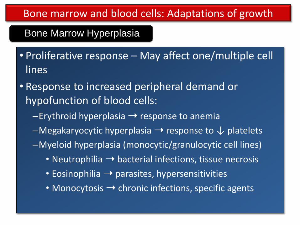
• Proliferative response – May affect one/multiple cell lines
• Response to increased peripheral demand or hypofunction of blood cells:
–Erythroid hyperplasia ➝ response to anemia
–Megakaryocytic hyperplasia ➝ response to ↓ platelets
–Myeloid hyperplasia (monocytic/granulocytic cell lines)
• Neutrophilia ➝ bacterial infections, tissue necrosis
• Eosinophilia ➝ parasites, hypersensitivities
• Monocytosis ➝ chronic infections, specific agents
Bone Marrow Hyperplasia
Bone marrow and blood cells: Adaptations of growth

Normal Hyperplasia
Gross lesions: •Red marrow
replaces the yellow marrow • Metaphyses • Endosteal surface of
diaphysis • Progresses to
occupy entire marrow cavity
Bone Marrow Hyperplasia
Bone marrow and blood cells: Adaptations of growth
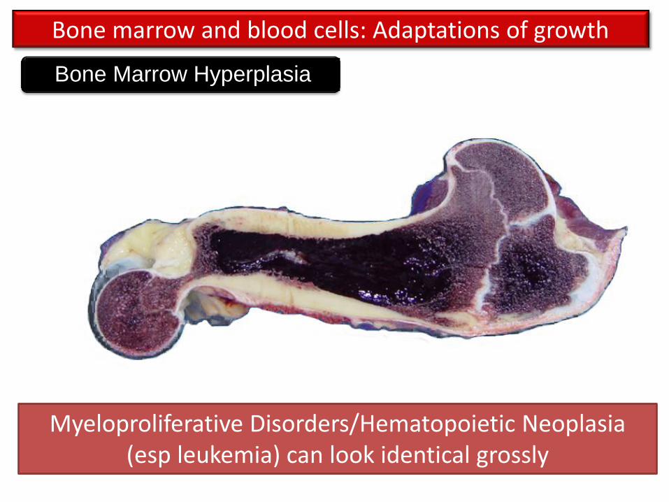
Bone Marrow Hyperplasia
Bone marrow and blood cells: Adaptations of growth
Myeloproliferative Disorders/Hematopoietic Neoplasia (esp leukemia) can look identical grossly

Histologic lesions: • Increased cellularity (decreased ratio of fat to hematopoietic cells)
• One or more cell lines can be affected
• Shift toward immaturity (eg left shift in neutrophils)
• Extramedullary hematopoiesis (spleen & liver) if severe
Normal bone marrow Hyperplastic bone marrow
Bone Marrow Hyperplasia
Bone marrow and blood cells: Adaptations of growth

• Clonal proliferative disorders of hematopoietic cell types • Affect primarily:
• Bone marrow • The circulating blood (leukemia) • Lymphoid tissue (lymph nodes, spleen, thymus, etc)
• Common associated features:
• Bone marrow hypercellularity • Anemia • Thrombocytopenia/neutropenia • +/- Leukemic cells in peripheral blood
• Divided into lymphoproliferative and myeloproliferative diseases:
• Lymphoid cells: Lymphocytes (B and T Cells) • Myeloid cells: granulocytes (neutrophils, eosinophils, basophils),
monocytes/macrophages, erythrocytes, and megakaryocytes
Primary Hematopoietic Neoplasia

Hematopoietic Neoplasia
Lymphoproliferative Disease
Lymphoma
Lymphoid leukemia
Plasma cell tumours
Myeloproliferative
Disease
Histiocytic Neoplasia
Myeloid leukemia
Myelodysplastic Syndrome
Mast cell tumour
Primary Hematopoietic Neoplasia
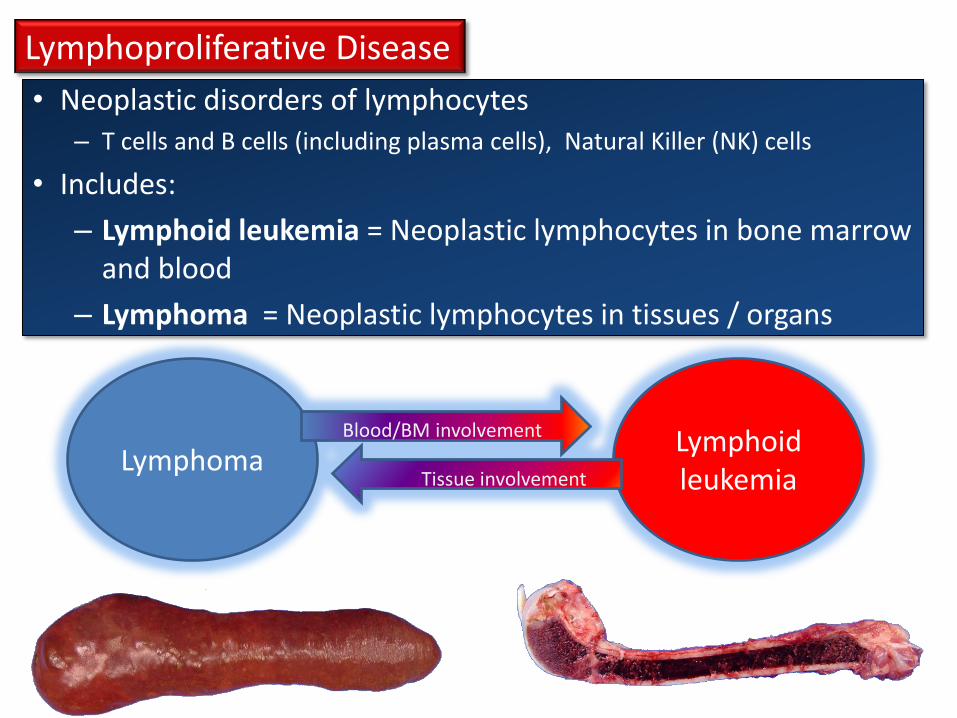
• Neoplastic disorders of lymphocytes – T cells and B cells (including plasma cells), Natural Killer (NK) cells
• Includes:
– Lymphoid leukemia = Neoplastic lymphocytes in bone marrow and blood
– Lymphoma = Neoplastic lymphocytes in tissues / organs
Lymphoproliferative Disease
Lymphoma Lymphoid leukemia Tissue involvement
Blood/BM involvement

Lymphoproliferative Disease: Lymphoma
*Lymphoma (lymphosarcoma) is one of the most common malignant tumours of domestic animals
Affects many species
Etiology: Idiopathic (sporadic), Viral infections, Hereditary

Several methods of classification of lymphomas:
Anatomical classification
• Multicentric
• Alimentary
• Thymic
• Cutaneous
• Misc.
• Leukemic
Cellular morphology
• Cell size
• Nuclear features
• Mitotic rate
Immuno-phenotype
• B -cell
• T - cell
• Non- B/T
Biologic behaviour
• Low grade (indolent)
• Intermediate grade
• High grade (aggressive)
Histologic pattern
• Diffuse vs follicular
• Newer classification systems use a combination of
these methods
• World Health Organization (WHO) system of
classification of canine lymphoma
• Also - clinical staging is important

Anatomical classification
• Multicentric
• Alimentary
• Thymic
• Cutaneous
• Misc.
• Leukemic
Cellular morphology
• Cell size
• Nuclear features
• Mitotic rate
Immuno-phenotype
• B -cell
• T - cell
• Non- B/T
Biologic behaviour
• Low grade (indolent)
• Intermediate grade
• High grade (aggressive)
• B-cell lymphomas may have better survival profiles and response to treatment (compared to T-cell lymphoma)
• Small cell lymphoma with low mitotic rate → slow progression, poor response to chemotherapy
• Large cell lymphoma with high mitotic rate → rapid progression, but do respond to chemotherapy
Histologic pattern
• Diffuse vs follicular
Several methods of classification of lymphomas:

• Non specific signs:
–Weight loss and loss of appetite
• Painless enlargement of lymph nodes
–Lymphadenopathy
• Other signs depend on anatomic location:
–Retrobulbar lymph nodes➝ exophthalmos
–Thymus ➝ dyspnea, esophageal obstruction
–Alimentary ➝ diarrhea, obstruction or melena
Clinical Signs of Lymphoma
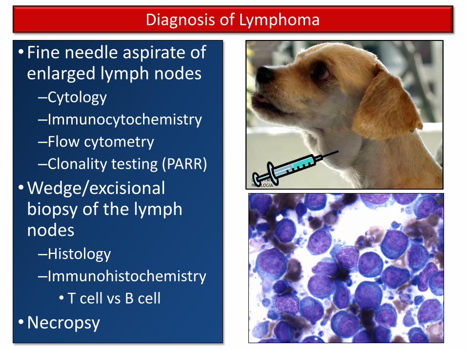
•Fine needle aspirate of enlarged lymph nodes –Cytology
–Immunocytochemistry
–Flow cytometry
–Clonality testing (PARR)
•Wedge/excisional biopsy of the lymph nodes –Histology
–Immunohistochemistry
• T cell vs B cell
•Necropsy
Diagnosis of Lymphoma
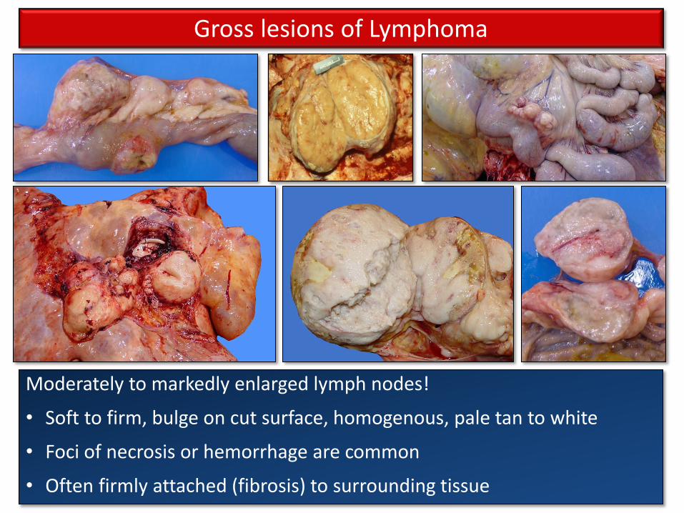
Moderately to markedly enlarged lymph nodes!
• Soft to firm, bulge on cut surface, homogenous, pale tan to white
• Foci of necrosis or hemorrhage are common
• Often firmly attached (fibrosis) to surrounding tissue
Gross lesions of Lymphoma
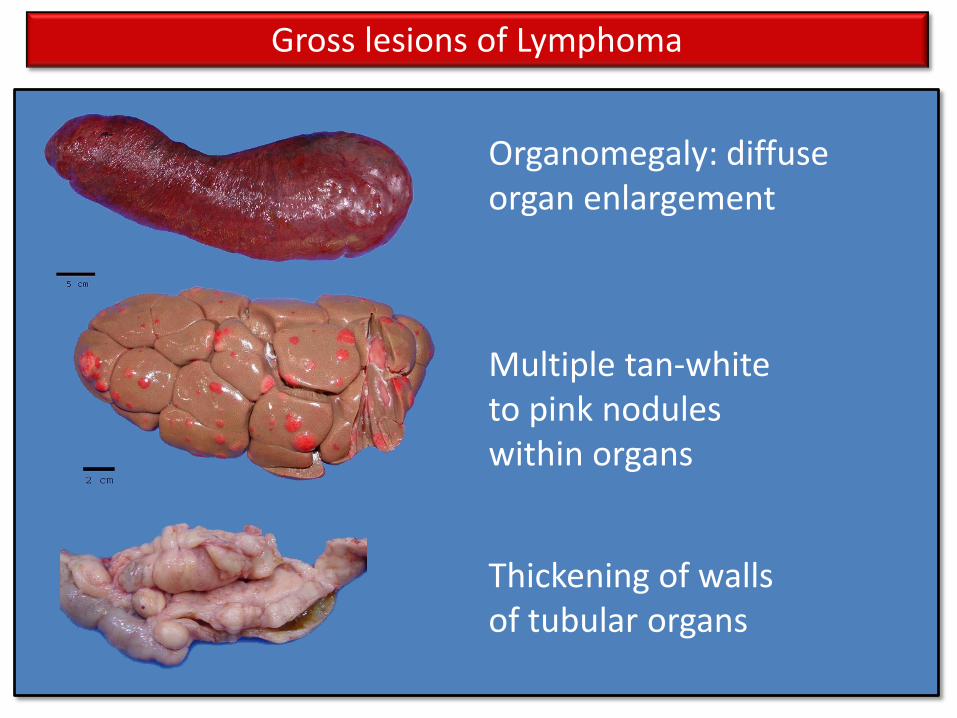
Organomegaly: diffuse organ enlargement
Multiple tan-white to pink nodules within organs
Thickening of walls of tubular organs
Gross lesions of Lymphoma

Neoplastic round cells efface the normal architecture
Uniform population of
small lymphocytes
Anaplastic round cells
with mitoses, anisocytosis, anisokaryosis
Low grade High grade
Microscopic lesions of Lymphoma

• Most common canine hematopoietic neoplasia
• Usually middle aged to older animals
• 80-85 % have multicentric lymphoma
• Peripheral LN involvement is common
• Usually medium to high grade
• Leukograms are usually normal
Multicentric Lymphoma
Canine Lymphoma
B cell lymphoma is most common
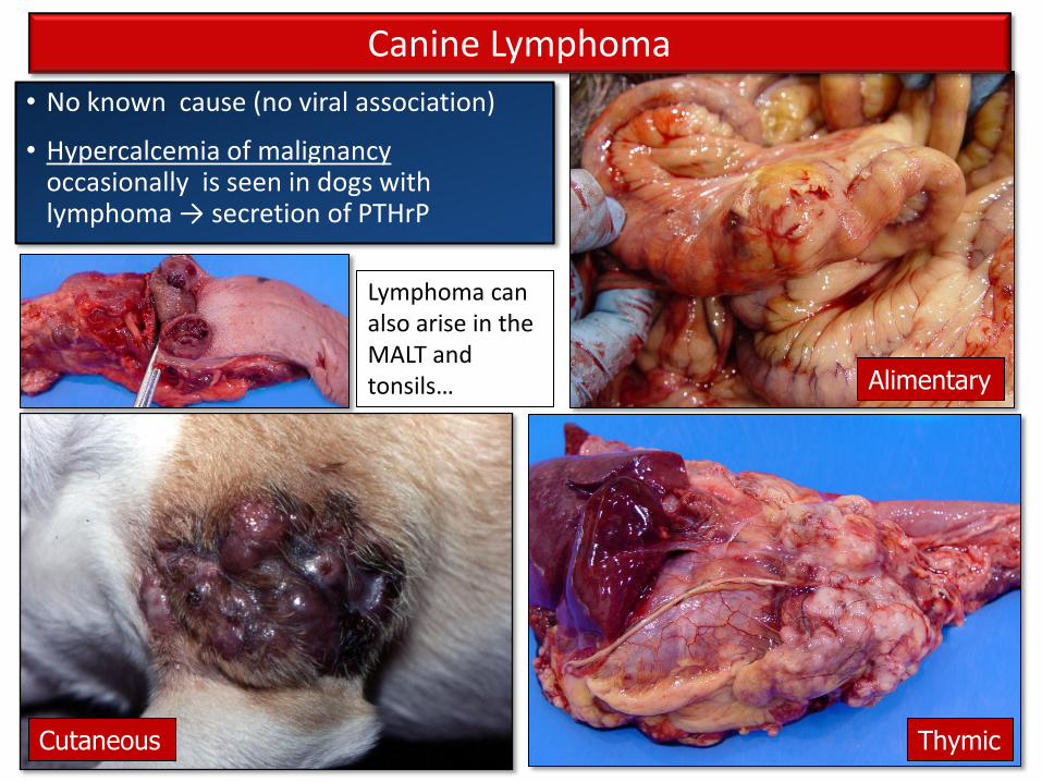
Cutaneous
Alimentary
Thymic
• No known cause (no viral association)
• Hypercalcemia of malignancy occasionally is seen in dogs with lymphoma → secretion of PTHrP
Lymphoma can also arise in the MALT and tonsils…
Canine Lymphoma
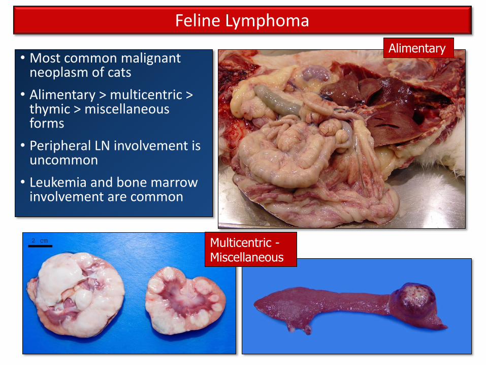
• Most common malignant neoplasm of cats
• Alimentary > multicentric > thymic > miscellaneous forms
• Peripheral LN involvement is uncommon
• Leukemia and bone marrow involvement are common
Alimentary
Multicentric - Miscellaneous
Feline Lymphoma

Thymic /Mediastinal
Association with Feline Leukemia Virus (FeLV):
• 10 -20 % of cats with lymphoma are FeLV +
• FeLV is associated with mediastinal and multicentric T cell lymphoma
• Young cats!
Feline Lymphoma

Questions?