PATHOLOGY OF THE FEMALE REPRODUCTIVE SYSTEM I MHD II March 19 th, 2015.
52
PATHOLOGY OF THE FEMALE REPRODUCTIVE SYSTEM I MHD II March 19 th , 2015
-
Upload
jemima-hicks -
Category
Documents
-
view
223 -
download
1
Transcript of PATHOLOGY OF THE FEMALE REPRODUCTIVE SYSTEM I MHD II March 19 th, 2015.
PATHOLOGY OF THE FEMALE REPRODUCTIVE SYSTEM I
MHD II
March 19th, 2015
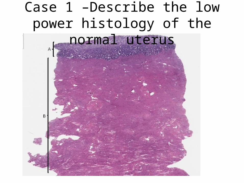
Case 1 –Describe the low power histology of the normal uterus
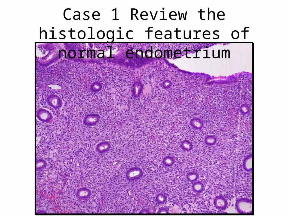
Case 1 Review the histologic features of normal endometrium

Case 1 - Compare and contrast the histology
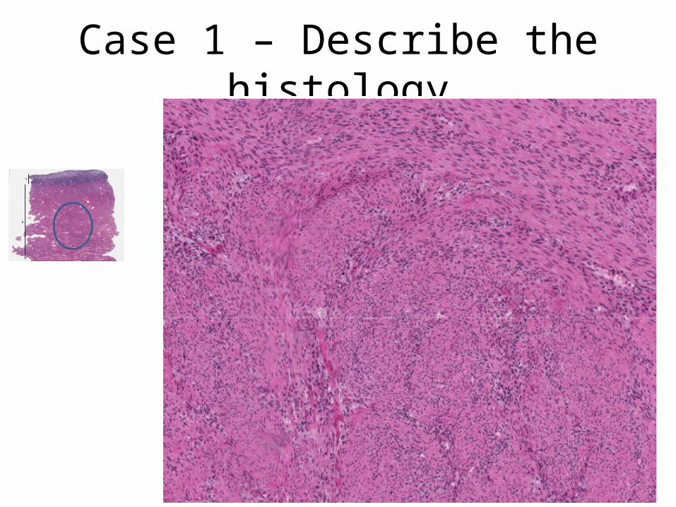
Case 1 – Describe the histology

Case 2 CHIEF COMPLAINT: “My husband and I have been trying to have a baby for the past five years.”HISTORY: Pt is a 27-year-old woman who presents with the inability to conceive a child during the past five years. She and her husband have sexual relations 1-2 times per week and very much desire to have children. Menstrual history
– Menarche occurred at age 13.– Menstrual cycles have been fairly regular since age 17.– Average cycle is 29 30 days, with menses being 5 days in duration. ‑– Menstrual flow has been consistent, requiring 3 - 4 pads initially and
gradually tapering off. – She has had premenstrual cramping and cramping pain the first day of
menses since her teen years.– For the past year or two she has experienced a more diffuse pain throughout
her lower pelvis and more severe menstrual pain.– Has nausea and low back pain during menstruation.

Case 2
Additional history– She notes abdominal pain at times during intercourse and when she
moves her bowels.– Intermittent vaginal spotting occurs but no vaginal discharge.– At age 24 she was involved in an automobile accident which required
abdominal surgery to stop bleeding from "my intestines.”

Case 2
PHYSICAL EXAMINATION:Patient is a thin, well nourished woman. She is alert and in no distress. Vital signs: Blood pressure 116/82, Apical heart rate 76/minute and regular, respiratory rate 12/minute, temperature 98.2°F. Abdomen – normal bowel sounds, no masses or tenderness13.5 cm long parasagital scar in the left lower quadrant represents a healed surgical incision. Several small nodules are felt below the scar (patient says her scar "hurts when I menstruate").
Case 2Pelvic exam reveals normal external genitalia. Speculum exam reveals normal vaginal and cervical mucosa. The external os is small and round. Bimanual exam reveals slight tender nodularity in cul-de-sac, recto-vaginal septum and uterosacral ligaments.The uterus is of normal size. The right adnexa is tender but the left is not. The rectal exam produces pain when the anterior wall is palpated.
Case 2
What are the major clinical problems?

Case 2
Develop a differential diagnosis for pelvic pain in a woman
What aspects of the history and physical may help differentiate the cause of pain?

Case 2 – List key clinical distinguishing features of the following:
• Ruptured ovarian cyst and twisted ovarian cyst
• Ovarian cancer
• Ectopic pregnancy
• Pelvic inflammatory disease
• Adenomyosis
• Endometriosis

Case 2
Based on the given data, what is this patient’s likely diagnosis?
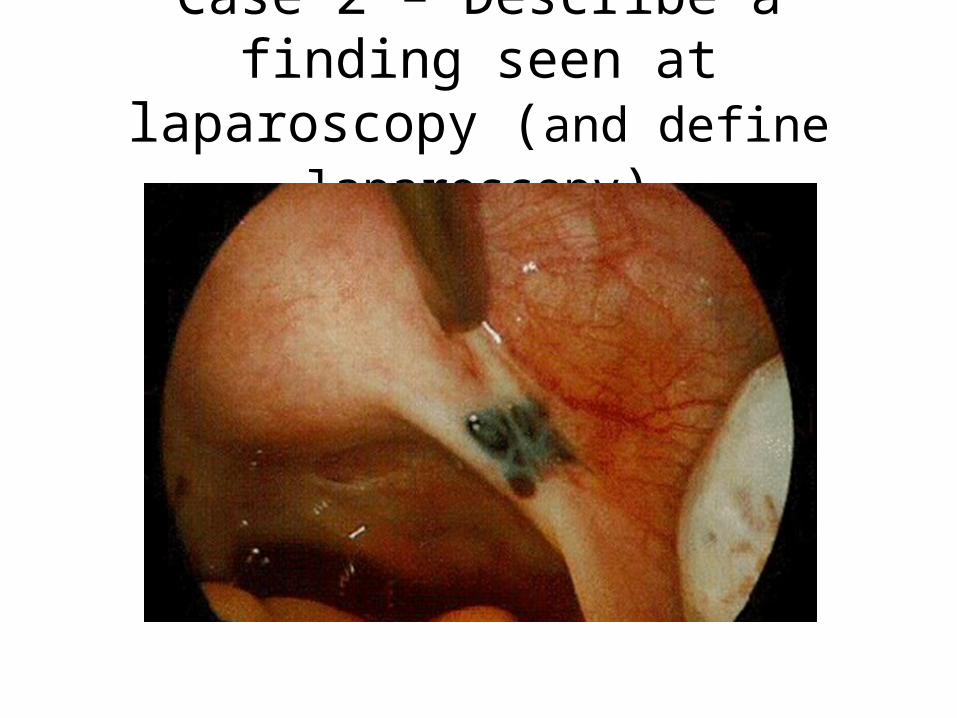
Case 2 – Describe a finding seen at laparoscopy (and define laparoscopy)
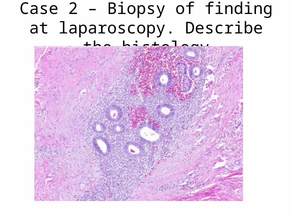
Case 2 – Biopsy of finding at laparoscopy. Describe the histology

Case 2 – Describe high power findings
Case 2
What is your diagnosis? Correlate the pathologic findings with the clinical findings.
Why did the patient’s surgical scar hurt when she menstruated?
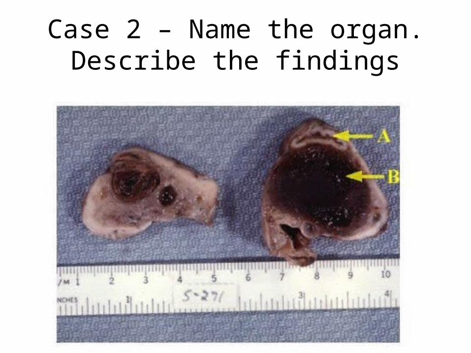
Case 2 – Name the organ. Describe the findings
Case 3CHIEF COMPLAINT: “I'm bleeding from my vagina.”HISTORY: Patient is a 69 year-old woman who presents with intermittent vaginal bleeding of 9 months duration. Initially she noted spotting of bright red blood which seemed to occur several times each month. No particular activity precipitates the bleeding and she experiences no other symptoms. She states, "I just thought my period came back." She is twelve years post menopause. ‑ She has never had children.After three months of spotting, she sought care from a physician who placed her on "estrogen pills to control my bleeding." He also gave her "iron pills”.

Case 3The vaginal bleeding continued and contained small clots. The bleeding was always irregularShe sought a second opinion because the bleeding progressed. She has been treated for diabetes mellitus, type 2 since age 52.
PHYSICAL EXAMINATION: The patient is obese, alert and in no distress. Vital signs: Blood pressure 146/90, pulse 90/minute, respiratory rate 18/minute, temperature 98° F.Abdomen is soft and round. Organomegaly and masses are not palpable. Pelvic exam reveals external genitalia consistent with the age of the patient. The vaginal introitus is narrowed and only admits two fingers. A small speculum allows visualization of the cervix. The ectocervix is smooth. Blood is noted in the os. Bimanual exam reveals a symmetrically enlarged, non-tender uterus. The adnexae are not palpable. A rectocele or cystocele are not present.
Case 3

Case 3
LAB TESTS: Hemoglobin 9.8 grams/dlHematocrit 30%MCV 79 fl

Case 3
What is the major clinical problem?
What are the common causes of this problem in post-menopausal women?
List factors to help differentiate the cause of bleeding.

Case 3- If this patient’s uterus would be removed. It might look like one of these. Describe the gross findings
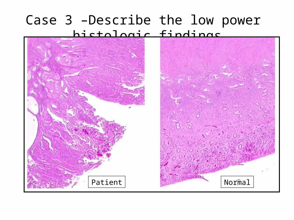
Case 3 –Describe the low power histologic findings
Patient Normal

Case 3 – Describe the high power histologic findings
PatientNormal
Case 3
What is your diagnosis?
From this patient’s history, explain why her initial management was not the standard of care.
Case 3 - Define the following terms:• Menorrhagia
• Hypomenorrhea
• Hypermorrhea
• Polymenorrhea
• Oligomenorrhea
• Metrorrhagia
• Menometrorrhagia
• Post-menopausal bleeding

Case 3
That patient is visibly upset when she learns of the diagnosis and asks “Why did this happen?”How would you respond?

Case 4CHIEF COMPLAINT: NoneHISTORY: A 38-year-old woman undergoes a routine, annual gynecologic examination. Her gestational history is as follows: gravida 4, para 3 with one spontaneous abortion. She menstruates every 28-31 days for 2-4 days. She experienced three or four episodes of metrorrhagia in the previous year, but no hypermenorrhea. She has a persistent, mild leukorrhea.PHYSICAL EXAMINATION: The patient is a moderately obese woman who is alert and in no distress. Vital signs are as follows: blood pressure 136/84, apical heart rate 80/minute and regular, respiratory rate 14/minute, temperature 97.9°F. Examination of the abdomen: It is soft and slightly round; no organomegaly.
Case 4PHYSICAL EXAMINATION: Pelvic examination reveals normal external genitalia. Speculum exam reveals normal vaginal and cervical mucosa. Bimanual exam reveals an asymmetrically enlarged uterus. The uterus is the size of a 2-3 month gestation and is freely movable. The uterus contains multiple nodules of varying size which are not tender. The adnexae are not palpable.

Case 4
What is the clinical problem?
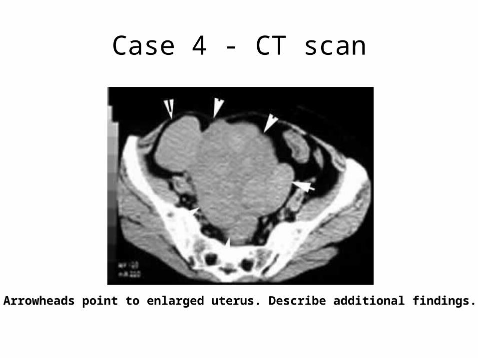
Case 4 - CT scan
Arrowheads point to enlarged uterus. Describe additional findings.
Case 4
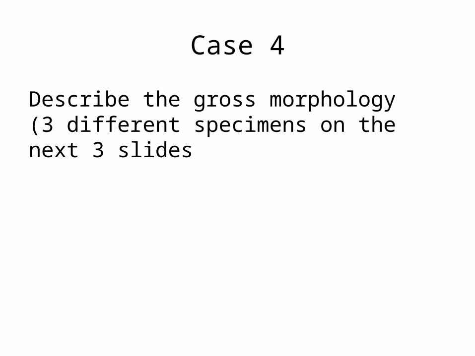
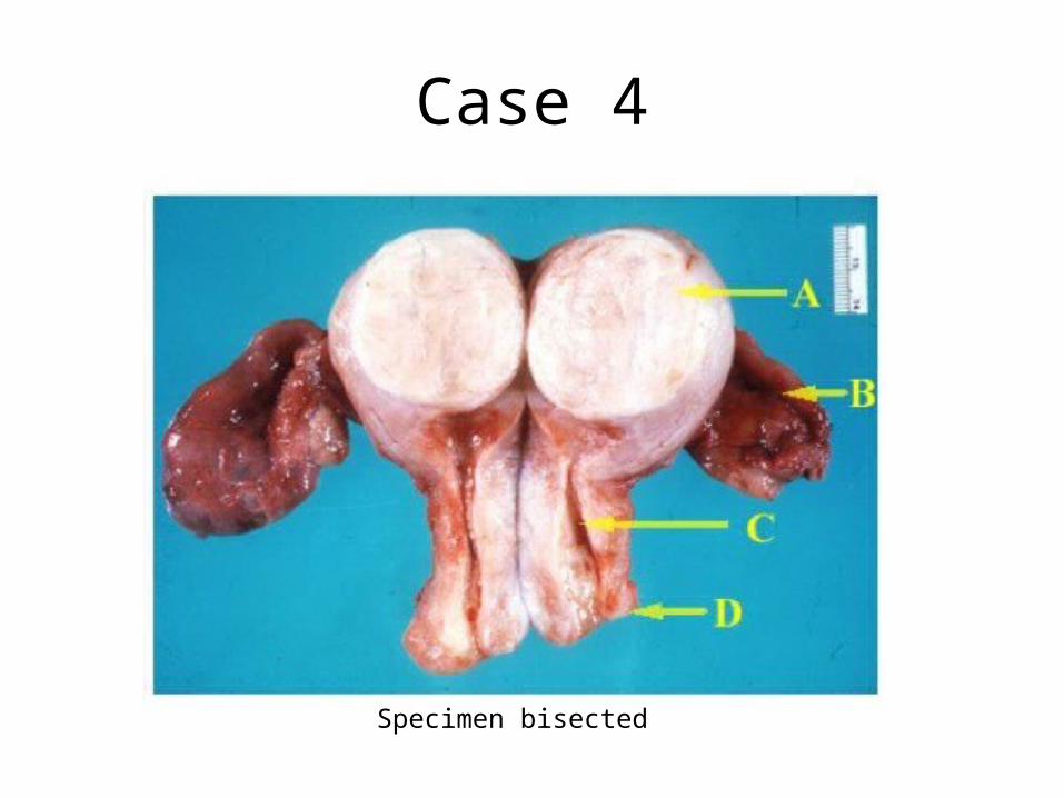
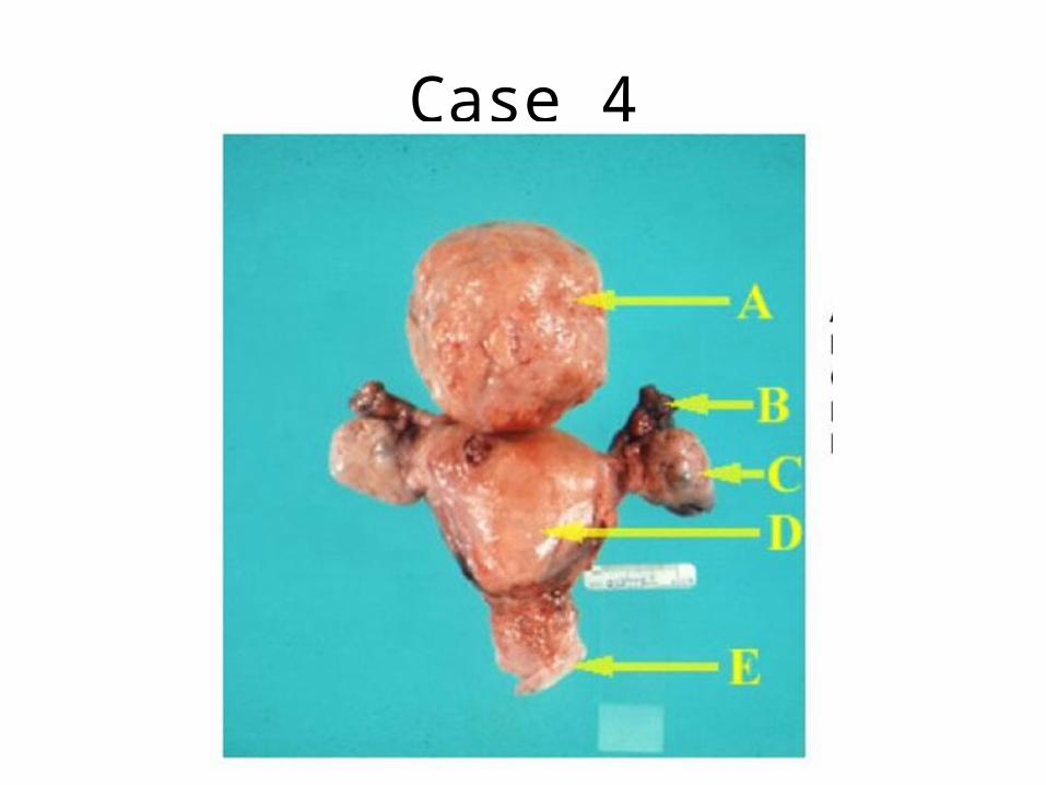
Describe the gross morphology (3 different specimens on the next 3 slides
Case 4
Specimen bisected
Case 4

Case 4

Case 4 – Describe the low power findings
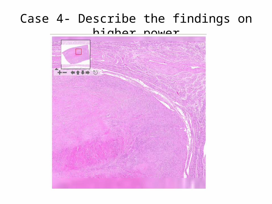
Case 4- Describe the findings on higher power
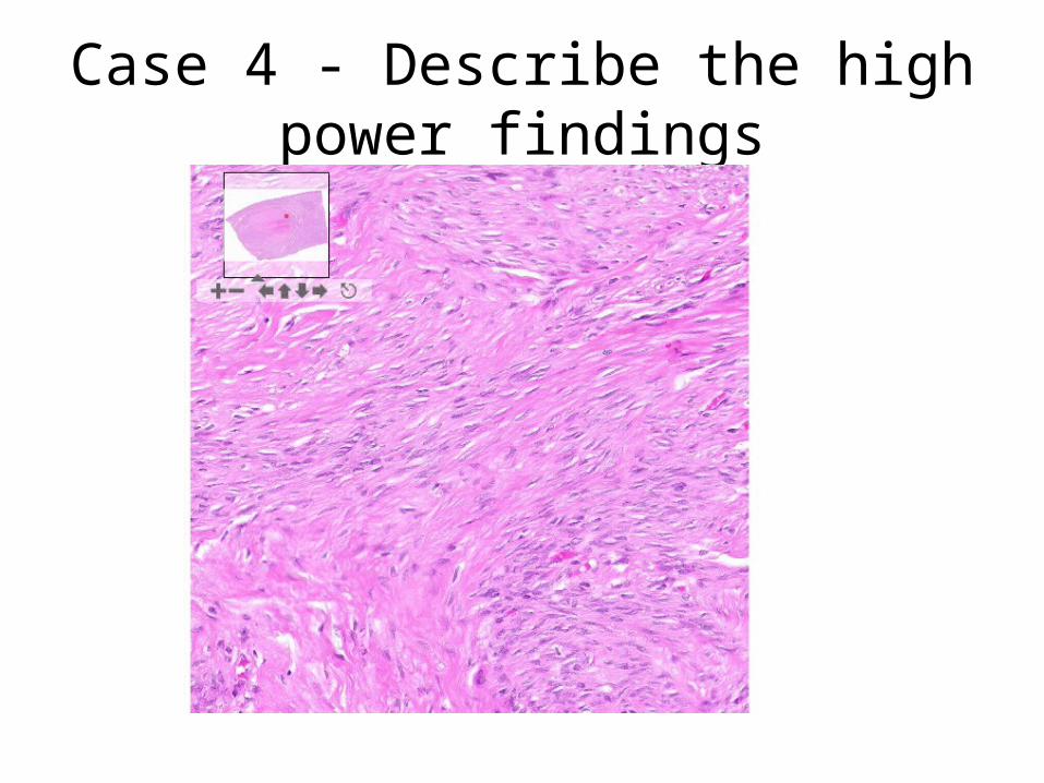
Case 4 - Describe the high power findings

Case 4
What is your diagnosis?
Correlate the clinical findings with the pathology.
Case 5HISTORY: The patient is a 25-year-old woman who presents with bilateral lower abdominal pain of 3 days duration. The pain is sometimes sharp but more often dull; it is moderate in intensity and continuous. Movement accentuates the pain which remains localized to the right and left lower quadrants of the abdomen. In addition to the abdominal pain, the patient has noticed a yellowish-white vaginal discharge for two weeks. She also has mild dysuria but no urgency or frequency. The patient relates that she had similar health problems in the past for which she had been treated. Treatment did not require hospitalization. Her gestational history is gravida 0.

Case 5PHYSICAL EXAMINATION: The abdomen is scaphoid and soft. Palpation reveals mild point tenderness in the right and left lower quadrants but no rebound tenderness. No organomegaly. The bowel sounds are normoactive. Pelvic exam reveals normal external genitalia. The vaginal mucosa is hyperemic and covered by a thin, yellow-white exudate. The same exudate flows from the cervical os. Bimanual examination of the corpus/cervix uteri demonstrates normal size but adnexal pain on motion of the cervix. The adnexae are tender by palpation.

Case 5 - Laboratory Data
• WBCs13,700/mm³ (reference range 4,500-11,000/ mm³)
• Urine HCG - negative

Case 5
What is the main clinical problem?
Develop a differential diagnosis.

Case 5 - Describe the gram stain of cervical discharge
Case 5
What is the clinical diagnosis?
What microorganism is the most likely etiologicagent in this case? What are other etiologic agents?
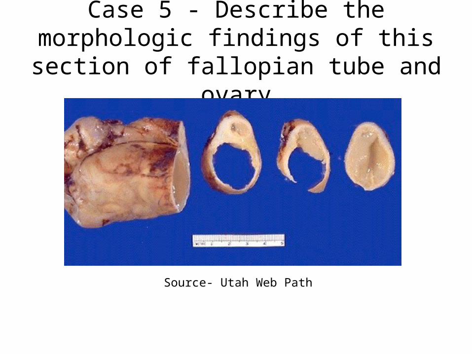
Case 5 - Describe the morphologic findings of this section of fallopian tube and ovary
Source- Utah Web Path

Case 5: What is the organ? Describe the histologic findings.
PatientNormal
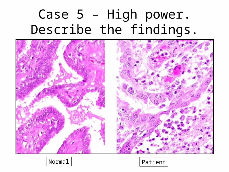
Case 5 – High power. Describe the findings.
Normal Patient

Case 5
What potential complications of this disease process may this patient experience in the future?
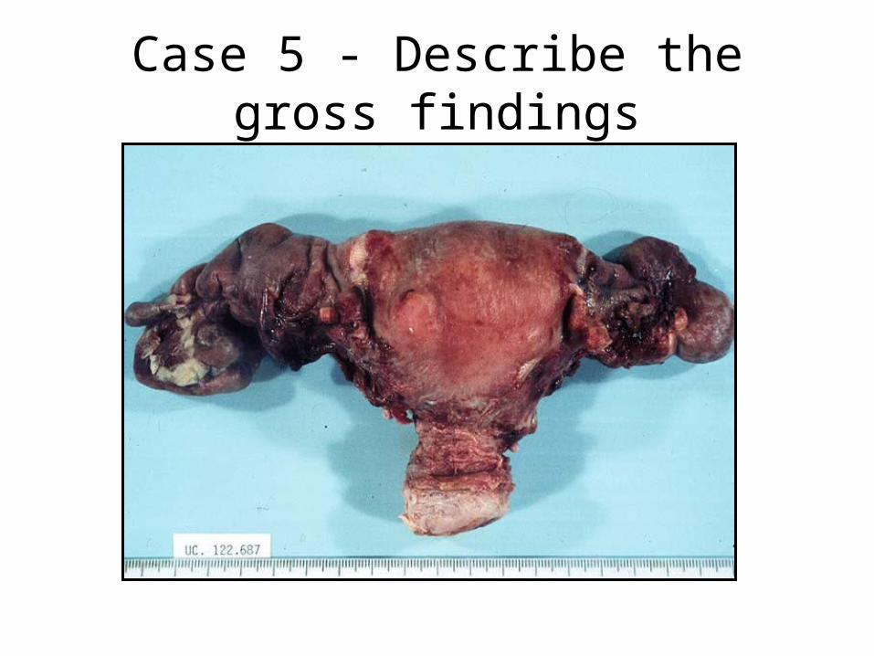
Case 5 - Describe the gross findings