Pathology of the Exocrine Pancreas and Extrahepatic ... · Pathology of the Exocrine Pancreas and...
58
Pathology of the Exocrine Pancreas and Extrahepatic Biliary Tract Robert Stern, MD Department of Pathology Special thanks to Eric Harp, DO
Transcript of Pathology of the Exocrine Pancreas and Extrahepatic ... · Pathology of the Exocrine Pancreas and...
Pathology of the Exocrine Pancreas and Extrahepatic Biliary Tract
Robert Stern, MDDepartment of Pathology
Spec i a l t h ank s to E r i c Harp, DO
Learning ObjectivesEXOCRINE PANCREAS
Acute Pancreatitis
oDefinition
oEpidemiology
oEtiopathogenesis (with Emphasis on Alcohol and Gallstones)
oGross and Microscopic Morphology
oClinicopathologic Degrees of Severity (Interstitial, Necrotizing, Hemorrhagic),
oClinical Manifestations
oDiagnostic Markers, (Prognosis, Treatment)
Learning ObjectivesEXOCRINE PANCREAS
Chronic Pancreatitis
oDefinition
oMorphology
oEtiopathogenesis
oCorrelation between Pathology and Radiologic Appearance
oClinical Manifestations and Complications
Learning Objectives
EXOCRINE PANCREAS
Pancreatic Adenocarcinoma
oMolecular/Morphological Progression from In-Situ to Invasive Cancer
oEpidemiology
oMorphology
oSpread
oClinical Manifestations and Prognosis
Learning ObjectivesBILIARY TRACT
Primary Biliary AtresiaoDefinition
oEpidemiology
oClassification into 4 types
oDefinition of Kasai procedure
CholelithiasisoDefinition
oEpidemiology
oClassification of Gallstones into Types (Cholesterol and Pigment Stones)
oDifferential Morphology and Pathogenesis of Cholesterol and Pigment Stones
oRisk Factors
oClinical Manifestations
Learning ObjectivesBILIARY TRACT
Complications of CholelithiasisoAcute Cholecystitis
oAcute Cholangitis
oEmpyema
oPerforation
oFistulas
oGallstone Ileus
oAcute Pancreatitis
oGallbladder Carcinoma
Acute CholecystitisoClinical Manifestations and Complications
Learning Objectives
BILIARY TRACT
Chronic CholecystitisoDefinition
oMain Pathological and Clinical Features
Acute CholangitisoDefinition
oCauses and Typical Clinical Manifestations (Charcot Triad)
Carcinoma of the GallbladderoEtiopathogenesis
oMode of Spread and Prognosis

ReadingROBBINS AND COTRAN 9TH ED. PP. 856-857, 875-880, 883-894THIS HANDOUT.
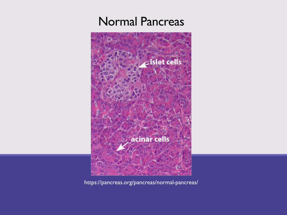
Exocrine Pancreas
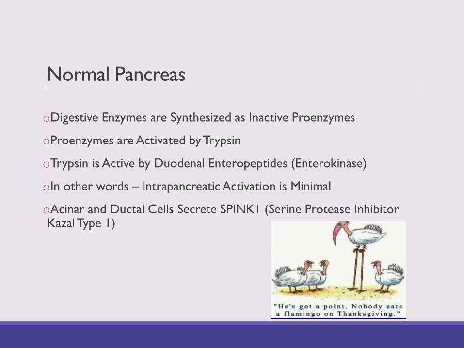
Normal Pancreas
oDigestive Enzymes are Synthesized as Inactive Proenzymes
oProenzymes are Activated by Trypsin
oTrypsin is Active by Duodenal Enteropeptides (Enterokinase)
oIn other words – Intrapancreatic Activation is Minimal
oAcinar and Ductal Cells Secrete SPINK1 (Serine Protease Inhibitor Kazal Type 1)
Acute Pancreatitis
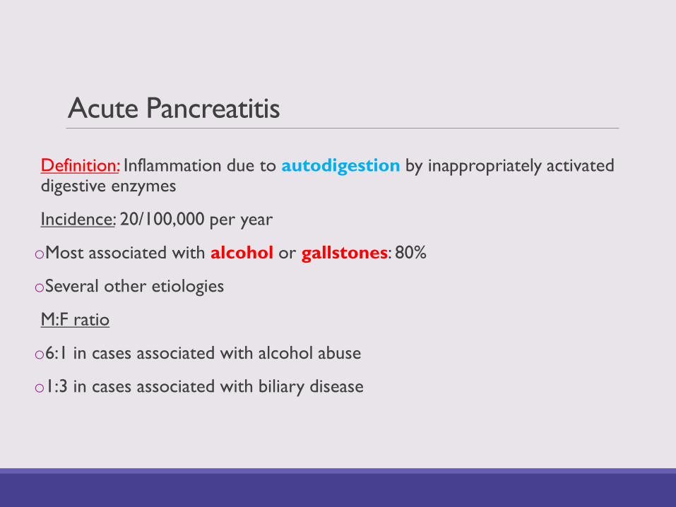
Definition: Inflammation due to autodigestion by inappropriately activated digestive enzymes
Incidence: 20/100,000 per year
oMost associated with alcohol or gallstones: 80%
oSeveral other etiologies
M:F ratio
o6:1 in cases associated with alcohol abuse
o1:3 in cases associated with biliary disease
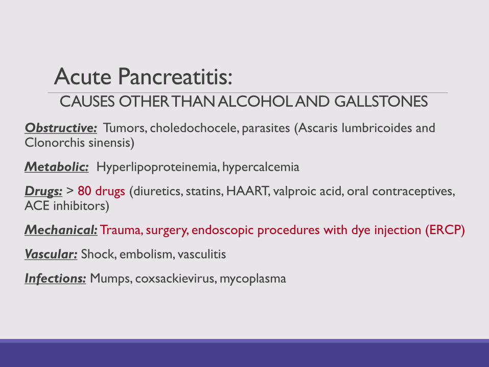
Acute Pancreatitis:CAUSES OTHER THAN ALCOHOL AND GALLSTONES
Obstructive: Tumors, choledochocele, parasites (Ascaris lumbricoides and Clonorchis sinensis)
Metabolic: Hyperlipoproteinemia, hypercalcemia
Drugs: > 80 drugs (diuretics, statins, HAART, valproic acid, oral contraceptives, ACE inhibitors)
Mechanical: Trauma, surgery, endoscopic procedures with dye injection (ERCP)
Vascular: Shock, embolism, vasculitis
Infections: Mumps, coxsackievirus, mycoplasma

Acute Pancreatitis Genetics
Genetics: Mutations of
o PRSS1 gene encoding cationic trypsinogen: Trypsin becomes resistant to inactivation by cleavage and will activate other proenzymes. Autosomal dominant
o SPINK1 encoding trypsin inhibitor: Loss of trypsin inhibition, with inappropriate activation of trypsin. Autosomal recessive
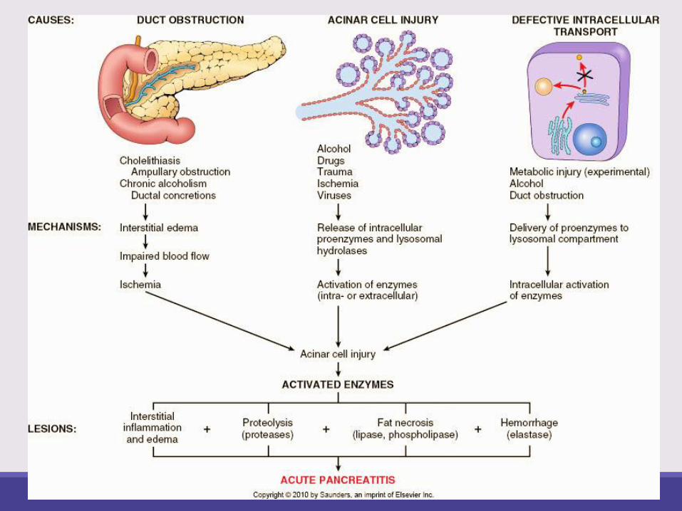
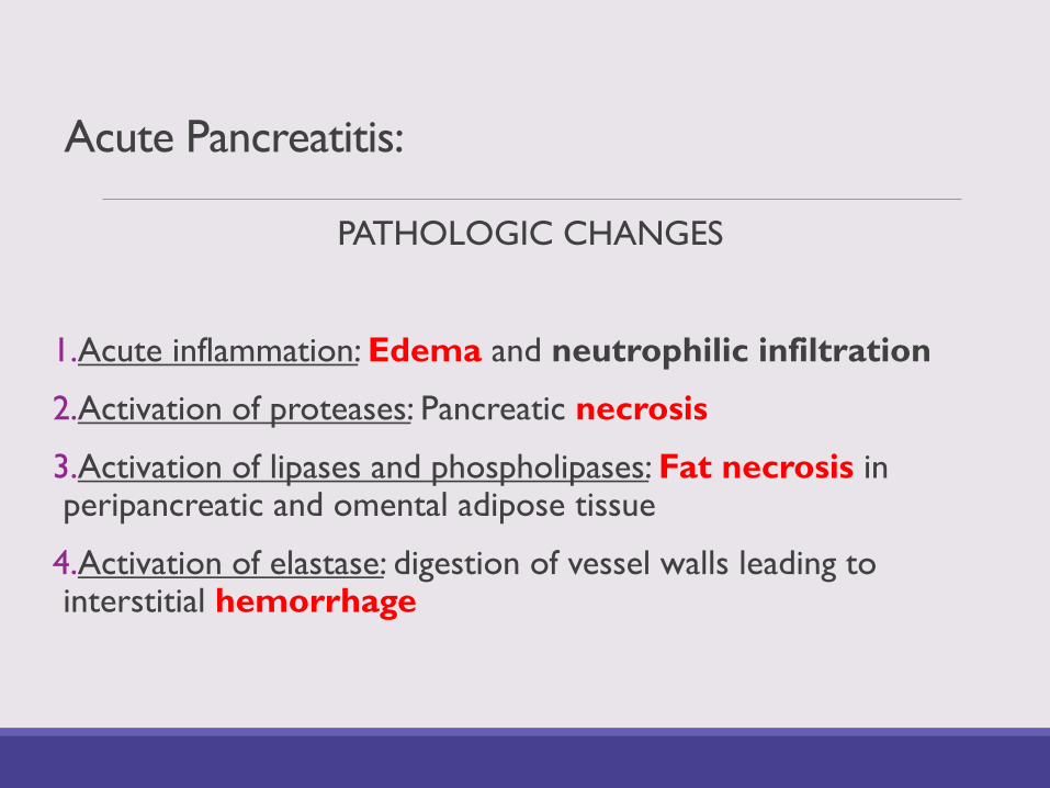
Acute Pancreatitis:
PATHOLOGIC CHANGES
1.Acute inflammation: Edema and neutrophilic infiltration
2.Activation of proteases: Pancreatic necrosis
3.Activation of lipases and phospholipases: Fat necrosis in peripancreatic and omental adipose tissue
4.Activation of elastase: digestion of vessel walls leading to interstitial hemorrhage
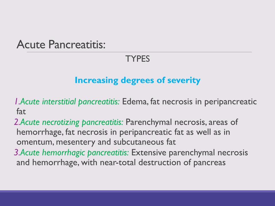
Acute Pancreatitis: TYPES
Increasing degrees of severity
1.Acute interstitial pancreatitis: Edema, fat necrosis in peripancreatic fat
2.Acute necrotizing pancreatitis: Parenchymal necrosis, areas of hemorrhage, fat necrosis in peripancreatic fat as well as in omentum, mesentery and subcutaneous fat
3.Acute hemorrhagic pancreatitis: Extensive parenchymal necrosis and hemorrhage, with near-total destruction of pancreas

Acute Pancreatitis:
Pictures from Rubin and Farber
Moderately advanced acute
pancreatitis: Numerous yellow-
white foci of fat necrosis
Severe acute pancreatitis: Large
areas of hemorrhage in the
pancreas
GROSS CHANGES
Normal Pancreas
https://pancreas.org/pancreas/normal-pancreas/
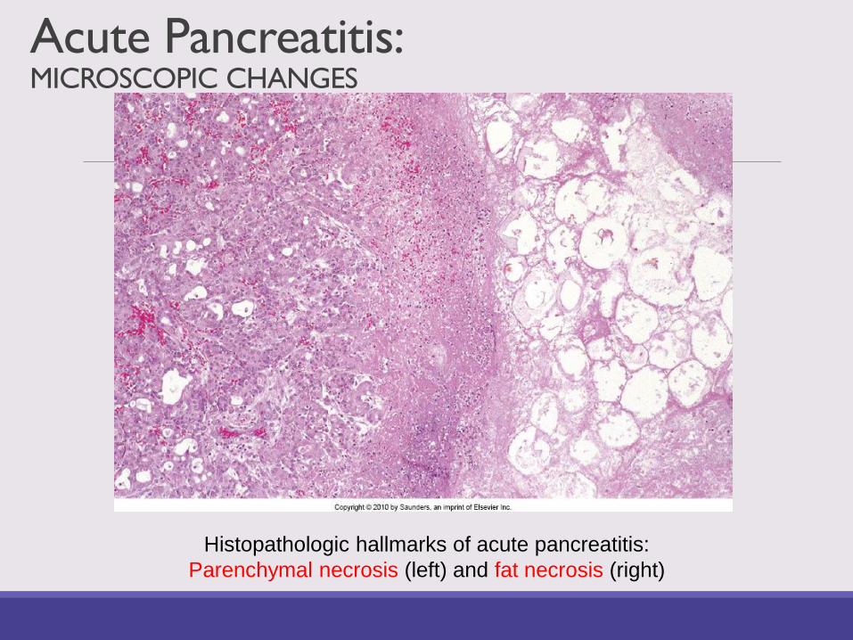
Acute Pancreatitis: MICROSCOPIC CHANGES
Histopathologic hallmarks of acute pancreatitis:
Parenchymal necrosis (left) and fat necrosis (right)
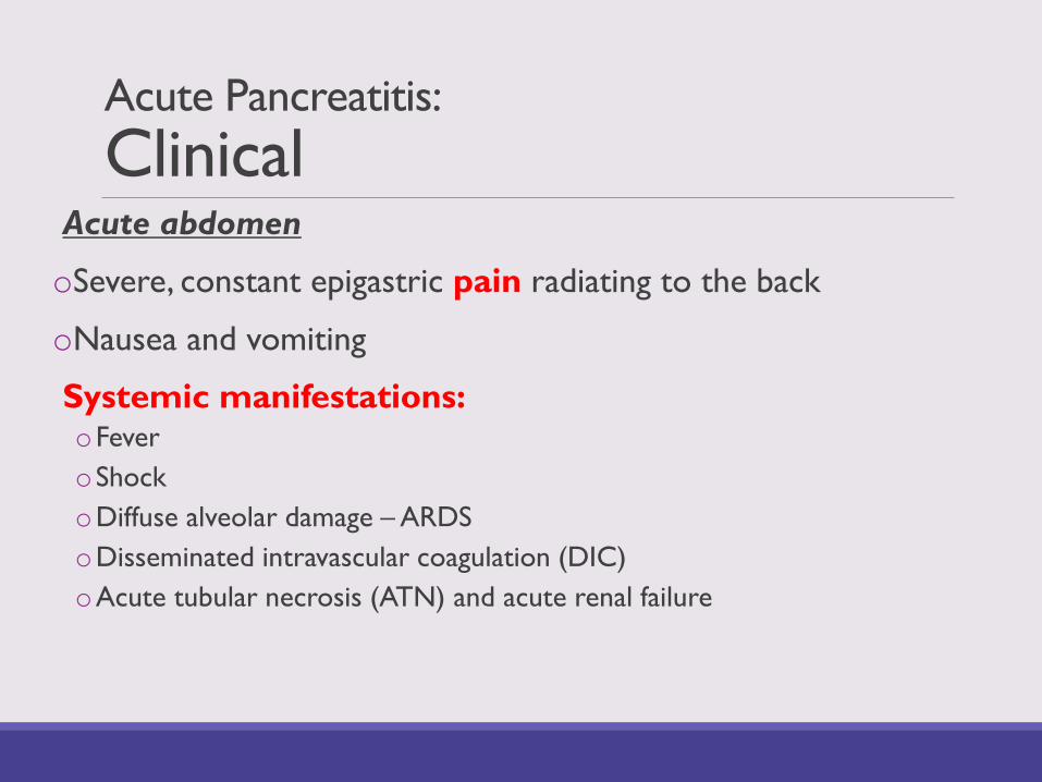
Acute Pancreatitis:
ClinicalAcute abdomen
oSevere, constant epigastric pain radiating to the back
oNausea and vomiting
Systemic manifestations:oFever
oShock
oDiffuse alveolar damage – ARDS
oDisseminated intravascular coagulation (DIC)
oAcute tubular necrosis (ATN) and acute renal failure
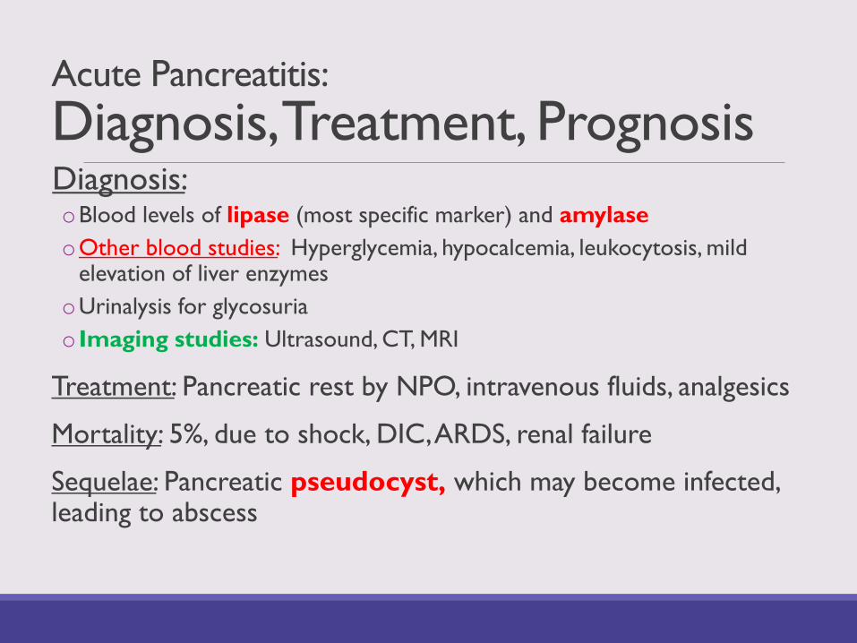
Acute Pancreatitis:
Diagnosis, Treatment, PrognosisDiagnosis:oBlood levels of lipase (most specific marker) and amylase
oOther blood studies: Hyperglycemia, hypocalcemia, leukocytosis, mild elevation of liver enzymes
oUrinalysis for glycosuria
o Imaging studies: Ultrasound, CT, MRI
Treatment: Pancreatic rest by NPO, intravenous fluids, analgesics
Mortality: 5%, due to shock, DIC, ARDS, renal failure
Sequelae: Pancreatic pseudocyst, which may become infected, leading to abscess
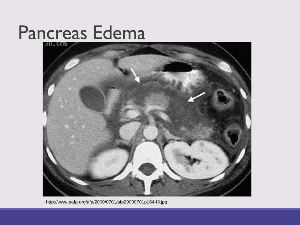
Pancreas Edema
http://www.aafp.org/afp/2000/0701/afp20000701p164-f3.jpg
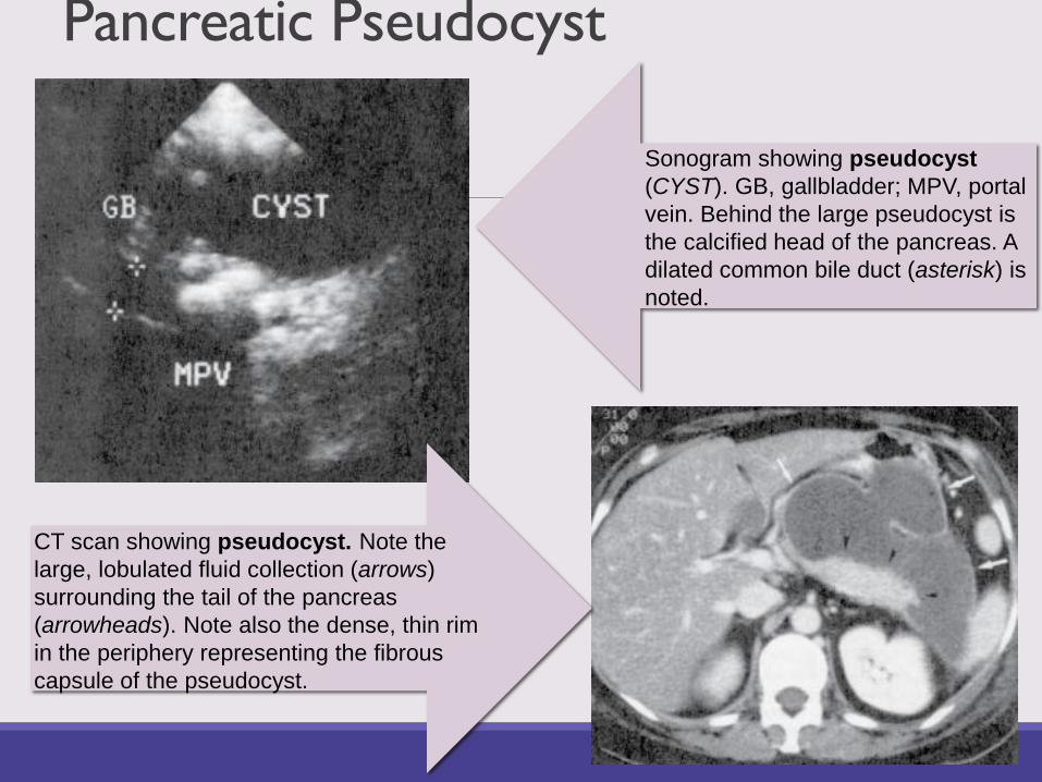
Pancreatic Pseudocyst
Sonogram showing pseudocyst
(CYST). GB, gallbladder; MPV, portal
vein. Behind the large pseudocyst is
the calcified head of the pancreas. A
dilated common bile duct (asterisk) is
noted.
CT scan showing pseudocyst. Note the
large, lobulated fluid collection (arrows)
surrounding the tail of the pancreas
(arrowheads). Note also the dense, thin rim
in the periphery representing the fibrous
capsule of the pseudocyst.
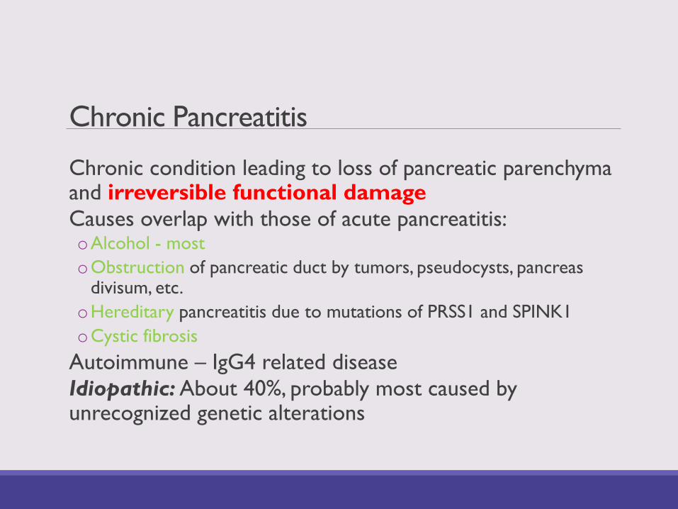
Chronic Pancreatitis
Chronic condition leading to loss of pancreatic parenchyma and irreversible functional damage
Causes overlap with those of acute pancreatitis:oAlcohol - most
oObstruction of pancreatic duct by tumors, pseudocysts, pancreas divisum, etc.
oHereditary pancreatitis due to mutations of PRSS1 and SPINK1
oCystic fibrosis
Autoimmune – IgG4 related disease
Idiopathic: About 40%, probably most caused by unrecognized genetic alterations
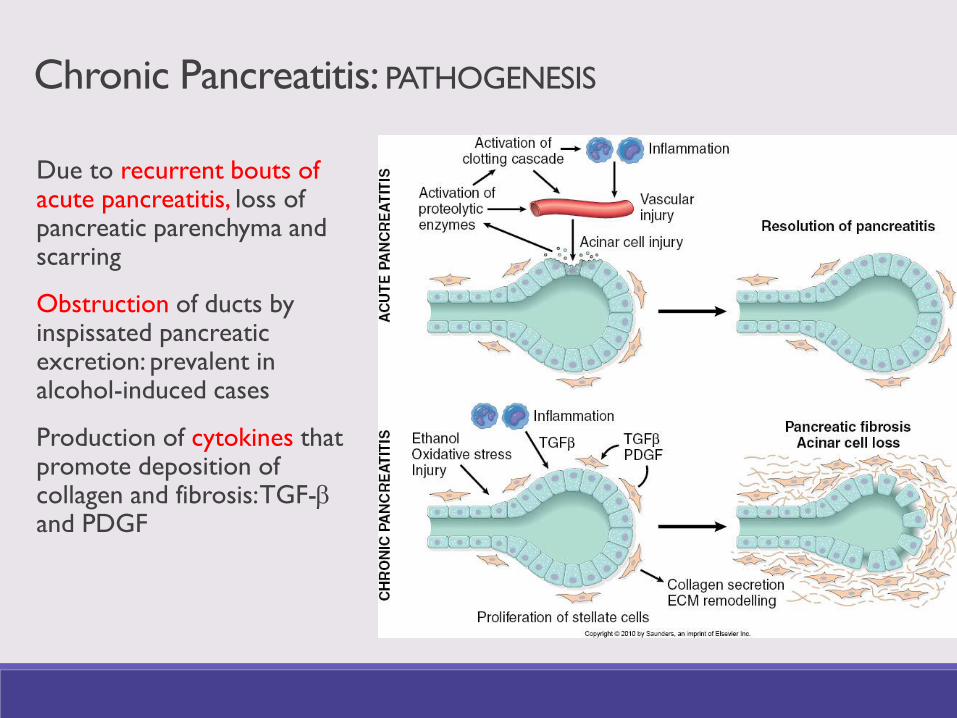
Chronic Pancreatitis: PATHOGENESIS
Due to recurrent bouts of acute pancreatitis, loss of pancreatic parenchyma and scarring
Obstruction of ducts by inspissated pancreatic excretion: prevalent in alcohol-induced cases
Production of cytokines that promote deposition of collagen and fibrosis: TGF-band PDGF
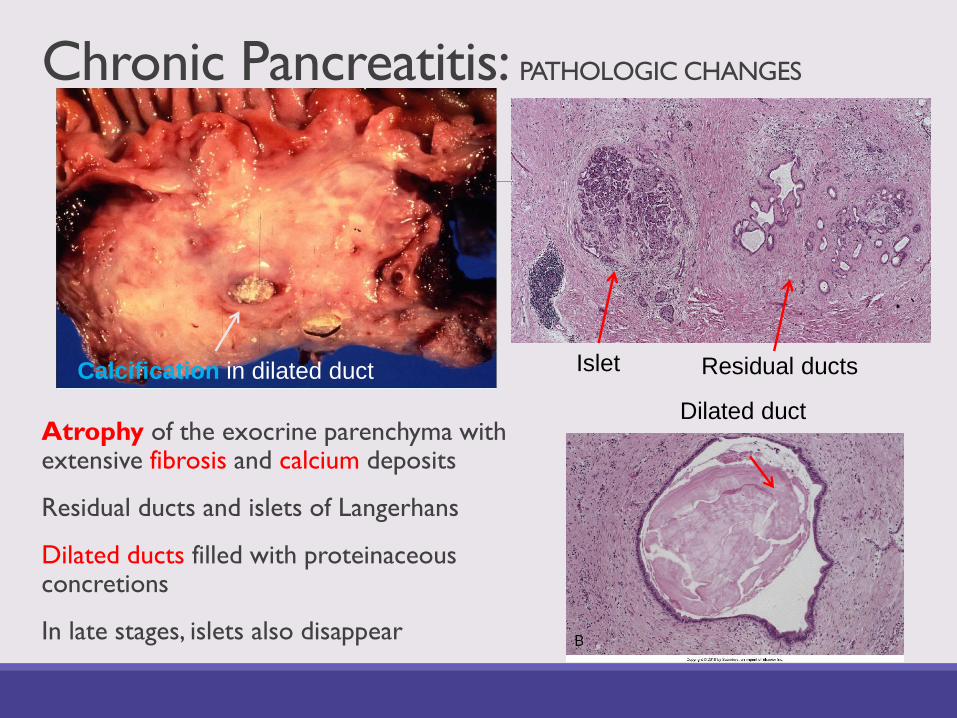
Chronic Pancreatitis: PATHOLOGIC CHANGES
Atrophy of the exocrine parenchyma with extensive fibrosis and calcium deposits
Residual ducts and islets of Langerhans
Dilated ducts filled with proteinaceous concretions
In late stages, islets also disappear
Islet Residual ducts
Dilated duct
Calcification in dilated duct
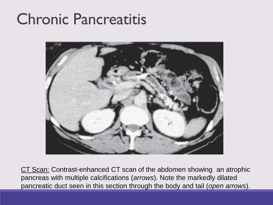
Chronic Pancreatitis
CT Scan: Contrast-enhanced CT scan of the abdomen showing an atrophic
pancreas with multiple calcifications (arrows). Note the markedly dilated
pancreatic duct seen in this section through the body and tail (open arrows).
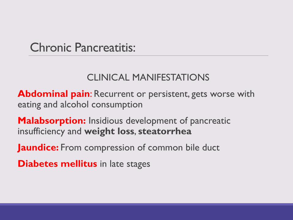
Chronic Pancreatitis:
CLINICAL MANIFESTATIONS
Abdominal pain: Recurrent or persistent, gets worse with eating and alcohol consumption
Malabsorption: Insidious development of pancreatic insufficiency and weight loss, steatorrhea
Jaundice: From compression of common bile duct
Diabetes mellitus in late stages
Pancreatic Adenocarcinoma
Fourth most frequent cause of cancer-related death in USA
Majority occur at 60-80 years of age
Risk factors:
oSmoking doubles the risk
oFat-rich diet
oChronic pancreatitis
oGene mutations: Peutz-Jeghers syndrome, hereditary chronic pancreatitis, p-16 mutations, BRCA2
Carcinogenesis:As in many other epithelial cancers, pancreatic adenocarcinoma is believed to progress from normal epithelium to precursor lesions (Pancreatic Intraepithelial Neoplasia – PanIN) to invasive carcinoma
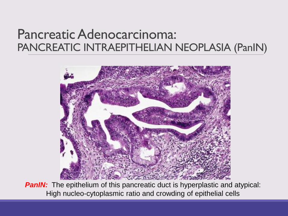
Pancreatic Adenocarcinoma: PANCREATIC INTRAEPITHELIAN NEOPLASIA (PanIN)
PanIN: The epithelium of this pancreatic duct is hyperplastic and atypical:
High nucleo-cytoplasmic ratio and crowding of epithelial cells
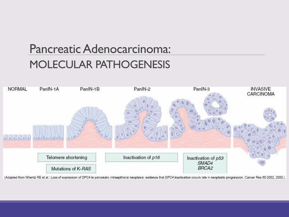
Pancreatic Adenocarcinoma:
MOLECULAR PATHOGENESIS

Pancreatic Adenocarcinoma: MORPHOLOGY
Poorly defined, firm, gray-white tumor in the
head of pancreas.
Desmoplastic reaction (formation of fibrous
tissue) is characteristic of this cancer.
Disorganized glandular structures
surrounded by fibrotic stroma. Nerve
infiltration is characteristic.
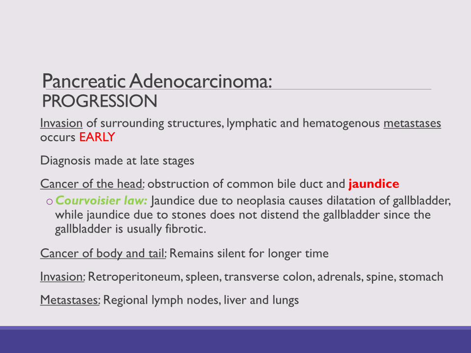
Pancreatic Adenocarcinoma: PROGRESSIONInvasion of surrounding structures, lymphatic and hematogenous metastasesoccurs EARLY
Diagnosis made at late stages
Cancer of the head: obstruction of common bile duct and jaundice
oCourvoisier law: Jaundice due to neoplasia causes dilatation of gallbladder, while jaundice due to stones does not distend the gallbladder since the gallbladder is usually fibrotic.
Cancer of body and tail: Remains silent for longer time
Invasion: Retroperitoneum, spleen, transverse colon, adrenals, spine, stomach
Metastases: Regional lymph nodes, liver and lungs
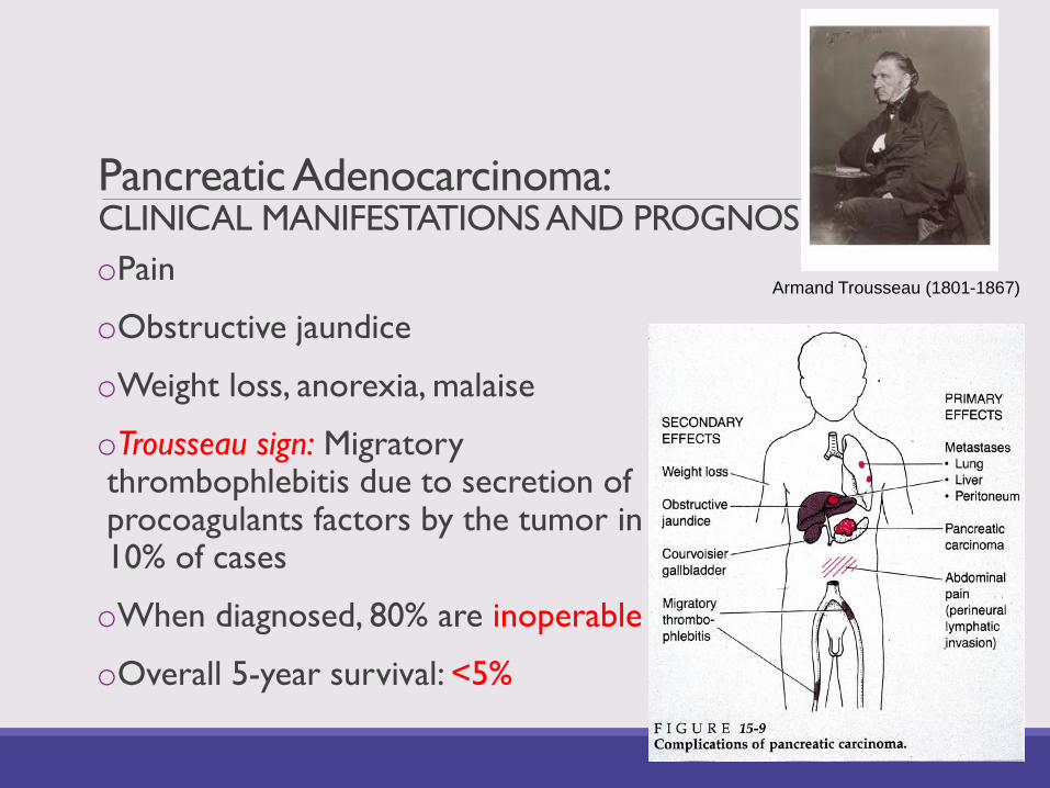
Pancreatic Adenocarcinoma: CLINICAL MANIFESTATIONS AND PROGNOSIS
oPain
oObstructive jaundice
oWeight loss, anorexia, malaise
oTrousseau sign: Migratory thrombophlebitis due to secretion of procoagulants factors by the tumor in 10% of cases
oWhen diagnosed, 80% are inoperable
oOverall 5-year survival: <5%
Armand Trousseau (1801-1867)
Extrahepatic Biliary System
Biliary Atresia
Definition: Partial/complete obstruction of extrahepatic biliary tree within 3 months of life
Most common cause of neonatal obstructive jaundice (1:12,000 live births) and of liver-related deaths in childhood
Accounts for 50% of liver transplantation in children
Fetal form (20%): Abnormal development of extrahepatic biliary tree, associated with other congenital anomalies
Perinatal form (80%): Inflammatory destruction of biliary tree by infectious agents (CMV, reovirus, rotavirus) or autoimmune
Leads to cirrhosis within 6 months
Cholelithiasis
In the US, >20 million people have gallstones, including 20% of women and 8% of men >40◦ 40% of women > 65 have gallstones
Cholesterol stones: 80% of gallstones (90% in the West), composed of >50% of cholesterol monohydrate
Pigment stones: 20% of gallstones, composed predominantly of bilirubin and calcium salts and < 20% of cholesterol
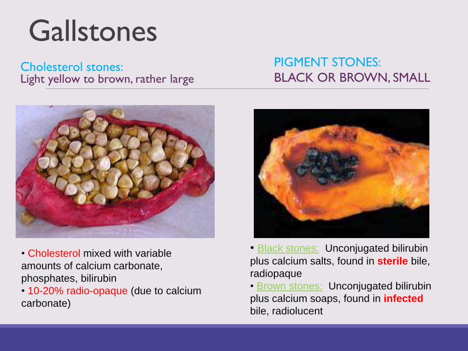
GallstonesCholesterol stones:Light yellow to brown, rather large
PIGMENT STONES:
BLACK OR BROWN, SMALL
• Cholesterol mixed with variable
amounts of calcium carbonate,
phosphates, bilirubin
• 10-20% radio-opaque (due to calcium
carbonate)
• Black stones: Unconjugated bilirubin
plus calcium salts, found in sterile bile,
radiopaque
• Brown stones: Unconjugated bilirubin
plus calcium soaps, found in infected
bile, radiolucent
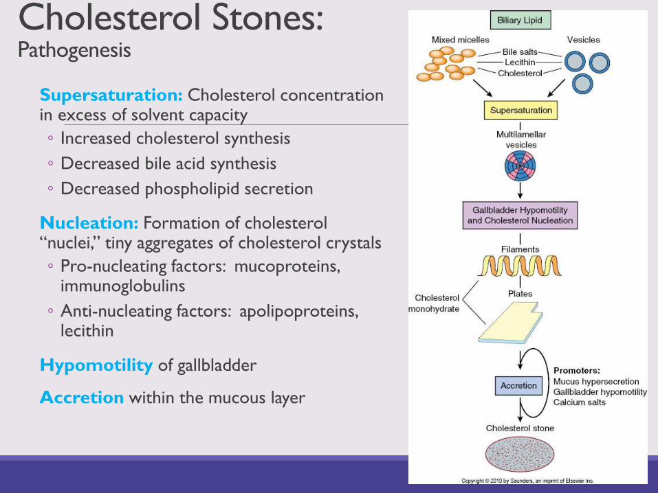
Cholesterol Stones: Pathogenesis
Supersaturation: Cholesterol concentration in excess of solvent capacity
◦ Increased cholesterol synthesis
◦ Decreased bile acid synthesis
◦ Decreased phospholipid secretion
Nucleation: Formation of cholesterol “nuclei,” tiny aggregates of cholesterol crystals
◦ Pro-nucleating factors: mucoproteins, immunoglobulins
◦ Anti-nucleating factors: apolipoproteins, lecithin
Hypomotility of gallbladder
Accretion within the mucous layer
Pigment Stones:
PathogenesisBlack stones: Occur in patients with chronic hemolytic conditions e.g. sickle cell anemia, thalassemia, megaloblastic anemia, etc.
◦ Other predisposing conditions: Cirrhosis, ileal disease or resection, cystic fibrosis
Brown stones: Result from deconjugation of bilirubin by b-glucuronidase produced by bacteria or parasites◦ Associated with chronic gallbladder infection or infestation (liver
flukes important in Eastern Asia)
Gallstones - Risk Factors
CHOLESTEROL STONES
Western countries, America
Age
Females, oral contraceptives, pregnancy
Obesity and rapid weight loss
Hyperlipidemia
Inborn errors of bile acid metabolism
PIGMENT STONES
Asians more than Westerners
Biliary infections (brown stones)
Chronic hemolysis (black stones)
Various GI disorders (black stones) – i.e. ileal resection or bypass, Crohn’s disease, cystic fibrosis
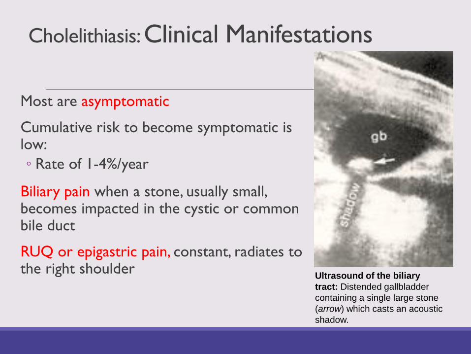
Cholelithiasis: Clinical Manifestations
Most are asymptomatic
Cumulative risk to become symptomatic is low:
◦ Rate of 1-4%/year
Biliary pain when a stone, usually small, becomes impacted in the cystic or common bile duct
RUQ or epigastric pain, constant, radiates to the right shoulder
Ultrasound of the biliary
tract: Distended gallbladder
containing a single large stone
(arrow) which casts an acoustic
shadow.

Cholelithiasis - Complications
1. Acute cholecystitis: Most frequent complication requiring surgery
2. Acute cholangitis: Infection of biliary tract, due to stones in the common bile duct
3. Empyema: Purulent infection of gallbladder, which becomes filled with pus
4. Perforation: Causes acute bilious peritonitis
5. Fistulas: Duodenum, colon, jejunum, stomach, abdominal wall, renal pelvis
6. Gallstone ileus: Bowel obstruction by a large stone that erodes directly into a bowel loop
7. Acute pancreatitis
8. Gallbladder carcinoma
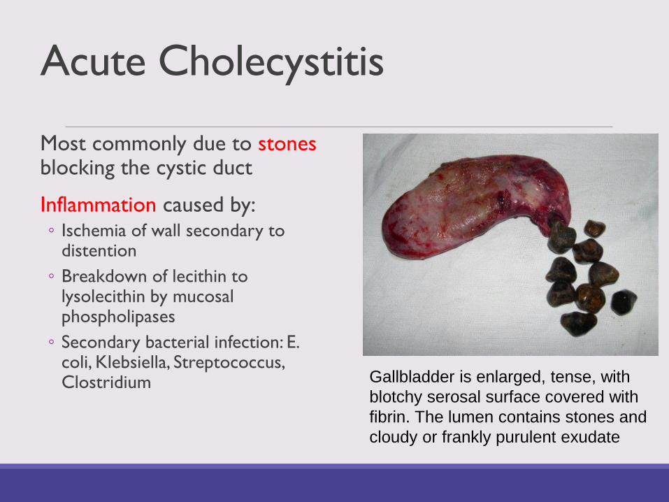
Acute Cholecystitis
Most commonly due to stonesblocking the cystic duct
Inflammation caused by:◦ Ischemia of wall secondary to
distention
◦ Breakdown of lecithin to lysolecithin by mucosal phospholipases
◦ Secondary bacterial infection: E. coli, Klebsiella, Streptococcus, Clostridium Gallbladder is enlarged, tense, with
blotchy serosal surface covered with
fibrin. The lumen contains stones and
cloudy or frankly purulent exudate
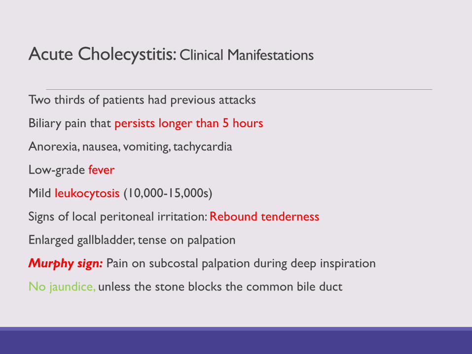
Acute Cholecystitis: Clinical Manifestations
Two thirds of patients had previous attacks
Biliary pain that persists longer than 5 hours
Anorexia, nausea, vomiting, tachycardia
Low-grade fever
Mild leukocytosis (10,000-15,000s)
Signs of local peritoneal irritation: Rebound tenderness
Enlarged gallbladder, tense on palpation
Murphy sign: Pain on subcostal palpation during deep inspiration
No jaundice, unless the stone blocks the common bile duct
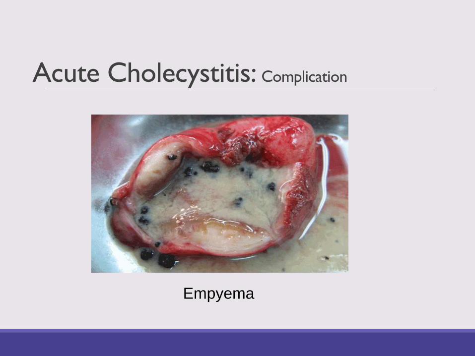
Acute Cholecystitis: Complication
Empyema
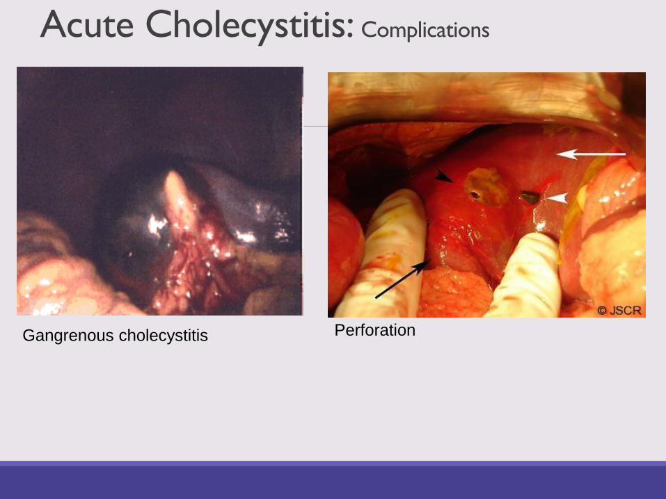
Acute Cholecystitis: Complications
Gangrenous cholecystitis Perforation

Acute Acalculus CholecystitisIschemia
◦ Cystic artery is an end artery
◦ Edema of the wall
◦ Hypomotility
Biliary sludge – cystic duct obstruction
Risk factors
◦ Sepsis with hypotension
◦ Immunosuppression
◦ Diabetes mellitus
◦ Major trauma and burns
◦ Infections
Acute Acalculus Cholecystitis
Symptoms often insidious
Symptoms obscured by predisposing condition
Maintain high risk of suspicion
Delay in diagnosis can lead to gangrenous changes and perforation
Chronic Cholecystitis
Associated with cholelithiasis in 90% of cases
Due to recurrent bouts of acute cholecystitis or manifesting without preceding attacks
Unclear pathogenesis: Bile supersaturationcommon cause of both chronic cholecystitis and stone formation and chronic low grade inflammation
In 1/3 of cases bacteria are present
Asymptomatic or manifests with biliary pain, acute cholecystitis or complications
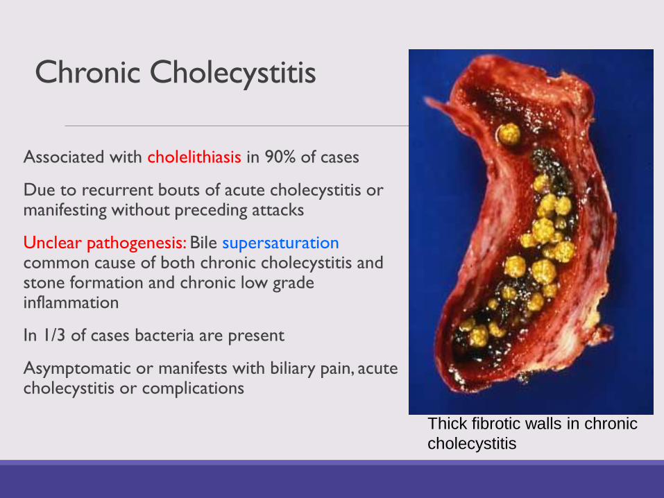
Thick fibrotic walls in chronic
cholecystitis
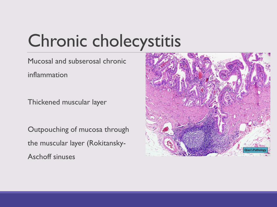
Chronic cholecystitisMucosal and subserosal chronic
inflammation
Thickened muscular layer
Outpouching of mucosa through
the muscular layer (Rokitansky-
Aschoff sinuses

Chronic Cholecystitis: Complication
Porcelain Gallbladder
Acute Cholangitis
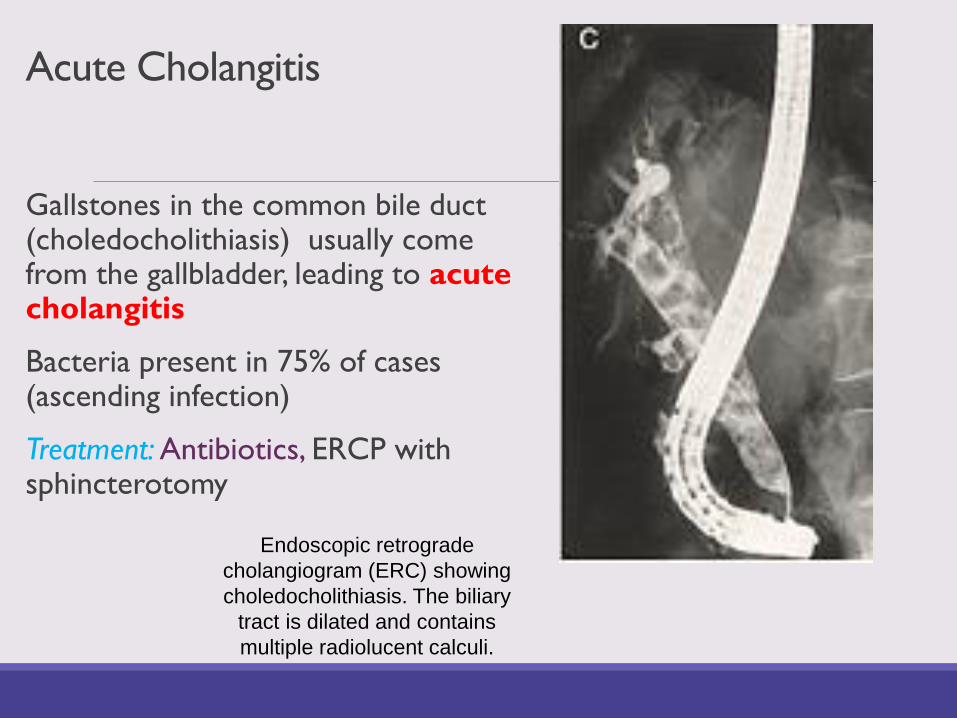
Gallstones in the common bile duct (choledocholithiasis) usually come from the gallbladder, leading to acute cholangitis
Bacteria present in 75% of cases (ascending infection)
Treatment: Antibiotics, ERCP with sphincterotomy
Endoscopic retrograde
cholangiogram (ERC) showing
choledocholithiasis. The biliary
tract is dilated and contains
multiple radiolucent calculi.
Carcinoma of the Gallbladder
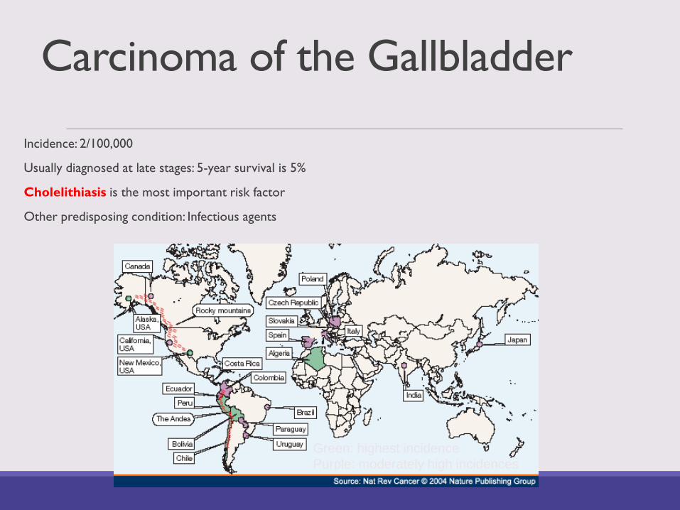
Incidence: 2/100,000
Usually diagnosed at late stages: 5-year survival is 5%
Cholelithiasis is the most important risk factor
Other predisposing condition: Infectious agents
Green: highest incidence
Purple: moderately high incidences
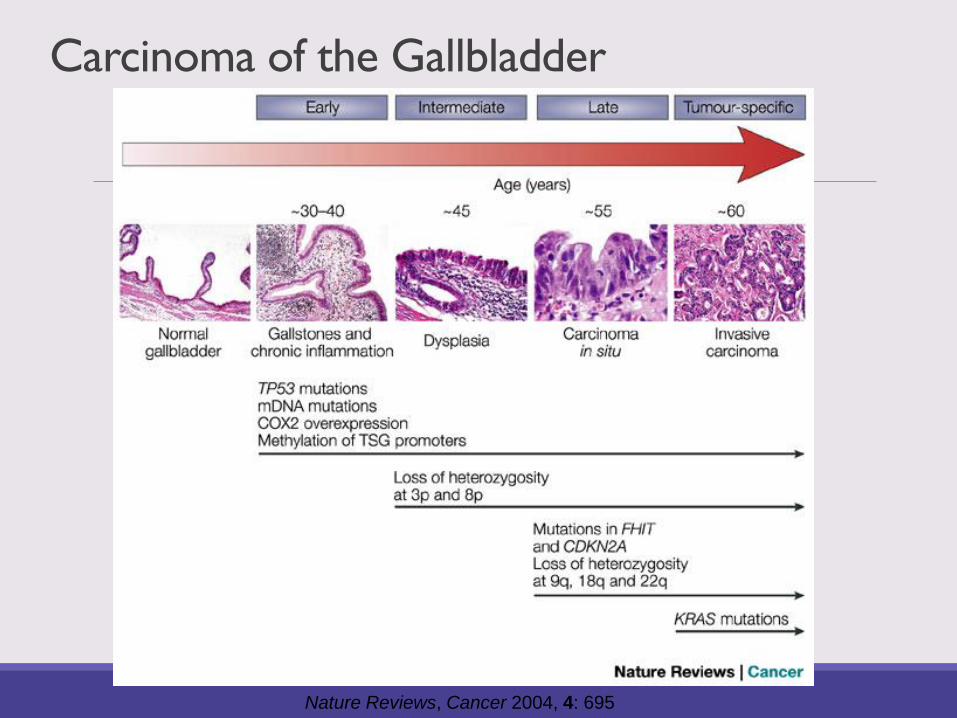
Carcinoma of the Gallbladder
Nature Reviews, Cancer 2004, 4: 695
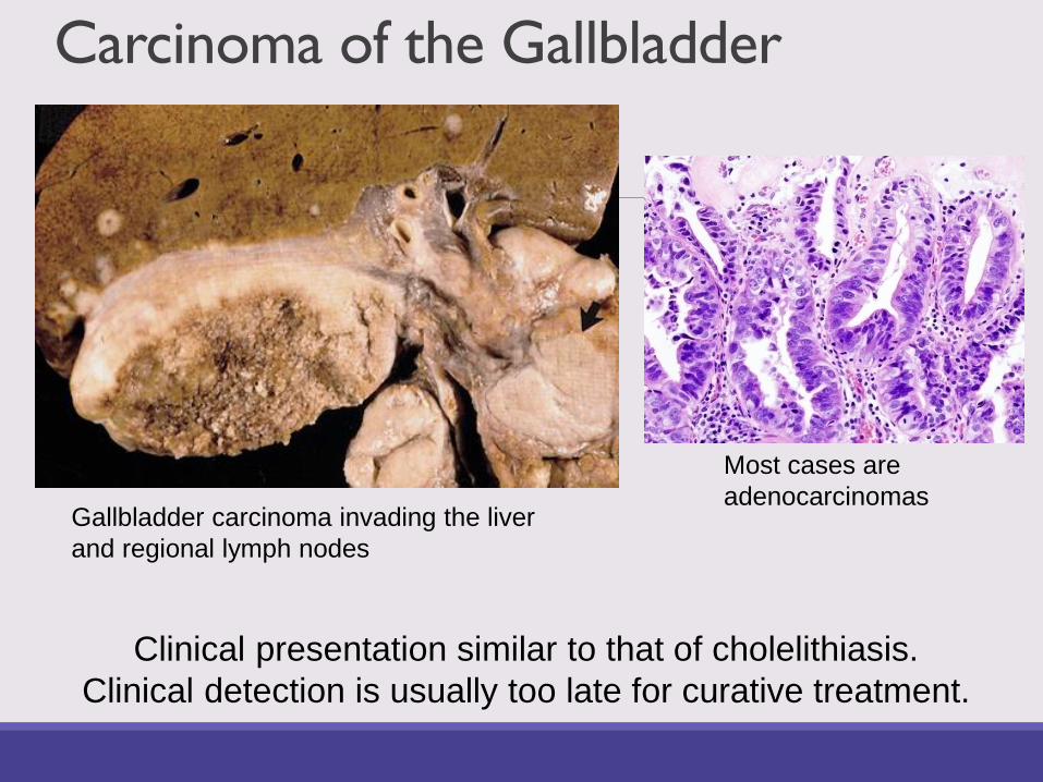
Carcinoma of the Gallbladder
Gallbladder carcinoma invading the liver
and regional lymph nodes
Most cases are
adenocarcinomas
Clinical presentation similar to that of cholelithiasis.
Clinical detection is usually too late for curative treatment.

QUESTIONS?