PATHOLOGY OF BLOOD VESSELS. …ustavpatologie.upol.cz/_data/section-1/167.pdfPATHOLOGY OF BLOOD...
27
PATHOLOGY OF BLOOD VESSELS. HAEMATOPATHOLOGY. II. practical training 3 rd year General Medicine
Transcript of PATHOLOGY OF BLOOD VESSELS. …ustavpatologie.upol.cz/_data/section-1/167.pdfPATHOLOGY OF BLOOD...

PATHOLOGY OF BLOOD
VESSELS.
HAEMATOPATHOLOGY.
II. practical training
3rd year General Medicine

Myeloproliferative syndrome, polycytemia vera

Myeloproliferative syndrome, polycytemia vera

Extramedullary haematopoiesis

Extramedullary haematopoiesis

Myeloproliferative syndrome, chronic myeloid leukaemia
(Ph+), liver

Myeloproliferative syndrome, chronic myeloid leukaemia
(Ph+), liver

Chronic myeloid leukaemia, lymph node

Myelodysplastic syndrome, bone marrow

Myelodysplastic syndrome, bone marrow
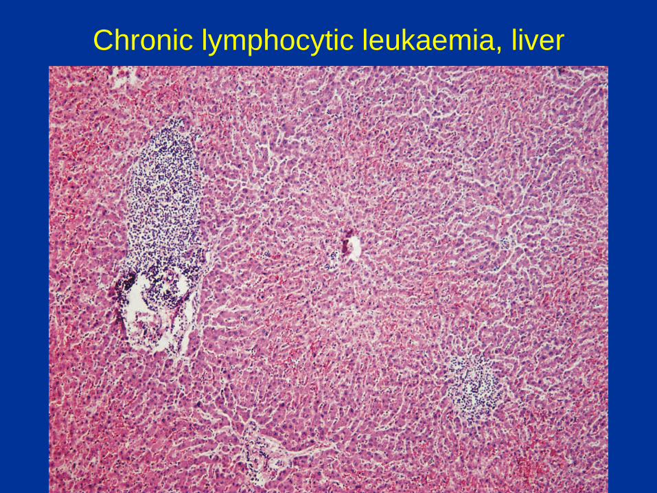
Chronic lymphocytic leukaemia, liver

Malignant lymphoma - classification
Hodgkin’s
non- Hodgkin’s lymphomas
o precursor cells
B
T/NK
o Mature cells
B
T/NK

Follicle center lymphoma
Morphology Mixture of germinal center blasts and cleaved cells (centroblasts and centrocytes). Immunology Surface Ig + CD5 – CD10 ± CD19, 20, 22, 79a + BCL-2 + Genetics t(14;18) & BCL-2 rearrange- ment in majority of cases. Clinical Adults. Indolent course (median survival 7-9 yrs).
Equivalent to
“centroblastic / centrocytic”
and “follicular centroblastic”
lymphomas in Kiel
scheme
H&E
BCL-2
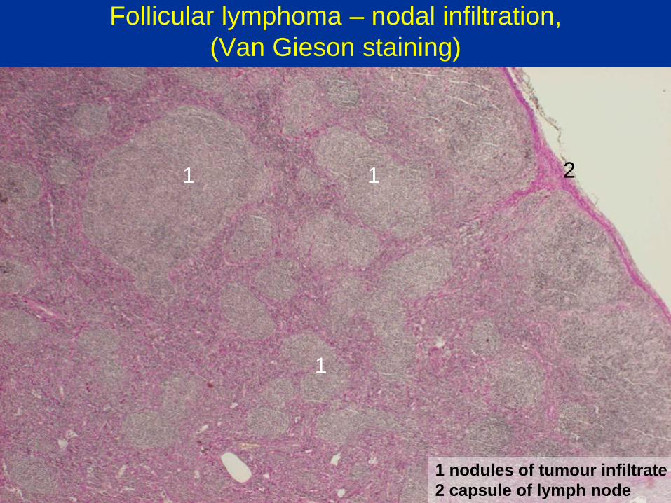
Follicular lymphoma – nodal infiltration,
(Van Gieson staining)
1 1
1
1 nodules of tumour infiltrate
2 capsule of lymph node
2

Follicular lymphoma – nodal infiltration, detail
Centroblasts
Centrocytes

Follicular lymphoma – nodal infiltration (anti-CD 20)

Mantle cell lymphoma
Morphology Small irregularly shaped centrocyte-like cells. Immunology Surface Ig (l>k) + CD5 – CD10 ± CD19, 20, 22, 79a + CD23 – Cyclin D1 + Genetics t(11;14). BCL-1 rearrangement. Clinical Adults. Moderately aggressive course (median survival 3-4 yrs). Equivalent to
“centrocytic lymphoma”
in Kiel scheme
Giemsa
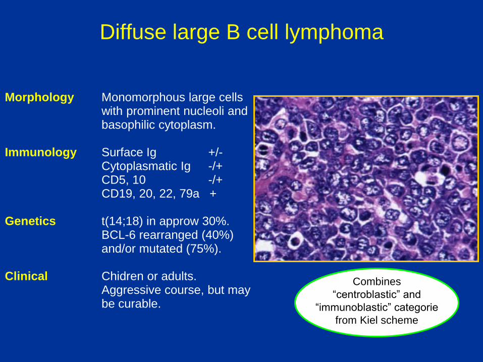
Diffuse large B cell lymphoma
Morphology Monomorphous large cells with prominent nucleoli and basophilic cytoplasm. Immunology Surface Ig +/- Cytoplasmatic Ig -/+ CD5, 10 -/+ CD19, 20, 22, 79a + Genetics t(14;18) in approw 30%. BCL-6 rearranged (40%) and/or mutated (75%). Clinical Chidren or adults. Aggressive course, but may be curable.
Combines
“centroblastic” and
“immunoblastic” categorie
from Kiel scheme

Diffuse large B cell lymphoma
1
2 3
1 gastric foveoles
2 gastric glands
3 tumour infiltrate
4 reactive lymphoid nodule
4
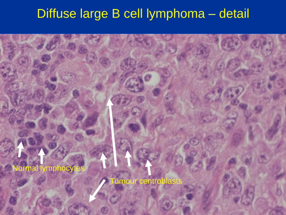
Diffuse large B cell lymphoma – detail
Normal lymphocytes
Tumour centroblasts

Hodgkin’s lymphoma
Classical – diagnostic cells, non-tumorous backround:
o Nodular sclerosis
o Mixed cellularity
o Lymphocyte depleted
o Lymphocyte rich
Nodular lymphocyte predominant: background with
lymphocytes and histiocytes

Diagnostic cells in classical Hodgkin’s lymphoma
Hodgkin’s cells – one nucleus with a large
eosinophilic nucleolus
Reed-Sternberg cells – two nuclei with nucleoli
Sternberg cells – multiple nuclei and nucleoli
Lacunar cells – clear large cytoplasma, small
nucleus
CD20-, CD 79a-, CD 30+, CD 15+, CD 45-, Ig-

Diagnostic cells
Hodgkin
Reed-Sternberg Lacunar
Sternberg
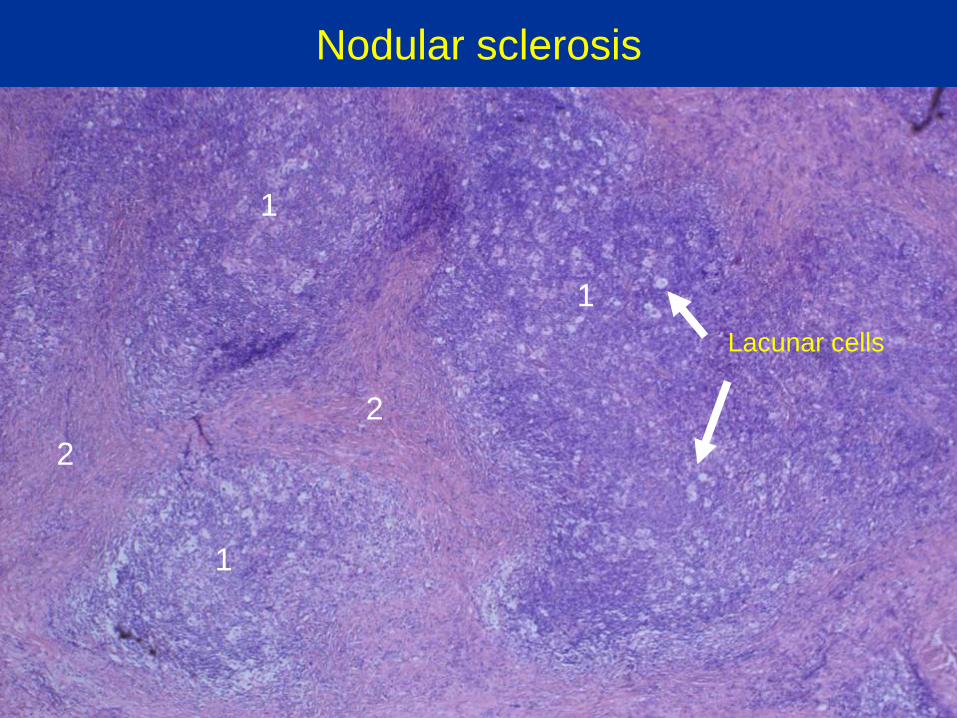
Nodular sclerosis
1
1
1
2
2
Lacunar cells

Mixed cellularity

HD – background
Plasmocyt
Lymfocytes
Eosinophils
Histiocyt
Fibroblast

Spleen infiltration by lymphoma