Pathology. History continues… - … · Pathology. History continues… M.B. 2017 Pathology...
16
1 Pathology. History continues… M.B. 2017 Pathology –what is it ? (Greek; pathos – suffering, disease logos – study, word The study of disease or Let’s talk about disease „a discipline that bridges clinical practice and basic science, involving causes and mechanisms of disease that results in the presenting symptoms of the patient” (by Robbins) Regressive lesions Changes in the structure of normally developed cells, tissues or organs, which usually interfere with their function • Cellular adaptation to injury – atrophy – intracellular accumulations (degeneration) • cell death (necrosis) • inborn abnormalities Developmental abnormalities (organ-specific malformations) • agenesia (a lack of organ anlage) • aplasia • hypoplasia (incomplete development) • atresia (complete failure of development of the intestinal/duct lumen) • double organs • ectopy (abnormally located tissue) Developmental abnormalities - hypoplastic kidney • Hepinstall’s Pathology of the Kidney Developmental abnormalities atresia
-
Upload
vuongquynh -
Category
Documents
-
view
218 -
download
1
Transcript of Pathology. History continues… - … · Pathology. History continues… M.B. 2017 Pathology...
1
Pathology. History continues…
M.B. 2017
Pathology –what is it ?
(Greek; pathos – suffering, disease
logos – study, word
The study of disease or
Let’s talk about disease
„a discipline that bridges clinical practice and basic science, involving causes and mechanisms of disease that results in the presenting symptoms of the patient” (by Robbins)
Regressive lesions
Changes in the structure of normally developed cells, tissues or organs, which usually interfere with their function
• Cellular adaptation to injury
– atrophy
– intracellular accumulations (degeneration)
• cell death (necrosis)
• inborn abnormalities
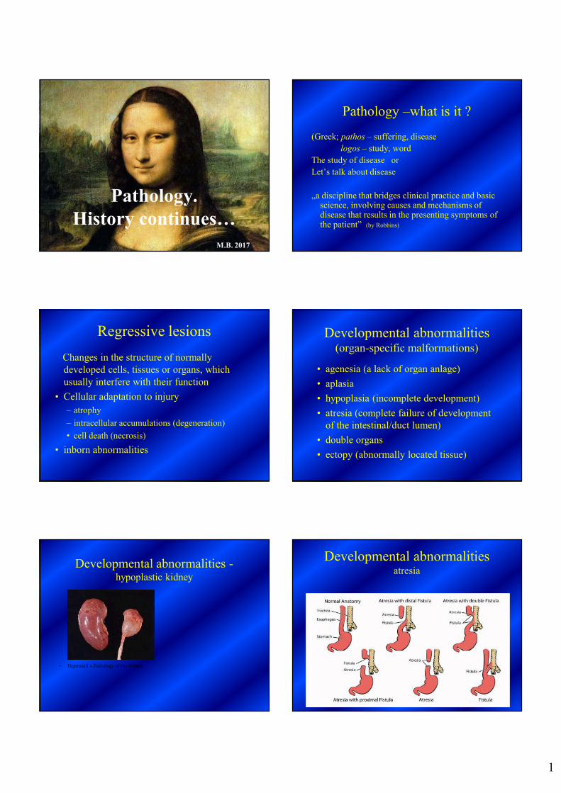
Developmental abnormalities (organ-specific malformations)
• agenesia (a lack of organ anlage)
• aplasia
• hypoplasia (incomplete development)
• atresia (complete failure of development of the intestinal/duct lumen)
• double organs
• ectopy (abnormally located tissue)
Developmental abnormalities -hypoplastic kidney
• Hepinstall’s Pathology of the Kidney
Developmental abnormalitiesatresia
2
Developmental abnormalitiesatresia
• Duodenal atresia frequently associated with Down's syndrome or other anomalies
• Presents relatively early in life with vomiting
• Treated surgically
Developmental abnormalitiesdouble organs
• Ureteral duplication
note the two ureters (arrows) from the right kidney.
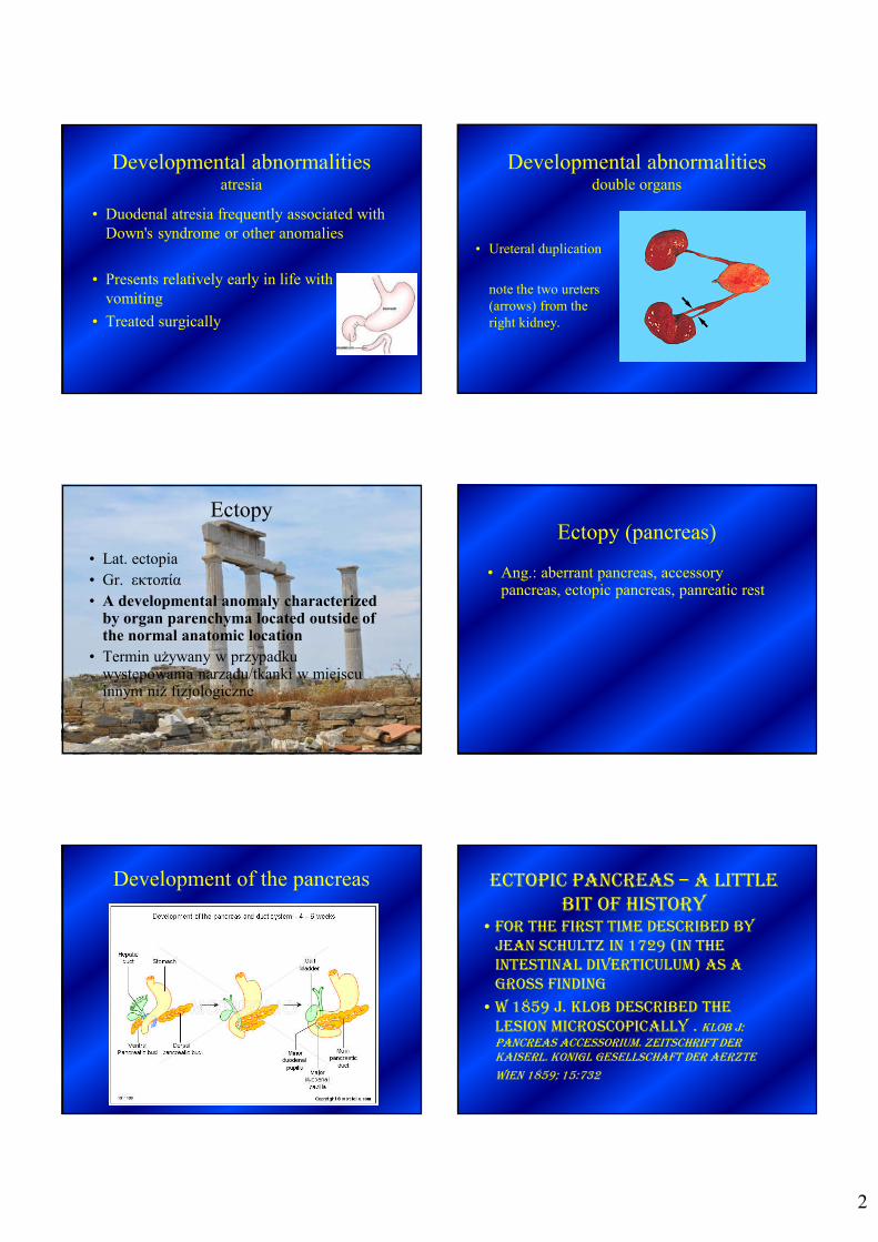
Ectopy
• Lat. ectopia
• Gr. εκτοπία
• A developmental anomaly characterized by organ parenchyma located outside of the normal anatomic location
• Termin używany w przypadku występowania narządu/tkanki w miejscu innym niż fizjologiczne
Ectopy (pancreas)
• Ang.: aberrant pancreas, accessory pancreas, ectopic pancreas, panreatic rest
Development of the pancreas ectopic pAncreAS – A little bit oF hiStory
• For the FirSt time deScribed by JeAn SchultZ in 1729 (in the inteStinAl diverticulum) AS A GroSS FindinG
• W 1859 J. Klob deScribed the leSion microScopicAlly . Klob J: pAncreAS AcceSSorium. ZeitSchriFt der KAiSerl. KoniGl GeSellSchAFt der AerZte
Wien 1859; 15:732

3
Histologic classification of heterotopic pancreatic tissue by Heinrich
• Type I – normal pancreatic tissue (preserved lobular structure, visible islets and pancreatic ducts)
• Type II – abnormal structure, no islets
• Type III – lesion composed only of pancreatic ducts
Ectopic pancreas in the duodenal wall
Duodenal heterotopic pancreas World J.Pediatr. 2009; 5(2):146
Degeneration = accumulation
• Intra- or extracellular accumulation of substances which under normal condition are there in small quantities or are absent
Accumulation of water
• Tubular epithelial vacuolar change, kidney (enlargement of the reticulum)
• This change can be seen in hypokalemia, ischemia, dextran and mannitol administration,and post intravenous pyelogram studies.
Protein degeneration
• Intracellular protein accumulations
– hyaline degeneration - Mallory bodies („alcoholic hyalin”) –aggregated intermediate filaments in hepatocytes
• extracellular
– collagen degeneration
– amyloidosis
Amyloidosis
An extracellular deposition of characterisitcproteins that may occur in a localized orsystemic manner, as interstitial, insolublefibrils. Amyloid progressively accumulatesand produces pressure atrophy of adjacentcells.
Etiology:
• Still not well understood.

4
Amyloid
• At the time of the first description
„..it was thought to be starchlike, hence the term amyloidosis (amylum=starch), because of staining properties (it is stained with iodine which in chemistry is used to indicate starch)
Rudolf Virchow
• Born , Oct. 13, 1821, Schivelbein (Świdwin), Pomerania, Prussia
• died Sept. 5, 1902, Berlin
Rudolf Virchow
• Virchow was known for constantly urging his students to „think microscopically”
Rudolf Virchow
• Pathologist and statesman, one of the most prominent physicians of the 19th century. He pioneered the modern concept of pathological processes by his application of the cell theory to explain the effects of disease in the organs and tissues of the body
• A father of cellular pathology
History of the cell theory• Until the 18th century, diseases were supposed to be
due to an imbalance of the four fluid humours of the body (blood, phlegm, yellow bile, and black bile): “humoral pathology” which dated back to the Greeks.
• In 1761 an Italian anatomist, Giovanni Battista Morgagni, showed that diseases were due to lesions in organs.
• Around 1800 a French anatomist, Xavier Bichat, demonstrated that the body was made up of 21 different kinds of tissues.
• The later event in the complex history of the cell theory : Virchow postulated that every cell originated from a preexisting cell (Omnis cellula e cellula )
Virchows medical investigations
• He demonstrated that masses in the blood vessels resulted from “thrombosis” (his term) and that portions of a thrombus could become detached to form an “embolus” (also his term).
• Virchow's triad — 3 groups of factors contributing toward venous thrombus formation.

5
The International Society onThrombosis and Haemostasis(ISTH) has declared Oct 13 to be World Thrombosis Day, to be held annually from 2014 onwards
This date has been chosen because it is the birthday of Rudolf Virchow (1821–1902)
Virchows medical investigations
• He devoted great attention to the pathology of tumors. He is cited as the first to recognize leukemia(1845).
• Virchow's node — the presence of metastatic cancer in a lymph node in the left supraclavicular fossa
Rudolf Virchow
• In 1869 he founded the Society for anthropology, ethnology and prehistory (Gesellschaft für Anthropologie, Ethnologie und Urgeschichte) which was very influential in coordinating German archaeological research
Rudolf Virchow
• Virchow's method of autopsy — A method of autopsy where each organ is taken out one by one.
• Other methods are Letulle's method, where they are taken out en bloc, Rokitansky's method, where they are examined in situ, and Ghon's method where they are usually taken out in three separate blocks
Rudolf Virchow
• Virchow shed new light on the process of inflammation, though he rejected the possibility of migration of the leukocytes
• He described fatty degeneration
• He introduced the modern conception of amyloid (starchy) degeneration
(After Encyclopedia Britannica)

6
Amyloidosis
Pathogenesis:
• Each type of amyloid is composed of aspecific protein made of polypeptide chainunits derived from proteolysis of specificprecursor proteins.
Amyloidosis
• Despite variety of different precursor proteins and types of resulting amyloid, various types share common features:
– Identical light microscopic appearance
– Selective staining with Congo red dye, with resulting diagnostic bright green fluorescence under polarized light.
Amyloidosis
• Note the amyloid deposits stain red with Congo red stain and exhibit an apple green birefringence with polarized light
The forms of amyloid
• There are 27 different human and 9 animal forms of amyloidosis based on classification of the precursors (immunoglobulin light-chain, serum amyloid A, transthyretin, apolipoprotein)
• „Twenty-seven proteins have been described as amyloidogenic precursors”
• Desport et al. Orphanet Journal of Rare Diseases 2012, 7:54
Epidemiology
• AL amyloidosis is the most common type of systemic amyloidois in developed countries with an estimated incidence of 9-10 cases/million inhabitant/year. The average age of diagnosed patients is 65 years
• approximately 5,000 new patients/year in the European Union.
The most common forms of amyloid
1/ AL (amyloid light chain, primary amyloid)- light chains of immunoglobulins: associated with multiple myeloma or macroglobulinemia of Waldenström.
Myeloma - neoplastic plasma cells secrete different immunoglobulins or only light chains
Macroglobulinemia (B-cell lymphoma) - cells secrete only IgM

7
The most common forms of amyloid
2/ AA (amyloid-associated, secondaryamyloid), nonimmunoglobulin proteinsynthesized by the liver; associated withchronic debilitating diseases, running withthe breakdown of tissues, e.g. infectious(TB), neoplastic (Hodgkins disease),autoimmune diseases (rheumatoid arthritis,Crohn’s disease).
The most common forms of amyloid
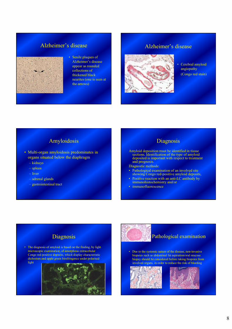
3/ Aß – found in the cerebral lesions of Alzheimer’s disease (a common form of dementia)
brain deposits are composed of an abnormally folded proteolytic fragment of amyloid precursor protein. Alzheimer’s disease is not a „pure” amyloidosis, as it is characterized not only by abnormal protein processing but also by intracellular accumulation of another protein – microtubule stabilizing tau protein in neuronal cells
Alzheimer’s disease
• Alzheimer's disease is a devastating diseasecausing loss of recent memory, followed byold memories or inability to carry out anormal procedure such as dressing, eatingetc. The patient is finally bedridden withmany neurological signs and usually dies ofpneumonia, heart attack, or stroke.
Alzheimer’s disease
The cause of Alzheimer's disease is unknown.
• May be due to deposition of amyloid in theparenchyma of the brain which is toxic to neuronsand oligodendroglia.
• An alternative hypothesis is that deposition ofamyloid is a secondary effect. The amyloiddeposition is thought to be due to an abnormality inthe amyloid precursor protein.
Alzheimer’s disease
• Alzheimer's disease is characterized by theformation of round senile plaques oftenwith an amyloid center in the cerebralcortex as well as neurofibrillary tangleswithin neurons.
8
Alzheimer’s disease
• Senile plaques of Alzheimer’s disease appear as rounded collections of thickened black neurites (one is seen at the arrows)
Alzheimer’s disease
• Cerebral amyloid angiopathy
(Congo red stain)
Amyloidosis
• Multi-organ amyloidosis predominates in organs situated below the diaphragm
– kidneys
– spleen
– liver
– adrenal glands
– gastrointestinal tract
Diagnosis
Amyloid deposition must be identified in tissue sections. Identification of the type of amyloid deposited is important with respect to treatment and prognosis.
Diagnostic methods:• Pathological examination of an involved site
showing Congo red-positive amyloid deposits, • Positive reaction with an anti-LC antibody by
immunohistochemistry and/or • immunofluorescence
Diagnosis
• The diagnosis of amyloid is based on the finding, by light microscopic examination, of amorphous extracellular Congo red positive deposits, which display characteristic dichroism and apple green birefringence under polarised light
Pathological examination
• Due to the systemic nature of the disease, non-invasive biopsies such as abdominal fat aspiration/oral mucosa biopsy should be considered before taking biopsies from involved organs, in order to reduce the risk of bleeding complications

9
Immunohistochemical diagnosis
• To detect immunoglobulin light chain amyloidosis (AL amyloidosis) in formalin-fixed, paraffin-embedded tissue sections by immunohistochemistry, polyclonal antibodies were generated against synthetic peptides corresponding to amino acids of the Ig light chain
• Pathology International 2006; 56: 324–330 Immunohistochemical study of immunoglobulin light chain amyloidosis with antibodies to the immunoglobulin light chain variable region
• Yoshinobu Hoshii, Makiko Kiyama, Dan Cui, Hiroo Kawano and Tokuhiro Ishihara
Renal amyloidosis
• Renal amyloidosis is common and clinically most important. Glomerular amyloidosis results in proteinuria and nephrotic syndrome. Chronic renal failure developes in advanced cases.
• The kidneys are enlarged, pale and firm („white large kidney”).
• Disease affects all glomeruli in both kidneys
Renal amyloidosis
• The amyloid fibrils deposit in extracellular regions such as the glomerular mesangia and glomerular basement membranes
Amyloidosis of the liver
• This is a gross specimen of a liver. In comparison to a normal liver, this one has a yellow and glassy appearance. If this liver had been stained with Lugol's solution, (an iodine containing solution) the amyloid would have stained a mahogany brown.
The current treatment of AA amyloidosis
• An longstanding overproduction in the liver cytokine-induced acute phase serum amyloid A protein – a key event in the pathogenesis of amyloidosis
• Treatment:
reducing the amyloid precursors load by intensive anti-inflammatory and immunosuppressive therapy
The eradication of an existing infectious focus
Dissolution of the amyloid with small molecules that interact with the deposits
Treatment
• Iniximab (IFX) is a monoclonal antibody against tumor necrosis factor alpha (TNF-) and is proven to induce clinical response and remission in CD patients.(Crohn disease)
• long term treatment with IFX caused a decrease in SAA (serum amyloid A) circulating levels, thereby stabilizing or even improving renal function.
• JGastrointestin Liver Dis, September 2013 Vol. 22 No 3: 333-336

10
Amyloidosis –recent experimental data
• „AA amyloidosis can be transferred by peripheral blood monocytes” !?
• Blood transfusion as a route for transfer of amyloidosis ?
Linkoping University, Sweden, Oct 2008
Mineral degenerations
Calcium degeneration (calcification)
• Metastatic calcification (deposition of calcium salts in normal tissues when the serum ionized calcium conentration is elevated.
– hyperparathyroidism
– overdosage of vitamin D
– destruction of bone
– alkalosis
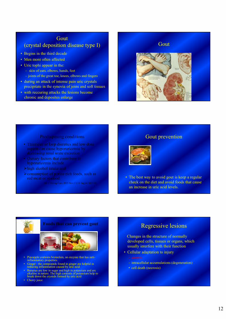
Gout(crystal deposition disease type I)
• A disorder of purine metabolism with an increased serum uric acid concentration and deposition of uric acid and urates in various connective tissues (crystals of monosodium urate)
• The result is
Painful attacks of gout
Urate nephropathy
Tophi formation
Gout - epidemiology
• Because of changing dietary habits, at least 1-2 % of all adults in the industrialized nations are now affected by gout (9%of men and 0,4% of women have hyperuricemia and 19% of these suffer from gout)
• According to recent studies, gouty arthritis is the most common form of arthritis seen in general practice in adults
Dtsch Arztbl Int 2009;106(34-35)
Gout
• Primary gout has a genetic background. There is a deficiency of the enzyme HGPRT (hypoxanthine-guanine phosphoribosyltransferase). This defect inhibits reconversion of hypoxanthine, which is further degraded to uric acid.

11
Gout(crystal deposition disease type I)
• Secondary gout accompanies cases of leukaemia or polycythemia and some lymphomas (these neoplasms produce large number of cell nuclei which provide a source of purines and consequently of uric acid).
Gout
• Uric acid (the end product of purine metabolism) precipitates out in the form of monosodium crystals in peripheral regions of the body (e.g.joints and extremities) when ambient temperatures are low
• The typical first manifesatation is an acute episode of monoarticular arthritis at the joint of the large toe (podagra) – very painful, lasts around a week and is self-limiting
Symptoms
• Symptoms of gout include excruciating pain, redness and swelling of affected joints and usually the joint of the big toe is affected (podagra)
Gout - pathophysiology
• Urate crystals lead to activation of
NLRP3 inflammasome (cryopyrin)with
Consequent activation of caspase and
release of proinflammatory cytokines(interleukins 1,18, 8 and TNF) and
Attraction of neutrophilic granulocytes
Gout
• The urate crystals become the centre of a foreign body granuloma with a rim of fibroblasts and multinucleated giant cells. This granuloma is called tophus (Latin tofus means „porous stone”)
Gout – histologic picture• Deposit of characteristic crystals in synovium
(arrow)
• crystal deposits surrounded by a scalloping of
palisaded histiocytes.
12
Gout(crystal deposition disease type I)
• Begins in the third decade
• Men more often affected
• Uric tophi appear in the:
– skin of ears, elbows, hands, feet
– joints of the great toe, knees, elbows and fingers
• during an attack of intense pain uric crystals precipitate in the synovia of joins and soft tissues
• with reccuring attacks the lesions become chronic and deposites enlarge
Gout
Predisposing conditions
• Thiazides or loop diuretics and low-dose aspirin can cause hyperuricemia by decreasing renal urate excretion
• Dietary factors that contribute to hyperuricemia include
high alcohol intake and
consumption of purine rich foods, such as red meat or seafood.
Am J Manag Care 2005 Nov;11(15 Suppl):S435-42
Gout prevention
• The best way to avoid gout is keep a regular check on the diet and avoid foods that cause an increase in uric acid levels.
Foods that can prevent gout
• Pineapple contains bromelain, an enzyme that has anti-inflammatory properties.
• Ginger : the compounds found in ginger are helpful in reducing inflammation caused by uric acid
• Bananas are low in sugar and high in potassium and are alkaline in nature. The high contents of potassium help to break down the crystals formed by uric acid
• Cherry juice
Regressive lesions
Changes in the structure of normally developed cells, tissues or organs, which usually interfere with their function
• Cellular adaptation to injury
– atrophy
– intracellular accumulations (degeneration)
• cell death (necrosis)

13
Atrophy
Regression of a tissue or organ because of a decrease in the size of cells
Causes of atrophy:
• hyponutrition (local or systemic)
• inactivity (due to a loss of innervation or endocrine stimulation)
• hyperactivity
• senescence
• autoimmunization
Cerebral atrophy
• Notice the widening of the sulci, dilatation of the ventricles and narrowing of the gyri in senile cerebral atrophy
Inactivity-a decreased workload
• Immobilization of a limb to permit healing of a fracture
• The loss of endocrine stimulation (Sheehan’s syndrome, named for Harold Sheehan; 1937)
Dr Harold Sheehan
• Harold Leeming Sheehan was the son of a general practitioner, and was born in 1900, in Carlisle, in the UK . He died in 1986.
• Sheehan was a dedicated pathologist with an outstanding intelligence and knowledge, He was an original thinker.
• Sheehan recorded many cases of acute postpartum pituitary necrosis and described in great detail the early and late histological changes
• He correlated clinical and morphological findings and assessed the incidence of the disease
• Kovacs, Lancet 2003; 361: 520–22
Sheehan syndrome
• Sheehan syndrome is the name given to postpartum hypopituitarism. The syndrome is caused by an infarction in the hypophysis, usually precipitated by uterine haemorrhage. Extensive destruction of cells results in varying degrees of hypopituitarism
• Deficient adenohypophysis endocrine activity can lead to secondary hypocortism, hypothyroidism, hypogonadism, and loss of growth hormone and prolactin production.
Sheehan syndrome symptoms
• Symptoms of patients with Sheehan syndrome include weakness, decreased muscular strength, dryness and wrinkling of the skin, premature ageing, hypotension, cold intolerance, constipation, pallor, anaemia, bradycardia, hypoglycaemia, insulin hypersensitivity, amenorrhoea, infertility, absence of lactation, breast atrophy, mental slowing, apathy, and psychiatric disturbances. Treatment aims to replace the missing hormones and restore endocrine homoeostasis.

14
Sheehan’s syndrome
• Postpartum pituitary necrosis: hypopituitarism caused by blood loss and/or hypovolemic shock during childbirth
• A rare complication of pregnancy
• (Hypertrophy during pregnancy results in the enlargement of the anterior pituitary, without a corresponding increase in blood supply)
Hyperactivity
• exhaustion of isletic beta cells in acquired diabetes
Autoimmunization
• Chronic thyroiditis (Hashimoto)
Dr Hakaru Hashimoto1881-1934
Hashimoto, as a first, identified lymphocytic thyroiditisIn 1912, he reported his findings as an independent new disease in Archiv Fur Klinishe Chirurgie,the German journal of clinical surgery.
Chronic thyroiditisLymphocytic, autoimmune, Hashimoto
• The most common cause of hypothyroidism in the USA
• Autoimmune inflammatory disorder
• Formation of CD8+ cytotoxic T cells distroying parenchyma
• B cells secret inhibitory anti-TSH receptor antibodies (hypothyroidism)
• Antithyroglobulin and antithyroid peroxidase antibodies

15
Chronic thyroiditismorphology
• Mononuclear inflammatory lymphocytic infiltrate with germinal centers
• Atrophic thyroid follicles
• Oxyphillic metaplasia of follicular cells (Hürthle, Askenazy cells, oncocytes)
• Fibrosis
Lymphocytic aggregation Persistent thyroid follicles
Lymphocytic (Hashimoto) thyroiditis
Hashimoto thyroiditisCytologic smear
Aspirate containing groups of oxyphillic cells and dispersed lymphocytes and plasma cells
Morphologic classification of atrophy
• simple (cells and normal stroma)
• brown
• with serous swelling of cells
• concentric
• eccentric (normal size and diminished mass)
Liver - brown atrophy
• Brown cytoplasmic granules of lipofuscin in hepatocytes
(lysosomes loaded with lipids and proteins deriving from focal cytoplasmic degradation)
Liver - brown atrophy

16
Medical eponyms used during this lecture
• Down syndrome (Down’s syndrome)• Virchow triad, Virchow node,Virchow method of
autopsy• Crohn disease, Mallory bodies• Hodgkin disease, macroglobulinemia of
Waldenström.• Alzheimer disease• Sheehan syndrome• Hashimoto thyroiditis• Hurthle cells
in possessive or nonpossessive form
Eponyms
• By definition, the term „eponym” derives from Greek: eponymos
• epi- upon, onyma – name
• Eponym indicates the name of a person after whom a disease is named to commemorate the importance of his/her contribution
• Most are derived from the names of those who first described a disease
• Along with eponyms derived from names, there are the names of mythical or biblical heroes, as well; some eponyms came from paintings
Should eponyms be abandoned ? A recent debate evoked strong responses both in favour
and against the motion:
Eponyms lack accuracy
lead to confusions
are not descriptive
sometimes are even misleading
Their main function is to immortalize the greats of the profession
They add color to medical communication
Enrich our dry medical language
Extend our knowledge beyond pure medicine
Embed medical tradition in our history BMJ 2007
Mona Lisa sydrome – the facial muscle contracture which develops after facial
nerve palsy


![HISTORY OF SPEECH-LANGUAGE PATHOLOGY [Autosaved]](https://static.fdocuments.net/doc/165x107/5886a5bd1a28ab0c1d8b70c1/history-of-speech-language-pathology-autosaved.jpg)