Pathological, immunohistochemical and microbiologicalal analysis of lacrimal sac biopsies in...
10
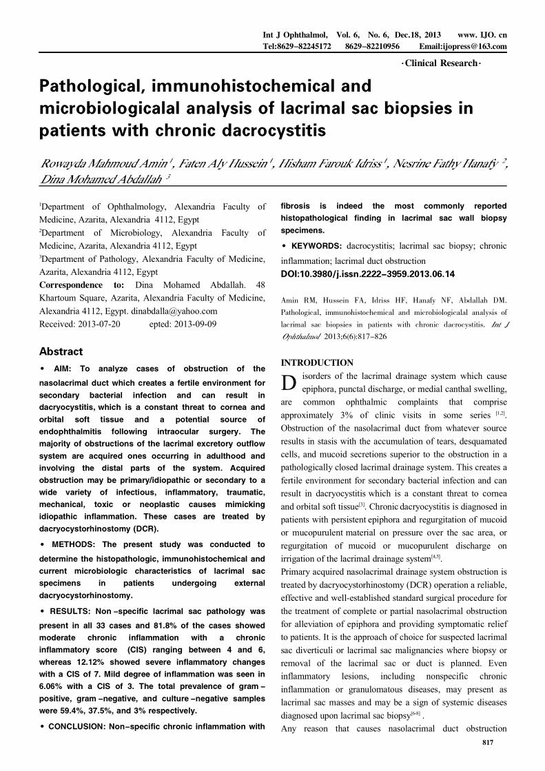
陨灶贼 允 韵责澡贼澡葬造皂燥造熏 灾燥造援 6熏 晕燥援 6熏 Dec.18, 圆园13 www. IJO. cn 栽藻造押8629原愿圆圆源缘员苑圆 8629-82210956 耘皂葬蚤造押ijopress岳员远猿援糟燥皂 Pathological, immunohistochemical and microbiologicalal analysis of lacrimal sac biopsies in patients with chronic dacrocystitis 1 Department of Ophthalmology, Alexandria Faculty of Medicine, Azarita, Alexandria 4112, Egypt 2 Department of Microbiology, Alexandria Faculty of Medicine, Azarita, Alexandria 4112, Egypt 3 Department of Pathology, Alexandria Faculty of Medicine, Azarita, Alexandria 4112, Egypt Correspondence to: Dina Mohamed Abdallah. 48 Khartoum Square, Azarita, Alexandria Faculty of Medicine, Alexandria 4112, Egypt. [email protected] Received: 2013-07-20 epted: 2013-09-09 Abstract · AIM: To analyze cases of obstruction of the nasolacrimal duct which creates a fertile environment for secondary bacterial infection and can result in dacryocystitis, which is a constant threat to cornea and orbital soft tissue and a potential source of endophthalmitis following intraocular surgery. The majority of obstructions of the lacrimal excretory outflow system are acquired ones occurring in adulthood and involving the distal parts of the system. Acquired obstruction may be primary/idiopathic or secondary to a wide variety of infectious, inflammatory, traumatic, mechanical, toxic or neoplastic causes mimicking idiopathic inflammation. These cases are treated by dacryocystorhinostomy (DCR). · METHODS: The present study was conducted to determine the histopathologic, immunohistochemical and current microbiologic characteristics of lacrimal sac specimens in patients undergoing external dacryocystorhinostomy. · RESULTS: Non -specific lacrimal sac pathology was present in all 33 cases and 81.8% of the cases showed moderate chronic inflammation with a chronic inflammatory score (CIS) ranging between 4 and 6, whereas 12.12% showed severe inflammatory changes with a CIS of 7. Mild degree of inflammation was seen in 6.06% with a CIS of 3. The total prevalence of gram - positive, gram -negative, and culture -negative samples were 59.4%, 37.5%, and 3% respectively. · CONCLUSION: Non-specific chronic inflammation with fibrosis is indeed the most commonly reported histopathological finding in lacrimal sac wall biopsy specimens. · KEYWORDS: dacrocystitis; lacrimal sac biopsy; chronic inflammation; lacrimal duct obstruction DOI:10.3980/j.issn.2222-3959.2013.06.14 Amin RM, Hussein FA, Idriss HF, Hanafy NF, Abdallah DM. Pathological, immunohistochemical and microbiologicalal analysis of lacrimal sac biopsies in patients with chronic dacrocystitis. 2013;6(6):817-826 INTRODUCTION D isorders of the lacrimal drainage system which cause epiphora, punctal discharge, or medial canthal swelling, are common ophthalmic complaints that comprise approximately 3% of clinic visits in some series [1,2] . Obstruction of the nasolacrimal duct from whatever source results in stasis with the accumulation of tears, desquamated cells, and mucoid secretions superior to the obstruction in a pathologically closed lacrimal drainage system. This creates a fertile environment for secondary bacterial infection and can result in dacryocystitis which is a constant threat to cornea and orbital soft tissue [3] . Chronic dacryocystitis is diagnosed in patients with persistent epiphora and regurgitation of mucoid or mucopurulent material on pressure over the sac area, or regurgitation of mucoid or mucopurulent discharge on irrigation of the lacrimal drainage system [4,5] . Primary acquired nasolacrimal drainage system obstruction is treated by dacryocystorhinostomy (DCR) operation a reliable, effective and well-established standard surgical procedure for the treatment of complete or partial nasolacrimal obstruction for alleviation of epiphora and providing symptomatic relief to patients. It is the approach of choice for suspected lacrimal sac diverticuli or lacrimal sac malignancies where biopsy or removal of the lacrimal sac or duct is planned. Even inflammatory lesions, including nonspecific chronic inflammation or granulomatous diseases, may present as lacrimal sac masses and may be a sign of systemic diseases diagnosed upon lacrimal sac biopsy [6-8] . Any reason that causes nasolacrimal duct obstruction 窑 Clinical Research 窑 817
-
Upload
mario-fernando-duenas-achicanoy -
Category
Health & Medicine
-
view
85 -
download
0
Transcript of Pathological, immunohistochemical and microbiologicalal analysis of lacrimal sac biopsies in...
陨灶贼 允 韵责澡贼澡葬造皂燥造熏 灾燥造援 6熏 晕燥援 6熏 Dec.18, 圆园13 www. IJO. cn栽藻造押8629原愿圆圆源缘员苑圆 8629-82210956 耘皂葬蚤造押ijopress岳员远猿援糟燥皂
Pathological, immunohistochemical andmicrobiologicalal analysis of lacrimal sac biopsies inpatients with chronic dacrocystitis
1Department of Ophthalmology, Alexandria Faculty ofMedicine, Azarita, Alexandria 4112, Egypt2Department of Microbiology, Alexandria Faculty ofMedicine, Azarita, Alexandria 4112, Egypt3Department of Pathology, Alexandria Faculty of Medicine,Azarita, Alexandria 4112, EgyptCorrespondence to: Dina Mohamed Abdallah. 48Khartoum Square, Azarita, Alexandria Faculty of Medicine,Alexandria 4112, Egypt. [email protected]: 2013-07-20 epted: 2013-09-09
Abstract· AIM: To analyze cases of obstruction of thenasolacrimal duct which creates a fertile environment forsecondary bacterial infection and can result indacryocystitis, which is a constant threat to cornea andorbital soft tissue and a potential source ofendophthalmitis following intraocular surgery. Themajority of obstructions of the lacrimal excretory outflowsystem are acquired ones occurring in adulthood andinvolving the distal parts of the system. Acquiredobstruction may be primary/idiopathic or secondary to awide variety of infectious, inflammatory, traumatic,mechanical, toxic or neoplastic causes mimickingidiopathic inflammation. These cases are treated bydacryocystorhinostomy (DCR).
· METHODS: The present study was conducted todetermine the histopathologic, immunohistochemical andcurrent microbiologic characteristics of lacrimal sacspecimens in patients undergoing externaldacryocystorhinostomy.
·RESULTS: Non -specific lacrimal sac pathology waspresent in all 33 cases and 81.8% of the cases showedmoderate chronic inflammation with a chronicinflammatory score (CIS) ranging between 4 and 6,whereas 12.12% showed severe inflammatory changeswith a CIS of 7. Mild degree of inflammation was seen in6.06% with a CIS of 3. The total prevalence of gram -positive, gram -negative, and culture -negative sampleswere 59.4%, 37.5%, and 3% respectively.
·CONCLUSION: Non-specific chronic inflammation with
fibrosis is indeed the most commonly reportedhistopathological finding in lacrimal sac wall biopsyspecimens.
·KEYWORDS: dacrocystitis; lacrimal sac biopsy; chronic
inflammation; lacrimal duct obstructionDOI:10.3980/j.issn.2222-3959.2013.06.14
Amin RM, Hussein FA, Idriss HF, Hanafy NF, Abdallah DM.
Pathological, immunohistochemical and microbiologicalal analysis of
lacrimal sac biopsies in patients with chronic dacrocystitis.
2013;6(6):817-826
INTRODUCTION
D isorders of the lacrimal drainage system which causeepiphora, punctal discharge, or medial canthal swelling,
are common ophthalmic complaints that compriseapproximately 3% of clinic visits in some series [1,2].Obstruction of the nasolacrimal duct from whatever sourceresults in stasis with the accumulation of tears, desquamatedcells, and mucoid secretions superior to the obstruction in apathologically closed lacrimal drainage system. This creates afertile environment for secondary bacterial infection and canresult in dacryocystitis which is a constant threat to corneaand orbital soft tissue[3]. Chronic dacryocystitis is diagnosed inpatients with persistent epiphora and regurgitation of mucoidor mucopurulent material on pressure over the sac area, orregurgitation of mucoid or mucopurulent discharge onirrigation of the lacrimal drainage system[4,5].Primary acquired nasolacrimal drainage system obstruction istreated by dacryocystorhinostomy (DCR) operation a reliable,effective and well-established standard surgical procedure forthe treatment of complete or partial nasolacrimal obstructionfor alleviation of epiphora and providing symptomatic reliefto patients. It is the approach of choice for suspected lacrimalsac diverticuli or lacrimal sac malignancies where biopsy orremoval of the lacrimal sac or duct is planned. Eveninflammatory lesions, including nonspecific chronicinflammation or granulomatous diseases, may present aslacrimal sac masses and may be a sign of systemic diseasesdiagnosed upon lacrimal sac biopsy[6-8] .Any reason that causes nasolacrimal duct obstruction
窑Clinical Research窑
817
(NLDO) generally converts the lacrimal sac into a reservoirof bacterial infection that may lead to chronic dacryocystitiswhich is a constant threat to cornea and orbital soft tissue[9].The macroscopic appearance of an inflamed lacrimal sacspecimen reveals a thickened and purulent or mucoidmaterial in the lumen. A non-specific chronic inflammationwith fibrosis and thickening of the wall due to submucosallymphocytic infiltration with follicle formation is the mostcommonly reported histopathological finding in lacrimal sacwall biopsy specimens[9].Thus, determining the incidence of primary lacrimalsac-specific pathology mimicking primary acquirednasolacrimal duct obstruction is important. It may haveimplications on whether routine biopsy during DCR iswarranted or not, and how great is a risk of missing aclinically non-suspected and intra-operatively non-visibleunderlying specific non-neoplastic or neoplastic processinvolving the lacrimal sac in patients not undergoing routinebiopsy during DCR[9,10]. The risk of overlooking a spectrum oflacrimal sac originated specific pathologies, particularlymalignancies that cause a nasolacrimal system obstruction,although very low, always exists[9,10].Neoplasms that affect the lacrimal drainage system are rare,but potentially life-threatening, so early diagnosis andtreatment are particularly important [11,12]. Epithelial neoplasmsare most common (73% ), including benign tumors(squamous cell papilloma, transitional cell papilloma,mixed-cell papilloma, oncocytoma) and malignant tumors(squamous cell carcinoma, transitional cell carcinoma,adenocarcinoma, mucoepidermoid carcinoma, oncocyticadenocarcinoma). Mesenchymal tumors such as fibroushistiocytoma, fibroma, hemangioma, hemangiopericytoma,angiosarcoma, or lipoma are less common (14%), and therarer tumors include malignant melanomas (4%), and neuraltumors (1%)[1,11].Primary lymphomas of the lacrimal sac are rare, but are agenuine cause of secondary acquired nasolacrimal ductobstruction. Lacrimal sac primary lymphomas are infrequent,not aggressive lesions that evolve rapidly and are not likely tobe associated with typical clinical symptoms and signs suchas mass extending above medial canthal tendon or bloodytearing suggestive of neoplastic pathology or malignancy.Secondary tumors originating in adjacent structures(paranasal sinuses, orbit and nose) may extend into thelacrimal sac. Metastatic neoplasms confined to the lacrimalsac are extremely rare, and most metastases also affectadjacent structures, such as the eyelid, nose, sinuses, andorbit[11,12].These findings have led to the recommendation that lacrimalsac biopsy specimens need not be routinely submitted forpathologic examination during DCR surgery, except foratypical clinical presentations or intraoperative findings,
although that recommendation is controversial . Anderson[13] thought that all lacrimal sac walls should be biopsied
at the time of DCR. Thus there has been debate in theliterature regarding the value of lacrimal sac biopsy at thetime of DCR surgery as the literature presents this dichotomyof views between "biopsy always" and "biopsy if the saclooks suspicious"[10,11].The lacrimal excretory system is prone to infection andinflammation for various reasons. This mucusmembrane-lined tract is contagious with two surfaces(conjunctiva and nasal mucosa) that are normally colonizedwith bacteria. Obstruction of the nasolacrimal duct fromwhatever source results in stasis with the accumulation oftears, desquamated cells, and mucoid secretions superior tothe obstruction in a pathologically closed lacrimal drainagesystem. This creates a fertile environment for secondarybacterial infection and can result in dacryocystitis.It is currently believed that the inflammation and fibrosis inpatients with nasolacrimal duct obstruction may be secondaryto coexisting infectious colonization within the lumen of thelacrimal sac. It is possible that many cases of primaryacquired nasolacrimal duct obstruction are in fact secondaryto unrecognized low-grade dacryocystitis [14]. Knowledge ofthe bacteriology of nasolacrimal duct obstruction contributessignificantly as well to the choice of prophylacticantimicrobial agents, and it would help in reducing theunnecessary load of anti-microbial agents with subsequentdevelopment of resistance patterns in the conjunctival florawith detrementous effects on regimens for prophylaxis onfurther intraocular surgery[15,16].During the past 20 years there have been only a few studieson the bacteriology of dacryocystitis in patients with NLDO.According to them,coagulase-negative (CoNS),
epidermidis and staphylococcus aureus arethe most frequently isolated organisms in lacrimal sacinfections [3]. Mixed bacterial isolates are more commonlyfound in cases of chronic dacryocostitis with thepredominance of streptococcus pneumoniae andstaphylococcus spp. fungal infections caused by candidaalbicans and aspergillus spp. occur infrequently[3].This study was conducted for a further understanding of thepathological changes and the incidence of specific pathologyin our cases of presumed primary acquired nasolacrimal ductobstruction, together with the spectrum and antimicrobialsusceptibility of microbiologic agents isolated from suchcases.SUBJECTS AND METHODSSubjects This prospective analysis included a total of 33lacrimal sac wall biopsies obtained from 33 patients eligiblefor the study who underwent external DCR for NLDO duringthe period from March 2012 to November 2012. Informedconsent was obtained from the patients that were included in
Pathology of lacrimal duct obstruction
818

陨灶贼 允 韵责澡贼澡葬造皂燥造熏 灾燥造援 6熏 晕燥援 6熏 Dec.18, 圆园13 www. IJO. cn栽藻造押8629原愿圆圆源缘员苑圆 8629-82210956 耘皂葬蚤造押ijopress岳员远猿援糟燥皂
the study after explanation of the details of the study and ofthe procedure to be performed.Methods All the patients underwent a complete preoperativeophthalmic evaluation and slit-lamp ophthalmologicexamination including visual acuity, ocular surfaceassessment and fundus biomicroscopy to report any posteriorsegment abnormalities and the demographic data (age andgender) and medical history of the patients and operativeinformation were regularly recorded.Assessment of lacrimal drainage system abnormalities usingprobing and syringing of the proximal lacrimal drainagesystem up to the nasal wall of the lacrimal sac together withlacrimal system irrigation for confirmation of NLDO. Allpatients underwent external dacryocystorhinostomy undergeneral anesthesia and operative data was recorded. Biopsyspecimens obtained from the posterior inferior flap of thelacrimal sac were examined under light microscopy by thesame pathologist after being fixated in 4% neutral bufferedformalin, paraffin embedded, cut at 5滋m and stained withconventional histological stain (haematoxylin and eosin,H&E). All the specimens were examined for the chronicinflammation related histopathological features(inflammatory cell infiltration, fibrosis and capillaryproliferation) and graded according to their severity using a"chronic inflammation score" established to determine theintensity of chronic inflammation using the grade ofhistopathological features [17,18]. These are: 1) The intensity ofinflammatory cell infiltration (number of lymphocytes,histiocytes and plasma cells in a HPF): mild<50 cells,moderate 50-200 cells, severe>200 cells; 2) The density offibrosis (the amount of fibrotic tissue in a HPF): mild <25%,moderate 25%-50%, severe>50%; 3) The degree of capillaryproliferation (number of capillary vessels in a HPF): mild<5,moderate 5-10, severe>10.In addition, to determine the intensity of chronicinflammation in the lacrimal sac, all these threehistopathological features were scored individually accordingto their severity (mild=1, moderate=2, and severe=3). Thus, atotal score (sum) was obtained for each case ranging between3 and 9 and named "chronic inflammatory score" (CIS).Finally, every case was grouped according to its CIS as: mildchronic inflammation (CIS<3), moderate chronicinflammation (3<CIS<6) and severe chronic inflammation(CIS>6).Immunohistochemical staining of the specimens using antiCD3 and CD 20 were done to determine the type of chronicinflammatory infiltrate using Avidin Biotin method accordingto the following procedurePositive and negative controls were included in all runs.External positive control cases were used as follow (asrecommended by manufacturer's protocols): tonsillar tissuesfor CD20, CD3. Immunostaining technique was done as
recommended by manufacturer's protocols [19,20]. Clones ofantibodies, antigen retrieval and dilutions were as follow:CD20: mouse monoclonal Ab, clone L26, no specialpretreatment, dilution 1:100. CD3: mouse monoclonal Ab,clone, antigen retrieval using citrate (10mM, pH6.0), dilution1:150. Expression of immunohistochemical markers wasvisualized using the stretavidin-biotin-immunoenzymaticantigen detection system which was performed according tomanufacturer's protocol [20]. Positive cell membranous stainingin tumor cells for CD3 antibody. Positive cytoplasmic andcell membranous staining in tumor cells for: CD20 antibody.Microbiology Specimens for microbiological analysis wereobtained by wiping a sterile broth-moistened swab overeverted puncta after applying pressure over the lacrimal sacarea and allowing the mucopurulent material to refluxthrough the lacrimal punctum, or by irrigating the lacrimaldrainage system with sterile saline and collecting the samplefrom the refluxing material in cases that did not haveregurgitation of mucus or mucopurulent discharge or pus onpressure on the lacrimal sac area encysted mucoceles andpyoceles. None of the patients had used either antibiotic eyedrops or systemic antibiotics for at least a week beforesample collection.Samples were incubated in 2mL of brain-heart infusion brothagar cultured onto blood agar, MacConkey, chocolate agar,Sabouraud dextrose agar and incubated for 4d. The specificidentification of bacterial isolates were performed based onmicroscopic morphology, staining characteristics, andbiochemical properties using standard laboratory criteria,such as catalase, oxidase, and coagulase tests. All inoculatedmedia were incubated aerobically. The inoculatedSabouraud's dextrose agar was incubated at 27℃ , examineddaily, and discarded at 3 weeks if no growth was seen. Theinoculated blood agar, chocolate agar, thioglycollate broth,brain-heart infusion broth were incubated at 37℃ , examineddaily, and discarded at 7d if growth was not seen.Microbial cultures were considered significant if growth ofthe same organism was demonstrated on more than onesolid-phase medium, and/or if there was confluent growth atthe site of inoculation on one solid medium, and/or if growthof one medium was consistent with direct microscopyfindings ( . appropriate staining and morphology with Gramstain), and/or if the same organism was grown from morethan one specimen.Antibiotic susceptibility testing was done through theconventional Kirby-Bauer disk diffusion method of in vitroantibacterial susceptibility testing for tobramycin, quinolones(ciprofloxacin and ofloxacin), chloramphenicol, doxycycline,vancomycin, amoxicillin clavulinate and cefoxitin.Statistical Analysis StatSoft, Inc. (2007). STATISTICA(data analysis software sustem), version 8.0. Chi-square ( 2)distribution was used to test the qualitative distribution. Thevalues of <0.05 were accepted as statistically significant.
819
RESULTSIn this prospective interventional case series, 33 lacrimal sacspecimens were obtained from a total of 33 consecutivepatients who underwent external DCR for clinicallypresumed acquired NLDO at the time interval from March toDecember 2012 at Alexandria Main University Hospital.The cases included 32 females (96.97%) and 1 male (3.03%).The female subjects with chronic dacryocystitis (78.1% )were more in number in than male subjects (0).The mean age of study group was 50.4 with a range between6 and 63 years. Patients above the age of 30 years (30 of 33;91%) were significantly more than patients below 30 years (3of 33; 9.1% ). Four of the 33 patients with lacrimal ductobstruction were bilateral cases (12.1%).Eight (24.2%) of the patients had previously had at least oneattack of acute dacryocystitis. A total of 25 (78.8%) of thecases had chronic dacryocystitis. These were furthersubdivided into two groups according to the nature of thelacrimal discharge. Fourteen cases (56% ) showed copiousthick mucopurulent discharge coming from the sac, whereas7 cases (28%) showed epiphora with clear tear fluid or minormucopurulent discharge. Four patients (16%) had swellingsover the lacrimal sac with characters consistent withmucoceles, of which 2 cases were expressible mucoceles.Of the 33 cases recruited in this study, 29 (87.9% ) hadcomplete NLDO, whereas 4 patients (12.1%) were diagnosedas having functional obstruction of the nasolacrimal passages.Pathological Findings Of all the study cases, none showednormal histology. Non-specific lacrimal sac pathology waspresent in all 33 cases including varying degrees ofnon-specific non-granulomatous chronic inflammation,whereas specific lacrimal sac pathology was not found in anyof the biopsied cases.All the obtained specimens were further examined for certainchronic inflammation related histopathological features(inflammatory cell infiltration, fibrosis and capillaryproliferation) and graded according to their severity.Moreover, a chronic inflammation score CIS was used todetermine the intensity of chronic inflammation using thegrade of these histopathological features (Table 1).In addition, to determine the intensity of chronicinflammation in the lacrimal sac, all these threehistopathological features were scored individually accordingto their severity (mild=1, moderate=2, and severe=3). Thus, atotal score (sum) was obtained for each case ranging between
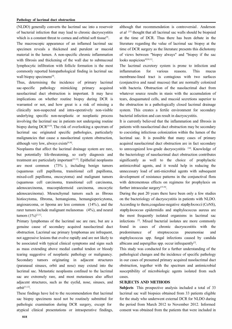
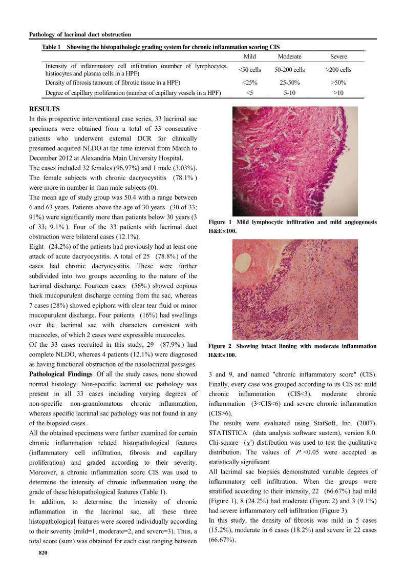
3 and 9, and named "chronic inflammatory score" (CIS).Finally, every case was grouped according to its CIS as: mildchronic inflammation (CIS<3), moderate chronicinflammation (3<CIS<6) and severe chronic inflammation(CIS>6).The results were evaluated using StatSoft, Inc. (2007).STATISTICA (data analysis software sustem), version 8.0.Chi-square (字2) distribution was used to test the qualitativedistribution. The values of <0.05 were accepted asstatistically significant.All lacrimal sac biopsies demonstrated variable degrees ofinflammatory cell infiltration. When the groups werestratified according to their intensity, 22 (66.67%) had mild(Figure 1), 8 (24.2%) had moderate (Figure 2) and 3 (9.1%)had severe inflammatory cell infiltration (Figure 3).In this study, the density of fibrosis was mild in 5 cases(15.2%), moderate in 6 cases (18.2%) and severe in 22 cases(66.67%).
Table 1 Showing the histopathologic grading system for chronic inflammation scoring CIS Mild Moderate Severe Intensity of inflammatory cell infiltration (number of lymphocytes, histiocytes and plasma cells in a HPF) <50 cells 50-200 cells >200 cells
Density of fibrosis (amount of fibrotic tissue in a HPF) <25% 25-50% >50% Degree of capillary proliferation (number of capillary vessels in a HPF) <5 5-10 >10
Figure 1 Mild lymphocytic infiltration and mild angiogenesisH&E伊100.
Figure 2 Showing intact linning with moderate inflammationH&E伊100.
Pathology of lacrimal duct obstruction
820
陨灶贼 允 韵责澡贼澡葬造皂燥造熏 灾燥造援 6熏 晕燥援 6熏 Dec.18, 圆园13 www. IJO. cn栽藻造押8629原愿圆圆源缘员苑圆 8629-82210956 耘皂葬蚤造押ijopress岳员远猿援糟燥皂
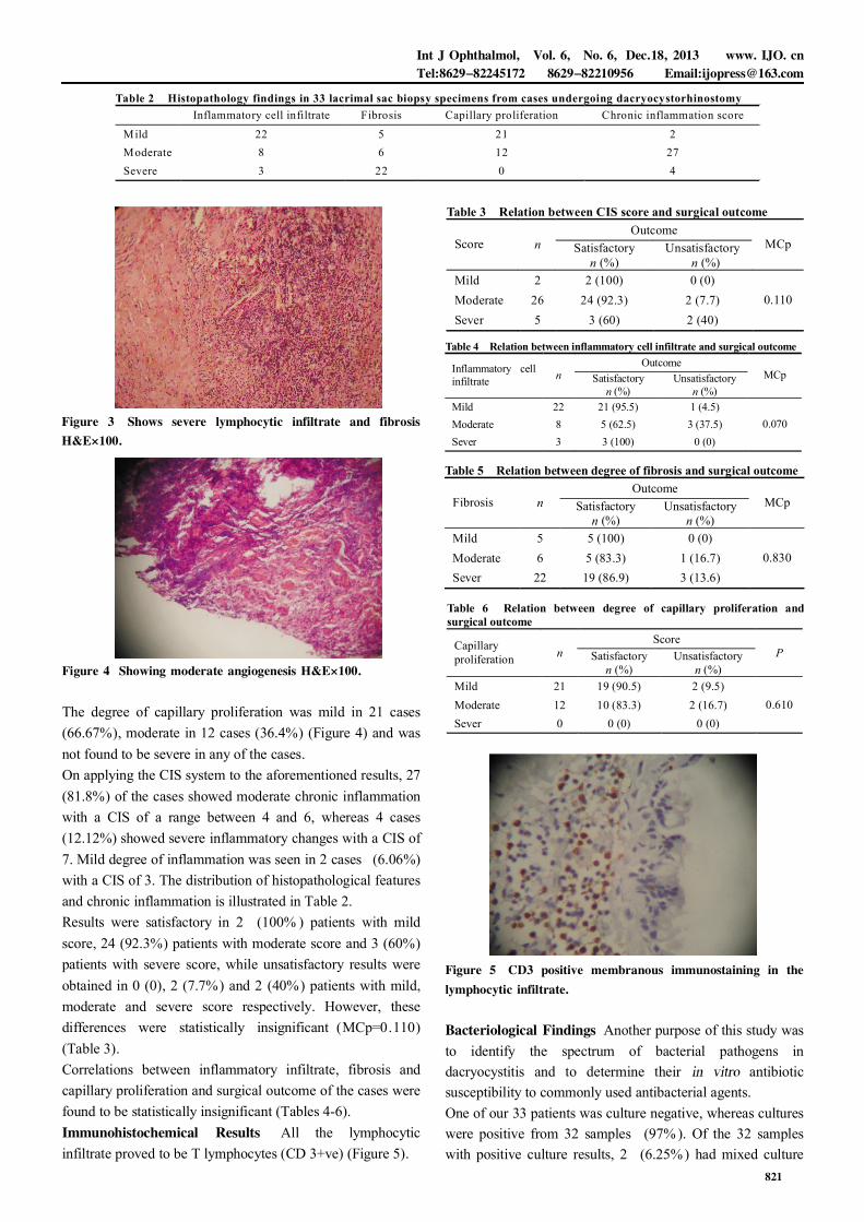
The degree of capillary proliferation was mild in 21 cases(66.67%), moderate in 12 cases (36.4%) (Figure 4) and wasnot found to be severe in any of the cases.On applying the CIS system to the aforementioned results, 27(81.8%) of the cases showed moderate chronic inflammationwith a CIS of a range between 4 and 6, whereas 4 cases(12.12%) showed severe inflammatory changes with a CIS of7. Mild degree of inflammation was seen in 2 cases (6.06%)with a CIS of 3. The distribution of histopathological featuresand chronic inflammation is illustrated in Table 2.Results were satisfactory in 2 (100% ) patients with mildscore, 24 (92.3%) patients with moderate score and 3 (60%)patients with severe score, while unsatisfactory results wereobtained in 0 (0), 2 (7.7%) and 2 (40%) patients with mild,moderate and severe score respectively. However, thesedifferences were statistically insignificant (MCp=0.110)(Table 3).Correlations between inflammatory infiltrate, fibrosis andcapillary proliferation and surgical outcome of the cases werefound to be statistically insignificant (Tables 4-6).Immunohistochemical Results All the lymphocyticinfiltrate proved to be T lymphocytes (CD 3+ve) (Figure 5).
Figure 5 CD3 positive membranous immunostaining in thelymphocytic infiltrate.
Bacteriological Findings Another purpose of this study wasto identify the spectrum of bacterial pathogens indacryocystitis and to determine their antibioticsusceptibility to commonly used antibacterial agents.One of our 33 patients was culture negative, whereas cultureswere positive from 32 samples (97% ). Of the 32 sampleswith positive culture results, 2 (6.25%) had mixed culture
Table 4 Relation between inflammatory cell infiltrate and surgical outcome Outcome Inflammatory cell
infiltrate n Satisfactory n (%)
Unsatisfactory n (%)
MCp
Mild 22 21 (95.5) 1 (4.5) Moderate 8 5 (62.5) 3 (37.5) Sever 3 3 (100) 0 (0)
0.070
Table 2 Histopathology findings in 33 lacrimal sac biopsy specimens from cases undergoing dacryocystorhinostomy Inflammatory cell infiltrate Fibrosis Capillary proliferation Chronic inflammation score
Mild 22 5 21 2 Moderate 8 6 12 27 Severe 3 22 0 4
Figure 3 Shows severe lymphocytic infiltrate and fibrosisH&E伊100.
Figure 4 Showing moderate angiogenesis H&E伊100.
Table 3 Relation between CIS score and surgical outcome Outcome
Score n Satisfactory n (%)
Unsatisfactory n (%)
MCp
Mild 2 2 (100) 0 (0) Moderate 26 24 (92.3) 2 (7.7) Sever 5 3 (60) 2 (40)
0.110
Table 5 Relation between degree of fibrosis and surgical outcome Outcome
Fibrosis n Satisfactory n (%)
Unsatisfactory n (%)
MCp
Mild 5 5 (100) 0 (0) Moderate 6 5 (83.3) 1 (16.7) Sever 22 19 (86.9) 3 (13.6)
0.830
Table 6 Relation between degree of capillary proliferation and surgical outcome
Score Capillary proliferation n Satisfactory
n (%) Unsatisfactory
n (%) P
Mild 21 19 (90.5) 2 (9.5) Moderate 12 10 (83.3) 2 (16.7) Sever 0 0 (0) 0 (0)
0.610
821
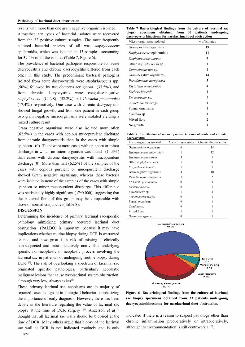
results with more than one gram negative organism isolatedAltogether, ten types of bacterial isolates were recoveredfrom the 32 positive culture samples. The most frequentlycultured bacterial species of all wasepidermidis, which was isolated in 13 samples, accountingfor 39.4% of all the isolates (Table 7, Figure 6).The prevalence of bacterial pathogens responsible for acutedacryocystitis and chronic dacryocystitis differed from eachother in this study. The predominant bacterial pathogensisolated from acute dacryocystitis were spp.(50%) followed by aeruginosa (37.5% ), andfrom chronic dacryocystitis were coagulase-negative
(CoNS) (52.2%) and(17.4% ) respectively. One case with chronic dacryocystitisshowed fungal growth, and from one patient in each grouptwo gram negative microorganisms were isolated yielding amixed culture result.Gram negative organisms were also isolated more often(62.5% ) in the cases with copious mucopurulent dischargefrom chronic dacryocystitis than in the cases with simpleepiphora (0). There were more cases with epiphora or minordischarge in which no micro-organism was found (14.3%)than cases with chronic dacryocystitis with mucopurulentdischarge (0). More than half (62.5%) of the samples of thecases with copious purulent or mucopurulent dischargeshowed Gram negative organisms, whereas these bacteriawere isolated in none of the samples of the cases with simpleepiphora or minor mucopurulent discharge. This differencewas statistically highly significant ( =0.000), suggesting thatthe bacterial flora of this group may be comparable withthose of normal conjunctiva(Table 8).DISCUSSIONDetermining the incidence of primary lacrimal sac-specificpathology mimicking primary acquired lacrimal ductobstruction (PALDO) is important, because it may haveimplications whether routine biopsy during DCR is warrantedor not, and how great is a risk of missing a clinicallynon-suspected and intra-operatively non-visible underlyingspecific non-neoplastic or neoplastic process involving thelacrimal sac in patients not undergoing routine biopsy duringDCR [9]. The risk of overlooking a spectrum of lacrimal sacoriginated specific pathologies, particularly neoplasticmalignant lesions that cause nasolacrimal system obstruction,although very low, always exists[21].These primary lacrimal sac neoplasms are in majority ofreported cases malignant in biological behavior, emphasizingthe importance of early diagnosis. However, there has beendebate in the literature regarding the value of lacrimal sacbiopsy at the time of DCR surgery [9]. Anderson [13]
thought that all lacrimal sac walls should be biopsied at thetime of DCR. Many others argue that biopsy of the lacrimalsac wall at DCR is not indicated routinely and is only
indicated if there is a reason to suspect pathology other thanchronic inflammation preoperatively or intraoperatively,although that recommendation is still controversial[13].
Table 7 Bacteriological findings from the culture of lacrimal sac biopsy specimens obtained from 33 patients undergoing dacryocystorhinostomy for nasolacrimal duct obstruction
Micro-organisms isolated n of isolates Gram positive organisms 19 Staphylococcus epidermidis 13 Staphylococcus aureus 4 Other staphylococcus sp. 1 Corynebacterium sp. 1 Gram negative organisms 14 Pseudomonas aeruginosa 5 Klebsiella pneumoniae 4 Escherichia coli 1 Enterobacter sp. 3 Acinetobacter lwoffii 1 Fungal organisms 1 Candida sp. 1 Mixed flora 2 No growth 1
Table 8 Distribution of microorganisms in cases of acute and chronic dacryocystitis
Micro-organisms isolated Acute dacryocystitis Chronic dacryocystitis
Gram positive organisms 6 14
Staphylococcus epidermidis 1 12
Staphylococcus aureus 3 1 Other staphylococcus sp. 1 0
Corynebacterium sp. 1 1 Gram negative organisms 4 10
Pseudomonas aeruginosa 3 2
Klebsiella pneumoniae 0 4 Escherichia coli 0 1
Enterobacter sp. 1 2
Acinetobacter lwoffii 0 1 Fungal organisms 0 1
Candida sp. 0 1 Mixed flora 1 1 No micro-organism 0 1
Figure 6 Bacteriological findings from the culture of lacrimalsac biopsy specimens obtained from 33 patients undergoingdacryocystorhinostomy for nasolacrimal duct obstruction.
Pathology of lacrimal duct obstruction
822
陨灶贼 允 韵责澡贼澡葬造皂燥造熏 灾燥造援 6熏 晕燥援 6熏 Dec.18, 圆园13 www. IJO. cn栽藻造押8629原愿圆圆源缘员苑圆 8629-82210956 耘皂葬蚤造押ijopress岳员远猿援糟燥皂
Nevertheless, some lacrimal sac tumors such as primarylymphomas, although infrequent, are not likely to beassociated with typical clinical symptoms and signs such asmass extending above medial canthal tendon or bloodytearing suggestive of neoplastic pathology or malignancy.Some other lesions are also so incipient to produce a grosslyvisible abnormality. The value of lacrimal sac biopsy andhistological examination of the lacrimal sac wall at DCR, inthose cases, is undoubtful [22]. This prospective study tests thepossible value of routine lacrimal sac biopsy during surgeryfor clinically presumed PALDO and also adds to ourknowledge more about the nature and prevalence of lacrimalsac non-specific and specific pathologic features in ourpatients.Our study is based on a consecutive series of lacrimal sacbiopsy specimens obtained from patients with clinicallypresumed nasolacrimal duct obstruction and submitted to asingle pathology laboratory. Non granulomatousinflammation was the most common histopathologicdiagnosis, as previously reported in many other similar series[21]. Of all the study cases, none showed normal histology.Non-specific lacrimal sac pathology was present in all 33cases including varying degrees of non-specificnon-granulomatous chronic inflammation, whereas specificlacrimal sac pathology was not found in any of the biopsiedcases.Specific pathology was found in 31 out of 377 specimens inAnderson's series [13] including eight sarcoidosis, sevenlymphoma, four papilloma, four lymphoplasmacyticinfiltrate, two transitional cell carcinoma, one oncocytoma,one granular cell tumour, one adenocarcinoma, one poorlydifferentiated carcinoma, one plasmacytoma, and oneleukaemia; and in 10 out of 302 specimens in Bernardini'sseries (four sarcoidosis, three squamous papilloma, twolymphoma, one leukaemia)[23].On combining the results from the previous studies, 50 out of1 294 specimens (3.9%) in these seven series showed specificpathology. Only seven out of 1 294 specimens (0.5%) in theseseven series showed specific pathology which was definitelyunsuspected preoperatively, and in only one of these was thismalignant. In Lindberg's series, of the two specimens withspecific pathology this was unsuspected in one specimen withsarcoidosis. In Tucker's series, of the four specimens withspecific pathology this was unsuspected in one specimen withoncocytoma. In Bernardini's series, of the 10 specimens withspecific pathology this was suspected in all specimens eitherbefore or during the surgery. Two of the 3 cases of specificpathology in Merkonidis' series was unsuspected [24]. InAnderson's series, of the 31 specimens with specificpathology it was stated this was unsuspected preoperativelyin at least three[13].
Findings from our study together with previous series fromthe literature have led to the recommendation that lacrimalsac biopsy specimens need not be routinely submitted forpathologic examination during DCR surgery, except foratypical clinical presentations or intraoperative findings.This recommendation may be argued by the small samplesize in some of the mentioned studies and the present study.Again, ethnic heterogeneity may contribute to the results insome studies [24,25]. Another limitation is the fact that it shouldalways be kept in mind that obtaining a representative biopsyof the lesion is not always easy and sometimes is challenging.If a peripheral portion of specific lesion or inadequatespecimen is taken, or if pathologic tissue is not recognized asabnormal, tumor or any other pathologic process may not bepresented in the biopsy specimen, so biopsy may yield afalse-negative result. In a case of lacrimal drainage systemobstruction, a misdiagnosis of chronic inflammation mayoccur. In addition, intra-operative normal appearance of thelacrimal sac is not an absolute guarantee that the sac isdevoid of pathological process other than chronicinflammation and/ or fibrosis in the early phase of clinicalevolution of lacrimal sac tumors (stage 1, according to Cookand Olver [26]) even in the eyes of experienced ophthalmiclacrimal surgeons. Because of the absence of definite tumoron palpation, it is difficult to clinically differentiate a lacrimalsac tumor from chronic dacryocystitis, so routine blindlacrimal sac wall biopsy during DCR may not be the bestchoice.To minimize the risk of overlooking specific pathology it isimportant to inquire about symptoms or history of systemicdisease preoperatively, to assess the appearance of thelacrimal sac intraoperatively, and to biopsy the lacrimal sacin those cases where specific pathology is suspected.In our study, the role of chronic inflammation on DCRoutcome was also evaluated, using histopathological featuresof chronic inflammation such as inflammatory cellinfiltration, fibrosis and capillary proliferation.All lacrimal sac biopsies demonstrated variable degrees ofinflammatory cell infiltration. When the groups werestratified according to their intensity, 22 (66.67%) had mild,8(24.2%) had moderate and 3(9.1%) had severe inflammatorycell infiltration.In this study, the density of fibrosis was mild in 5 cases(15.2%), moderate in 6 cases (18.2%) and severe in 22 cases(66.67%). It was noted that in our two cases of revision DCR,there was a marked increase in the density of fibrosis, whichis consistent with previous findings in a study that examinedthe silicone tube intubation related histopathological changesin revision DCR cases, where a marked increase in thedensity of fibrosis was observed and the role of fibrosis onrecurrence was emphasized [27]. Our cases had had silicone
823
tubes inserted during their primary surgeries. Nevertheless,we believe that further studies are required to determine theeffect of fibrosis on DCR outcome.The degree of capillary proliferation was mild in 21 cases(66.67%), moderate in 12 cases (36.4%) and was not foundto be severe in any of the cases.On applying the CIS system to the aforementioned results, 27(81.8%) of the cases showed moderate chronic inflammationwith a CIS of a range between 4 and 6, whereas 4 (12.1%)cases showed severe inflammatory changes with a CIS of 7.Mild degree of inflammation was seen in 2 cases (6.06) witha CIS of 3. A quantitative and statistical analysis ofhistopathological features and chronic inflammation wasperformed between patients with satisfactory andunsatisfactory outcome, and of patients with unsatisfactoryoutcome.Another purpose of this study was to identify the bacterialaetiology behind dacryocystitis and to determine the in vitroantibacterial susceptibility and resistance of bacterialpathogens to commonly used antibacterial agents.Using direct biopsy methods, we found culture-positivelacrimal sac specimens in a large proportion of patientsundergoing DCR surgery. These organisms were found to bepresent in patients with and without a history of infection.In this study Gram positive bacteria were found in 59.4% ofthe isolates. This is in close agreement with the observationof 65% of Gram positive organisms by Coden [28] Themost common organisms cultured in our study wereStaphylococcus species, accounting for 54.5% of the isolates.This percentage compares fairly well with the results ofThicker and Buffam [29], Huber-Spitzy [30] and Coden
[28] (their percentages being 73% , 51% , and 49%respectively). epidermidis and staphylococcusaureus represented 40% and 12.1% of all the isolates in ourstudy, which is higher than what was reported in previousstudies for epidermidis, and the same for
(their percentages being 26.9% and12.3% respectively)[5].Gram negative organisms represented 37.5% of the isolatesof the total material in this study, the most frequently isolatedspecies being (38.5%) followed by
spp. (30.8%). Previously, Huber-Spitzy[30] reported Gram negative organisms accounting for
26% of isolates, the most frequent species being escherichiacoli (12%). Coden [28] observed Gram negative organismsin 27% of all isolates, including in9% and species in 6% of isolates.These findings demonstrate some discrepancy in thespectrum of gram negative isolates cultured from our study ascompared to other similar series. This may be attributed tothe injudicious use of antibiotics in our community,
particularly broad spectrum antibiotics, in the treatment ofnon-infectious conditions. More virulent organisms havereplaced the flora and anticipated organisms in culture resultsfrom such cases.One of the cases showed fungal growth in the form ofcandida albicans. Isolation of fungi from the normalconjunctival sac occurs in about 6 to 25% of normal patients[31].Perhaps the high incidence of fungal isolation from theconjunctiva of humans is related to the frequency ofcosmetics use and the chronic use of topical ophthalmicantibiotics, which predisposes humans to fungal carriage inthe conjunctiva. This change in flora may be important iffollowed by trauma or contact lens wear, thus allowingsaprophytic fungi direct ingress to the cornea. Infection andobstruction of the lacrimal duct system or dacryocystitis maybe due to fungal infection as well. Yeasts such as candidaspp. have been implicated [32]. Fungi can be isolated fromapproximately 30% of eyes with congenital dacryocystitis,and candida is most often cultured[33].Three cases showed spores from their cultured swab samples,the significance of which still remains unclear. It ispostulated that contamination may have occurred during theswab collection process, spores being saprophytes normallyoccurring on the skin and adnexa. These results, afterconducting tests of agreement, suggest that sample collectionby either way be recommended with care for cleaning of theskin and adnexa before swab collection.Another interesting finding in our study was that Gramnegative organisms occurred with high statistical significancemore frequently in cases with copious purulent or mucousdischarge than in cases with minor discharge. All thesefindings suggest that the antibiotic treatment protocol beforeand after lacrimal surgery should be reconsidered accordingto the subgroup of patients.The lack of Gram negative bacteria in the cases withepiphora or minor discharge, suggested that the bacterialflora of this group are comparable with those of normalconjunctiva. Normal flora of human conjunctiva mostlyconsist of Gram positive bacteria, which represent up to 97%of cultured aerobic isolates[33]. The most common bacterium is
epidermidis, accounting for 57% -87% ofisolates, while species account only for 6% ofall aerobic isolates of normal conjunctiva. Gram negativebacteria represent 0-5% of aerobic isolates [34]. Thus theincreased proportion of Gram negative bacterial isolates fromcases of chronic dacryocystitis in adults is clearly not relatedto conjunctival flora.These Gram negative bacteria are potential pathogens inpostoperative infections, both in intraocular and lacrimaldrainage surgery. This conforms to the practice that chronicdacryocystitis with mucous or purulent discharge is a
Pathology of lacrimal duct obstruction
824
陨灶贼 允 韵责澡贼澡葬造皂燥造熏 灾燥造援 6熏 晕燥援 6熏 Dec.18, 圆园13 www. IJO. cn栽藻造押8629原愿圆圆源缘员苑圆 8629-82210956 耘皂葬蚤造押ijopress岳员远猿援糟燥皂
contraindication for elective intraocular surgery, whereaspatients with simple epiphora did not appear to have anincreased risk for endophthalmitis after intraocular surgery inprevious studies [34]. For this reason, in lacrimal drainagesurgery of such cases, the antimicrobial prophylaxis shouldalso cover Gram negative organisms.The analysis of the in vitro susceptibility showed that thehighest percentages of bacterial isolates were mostsusceptible to vancomycin (95.1%), gatifloxacin (91.8%),cefotaxime (91.8% ), and amikacin (91.1% ), tobramycin(88.5%) and ofloxacin (88.5%), while the highest percentageof bacterial isolates were resistant to macrolides (42.3%) andamoxicillin (37.7% ). Of all antibacterial agents tested,gatifloxacin and ofloxacin showed lowest percentage ofresistance to all categories of bacterial species recoveredfrom both acute and chronic infections of the lacrimalapparatus (8.2% and 11.5% respectively)The analysis of the resistance pattern showedvariation in the resistance of isolates recovered from acuteand chronic dacryocystitis. The percentage of resistance ofbacterial isolates recovered from chronic infections totobramycin (5 of 35; 14.3%), gatifloxacin (5 of 35; 14.3%),ciprofloxacin (8 of 35; 22.9%), ofloxacin (7 of 35; 20%) andamoxicillin clavulinate (22.9% 8 of 35) was found to behigher than the percentage of resistance of bacterial isolatesrecovered from acute infection to tobramycin (12.5% 2of 16),gatifloxacin (0), ciprofloxacin (0), ofloxacin (0) andamoxicillin clavulinate (3 of 16 18.75%) .Thus our data revealed that the emergence of drug-resistancetakes place among bacterial isolates recovered from chroniccases. The reason for the emergence of resistance may beattributed to the prophylactic use of antibiotics for longerperiods of time, or using different antibiotics for differentocular infections, chronic antibiotic therapy fornon-infectious ocular diseases, and prolonged unnecessarytherapy before (several days) and after (weeks) surgery [10].Bacterial flora is abundant at the eyelid margin, and thesetting is conducive to a possible spontaneous mutation thatcan cause antibiotics resistance[34].It must be noted that in this present study, all inoculatedculture media were incubated at aerobic conditions. Thus, thespectrum of bacterial pathogens recovered from eyes withacute and chronic dacryocystitis in this study showed thecomplete profile of aerobic and facultative organisms.Although the bacterial aetiology of dacryocystitis includes aspectrum of bacterial species belonging to both aerobic andanaerobic group, the present study highlights the potentialimportance of aerobic bacterial pathogens and theirsusceptibility to commonly used antibacterial agents[10].It must also be noted that the conventional Kirby-Bauer discdiffusion method of in vitro antibacterial susceptibility testing
may not directly apply to ocular pathogens, since the ocularantibacterial level achievable by topical administration maybe considerably higher than the level attained at the oculartissue by systemic administration. Indeed, there have beenmany studies that have reported susceptible and resistantpattern of ocular pathogens with conventional in vitroantibacterial susceptibility testing, and these in vitrosusceptible and resistant patterns have been successfullytreated by those antibacterials[35]. These results still doprovide information that allows a clinician to makerationale-based decisions in choosing a primary treatmentregimen which provide coverage for common ocularpathogens in each subgroup of patients[10].In conclusion, the proportion of and
spp. is higher in causing acute dacryocystitis,while CoNS are frequently associated with chronicdacryocystitis. Of all antibacterials tested, gatifloxacin,ofloxacin, and amikacin show greater efficacy againstbacterial isolates from dacryocystitis. Bacterial speciesisolated from chronic dacryocystitis show higher resistance tobroad-spectrum antibiotics than those from acute cases.REFERENCES1 Ludwig M. Heindl Dr. med.Surgical Anatomy and Pathology in Surgery of
the Eyelids, Lacrimal System, Orbit and Conjunctiva. In: Naumann GOH,
Holbach L, Kruse FE, eds .
Berlin: Springer, 2008:29-75
2 Mandal R, Banerjee AR, Biswas MC, Mondal A, Kundu PK, Sasmal NK.
Clinicobacteriological study of chronic dacryocystitis in adults
2009;106(5):296-298
3 Iliff NT. Infections of the lacrimal drainage system. In: Peopse JS,
Holland GN, Wilhelmus KR (eds). Mosby:
St Louis, MO, 2010:1346-1355
4 Bartley GB. Acquired lacrimal drainage obstruction: an etiologic
classification system, case reports, and a review of the literature. Part 1.
2003;8(4):237-242
5 Hartikainen J, Lehtonen OP, Saari KM. Bacteriology of lacrimal duct
obstruction in adults. 2009;81(1):37-40
6 Stefanyszyn MA, Hidayat AA, Pe'er JJ, Flanagan JC. Lacrimal sac
tumors. 2010;10(3):169-184
7 Tucker N, Chow D, Stockl F, Codère F, Burnier M. Clinically suspected
primary acquired nasolacrimal duct obstruction. Clinicopathologic review of
150 patients. 2009;104(11):1882-1886
8 Valenzuela AA, McNab AA, Selva D, O'Donell BA, Whitehead KJ,
Sullivan TJ. Clinical features and management of tumors affecting the
lacrimal drainage apparatus. 2009;22 (2):
96-101
9 Salour H, Hatami MM, Parvin M, Ferdowsi AA, Abrishami M, Bagheri A,
Aletaha M, Yazdani S. Clinicopathological study of lacrimal sac specimens
obtained during DCR. 2010;29(5):250-253
10 Altan-Yaycioglu R, Canan H, Sizmaz S, Bal N, Pelit A, Akova YA.
Nasolacrimal duct obstruction: clinicopathologic analysis of 205 cases.
2010;29(5):254-259
11 Parmar DN, Rose GE. Management of lacrimal sac tumors.
2009;17(5):599-606
12 Valenzuela AA, McNab AA, Selva D, O'Donnell BA, Whitehead KJ,
825
Sullivan TJ. Clinical features and management of tumors affecting the
lacrimal drainage apparatus. 2010;22 (2):
96-101
13 Anderson NG, Wojno TH, Grossniklaus HE. Clinicopathologic findings
from lacrimal sac biopsy specimens obtained during dacryocystorhinostomy.
2009;19(3):173-176
14 Das JK, Deka AC, Kuri GC, Bhattacharjee K, Das D, Gogoi K.
Bacteriology of chronic dacryocystitis in adult population of northeast india.
2008;27(4):243-247
15 Chaudhary M, Bhattarai A, Adhikari SK. Bacteriology and antimicrobial
susceptibility of adult chronic dacryocystitis 2010;2 (4):
105-113
16 Walland MJ, Rose GE. Soft tissue infections after open lacrimal surgery.
2011;101(3):608-11
17 Ozer O, Eskiizmir G, Unl俟 H, I鬤isag A, Aslan A. Chronic inflammation:
a poor prognostic factor for endoscopic dacrocystorhinostomy.
2012;269(3):839-845
18 Taylor CR, Levenson RM. Quantification of immunohistochemistry-
issues concerning methods, utility and semiquantitative assessment II.
200;49(4):411-424
19 Chris van der Loos. User protocol for multi-color immunohistochemistry
staining in intact tissue. 2009;35
20 Mason D, Andre P, Bensussan A, Civin C, Clark E, de Haas M, Goyert
S, Hadam M, Hart D, Horejs侏 V, Meuer S, Morrissey J, Schwartz-Albiez R,
Shaw S, Simmons D, Uguccioni M, van der Schoot E, Vivier E, Zola H. CD
antigens 2001. 2009;15(1);71-76
21 Boboridis KG, Bunce C, Rose GE. Outcome of external
dacryocystorhinostomy combined with membranectomy of a distal
canalicular obstruction. 2011;139(6):051-5
22 Francis IC, Wilcsek G. Expect the unexpected. 2008;
90(8):936-937
23 Bernardini FP, Moin M, Kersten RC, Reeves D, Kulwin DR. Routine
histopathologic evaluation of the lacrimal sac during
dacryocystorhinostomy: how useful is it? 2009;109 (7):
1214-1417
24 Merkonidis C, Brewis C, Yung M, Nussbaumer M. Is routine biopsy of
the lacrimal sac wall indicated at dacryocystorhinostomy. A prospective
study and literature review. 2009;89(12):1589-1591
25 Kne觩evic M, Stojkovic M, Jovanovic M, Stankovic Z, Ra觢ic DM. A
7-year prospective study of routine histopathological evaluation of the
lacrimal sac wall incisional biopsy specimens obtained during external
dacryocystorhinostomy in adults and a review of the literature.
2012;29(1):396-400
26 Cook HL, Olver JM. Dacryocystectomy as treatment of chronic
dacryocystitis in a frail, elderly patient 2011;18(3):334-336
27 覶ift觭i F, Er鬤anli D, Civelek L, Baloglu H, Karadayi K, Güng觟r A. .
Histopathologic changes in the lacrimal sac of dacryocystorhinostomy
patients with and without silicone intubation.
2009;21(1):59-64
28 Coden DJ, Hornblass A, Haas BD. Clinical bacteriology of dacryocystitis
in adults. 2010;9(2):125-131
29 Thicker JA, Buffam FV. Lacrimal sac, conjunctival, and nasal culture
results in dacryocystorhinostomy patients.
2009;9(1):43-46
30 Huber-Spitzy V, Steinkogler FJ, Huber E, Arocker-Mettinger E,
Schiffb覿nker M. Acquired dacryocystitis: microbiology and conservative
therapy. 1992;70(6):745-749
31 Bartley GB. Acquired lacrimal drainage obstruction: an etiologic
classification system, case reports, and a review of the literature. Part 1.
2010;8:237-242
32 Klotz SA, Penn CC, Negvesky GJ, Butrus SI. Fungal and Parasitic
Infections of the Eye. 2009;13(10): 662-685
33 Purgason PA, Hornblass A, Loeffler M. Atypical presentation of fungal
dacryocystitis. A report of two cases 1992;99 (9):
1430-1432
34 Ghose S, Mahajan V M. Fungal flora in congenital dacryocystitis.
1990;38(4):189-190
35 Chaudhary M, Bhattarai A, Adhikari SK, Bhatta DR. Bacteriology and
antimicrobial susceptibility of adult chronic dacryocystitis
2010;2(2):105-113
Pathology of lacrimal duct obstruction
826