Pathogens Associated with Fishers (Martes pennanti · Pathogens Associated with Fishers (Martes...
101
Pathogens Associated with Fishers (Martes pennanti) and Sympatric Mesocarnivores in California ---Final Report submitted to the USFWS, Yreka, CA--- for USFWS Grant # 813335G021 Submitted: 13 May 2008
Transcript of Pathogens Associated with Fishers (Martes pennanti · Pathogens Associated with Fishers (Martes...

Pathogens Associated with Fishers (Martes pennanti)
and Sympatric Mesocarnivores in California
---Final Report submitted to the USFWS, Yreka, CA---
for USFWS Grant # 813335G021
Submitted: 13 May 2008

Contents
Chapter 1. Pathogens Associated with Fishers ..…………………..…..…………...… 2 Richard N. Brown,
1,2,7Mourad W. Gabriel,
2,3Greta M. Wengert,
2,4
Sean Matthews,5,6
J. Mark Higley,6
and Janet E. Foley7
Chapter 2. Pathogens Associated with Mesocarnivores Sympatric with Fishers in Northwestern California .……………………………...… 49 Mourad W. Gabriel,
2,3Greta M. Wengert,
2,4Janet E. Foley,
7
J. Mark Higley,6 Sean Matthews,
5,6and Richard N. Brown
1,2,7
Acknowledgements …………………………….…………..…….……………………..… 74
Literature Cited ………………………………………..….……….……………………..... 75
Appendix A. ………………………………………..……….……………………...……..... 91
Author Affiliations
1Department of Wildlife, Humboldt State University, Arcata, CA 95521 Phone: (707) 826-3320; Fax: (707) 826-4460; E-mail: [email protected]
2Integral Ecology Research Center, 102 Larson Heights Rd., McKinleyville, CA 95519
3Graduate Group of Comparative Pathology, Department of Veterinary Medicine and Epidemiology, Davis, CA 95616 Phone: (707) 845-7847; E-mail: [email protected]
4Graduate Group of Ecology, Department of Veterinary Medicine and Epidemiology, Davis, CA 95616
5Wildlife Conservation Society, Hoopa, CA
6Wildlife Section, Hoopa Tribal Forestry, Hoopa, CA
7Center for Vector-borne Diseases and Department of Veterinary Medicine and Epidemiology, University of California, Davis, CA
Brown et al. 2008. Final Report – Chapter 1. Pathogens Associated with Fishers
2
Chapter 1. Pathogens Associated with Fishers
Richard N. Brown, Mourad W. Gabriel, Greta M. Wengert, Sean Matthews, J. Mark Higley, and Janet E. Foley
ABSTRACT
We report disease exposures of fishers trapped on the Hoopa Valley
Indian Reservation (HVIR), in northwestern California, near Redding, in north-
central California, and along the west facing slopes of the southern Sierra
Nevada Mountains. Samples from HVIR were collected from 76 individual
fishers captured a total of 115 times between December 2004 and March 2007.
Four (4%) of 99 serum samples from HVIR fishers had been exposed to West
Nile virus (WNV), five (5%) of 98 fishers had been exposed to canine distemper
virus (CDV), 28 (31%) of 90 had been exposed to canine parvovirus (CPV), four
(4%) of 95 had been exposed to canine adenovirus (CAV2), five (5%) of 96 had
been exposed to canine herpes virus (CHV), 24 (24%) of 102 had been exposed
to Borrelia burgdorferi sensu lato, 60 (76%) of 79 had been exposed to
Anaplasma phagocytophilum, 28 (51%) of 55 had been exposed to an agent
similar to Rickettsia rickettsii, and 45 (58%) of 77 had been exposed to
Toxoplasma gondii. None of 34 samples from HVIR fishers had detectable
antibodies against Yersinia pestis and none of 35 had antibodies against
Bartonella spp. DNA extracted from samples was amplified using polymerase
chain reaction (PCR) and sequenced confirming current infections of CPV in 18
(18%) of 98 fecal samples and of A. phagocytophilum in three (4%) of 78 blood
samples. Exposures of fishers to T. gondii were more common in female (69%)

Brown et al. 2008. Final Report – Chapter 1. Pathogens Associated with Fishers
3
than in male (49%) fishers (P = 0.054), but exposures to other pathogens did not
vary significantly between males and females. All three fishers that were PCR-
positive for A. phagocytophilum were female, but we found no significant
differences between genders (P = 0.08). Exposures to CPV varied with age (P =
0.003); 20% of fishers aged 0-1 yr and 49% of fishers aged >2 yrs were exposed.
Likewise, exposures to A. phagocytophilum varied with age (P = 0.01) with 68%
of fishers aged 0-1 yr and 93% of fishers aged >2 yrs exposed. Other pathogen
exposures did not vary with age or sex. Fifteen radio-collared fishers were found
dead on the HVIR during this period. Two of the mortalities had been exposed to
CDV, six to CPV, 10 to A. phagocytophilum, nine to T. gondii, and nine to three
or more pathogens. However, there were no clear associations of co-exposures
nor demonstrated association of pathogen exposure with mortality risk. Fishers
trapped near Redding were less likely to be exposed to CPV (10% of 19; P =
0.052), A. phagocytophilum (25% of 19; P = 0.00006) and B. burgdorferi (5% of
19; P= 0.055) than were fishers from the HVIR. Additionally, 33 fecal samples
collected from fishers in the southern Sierras were evaluated for CPV-DNA using
PCR, of which none were positive for CPV, a significant difference (P = 0.013)
compared to findings from the HVIR. Although little is known about diseases in
fishers, many of the pathogens being evaluated cause morbidity or mortality in
susceptible carnivores. We have demonstrated exposures to several serious
pathogens and we believe ours to be the first report of exposures to West Nile
virus in a mustelid. Several of the pathogens discussed commonly cause
immunosuppression in other species, suggesting the potential for synergistic

Brown et al. 2008. Final Report – Chapter 1. Pathogens Associated with Fishers
4
effects of pathogen exposures. Several of the viruses can be transmitted via
saliva, oculonasal discharges, or feces. Traps and handling equipment used to
capture fishers and other mesocarnivores should be disinfected after each use to
minimize risks of spreading viruses throughout fisher populations, and disease
issues should be considered in any management strategy for fishers in the
western states.
INTRODUCTION
Fishers (Martes pennanti) are mid-sized, forest-dwelling carnivores in the
family Mustelidae. In western North America, the geographic distribution of the
species historically included forests of southwestern Canada southward through
the Cascade Range and coastal mountains along the Pacific Ocean, through
northwestern California, and along the western slopes of the Sierra Nevada
Mountains (Gibilisco 1994). However, forest management practices combined
with the effects of over-trapping during the early 1900s resulted in low population
densities throughout the western states of Washington, Oregon, and California
and may have contributed to the isolation of the populations in the southern
Sierra Nevada Mountains (United States Fish and Wildlife Service 2004). In
2004, the US Fish and Wildlife Service (USFWS) designated a “distinct
population segment” (DPS) in California, Oregon, and Washington, that merited
listing under the federal Endangered Species Act (United States Fish and Wildlife
Service 2004). In the finding, the USFWS considered disease to be one of five

Brown et al. 2008. Final Report – Chapter 1. Pathogens Associated with Fishers
5
threats to fishers in the western region (United States Fish and Wildlife Service
2004).
Relatively little is known about the diseases of fishers. Lists of
macroparasites found in fishers are available (Douglas and Strickland 1999,
Powell 1993), but few references mention exposures to viruses or bacteria or the
diseases that such pathogens cause ( Philippa et al. 2004,Brown et al. 2006).
The diseases most commonly reported in small and medium-sized wild mammals
are those that cause widespread epizootics with noticeably high rates of
mortality, such as plague (caused by Yersinia pestis) and distemper (caused by
canine distemper virus; CDV). Other pathogens that cause chronic disease or
that cause pathology synergistically with other pathogens often go unnoticed;
animals that fail to thrive may be more likely to succumb to other mortality factors
or may disappear and not be counted as disease-related mortalities (Cleaveland
et al. 2002). Most important may be those pathogens that cause subclinical
disease that affect fitness by reducing energy available for maintenance or
growth, by decreasing mating success, or by causing reproductive failure
(Addison et al. 1999, Cleaveland et al. 2002).
Controlled studies of exposures of fishers to pathogens are lacking, but by
extrapolating from closely related species, some disease outcomes can be
predicted. Pathogens known to cause severe disease in mustelids include rabies
virus (Ruprecht et al. 2001), CDV ( Williams 2001,Langlois 2005), parvoviruses
(PV; especially those causing Aleutian disease of mink [ADV], mink virus enteritis
[MEV], and feline panleukopenia virus [FPV]) (Barker and Parrish 2001, Langlois

Brown et al. 2008. Final Report – Chapter 1. Pathogens Associated with Fishers
6
2005), influenza viruses; corona viruses; Brucella spp.; Y. pestis (Williams et al.
1994), Toxoplasma gondii (the causative agent of toxoplasmosis) (Burns et al.
2003; Dietz et al. 1993, Frank 2001, Philippa et al. 2004), and the nematode
Trichinella spiralis (Dick et al. 1986, Dick and Leonard 1979). In addition, West
Nile virus (WNV), Anaplasma phagocytophilum (the agent causing granulocytic
anaplasmosis) (Foley et al. 2004), Borrelia burgdorferi sensu lato (sl; meaning in
the broad sense; members of this group cause Lyme borreliosis) (Brown and
Burgess 2001, Foley et al. 2004, Lane et al. 2001, Lane et al. 2004), and
Bartonella spp. (including several emerging zoonotic pathogens) (Chang et al.
1999, Chang et al. 2000, Gabriel et al. In Preparation) are of regional importance
and their effects in fishers remain unknown. In addition, canine adenovirus
(CAV2; a cause of infectious hepatitis) has been associated with mortality of
striped skunks (Mephitis mephitis) as well as members of the family Canidae
(Karstad et al. 1975, Woods 2001).
The current study was undertaken to (1) determine rates of exposure of
fishers in northwestern California to pathogens, (2) characterize selected
pathogens of fishers using molecular techniques, and (3) compare exposures of
fishers from different populations to provide disease-related information relative
to the conservation of fishers in the western DPS.
STUDY AREAS
Fishers were sampled from three study areas: blood and fecal samples
were collected from fishers on the Hoopa Valley Indian Reservation (HVIR) in

Brown et al. 2008. Final Report – Chapter 1. Pathogens Associated with Fishers
7
northeastern Humboldt County, California. Blood samples were collected from a
population near Redding, California, approximately 85-100 km east of the HVIR
and fecal samples were analyzed from fishers from the western slopes of the
southern Sierra Nevada Mountains.
The HVIR occupies approximately 362 km2, with elevations ranging from 76
m to 1170 m. The landscape was a heterogeneous mix of habitats, but dominant
forest species included Douglas fir (Pseudotsuga menziesii) and tan oak
(Lithocarpus densiflorus) (Singer and Begg1975, Mayer and Laudenslayer
1988;). The HVIR fisher population sampled was thought to be a subset of a
fairly continuous fisher population in northwestern California and southern
Oregon (Zielinski et al. 1995).
Fishers also were sampled on Sierra Pacific Industries properties near
Redding, California, from “Klamath mixed-conifer” forests with a predominance of
ponderosa pine (Pinus ponderosa) and sugar pine (Pinus lambertiana) (Mayer
and Laudenslayer 1988; Self and Kerns 2001). Other common forest trees
included white fir (Abies concolor), incense cedar (Calocedrus decurrens),
Jeffrey pine (Pinus jeffreyi), Douglas fir, California black oak (Quercus kelloggii),
and western juniper (Juniperus occidentalis). These sites were dryer, ranged to
higher elevations, and had more open habitat than sites near Hoopa (Mayer and
Laudenslayer 1988, Self and Kerns 2001).
Fishers were sampled from the Sierra National Forest in the southern Sierra
Nevada Mountains in typical “Sierra mixed-conifer” habitat at elevations ranging
from 1,200 m to 2,133 m (Dr. Mark Jordan, University of California, Berkeley,

Brown et al. 2008. Final Report – Chapter 1. Pathogens Associated with Fishers
8
personal communication). Plant communities in this area ranged from mixed
chaparral (Adenostema spp., Cercocarpus spp., Arctostaphylos spp., Ceanothus
spp.) at the lower elevations, through montane hardwood forest dominated by
canyon live oak (Quercus chrysolepis) and California black oak, to mixed conifer
forest dominated by ponderosa pine, white fir, incense cedar, sugar pine (Pinus
lambertiana), and giant sequoia (Sequoiadendron giganteum) (Zielinski et al.
1999).
METHODS
Capture, Handling, and Sampling
Fishers were trapped on the HVIR and at sites near Redding in wire mesh
live traps (81 x 25 x 31cm) (Tomahawk Live Trap Company, Tomahawk,
Wisconsin) baited with chicken during the months of December 2004 to April
2005, September 2005 to April 2006, and September 2006 to March 2007; traps
were not set during Spring and early Summer to avoid capture of lactating
females and young kits. Wooden nest boxes were attached to traps to provide
security, reduce environmental stressors, and facilitate handling of trapped
animals (Gabriel and Wengert 2005). Fishers were trapped and sampled near
Redding from January to March 2006. Archived fisher fecal samples from
animals trapped in the southern Sierras between October 1999 and August 2004
were saved frozen until sent to the University of California at Davis (UCD) for
analysis.

Brown et al. 2008. Final Report – Chapter 1. Pathogens Associated with Fishers
9
Body weights of fishers trapped at Hoopa were measured to the nearest 0.2
kg, and fishers were handled using standard protocols, including the use of a
handling cone with welded metal rods. Anesthesia was induced using 20mg/kg
ketamine (Fort Dodge Animal Health, Fort Dodge, Iowa, USA) and 0.5mg/kg
diazepam (Hospira, Inc., Lake Forest, Illinois, USA). A brief physical exam was
performed and anesthetized animals were monitored continuously throughout
anesthesia. Passive integrated transponders (PIT tags) were injected
subcutaneously, and numbered plastic tags were placed in both ear pinnae for
future identification. The dentition was evaluated to allow field estimation of the
age category of each animal and a first, upper premolar was removed for aging
by cementum annuli (Matson’s Laboratory, Milltown, Montana) (Poole et al.
1994). All fishers were released after recovery from anesthesia at their sites of
capture.
Approximately 1-3 ml of whole blood was collected by venapuncture from
fishers trapped at Hoopa and near Redding and stored frozen in ethylene
diamine tetraacetic acid (EDTA) until shipped to the UCD for further evaluation.
Blood was thawed and centrifuged to separate the plasma from the cells, and the
plasma was serially diluted in 10 mM phosphate-buffered saline. All trapping and
handling of fishers at Hoopa, and the reception of samples from collaborators,
followed protocols approved by Humboldt State University’s Institutional Animal
Care and Use Committee.

Brown et al. 2008. Final Report – Chapter 1. Pathogens Associated with Fishers
10
Assays for Exposure to Pathogens
Plasma samples were assayed for presence of antibodies that bound to
antigens of different pathogens by indirect immunofluorescence assays (IFAs).
Antigen for the CDV IFA was from kidney cells of CDV-infected domestic ferrets
(Mustela furo) (American Bioresearch, Milton, Tennessee). Positive controls
were serum samples from domestic dogs (Canis familiaris) previously shown to
have high, standardized titers, and negative controls were from dogs maintained
at a specific pathogen-free (SPF) colony. Positive results were defined as those
reacting at a dilution of >1:8. Antibodies specific to CPV, CAV2 and CHV were
detected with commercially available antigen slides (Veterinary Medical
Research and Development, Pullman, Washington). Positive controls for CPV,
CAV2 and CHV tests were blood samples from dogs previously shown to have
high standardized titers, and negative controls were from SPF colony dogs.
Positive results were defined as those reacting at a dilution of >1:25. Exposure
to WNV and Toxoplasma gondii was evaluated by the Animal Health Diagnostic
Center (Cornell University, Ithaca, New York) using serum neutralization for WNV
with positive results defined as those reacting at a dilution of >1:4, and indirect
hemagglutination for T. gondii with positive results defined as those reacting at a
dilution of >1:128. Antigen for the A. phagocytophilum IFA was from cells
isolated from infected horse blood. Positive controls were serum samples from
dogs of known infection history with high, standardized titers, and negative
controls were from dogs from an SPF colony. Positive results were defined as
those reacting strongly at a dilution of >1:25. Exposure to Bartonella spp. was

Brown et al. 2008. Final Report – Chapter 1. Pathogens Associated with Fishers
11
determined using antigen prepared from B. clarridgeiae and B. henselae and
positive reactions were defined as those reacting at a dilution of >1:64. The IFA
tests for exposure to Borrelia burgdorferi sl and Rickettsia sp. were conducted
using commercial antigen slides (Protatek, St. Paul, Minnesota) with serum
samples from domestic dogs with either known positive titers or known to have
been unexposed, used as positive and negative controls, respectively; dilutions
of 1:80 were used as the cutoff titers differentiating samples considered to be
positive or negative for both pathogens. Exposures to B. burgdorferi were
confirmed by Western blot. Tests for exposure to Y. pestis were performed by
Dr. Bruno Chomel’s lab (University of California, Davis) using a passive
hemagglutination test to identify anti-F1 antibodies (IHA), and positive results
were defined as those reacting at a dilution of >1:64.
Molecular Assays
DNA was extracted from whole blood using a Qiagen kit (Qiagen, Valencia,
California) following manufacturer’s instructions and from feces using a modified
Boom method of silica extraction (Cheung et al. 1994). The polymerase chain
reaction (PCR) was used to amplify DNA; thus identifying current infections of,
rather than prior exposure to, a pathogen. TaqMan real-time PCR for the A.
phagocytophilum p44 gene was performed as described in Drazenovich et al.
(2006). Nested-PCR was performed as described in Hirasawa et al. (1994) for
canine parvovirus, feline panleukopenia virus, and mink enteritis virus.
Amplicons from the nested-PCR assay were cloned into a pCR2.1 vector using

Brown et al. 2008. Final Report – Chapter 1. Pathogens Associated with Fishers
12
the TOPO cloning kit (Invitrogen, Carlsbad, California) and amplified in TOP10F -
competent cells of Escherichia coli (Invitrogen, Carlsbad, California). The
plasmid was purified using a Plasmid Miniprep kit (BioRad, Hercules, California)
and the vector insert was sequenced with the plasmid M13f primer (Davis
Sequencing, Davis, California) using the Big Dye Terminator cycle sequencing kit
(Applied Biosystems, Foster City, California). The sequences were determined
to be from canine parvovirus by comparison with previously published sequences
using the BLAST database search program (Altschul et al. 1990).
Statistics and Data Exclusion Rules
Co-exposures and prevalence of exposures in males vs. females, subadults
vs. adults, and among seasons were assessed using Fisher’s exact tests.
Comparison of mean ages of male and female fishers was performed with a t-
test for samples with unequal variance. Repeated sampling from individuals was
included in the analyses of prevalence only when we considered the results to be
independent to reduce potential errors associated with repeated sampling. Thus,
data were excluded when (1) results followed a previously positive test result, or
(2) when negative IFA samples were separated by less than nine months or
negative PCR results were separated by less than six months. However, data
from two repeated samples were included when negative results were followed
by a positive result indicating recent infection. A nine-month interval was
considered sufficient to allow exposures during the three seasons following
collection of an initially negative sample. Lastly, the discounting of previously

Brown et al. 2008. Final Report – Chapter 1. Pathogens Associated with Fishers
13
positive samples insures that single exposures were not counted twice and that
the prevalence reported was a minimum (i.e. we avoided inflation of our
estimates of prevalence that could result from repeatedly reporting positive
animals). Likewise, pairs of data for the analysis of co-exposure were included
only when sampling occurred at an interval of greater than six or nine months (for
PCR and IFA, respectively) or when one of the results changed from a negative
to a positive. Total sample size varied among pathogens in the analyses of
prevalence and between the analyses of prevalence and co-exposures because
some laboratory tests were not performed or because of exclusion of individual
results based on the criteria noted above.
RESULTS
Fishers from Hoopa
A total of 115 samples from 76 individual fishers trapped on the HVIR were
included in analyses. The mean age of male fishers (1.8 years; N = 56) was less
than the mean age of females (2.4 years; N = 49) (t stat = -1.56; p = 0.06)
(Figure 1); ages of individual fishers sampled during multiple years were counted
once per year.
Serologic evaluation of fisher plasma samples revealed that four (4%) of 99
had been exposed to WNV, five (5%) of 98 had been exposed to CDV, 28 (31%)
of 90 had been exposed to CPV, four (4%) of 95 had been exposed to CAV2, five
(5%) of 96 had been exposed to CHV, 28 (51%) of 55 had been exposed to a

Brown et al. 2008. Final Report – Chapter 1. Pathogens Associated with Fishers
14
Rickettsia sp. (either R. rickettsii or a species that causes cross reactions with
this species), 24 (24%) of 102 had been exposed to B. burgdorferi sl, 60 (76%) of
02468
101214161820
0 1 2 3 4 5 6 7 8Age (years)
Num
ber
Males Females
Figure 1. Age distribution, based on cementum annuli, of 56 male and 49 female fishers sampled on the Hoopa Valley Indian Reservation during December 2004 through March 2007.
79 had been exposed to A. phagocytophilum, 45 (58%) of 77 had been exposed
to T. gondii, and none had been exposed to Bartonella spp. (N = 35) or Y. pestis
(N = 34); data concerning fecally-transmitted viruses from the first 31 of these
fishers were reported previously (Brown et al. 2006). All three DNA sequences
amplified with primers specific to A. phagocytophilum matched previously
reported sequences available for this pathogen on GenBank. Eighteen (18%) of
98 fecal samples were PCR-positive with primers specific to parvoviruses, and
sequences of DNA amplicons were found to match CPV; no PCR-amplified DNA
matched the sequences of any of the other parvoviruses.

Brown et al. 2008. Final Report – Chapter 1. Pathogens Associated with Fishers
15
A greater proportion of females (25 of 36) than males (20 of 41) were
exposed to T. gondii (Fisher’s exact test, P = 0.05). However, none of the other
exposures to, or rates of infection by, pathogens differed significantly between
males and females (Table 1-1).

Brown et al. 2008. Final Report – Chapter 1. Pathogens Associated with Fishers
16
Table 1-1. The proportion of male and female fishers (Martes pennanti) exposed to pathogens at the Hoopa Valley Indian Reservation, December 2004, through March 2007.
Number (%) positive of No. Sampled
Pathogens Male Fishers Female Fishers P-value
West Nile Virus 2 (4%) of 53 2 (4%) of 46 P = 0.64
Canine Distemper Virus 2 (4%) of 52 3 (6%) of 46 P = 0.45
Canine Parvovirus 14 (29%) of 48 14 (33%) of 42 P = 0.45
Canine adenovirus-2 1 (2%) of 52 3 (7%) of 43 P = 0.25
Canine herpes virus 2 (4%) of 50 3 (6%) of 46 P = 0.67
Rickettisia spp. 16 (53%) of 30 12 (48%) of 25 P = 0.15
B. burgdorferi sl 11 (21%) of 53 13 (27%) of 49 P = 0.66
A. phagocytophilum 33 (77%) of 43 27 (75%) of 36 P = 0.45
Y. pestis 0 of 17 0 of 17 P = 1
Bartonella spp. 0 of 18 0 of 17 P = 1
T. gondii 20 (29%) of 41 25 (69%)of 36 P = 0.05 **
A. phagocytophilum – PCR* 0 of 43 3 (9%) of 35 P = 0.08 **
Parvovirus - PCR 11 (19%) of 59 7 (18%)of 39 P = 1
* PCR amplifies pathogen DNA, indicating current infections, whereas the other tests mentioned indicate prior exposure to the pathogen; ** statistical significance determined with Fisher’s exact tests.
Exposure rates to two pathogens were higher in adult fishers than in
juveniles and yearlings. Adult fishers (18 of 37) were more likely (P = 0.005) than
subadults (10 of 51) to have had detectable levels of antibodies against CPV at
the time of capture. Although a higher percentage of adults (24% of 42) were
found to be actively shedding CPV in their feces than were subadults (14% of

Brown et al. 2008. Final Report – Chapter 1. Pathogens Associated with Fishers
17
56), this difference was not significant (P = 0.17). Likewise, the probability of
exposure to A. phagocytophilum was greater (P = 0.02) for adults (26 of 28) than
subadults (34 of 50). Nonetheless, all three (N = 52) PCR-positive results were
obtained from samples from juveniles; no current infections of A.
phagocytophilum were found in adults (N = 26). No other pathogen exposures
were found to vary by fisher age.
Co-exposures were evaluated in a pair-wise fashion for each pair of
potential exposures for each of the 115 samples (Table 1-2; with some data pairs
excluded as described). We found few clear patterns of co-exposure. Fishers
that were seropositive for CPV were more likely to be exposed to A.
phagocytophilum than expected; 92% of 36 CPV-positive samples were also
positive for A. phagocytophilum whereas only 76% of 63 samples negative for
CPV were positive for A. phagocytophilum (P = 0.05). Fishers that were
seropositive for CAV2 were more likely to be exposed to CHV than expected;
43% of seven CAV2-positive samples were also positive for CHV whereas only
4.5% of 88 samples negative for CAV2 were positive for CHV (P = 0.008).
Fishers that were seropositive for T. gondii were more likely to be exposed to B.
burgdorferi sl than expected; 43% of 47 T. gondii-positive samples were also

Brown et al. 2008. Final Report – Chapter 1. Pathogens Associated with Fishers
18
Table 1-2. Matrix of co-exposures. The numbers on the diagonal indicate the number of positive samples included in the analysis from 115 possible samples from fishers (Martes pennanti) trapped on the Hoopa Valley Indian Reservation, December 2004 through March 2007, for each pathogen listed. The numbers in other cells indicate the number of samples positive for exposure for both of two pathogens indicated along the borders.
Agents CDV 1 CPV 2 WNV 3 CAV2 4 CHV 5 T g 6 R sp7 B b sl 8 A p 9 A p PCR10
CDV 5 1 0 0 0 3 2 1 4 0
CPV 28 3 3 3 22 12 8 33 0
WNV 4 2 0 3 0 0 3 0
CAV2 4 3 4 2 0 7 0
CHV 5 3 3 0 9 0
T g 45 7 20 41 3
R sp 28 0 29 0
B b sl 24 17 2
A p 60 2
A p PCR 3
1 canine distemper virus; 2 canine parvovirus; 3 West Nile virus; 4 canine adenovirus-2; 5 herpes virus reacting with canine herpesvirus; 6 Toxoplasma gondii; 7 Rickettsia sp. reacting to R. rickettsia antigen; 8 Borrelia burgdorfier sl; 9Anaplasma phagocytophilum; 10 results of amplifying DNA of Anaplasma phagocytophilum using the polymerase chain reaction.

Brown et al. 2008. Final Report – Chapter 1. Pathogens Associated with Fishers
19
positive for B. burgdorferi sl whereas only 13% of 30 samples negative for T.
gondii were positive for B. burgdorferi sl (P = 0.01). Lastly, the proportion of
fishers that were seropositive for both A. phagocytophilum and B. burgdorferi sl
(21% of 81 A. phagocytophilum-positive samples) was somewhat less than the
proportion of fishers seropositive for only B. burgdorferi sl (44% of 18 A.
phagocytophilum-negative samples were positive for B. burgdorferi sl), but this
difference was not statistically significant (P = 0.07). Cumulative exposure
histories indicated that seven (9%) of the 76 individual fishers sampled on the
HVIR were exposed or infected with five pathogens at some point during the
study; 20 (26%) were exposed to four pathogens; 21 (28%) were exposed to
three pathogens; 16 (21%) were exposed to two pathogens; and 20 (26%) were
exposed to only a single pathogen. A single fisher tested negative to four
pathogens, but the volume of the sample was inadequate for other tests, and no
fishers from which adequate blood volumes were obtained were free of evidence
of prior exposures to all pathogens studied.
Fifteen radio-collared fishers were found dead during this study. Prior
exposures of fishers found dead included 10 (67%) seropositive for A.
phagocytophilum, nine (60%) seropositive for T. gondii, six (40%) seropositive for
B. burgdorferi sl, six (40%) seropositive for Rickettsia sp., four (27%) seropositive
for CPV, two (27%) PCR-positive for CPV, two (13%) seropositive for CDV, two
(13%) seropositive for a CHV, one (7%) PCR-positive for A. phagocytophilum,
one (7%) seropositive for CAV2, and none of the dead fishers were among those
previously exposed to WNV. None of these prevalence values differed from

Brown et al. 2008. Final Report – Chapter 1. Pathogens Associated with Fishers
20
corresponding prevalences from the fishers that were not found dead; i.e. there
was no single pathogen exposure clearly associated with the fisher mortalities at
Hoopa.
Fishers Trapped near Redding
Nineteen fishers were sampled near Redding, California, by Steve Self
(Sierra Pacific Industries) and Richard Callas (California Department of Fish and
Game). Exposures to several pathogens differed between samples from these
study areas and from fishers sampled on the HVIR (Table 1-3). Most
importantly, fewer fishers from this group (2 of 19; 11%) had antibodies to CPV
compared to 28 (31%) of fishers from Hoopa (P = 0.05). Likewise, the
prevalences of tick-borne pathogens (B. burgdorferi sl, A. phagocytophilum, and
Rickettsia sp.) were higher at Hoopa than in the fishers trapped near Redding (P
= 0.05, 0.00009, and 0.02, respectively). The prevalence of WNV was
numerically higher in the fishers trapped near Redding (3 of 19) than in fishers
sampled at Hoopa (4 of 99), but this difference was not statistically significant (P
= 0.08). In contrast to exposures of fishers at Hoopa, the fishers trapped near
Redding did not show signs of prior exposure to CDV or CHV, but the small
sample size makes these comparisons difficult to interpret.
Brown et al. 2008. Final Report – Chapter 1. Pathogens Associated with Fishers
21
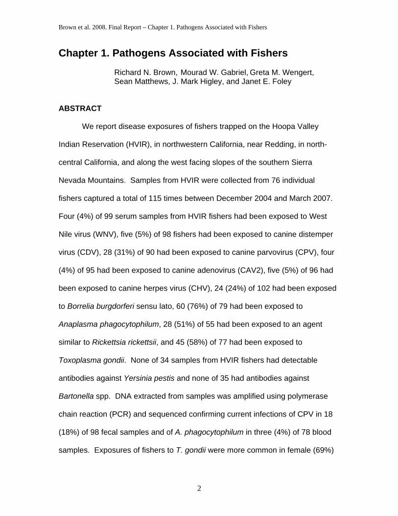
Table 1-3. The proportion of fishers (Martes pennanti) exposed to pathogens from the Hoopa Valley Indian Reservation (HVIR) vs. fishers trapped near Redding, California, December 2004 through March 2007.
Number (%) positive of No. Sampled
Pathogens Fishers from
HVIR 1Fishers from near
Redding P-value
West Nile virus 4 (4%) of 99 3 (16%) of 19 P = 0.08
Canine distemper virus 5 (5%) of 98 0 of 19 P = 0.59
Canine parvovirus 28 (31%) of 90 2 (11%) of 19 P = 0.05 **
Canine adenovirus-2 4 (4%) of 95 1 (5%) of 19 P = 1
Canine hepatitis virus 5 (5%) of 96 0 of 19 P = 0.59
Rickettisia spp. 28 (51%) of 55 6 (32%) of 19 P = 0.02 **
B. burgdorferi sl 24 (24%) of 102 1 (5%) of 19 P = 0.05 **
A. phagocytophilum 60 (76%) of 79 5 (26%) of 19 P = 0.00009 **
Y. pestis 0 of 34 0 of 12 P = 1
Bartonella spp. 0 of 35 0 of 15 P = 1
T. gondii 45 (58%) of 77 6 (46%) of 13 P = 0.55 1 Data on fecally-transmitted viruses from the first 31 of these fishers was reported previously (Brown et al., 2006); ** indicates statistical significance.
Fishers Trapped in the Southern Sierras
Fecal samples from 33 individual fishers trapped in the Sierra National
Forest were analyzed. Interestingly, none of these samples tested PCR-positive
for CPV DNA. Hoopa fishers were more likely (18 of 98) to be actively shedding
CPV DNA in their feces than were fishers trapped in the Sierra National Forest (0
of 33; P = 0.006).

Brown et al. 2008. Final Report – Chapter 1. Pathogens Associated with Fishers
22
DISCUSSION
Fishers on the HVIR were found to be exposed to five viruses (WNV,
CDV, CAV2, CHV, and CPV), three tick-borne bacteria (B. burgdorferi sl, A.
phagocytophilum, and Rickettsia sp.), and the protist T. gondii; but not Bartonella
spp. or Y. pestis.
Fishers trapped near Redding were exposed to many of the same
pathogens as were those on the HVIR. For instance, four and 16 percent
(respectively) of fishers sampled from the HVIR and from sites near Redding had
been exposed to WNV. The somewhat (although not significantly) higher
prevalence of exposure to WNV from sites near Redding may reflect higher rates
of transmission in the northern Sacramento Valley than in the Klamath Mountains
near Hoopa. Prevalences to several pathogens, including CPV and the tick-borne
bacteria (A. phagocytophilum, Rickettsia sp. and B. burgdorferi sl), were lower in
the fishers sampled near Redding than in those from the HVIR. Likewise,
although no serum or plasma samples were available to compare serology of
fishers from the southern Sierras with those from northern California, the PCR-
prevalence of CPV in the fishers sampled from the southern Sierra Nevada
Mountains was low compared to fishers from the HVIR. The fisher population in
the southern Sierras is isolated from all other fisher populations by a broad gap
extending approximately 420 km through the northern and central Sierra Nevada
Mountains (Zielinski et al. 1995), and such isolation may influence risks of
exposures to some pathogens.

Brown et al. 2008. Final Report – Chapter 1. Pathogens Associated with Fishers
23
The abundance of fishers on the HVIR appears to be greater than found
previously in the Shasta-Trinity National Forest (Yaeger 2005; at sites west of the
study areas near Redding), and higher densities of animals are expected to
maintain higher prevalences of directly transmitted viruses such as CPV.
However, we doubt that CPV cycles independently through fisher populations,
but instead cycles through the multiple sympatric species of carnivores (see
Chapter 2). The carnivore community on the HVIR is anecdotally quite diverse;
commonly observed species include American black bears (Ursus americanus),
gray foxes (Urocyon cinereoargenteus), ringtails (Bassariscus astutus), raccoons
(Procyon lotor), striped skunks, western spotted skunks (Spilogale gracilis),
fishers, bobcats (Lynx rufus), and cougars (Puma concolor). However, detailed
comparisons of carnivore community diversity at the HVIR vs. the study areas
near Redding are not available, and the details of community structure would be
necessary to evaluate the role of community diversity in maintaining these
pathogens.
Non-vaccinated domestic dogs might be a source of infection for wildlife.
Domestic dogs are commonly encountered in the rural areas surrounding the
HVIR, and they may be less common on large tracts of land owned or managed
by timber agencies (including lands near Redding owned by Sierra Pacific
Industries). If proximity to humans and their pets were responsible for the
difference in prevalence between these sites then one would expect that
prevalence in fishers found further from high densities of human habitation in the
valley at Hoopa would be lower than in those fishers living closer to human

Brown et al. 2008. Final Report – Chapter 1. Pathogens Associated with Fishers
24
habitation, and this was not substantiated (see Chapter 2). Alternatively, higher
exposures to CPV at Hoopa might indicate a random difference in fluctuating
prevalences, and studies designed to compare community level exposures will
be necessary to clarify the cause of high CPV prevalence at Hoopa.
Differences in exposures to tick-borne pathogens likely result from
differences in the risk of transmission from ticks. The abundance and distribution
of Ixodes pacificus, the primary tick vector of A. phagocytophilum and B.
burgdorferi sl, has been well established in the inland regions of the north-coastal
mountains of California (Lane et al. 2001, Lane et al. 2004, Brown et al. 2005),
and patterns of vector density and distribution, as well as host community
structure, likely influence the prevalence of exposures to these pathogens.
The sections that follow describe our results for each pathogen, disease
signs observed in other species infected with each pathogen, exposures of these
pathogens in populations of other carnivores, and perceived risks of transmission
among fishers. We also discuss co-exposures, important potential differences
among populations, and management implications of our work.
West Nile Virus
West Nile virus infects a broad range of birds and mammals, but mortality
is most often seen in birds, horses, and people. Animals with disease show
fever, muscle aches, lethargy, inappetence, diarrhea, vomiting, muscle stiffness,
abdominal pain, blindness, head tremors, cardiac arrhythmias, coma, and death.

Brown et al. 2008. Final Report – Chapter 1. Pathogens Associated with Fishers
25
West Nile virus is transmitted mainly by ornithophilic mosquitoes (Hubalek
and Halouzka 1999, Petersen and Roehrig 2001, Turell et al. 2001, Apperson et
al. 2004), but some tick species also have been shown to be competent vectors
(Lawrie et al. 2004, Hutcheson et al. 2005) and carnivores can become exposed
during consumption of prey (Austgen et al. 2004, Kuno 2001). It is logical that
species such as bats that feed on mosquitoes are exposed during feeding
(Pilipski et al. 2004,Davis et al. 2005, DeKrey and Adams 2005,). Although many
carnivores may be resistant, viremia and the ability to infect mosquito vectors has
been documented in domestic cats, and cats have become infected through
consumption of infected prey (Austgen et al. 2004). Thus, the fishers reported
exposed in this report may have been exposed by mosquito bite or, perhaps, by
feeding on infected birds.
Although WNV has not been documented in mustelids previously,
exposures to the virus, or disease, have been verified in numerous other
carnivores including: striped skunks (Centers for Disease Control and Prevention
2000); raccoons (Centers for Disease Control and Prevention 2000, Dietrich et
al. 2005, Root et al. 2005); American black bears (Farajollahi et al. 2003); brown
bears (Ursus arctos) (Madi et al. 1993); red pandas (Ailurus fulgens) (Ludwig et
al. 2002); snow leopards (Uncia uncia) (Ludwig et al. 2002); a harbor seal (Phoca
vitulina) (Del Piero et al. 2006); wolves (Canis lupus) (Lichtensteiger et al. 2003,
Lanthier et al. 2004); domestic dogs (Blackburn et al. 1989, Komar et al. 2001,
Lichtensteiger et al. 2003, Centers for Disease Control and Prevention 2005, Kile
et al. 2005); and domestic cats (Komar et al. 2001, Centers for Disease Control

Brown et al. 2008. Final Report – Chapter 1. Pathogens Associated with Fishers
26
and Prevention 2003, Austgen et al. 2004, Kile et al. 2005,). Other wild mammals
associated with WNV in the US include tree squirrels (Sciurus niger and Sciurus
carolinensis) ( Heinz-Taheny et al. 2004, Dietrich et al. 2005, Root et al. 2005),
white-tailed deer (Odocoileus virginianus) (Farajollahi et al. 2004) and bats
(Eptesicus fuscus; Myotis lucifugus; Myotis septentrionalis) (Pilipski et al. 2004,
Davis et al. 2005, DeKrey and Adams 2005).
Few studies have reported population level exposures of wild carnivores
to WNV. As examples, 6% of samples from a population of black bears in New
Jersey (Farajollahi et al. 2003), 35% of a small sample of brown bears in Croatia
(Madi et al. 1993), and 11% of gray foxes from the HVIR (Gabriel 2006), tested
positive for exposure to WNV and our results are similar to these exposures from
other species. To our knowledge, there is no documentation that WNV causes
disease or death in any mustelid, including fishers. More work is necessary to
determine the effects of WNV exposures in fishers and the likely route of
exposures.
Canine Distemper Virus
Mustelids infected with CDV suffer from rash, often with a thickening of the
muzzle and footpads, respiratory disease, immunosuppression, neurologic
disease, and often death (Deem et al. 2000, Williams 2001, Langlois 2005).
Survivors may suffer lingering effects from damage to organs long after the virus
is eliminated from the host. Canine distemper virus causes disease in stone
martens (Martes foina), black-footed ferrets (Mustela nigripes), mink (Mustela

Brown et al. 2008. Final Report – Chapter 1. Pathogens Associated with Fishers
27
vison), polecats (Mustela putorius), domestic ferrets, European badgers (Meles
meles), weasels (Mustela spp.), striped skunks, domestic dogs, wolves, coyotes
(Canis latrans), gray foxes, red foxes (Vulpes vulpes), African wild dogs (Lycaon
pictus), raccoon dogs (Nyctereutes procyonoides), raccoons, coatis (Nasua
narica), greater pandas (Ailuropoda melanoleuca), red pandas, African lions
(Panthera leo), and probably many other carnivores (Addison et al. 1999, Deem
et al. 2000, Williams 2001).
Although the host range is broad, susceptibility varies among species, with
high rates of mortality in black-footed ferrets, gray foxes, and lesser pandas and
some level of resistance in striped skunks and red foxes. Other carnivores,
including black bears, develop antibodies when exposed to CDV but rarely suffer
severe disease (Williams 2001). All species of mustelids probably are
susceptible to severe clinical distemper (Deem et al. 2000), and this presumably
includes fishers. Addison et al. (1999) stated that fishers are susceptible to CDV,
but they did not present data concerning the severity of disease nor was a
reference provided.
Exposure to CDV was evident in samples from five (5%) of 97 fishers from
the HVIR, including positive samples from two male and three female fishers.
Canine distemper virus is transmitted via respiratory droplets, contact with virus
laden discharges from the eyes or nose, in urine, or via fecal material (Williams
2001, Langlois 2005). Although the virus does not persist for prolonged periods
in the environment, it does persist as long as the contaminant remains fresh and
moist. Thus, contamination of equipment used for trapping and handling of

Brown et al. 2008. Final Report – Chapter 1. Pathogens Associated with Fishers
28
fishers could provide a source of infection for subsequent captures (Brown et al.
2006).
Lack of antibody response (negative IFA) in individuals must be combined
with other data to provide meaningful interpretations. Low levels of antibody
response in a population can be interpreted as low levels of exposure to a
pathogen, as too much time elapsed between exposure and sampling, as
exposures too recent to have stimulated a detectable immune response, or as
evidence that exposed animals have died prior to sampling (Delahay and Frolich
2000, Courtenay et al. 2001, Hanni et al. 2003). We know that CDV is present in
the wildlife surrounding Hoopa because 24% of 80 black bears sampled were
found to be exposed to CDV prior to 2001 (Brown et al. 2003). Also, exposure to
CDV is common in unvaccinated dogs (R. N. Brown et al., unpublished data),
and, anecdotally, because local veterinarians recognize distemper in domestic
dogs throughout the region. The prevalence of exposures among fishers from the
HVIR was lower than might be expected based on exposures of bears and
unvaccinated dogs, but this lower prevalence remains difficult to interpret. If
most fishers exposed to CDV die of acute disease, as reported from epizootics of
black-footed ferrets (Williams et al. 1988, Williams and Thorne 1996, Greenacre
2003), then few living animals would be expected to show evidence of prior
exposure. However, the relatively low prevalence might simply result from lower
rates of exposure among fishers than among dogs or bears.
Additional work with CDV in fishers, as well as in the sympatric
mesocarnivore community (see Chapter 2) and local domestic dogs, is needed to
Brown et al. 2008. Final Report – Chapter 1. Pathogens Associated with Fishers
29
allow interpretation of routes of exposure and risks to fishers. The lack of CDV in
the populations near Redding may indicate that CDV was less prevalent in their
local carnivore communities or that larger sample sizes would be necessary to
evaluate the expectedly low seroprevalence.
Canine Adenovirus
Canine adenoviruses are highly contagious and can be shed in urine of
apparently healthy animals. As disease develops, animals may shed virus in
discharges from the eyes and nose, in vomitus, as well as in feces. This virus is
easily transmitted via direct contact or by contact with contaminated inanimate
objects (e.g. fomites) (Cabasso 1981, Woods 2001). Pathology caused by CAV2
infection of fishers has not been reported, but the potential for disease should not
be ignored.
Five (4%) of 114 fisher samples had antibodies that bound to CAV2.
Adenoviruses, including both CAV1 and CAV2, cause infectious hepatitis in
domestic dogs and other susceptible carnivores, including mink, domestic ferrets,
North American river otters (Lontra canadensis), skunks, raccoons, wolves,
coyotes, red foxes, island foxes (Urocyon littoralis) and bears (Garcelon et al.
1992, Woods 2001). This virus can also cause encephalitis with inappetence,
oculonasal discharge, vomiting, mucoid or bloody diarrhea, hyperexcitability,
paralysis, depression, seizures, coma and death; the course of disease in striped
skunks can be so short that no signs are observed before sudden death (Karstad
et al. 1975, Cabasso 1981, Woods 2001). Other species, including mink, are

Brown et al. 2008. Final Report – Chapter 1. Pathogens Associated with Fishers
30
susceptible to canine adenoviruses, but they tend to develop mild, or at least
non-fatal, clinical disease (Addison et al. 1999).
Our findings of 4% and 5% seroprevalence of populations of fishers from
Hoopa and near Redding are comparable to a reported exposure of 4% of 28
fishers from British Columbia, Canada (Philippa et al. 2004). High
seroprevalence of CAV1 and CAV2 has been reported from some populations of
carnivores, including 50% of coyotes from Texas and 63% of striped skunks from
Maryland (Jamison et al. 1973, Woods 2001). Although CAV2 may occur in
many populations, actual disease has been reported rarely from wildlife and
some authors believe that this virus has little impact on wildlife populations
(Addison et al. 1999). Contamination of trapping and handling gear used in
research or management of fishers could easily provide a source of infection for
subsequently captured fishers (Brown et al. 2006).
Canine Herpesvirus
Canine herpesvirus causes respiratory disease, inappetence, abdominal
pain, and lesions of the genitalia in juvenile and adult domestic dogs,
reproductive failure and abortion in female dogs, and, rarely, a systemic
hemorrhagic syndrome and death of pups less than four weeks of age (Gaskell
and Willoughby 1999, Murphy et al. 1999).
Although it was originally thought that this virus was infectious only for the
family Canidae (Gaskell and Willoughby 1999), CHV infects, survives, and
replicates in fetal mink cells (McDonald and Lariviere 2001). Herpesviruses also

Brown et al. 2008. Final Report – Chapter 1. Pathogens Associated with Fishers
31
cause encephalitis and have been associated with pathology of the liver, spleen,
and lung of skunks (Charlton et al. 1977, Diters and Nielsen 1978). Herpesvirus
infections are exacerbated by physiologic and emotional stress in a wide range of
species, and CHV (or related herpesviruses) would be expected to cause
disease mainly in young, old, or otherwise immunocompromised animals.
Antibodies from five (5%) of 95 of the fisher samples from the HVIR reacted
with CHV, and we’ve found no reports of exposures of fishers to herpesviruses
for comparison in the literature. Herpesviruses typically are transmitted by direct,
animal to animal contact (Gaskell and Willoughby 1999), but infectious animals
may shed virus in oculonasal discharge, saliva or feces when the virus is actively
causing signs of disease (Gaskell and Willoughby 1999). Although CHV is
relatively fragile, infectious virus will persist for 1-2 days in moist fecal material
(Pesaro et al. 1995). Therefore, there is some risk for researcher-enhanced
transmission of CHV when trapping and handling mesocarnivores.
We have no evidence that CHV is causing serious disease in fishers, but
seroprevalence in the fishers sampled at Hoopa suggests infection and
replication of the virus in fisher cells. Most herpesviruses cause persistent latent
infections and might be expected to cause disease synergistically in combination
with other infectious organisms, if at all.
The potential exists for CHV to be transmitted via contaminated equipment
used for trapping and handling, and we have not characterized the virus infecting
fishers. Directed research would be necessary to clearly determine the risks of
herpesvirus infections in fishers, but we have no reason to suggest that CHV be

Brown et al. 2008. Final Report – Chapter 1. Pathogens Associated with Fishers
32
considered a conservation concern for fisher populations in the western DPS
(Brown et al. 2006).
Canine Parvovirus
Susceptibility of mustelid species to the feline parvovirus subgroup
(including feline panleukopenia virus (FPV), CPV, mink enteritis virus (MEV),
Aleutian disease of mink virus (ADV), and raccoon parvovirus (RPV)) probably
depends on characteristics of specific strains of the viruses and species
characteristics of the hosts. The most virulent parvoviruses in some mustelid
species have been shown to be ADV and MEV (Barker and Parrish 2001;
Langlois 2005). Canine parvovirus replicates in mink and ferret cells, and causes
seroconversion, without causing severe disease in these species (Barker and
Parrish 2001). Domestic ferrets appear to be susceptible only to disease caused
by ADV and not those of other parvoviruses, and both North American river
otters and domestic ferrets have been shown to be resistant to infection by CPV
(Fox 1998, Kimber et al. 2000). In captive studies with striped skunks, CPV,
FPV, and MEV failed to replicate (therefore causing no disease) and caused only
minimal antibody response (Barker and Parrish 2001).
The disease manifestations caused by this group of parvoviruses result from
infection and lysis (cell death) of rapidly multiplying cells; thus, many
parvoviruses (including CPV, FPV, MEV and RPV) cause pathology in lymphoid
tissues, tissues that produce blood cells, and the intestinal epithelium (Murphy et
al. 1999). Disease syndromes observed in species other than fishers include

Brown et al. 2008. Final Report – Chapter 1. Pathogens Associated with Fishers
33
fever, reduction of white blood cells, immunosuppression, cerebellar hypoplasia
(FPV) and hemorrhagic diarrhea (with or without vomiting) (Murphy et al.
1999,Barker and Parrish 2001, Langlois 2005). Inflammation and hemorrhaging
of the intestinal epithelium of animals suffering from parvovirus enteritis often
allows bacterial leakage into the blood vascular system, leading to sepsis, and
endotoxic shock. The combination of weakness, dehydration from the diarrhea
and endotoxic shock causes some animals to die before outward signs are
apparent. Animals that survive acute infections typically are immune to further
infections. Like distemper, disease caused by CPV should be expected to be
most severe in young animals.
Fishers from the HVIR were commonly exposed (31% of 90) to CPV, and
CPV-DNA was amplified from 19% (N=59) of the fisher fecal samples analyzed.
A somewhat lower prevalence of exposure to CPV, two (11%) of 19, was
detected in samples from fishers trapped near Redding, but a similar prevalence
(18% of 39) was determined using PCR of DNA from fecal material from the two
study areas. None of the fecal samples from fishers trapped previously in the
southern Sierra Nevada Mountains were PCR positive to CPV. Although
serology alone is insufficient to differentiate among related parvoviruses, PCR-
amplification and sequencing of DNA from fisher scat proved that sequences
evaluated were those of CPV, and no fishers were confirmed to have been
infected with any other parvovirus. We interpreted these findings as suggesting
that CPV was probably common in the carnivore communities on the HVIR and

Brown et al. 2008. Final Report – Chapter 1. Pathogens Associated with Fishers
34
near Redding, but we recognized that other parvoviruses might also have
occurred in this region.
The prevalence of exposures to CPV was greater in adult fishers (49% of
HVIR fishers > 2 years) than in subadults (20% of HVIR fishers 0-1 year of age),
and there are several possible explanations for this pattern. Older fishers may
have simply had longer to accumulate exposures than have younger animals.
Adult fishers may experience a higher rate of exposure to CPV than did subadult
fishers, especially if exposures are generated from a single source (such as
domestic dogs or gray foxes) to which subadult fishers might not be exposed.
Lastly, disease caused by CPV may be more severe in juveniles, and adult
fishers may have subclinical infections, as do adult mink and domestic ferrets.
Assuming that survival of infection confers immunity, as it does in other
carnivores, many adult fishers might be expected to have antibodies present in
their blood but not to be actively shedding virus in their feces (i.e. PCR-negative).
The virulence of the different parvovirus strains, including CPV, for fishers
remains unknown, and few of the fishers examined showed signs of bloody
diarrhea, severe dehydration or sepsis during handling; two fishers at necropsy
appeared to have signs consistent with hemorrhagic enteritis, but the results of
histological examination remained equivocal (Gabriel et al. unpublished data).
Nonetheless, the finding that 18% of individuals sampled were actively shedding
virus in their feces at some point during the study suggests that CPV infections
(with associated lysis of gut epithelium cells) occur commonly in the population of
fishers at Hoopa. Canine parvovirus is spread via direct contact with feces or

Brown et al. 2008. Final Report – Chapter 1. Pathogens Associated with Fishers
35
other body fluids, but, unlike CDV, CAV2, CHV and WNV, CPV remains
infectious for prolonged periods of time in the environment (Gaskell and
Willoughby 1999, Murphy et al. 1999). Therefore, like CDV, CAV2, and possibly
CHV, there is some potential for researcher enhanced transmission of CPV
within and between fisher populations (Brown et al. 2006).
Borrelia burgdorferi sl
Lyme borreliosis is caused by members of a group of related spirochetes
referred to as B. burgdorferi sl, all of which are transmitted by ixodid ticks. In
California, the most important vector is the western black-legged tick (Ixodes
pacificus )(Brown et al. 2005).
There are 12 described genospecies within the B. burgdorferi sl group, and
many strains within California and elsewhere that remain inadequately
characterized (Brown and Burgess 2001, Brown et al. 2006). Moreover, these
different genospecies or strains may all differ in the tendency to cause disease in
different species of hosts. Domestic dogs, horses, humans and occasionally
other species develop signs of disease related to infection by B. burgdorferi sl,
but no wild populations of carnivores are reported to have been adversely
impacted by this group of borreliae (Brown and Burgess 2001).
Although we can not predict exactly how individual fishers will be affected by
infection by a given strain of spirochetes, we have no reason to suspect that
fisher populations are threatened by these pathogens in California. However,
carnivores can be very useful as sentinels of borrelial infections because many

Brown et al. 2008. Final Report – Chapter 1. Pathogens Associated with Fishers
36
species range widely and are exposed to relatively large numbers of potential
vectors (Foley et al. 2004). Since our current analyses did not differentiate
among the variety of strains and genospecies of borreliae present in California,
we cannot interpret these findings beyond noting the prevalence of exposures in
the different populations.
Rickettsia sp.
The principal members of the spotted fever group of rickettsial pathogens
include Rickettsia rickettsii (the cause of Rocky Mountain spotted fever), the
Rickettsia conorii-complex (the cause of Mediterranean spotted fever and related
infections), Rickettsia sibirica (the cause of North Asian tick typhus), and,
Rickettsia australis (the cause of Queensland tick typhus). The Rocky Mountain
wood tick (Dermacentor andersoni) transmits R. rickettsii throughout the Rocky
Mountain regions of Canada and the US, in mountainous areas of the Great
Basin, and in northeastern California along the east facing slopes of the northern
Sierra Nevada Mountains ( Sonenshine 1993, Brown et al. 2005). However, D.
andersoni is not reported to occur within the geographic range of fishers in
California. A related vector, Dermacentor variabilis, is an important vector of R.
ricketsii in its eastern and southern range within the US. This species occurs at
the sites sampled for this study, but it is not confirmed as a vector in the far
western US. Dermacentor occidentalis, the Pacific Coast tick, is considered a
potential vector of this agent in California ( Philip et al. 1981, Lane et al. 1982),
and it also occurs in each of the areas from which fishers were reported herein.

Brown et al. 2008. Final Report – Chapter 1. Pathogens Associated with Fishers
37
However, human cases of rickettsial disease are exceedingly rare throughout
most of its distribution. Disease caused by R. rickettsii occurs in people and
dogs (Kidd et al. 2006), but disease in other carnivores remains unreported.
We found serologic evidence of antibodies in fishers from the HVIR that
react with R. rickettsii-antigen. Unfortunately, we can’t confirm from our studies
that the infecting agent is truly R. rickettsii because cross reactions are common
within this group and because this group is rapidly expanding as people evaluate
new sources of hosts and vectors. More work will be necessary to interpret our
findings concerning this pathogen. We do note that serologic exposures were
higher among fishers from the HVIR than among fishers near Redding (51% vs
32%; P = 0.02).
Anaplasma phagocytophilum
Sixty (76% of 79) of the fishers from the HVIR were exposed to A.
phagocytophilum and this prevalence was significantly higher (P = 0.00009) than
found in the fishers trapped near Redding (26% of 19). This difference is likely
related to higher densities of the vector, I. pacificus, in the more mesic forests of
the HVIR compared to the dryer forests surrounding the Sacramento Valley,
which is near the Redding site (Foley et al. 2004). The difference in prevalences
of exposure of adult and subadult fishers (93% and 68%, respectively) likely
results from accumulated exposure to ticks through time.
Clinical manifestations of infections of A. phagocytophilum in horses, cats,
and dogs include immunosuppression, fever, incoordination, swelling of the

Brown et al. 2008. Final Report – Chapter 1. Pathogens Associated with Fishers
38
ventral limbs, liver disease, and the reduction of white blood cells and
thrombocytes (e.g. immunosuppression) (Madigan and Gribble 1987, Foley et al.
2003, Foley et al. 2004), but we have found no reference demonstrating clinical
disease associated with infection of this bacterium in wild carnivores. Exposures
of other carnivore populations to A. phagocytophilum include 46% of coyotes
from central California (Pusterla et al. 2000), 17% of mountain lions from the
Sierra Nevada mountains (Foley et al. 1999), 86% of black bears from the HVIR
(Brown et al. 2003), and 50% of gray foxes from the HVIR (Gabriel 2006). In
California, A. phagocytophilum is transmitted to wildlife, people, and domestic
animals by the western black-legged tick as is B. burgdorferi sl (Lane et al. 2001,
Holden et al. 2003, Foley et al. 2004, Lane et al. 2004), and carnivore
populations exposed to large numbers of infected ticks are expected to have high
exposures to these pathogens (Foley et al. 2004). Nymphs and adults of several
species of ticks, including I. pacificus, have been removed from fishers captured
at Hoopa (Brown et al., personal unpublished data). Disease caused by infection
of fishers with A. phagocytophilum is unknown, and we expect outward signs of
disease to be mild and of short-term duration with the potential complication of
immunosuppression.
Yersinia pestis
Infamous as the agent that causes plague,Y. pestis is well established in
California wildlife (Ruppanner et al. 1982, Clover et al. 1989, Chomel et al. 1994).
This pathogen is usually associated with communities of rodents, some of which

Brown et al. 2008. Final Report – Chapter 1. Pathogens Associated with Fishers
39
die quickly (including prairie dogs, Cynomys spp., and ground squirrels,
Spermophilus spp.) and some of which are capable of some level of resistance
including some voles (Microtus spp.), some deer mice (Peromyscus spp.),
marmots, (Marmota spp.), and gerbils, (Rhombomys opimus and Meriones spp.)
( Gasper and Watson 2001, Gage and Kosoy 2005). Primates and many rodent
species are susceptible to Y. pestis (Gasper and Watson 2001). Other
mammals, including ungulates and most carnivores are resistant to Y. pestis
(Gasper and Watson 2001), but domestic cats and black-footed ferrets are
susceptible and may die (Williams et al. 1994, Williams et al. 1991, Gasper and
Watson 2001, Gage and Kosoy 2005). Prevalence of exposures to Y. pestis
have been reported from carnivores in northern California (Brown et al. 2003,
Chomel et al. 1994, Hoar et al. 2003, Ruppanner et al. 1982, Zielinski 1984) and
the susceptibility of some mustelids (Gage and Kosoy 2005, Gasper and Watson
2001, Williams et al. 1994, Williams et al. 1991) led us to evaluate exposures in
the fisher populations associated with this study. Nonetheless, we did not detect
exposures to Y. pestis in any of the sampled fishers. Until data suggest
otherwise, plague should not be considered a significant threat to fisher
populations in the western DPS.
Bartonella sp.
Bartonella spp. occur commonly in California (Yamamoto et al. 1998, Chang
et al. 1999, Hoar et al. 2003, MacDonald et al. 2004b, Riley et al. 2004,
Beldomenico et al. 2005, Henn et al. 2005), prevalence was found to be high in

Brown et al. 2008. Final Report – Chapter 1. Pathogens Associated with Fishers
40
gray foxes trapped on the HVIR (Henn et al. 2007), and both exposure rates and
Bartonella-associated disease have been found in wild mustelids (McDonald et
al. 2001, McDonald and Lariviere 2001). However, none of 60 fishers sampled
had been previously exposed to these bacteria. Unless future data suggest
otherwise, these data do not suggest that Bartonella should be considered an
immediate threat to fisher management or conservation.
Toxoplasma gondii
Toxoplasma gondii is a ubiquitous organism that utilizes both domestic
and wild felids as its definitive hosts and many species of mammals (and even
some birds) as its intermediate hosts. This species invades and multiplies in the
cells lining the intestines of cats, from which oocysts are shed in the feces.
Intermediate hosts do not shed oocysts in their feces, but rather develop
intracellular tissue cysts in a variety of organs including skeletal muscle,
pancreas, liver, heart, brain, eye, uterus, and placenta.
Intermediate hosts become infected either by exposure to sporulated
oocysts, while feeding on tissue cysts infecting other intermediate hosts, or
transplacentally prior to birth. Thus, carnivores become infected from a variety of
sources and other serologic surveys have indicated that infections of T. gondii
are common in carnivores (Dubey and Odening 2001). Although infections are
common, most infections remain asymptomatic, with disease severity dependent
upon the strain of the infecting parasite, the immune status of the host, and the
age of the host; many species develop severe disease as young individuals prior

Brown et al. 2008. Final Report – Chapter 1. Pathogens Associated with Fishers
41
to development of immuno-competence, but are typically resistant if infected as
adults (Dubey and Odening 2001).
Clinical toxoplasmosis is associated with the specific organ tissues
infected in susceptible hosts that fail to immunologically control the development
of tissue cysts. Thus, animals develop encephalitis, myocarditis, myositis,
pancreatic necrosis, hepatitis, retinal inflammation, etc., depending on where the
cysts localize within the individual. Infected animals may become anorexic,
depressed, and suffer signs of pneumonia and meningoencephalitis (respiratory
distress, disorientation, uncoordinated movements, paralysis). Infected mink and
ferrets develop hind limb paralysis. Placental infections cause local edema and
necrosis, and transmission to fetuses across the placenta allows uncontrolled
multiplication due to the lack of an intact fetal immune system. Congenital
infections are commonly associated with fetal deaths, abortions, stillbirths,
neonatal deaths, and production of small, weak survivors (Dubey and Odening
2001, Frank 2001).
We report that 57% (51 of 90) of fisher plasma samples had detectable
levels of antibodies reactive with T. gondii antigen and that exposures did not
vary significantly between the populations sampled on the HVIR and near
Redding. This compares to 18% of 28 fishers surveyed in British Columbia
(Philippa et al. 2004), 41% of 379 fishers surveyed in Ontario (Tizard et al. 1976),
and 41% (33 of 80) of black bears trapped at Hoopa (Brown et al. 2003). For
comparison, examples of exposures in a few other (non-felid) mammalian
carnivores include: 5 and 11% of martens (Martes americana) (Tizard et al. 1976,
Brown et al. 2008. Final Report – Chapter 1. Pathogens Associated with Fishers
42
Hurkova and Modry 2006); 100% of American mink (Tizard et al. 1976); 17 and
45% of northern river otters (Tocidlowski et al. 1997, Gaydos et al. 2007); 28 to
56% of striped skunks (Quinn et al. 1976, Tizard et al. 1976, Smith et al. 1992,
Hill et al. 1998, Mitchell et al. 2006); 15 to100% of black bears (Quinn et al. 1976,
Burridge et al. 1979, Chomel et al. 1995, Nutter et al. 1998, Zarnke et al. 2000);
15 to 100% of raccoons (Tizard et al. 1976, Dubey et al. 1992, Hill et al. 1998,
Mitchell et al. 2006); 64 and 78% of coyotes (Quinn et al. 1976, Tizard et al.
1976); and 9% of wolves (Zarnke et al. 2000). However, since most infections
remain asymptomatic, serologic results alone do not indicate population level
risks. That said, several mustelid species and skunks have been shown to suffer
severe disease and mortality associated with toxoplasmosis.
Black-footed ferrets were shown to be highly susceptible to T. gondii when
a captive breeding colony at the Louisville Zoological Garden suffered severe
disease and many deaths (Burns et al. 2003). Domestic ferrets and captive mink
also have been shown to be highly susceptible to toxoplasmosis and both
species transmit infections congenitally from infected females to their kits (Dietz
et al. 1993, Fox 1998, Frank 2001, Thornton 1990). Frank (2001) reported that
1,976 (26%) of 7,800 captive female mink lost their litters either from abortion or
neonatal mortality caused by T. gondii. Although a wild population might
withstand such a high percentage of reproductive failure, T. gondii would be only
one factor limiting fitness and the cumulative effects of multiple pressures might
be expected to limit population growth.

Brown et al. 2008. Final Report – Chapter 1. Pathogens Associated with Fishers
43
The southern sea otter (Enhydra lutris nereis) provides another example
of a mustelid that appears to be suffering significantly from toxoplasmosis. This
subspecies of sea otters has failed to thrive and expand following the cessation
of harvests, and several authors have recently suggested that such failure may
result from epizootics of T. gondii (Miller et al. 2002, Hanni et al. 2003, Miller et
al. 2004, Conrad et al. 2005,). In recent reports of infections in live and dead sea
otters from California, Miller et al. (2002) reported 42% (49/116) seroprevalence
for live otters and 62% (66/107) for dead otters and Conrad et al. (2005) reported
that 38% of 257 live sea otters and 52% of 305 freshly dead sea otters were
infected with T. gondii. Studies with southern sea otters has also identified a new
strain, Type X, that appears to be more heterogeneous and virulent than the
previously described terrestrial strains, Types I, II, and III (Cole et al. 2000, Miller
et al. 2002, Conrad et al. 2005,).
At this point, we can only speculate about the potential disease caused by
T. gondii in fishers. Our lack of association of age of fishers and seroprevalence
as shown in other species (Quinn et al. 1976) suggests that fishers are either
exposed very early in life or acquire infections congenitally. Congenital infections
would be more likely to cause abortions, still births, or neonatal deaths than
would infections acquired once the immune systems of the kits had fully
developed. Future studies should attempt to molecularly characterize the
infecting strains in fishers, assess neural pathology associated with protozoal
cysts in animals that are necropsied, and describe the ecological patterns of
disease transmission within fisher populations.

Brown et al. 2008. Final Report – Chapter 1. Pathogens Associated with Fishers
44
Sources of Mortality and Co-exposures
We did not identify clear associations between pathogen exposures and
any of the fifteen radio-collared fishers found dead during this study. However, it
should be noted that our ability to determine cause of death was compromised by
the fact that most carcasses were recovered days after death and several were
mutilated by predators or scavengers. We also had limited ability to evaluate
fishers that died without radio-collars because we did not recover the carcasses.
We did necropsy a few fishers in good enough condition to allow cellular
examination (Gabriel et al., unpublished data). From these animals, we have
identified several signs that appear highly suggestive of infectious disease
(including severe hemorrhagic diarrhea, a perforated hemorrhagic intestine,
protozoal cysts in the heart, and apparent high limb paralysis similar to
descriptions of toxoplasmosis in mink). However, we have not yet found
definitive evidence of severe disease caused by any specific pathogen (Gabriel
et al., unpublished data). We noted that most fishers found dead were female,
but this difference likely resulted from the study design of a parallel study in
which most radio-collars were placed on females to monitor den sites and
juvenile dispersal (Matthews et al., unpublished data).
Several of the pathogens discussed have the potential to cause
immunosuppression, and disease may be most severe in hosts simultaneously
infected with multiple pathogens (Diters and Nielsen 1978, Graham, 2002, Chen
et al. 2005, Holden et al. 2005, Cho et al. 2006, Hofmann-Lehmann et al. 2004,

Brown et al. 2008. Final Report – Chapter 1. Pathogens Associated with Fishers
45
Davidar and Morton 2006). However, coinfection of some pathogens confers
resistance rather than enhances severity ( Gale et al. 1997, Furze et al. 2006);
therefore, coinfection is a complicated issue and generalities may not always
apply (Mosquera and Adler 1998, Foley et al. 2003, Lively 2005). Positive
serology indicates exposure at some time in the past, and it does not necessarily
indicate current infections. The most worrisome co-exposure reported is that of
CPV and A. phagocytophilum, both of which cause immunosuppression in
susceptible species. Immunosuppression would likely make fishers more
susceptible to other pathogens, including potentially severe disease caused by
CDV and toxoplasmosis. Co-exposure to CAV and CHV may indicate a focused
source of infection to these pathogens such as provided by unvaccinated
domestic dogs. Other co-exposure results, including those between T. gondii
and B. burgdorferi, are difficult to explain.
Lack of co-exposure among the pathogens suggests that differences in
routes of transmission led to a random pattern of exposures in the population, as
might be predicted. Although the co-exposure of the pathogens mentioned
above is interesting, the data do not appear to indicate clear risks for fisher
populations. Co-exposure might be caused by similar routes of transmission or,
indirectly, by exposure rates that vary with habitat or distance from humans, etc.
The sources of directly transmitted infections of fishers remain unknown,
and the pathogens appear to be transmitted within the greater wildlife community
(see Chapter 2). Domestic dogs also are a logical source of CDV, CAV, CDV,
and CPV, and potentially represent the ultimate source of the fisher exposures

Brown et al. 2008. Final Report – Chapter 1. Pathogens Associated with Fishers
46
we’ve reported; unleashed dogs are very common in areas near houses, but less
common in the backcountry of the HVIR (Gabriel 2006). If dogs are a proximate
source of infections, then fishers living near humans should be more likely to be
exposed than fishers with home ranges at greater distance from human
habitation. Proximity to humans has been correlated with CPV infections in both
spotted hyenas (Crocuta crocuta) sampled in the Masai Mara of Kenya and gray
foxes living near parks in central California (Riley et al. 2004). However, if dogs
represent only the ultimate source and transmission among other carnivores is
high, then the association of exposures of fishers with people and dogs may not
be obvious. Molecular analysis of a large number of samples from individually
marked fishers and sympatric carnivores would be necessary to allow spatial
analysis to help evaluate such patterns of abundance and distribution of
pathogens within the carnivore community.
MANAGEMENT IMPLICATIONS
All animal populations suffer from infections of parasites and diseases, but
the potential influence of disease to negatively impact populations increases in
importance as populations become small and isolated. The pathogens discussed
cause disease of unknown severity in fishers; we found few reports of exposures
of fishers to pathogens in the literature, and no one has reported laboratory
studies of the effects of these pathogens in fishers. At this time, severity can
only be predicted based on extrapolation of disease in related species. Some
disease agents, including West Nile virus, Bartonella spp., Y. pestis, B.

Brown et al. 2008. Final Report – Chapter 1. Pathogens Associated with Fishers
47
burgdorferi, and A. phagocytophilum are vector-borne and therefore risks
associated with these pathogens will be difficult to manage.
Translocations of wildlife move pathogens along with their hosts. It is
generally recommended that translocations be planned carefully and monitored
throughout all phases to minimize risks associated with moving infected animals
or moving naïve animals into areas in which epizootics are cycling (Nettles et al.
1980, Woodford and Rossiter 1993, Cunningham 1996, Kimber et al. 2000,
Leighton 2002, Wobeser 2002,). Exposures of CDV in black-footed ferrets are
managed by vaccination and risk monitoring, and vaccination of fishers to CDV,
rabies virus, and feline panleukopenia virus (which would cross protect for CPV
infections) should be considered during translocation events.
Some pathogens, including CDV, CAV2, CHV, and CPV are transmitted via
feces or other body fluids, and CPV remains infectious for prolonged periods in
the environment. Trapped animals often contaminate equipment used for
trapping or handling. Unless trapping gear is cleaned and disinfected between
captures, subsequently captured animals may be exposed to viruses shed from
previous animals. Due to the documented morbidity and mortality of
parvoviruses, distemper viruses, adenoviruses, and influenza viruses for related
species of mustelids, all equipment in contact with trapped fishers should be
cleaned thoroughly and sprayed with appropriate anti-viral disinfectants after
each capture; all visible fecal material should be removed, surfaces should be
wiped clean, and equipment should be generously sprayed with a disinfectant
rated to kill parvoviruses. While this will not eliminate the potential for

Brown et al. 2008. Final Report – Chapter 1. Pathogens Associated with Fishers
48
researcher-facilitated transmission of these viruses, it should minimize the risks
significantly.

Chapter 2. Pathogens Associated with Mesocarnivores Sympatric with Fishers
Mourad W. Gabriel, Greta M. Wengert, Janet E. Foley,J. Mark Higley, Sean Matthews, and Richard N. Brown
ABSTRACT
Conservation efforts often require an understanding of the threats
imposed by sympatric species on the species of concern, and transmission of
disease among sympatric species should be considered in any management
scenario for species conservation. Monitoring ecological communities allows for
a logistically feasible assessment of pathogen prevalence than monitoring a
sensitive or rare species. We investigated the prevalence of several pathogens
within a mesocarnivore community that is sympatric with a fisher population
within the West Coast Distinct Population Segment of fishers (Martes pennanti).
A combined total of 63 gray foxes, ringtails, raccoons and spotted and striped
skunks showed prior exposures to canine distemper virus (2%), canine
parvovirus (30%), canine adenovirus (9%), West Nile virus (6%), Anaplasma
phagocytophilum (50%), and Toxoplasma gondii (40%). No individuals were
seropositive for canine herpesvirus. Of 20 individuals tested for active infections
using the polymerase chain reaction to amplify pathogen DNA, 15% were found
infected by A. phagocytophilum, and 19% were found to be actively shedding
canine parvovirus. Exposure to A. phagocytophilum was higher in fishers than in
other mesocarnivores (p = 0.001), but no other differences in exposures of
fishers and sympatric mesocarnivores were found. Our results indicate that
49
Gabriel et al. 2008. Final Report – Chapter 2. Pathogens Associated with Mesocarnivores

Gabriel et al. 2008. Final Report – Chapter 2. Pathogens Associated with Mesocarnivores
50
fishers and their sympatric mesocarnivores share several pathogens in similar
prevalence, suggesting a potential risk of disease transmission within this
community. Additionally, we suggest that sympatric mesocarnivores be
monitored to determine disease prevalence across the community and to help
determine risks for fishers.
INTRODUCTION
The conservation of a species entails the understanding of many
ecological factors, one of which involves health risks from infectious pathogens
with the potential to limit populations (Scott 1988, Thorne and Williams 1988,
Deem et al. 2001, Cleaveland et al. 2002, Mathews et al. 2006). Additionally,
interactions between sympatric wild and domesticated animals may increase the
risk of transmission of a pathogen within a sensitive species’ established or
potential range (Hudson and Greenman 1998, Deem et al. 2001, Macdonald et
al. 2004, Mathews et al. 2006).
Monitoring common sympatric mesocarnivores that overlap or inhabit the
same spatial niche as high-profile or sensitive species may provide an indication
of ecosystem health and may also be necessary when monitoring a focal species
with has low population numbers or is otherwise not feasible to investigate
(Munson and Karesh 2002, Wobeser 2006). Unfortunately, research of many
high-profile species or their sympatric community is not conducted until after an
unexpected epidemic moves through a threatened population and creates a high

Gabriel et al. 2008. Final Report – Chapter 2. Pathogens Associated with Mesocarnivores
51
level of morbidity or mortality (Woodroffe 1999, Munson and Karesh 2002,
Mathews et al. 2006)
In this chapter we compare the serological exposure and rates of
infections of potentially limiting viral, bacterial, and protozoal pathogens in fishers
and their sympatric mesocarnivores to determine whether information from
sympatric communities might provide valuable information for fisher conservation
efforts. We discuss the feasibility of proactive disease monitoring of the
mesocarnivores sympatric with a subset of the West Coast fisher Distinct
Population Segment (DPS).
STUDY AREA
Field sampling was conducted on the Hoopa Valley Indian Reservation
(HVIR) in northeastern Humboldt County, northwestern California as described in
Chapter 1. The valley floor and the outlying areas were distinguished as the
valley and backcountry zones, respectively. Most human HVIR inhabitants reside
in a residential zone near the valley floor (Figure 2-1).
Mid-sized to large mammals that inhabit the HVIR include Columbian
black-tailed deer (Odocoileus hemionus columbianus), Roosevelt elk (Cervus
elaphus roosevelti), American black bears (Ursus americana), gray foxes (Urocyon
cinereoargenteus), coyotes (Canis latrans), fishers, bobcats (Lynx rufus),
mountain lions (Felis concolor), raccoons (Procyon lotor), ringtails (Bassariscus
astutus), striped skunks (Mephitis mephitis), spotted skunks (Spilogale gracilis),

Gabriel et al. 2008. Final Report – Chapter 2. Pathogens Associated with Mesocarnivores
52
porcupines (Erethizon dorsatum), domestic cats (Felis domesticus), and domestic
dogs (Canis domesticus) (M. W. Gabriel and J. M. Higley, personal observation).
Figure 2-1. Hoopa Valley Indian Reservation, Humboldt County, California, USA, the study area for disease exposures in fishers and their sympatric mesocarnivores from February 2004 to February 2007.
Hoopa
Hoopa ValleyIndian Reservation
Trinity River
Valley Floor
Gabriel et al. 2008. Final Report – Chapter 2. Pathogens Associated with Mesocarnivores
53
METHODS
Capture, Handling, and Sampling
Five mesocarnivore species, including gray fox, ringtail, raccoon, striped
skunk, and spotted skunk were opportunistically trapped from February 2004
through February 2007 in parallel with an ongoing fisher project at the HVIR.
Carnivores were trapped with wire mesh live traps (81 x 25 x 31cm) (Tomahawk
Live Trap Company, Tomahawk, Wisconsin) baited with chicken. Insulated
wooden nest boxes were attached to traps to provide security, reduce
environmental stressors, and facilitate handling of trapped animals (Wilbert 1992,
Gabriel and Wengert 2005).
Captured foxes were moved to handling cones made of cloth mesh,
weighed and then anesthetized with 20 mg/kg ketamine (Fort Dodge Animal
Health, Fort Dodge, Iowa, USA) and 0.5mg/kg diazepam (Hospira, Inc., Lake
Forest, Illinions, USA) via intramuscular injection. Ringtails were similarly
processed but were intramuscularly injected with 25 mg/kg ketamine. Both
species of skunks were lightly anesthetized with isoflurane- (Abbott Laboratories,
Abbott Park, Illinois, USA) saturated cotton balls that were placed in the nest box
2 minutes prior to hand injection with 25 mg/kg ketamine to facilitate safe
handling and reduce the likelihood that skunks would spray. Weights of
raccoons were estimated to the nearest 0.2 kg before being moved to a handling
cone made of welded metal rods and injected with a mixture of 25 mg/kg
ketamine and 1.0 mg/kg diazepam.
Gabriel et al. 2008. Final Report – Chapter 2. Pathogens Associated with Mesocarnivores
54
Each anesthetized animal was maintained in lateral recumbency and a
brief physical exam was performed. Passive integrated transponder (PIT) tags
(Biomark Inc., Boise, Idaho, USA) were implanted subcutaneously for future
identification of each individual, and relevant morphometrics were recorded.
Animals were allowed to recover from anesthesia in the nest box of the traps in
which they were caught, and were released at the site of capture upon full
recovery.
Fecal samples were collected from each mesocarnivore using a fecal
loop, a Dacron® swab (Fisher Scientific, Pittsburgh, PA, USA) or directly from the
animal. Fecal samples were stored at -20°C until analysis. Approximately 1-3 ml
of whole blood was collected by venapuncture and stored frozen in the
anticoagulant ethylene diamine tetraacetic acid (EDTA) stored at -20°C until
analysis. Blood was thawed and centrifuged to separate plasma from cells.
Plasma samples were serially diluted in 10 mM phosphate-buffered saline (PBS;
138 mM NaCl, 2.7 mM KCl), then refrozen until thawed for analysis.
Assays for Exposure to Pathogens
Plasma samples were analyzed for presence of antibodies that bound to
antigens of the different pathogens by indirect immunofluorescence assays
(IFAs). Antigen for the canine distemper virus (CDV) IFA was from kidney cells of
CDV-infected domestic ferrets (Mustela furo) (American Bioresearch, Milton,
Tennessee, USA). Positive controls were serum samples from domestic dogs
previously shown to have high, standardized titers, and negative controls were
Gabriel et al. 2008. Final Report – Chapter 2. Pathogens Associated with Mesocarnivores
55
from dogs maintained at a specific pathogen-free (SPF) colony. Positive results
were defined as those reacting at a dilution of 1:8. Antibodies specific to
canine parvovirus-2 (CPV), canine adenovirus (CAV-2) and canine herpesvirus
(CHV) were detected with commercially available antigen slides (Veterinary
Medical Research and Development, Pullman, Washington, USA). Positive
controls for CPV, CAV2 and CHV tests were blood samples from dogs previously
shown to have high, standardized titers, and negative controls were from SPF
colony dogs. Positive results for these tests were defined as those reacting at a
dilution of 1:25. Exposure to West Nile virus (WNV) was evaluated by the
Animal Health Diagnostic Center (Cornell University, Ithaca, New York, USA) via
serum neutralization and positive results were defined as those reacting at a
dilution of 1:4. Antigen for the Anaplasma phagocytophilum IFA was from cells
isolated from infected horse (Equus callabus) blood. Positive controls were
serum samples from dogs of known infection history with high, standardized
titers, and negative controls were from SPF colony dogs. Positive results were
defined as those reacting at a dilution of 1:25. Antigen for Toxoplasma gondii
was detected with indirect hemagglutination tests and positive results were
defined as those reacting at a dilution of 1:64.
Molecular Assays
DNA was extracted from whole blood using a Qiagen kit (Qiagen,
Valencia, California, USA) following manufacturer’s recommendations and from
feces using a modified Boom method of silica extraction (Cheung et al. 1994).
Gabriel et al. 2008. Final Report – Chapter 2. Pathogens Associated with Mesocarnivores
56
TaqMan real-time polymerase chain reaction (PCR) for the A. phagocytophilum
p44 gene was performed as described previously (Drazenovich et al. 2006). A
nested PCR was performed for CPV2 as described previously (Hirasawa et al.
1994). Amplicons from the nested-PCR assay were cloned into a pCR2.1 vector
using the TOPO cloning kit (Invitrogen, Carlsbad, California, USA) and amplified
in TOP10F - competent cells of Escherichia coli (Invitrogen, Carlsbad, California,
USA). The plasmid was purified using a Plasmid Miniprep kit (BioRad, Hercules,
California, USA) and the vector insert was sequenced with the M13f primer
(Davis Sequencing, Davis, California, USA) using the Big Dye Terminator cycle
sequencing kit (Applied Biosystems, Foster City, California, USA). The
sequences were determined using the BLAST database search program and
GenBank (National Center for Biotechnology Information, Bethesda, Maryland,
USA) (Altschul et al. 1990).
Statistics and Data Exclusion Rules
Comparisons of exposures and active infections between fishers and all
mesocarnivores as a single group, were assessed using two-tailed Fisher’s exact
tests of association with a cutoff of p < 0.05 using NCSS (Number Cruncher
Statistical Software, Kaysville, UT, USA). Analyses included comparisons
between fisher and all mesocarnivores as a single group, and between fishers
and the group of mesocarnivores that were captured in the backcountry only.
Data from recaptures were included in analyses involving seroprevalence except
when individuals were resampled < 9 months after the initial capture or when the

Gabriel et al. 2008. Final Report – Chapter 2. Pathogens Associated with Mesocarnivores
57
first sample tested seropositive in order to reduce potential errors associated with
repeated sampling of seropositive animals. The 9-month interval was considered
sufficient to avoid counting repeated negative samples. All samples from which
DNA was amplified to determine active infections were incorporated irrespective
of sample date. In order to detect spatial patterns in disease prevalence among
fishers and their sympatric mesocarnivores, animal locations, and disease
exposures were spatially displayed using ArcMap 9.2 (ESRI, Redlands,
California, USA).
All trapping and handling of mesocarnivores for this project followed
protocols approved by Humboldt State University’s Institutional Animal Care and
Use Committee.
RESULTS
Capture Demographics
A total of 63 sympatric mesocarnivores were captured and processed
February 2004 though February 2007. This total included 27 gray foxes, 16
ringtails, four raccoons, 10 striped skunks, and six spotted skunks. There were
two male gray foxes and two male ringtails that were recaptured and processed
twice each. No striped skunks, spotted skunks, or raccoons were processed
more than once. Twenty-three percent of all sympatric mesocarnivores were
captured within the valley floor of the reservation (all striped skunks, 11 gray
foxes, and two raccoons); all other captures occurred in the backcountry.

Gabriel et al. 2008. Final Report – Chapter 2. Pathogens Associated with Mesocarnivores
58
Of the 27 gray foxes, 20 (74%) of 27 were male and seven (26%) of 27
were female. Eleven (69%) of 16 ringtails were male, four (25%) of 16 ringtails
were female, and the sex of one ringtail was not recorded. Of the four raccoons,
two were male and two were female. Of the 10 striped skunks eight (80%) were
male and two (20%) were female. All six spotted skunks captured were males.
Pathogen Exposures
Serologic examination of plasma samples revealed that 2% (1 of 63) of
animals sampled had been exposed to CDV, 30% (19 of 63) to CPV, 10% (6 of
63) to CAV2, 0% (0 of 63) to CHV, 6% (4 of 63) to WNV, 50% (32 of 63) to A.
phagocytophilum, and 40% (8 of 20) to T. gondii. Exposures to pathogens within
species were variable (Table 2-1).
Active Infections
From January 2006 to February 2007, a total of 47 fecal samples from 46
individual mesocarnivores were analyzed for CPV DNA. One gray fox was
sampled twice within an approximately nine month interval. We amplified CPV
DNA from 19% (9 of 38) of these samples using PCR. Of the nine positive
samples, five were from gray foxes (N = 24), two were from ringtails (N = 12),
one was from a raccoon (N = 3), and one was from a spotted skunk (N = 5); none
of the three striped skunks sampled were PCR-positive for CPV. Sequences of
DNA amplicons from fecal samples of the positive spotted skunk, the positive
raccoon, and two positive gray foxes were found to match CPV, and no PCR-

Gabriel et al. 2008. Final Report – Chapter 2. Pathogens Associated with Mesocarnivores
59
Table 2-1. Results of serologic evaluation of plasma samples from gray fox (Urocyon cinereoargenteus), ringtail (Bassariscus astutus), spotted skunk (Spilogale gracilis), striped skunk (Mephitis mephitis), and raccoon (Procyonlotor) trapped on the Hoopa Valley Indian Reservation, February 2004 – February 2007. Pathogens evaluated include canine parvovirus (CPV), canine distemper virus (CDV), canine herpes virus (CHV), canine adenovirus (CAV2), West Nile virus (WNV), Anaplasma phagocytophilum and Toxoplasma gondii.
Gray Fox Ringtail Raccoon Spotted Skunk
StripedSkunk
Pathogen NNo. (%) Positive N
No. (%) Positive N
No. (%) Positive N
No. (%) Positive N
No. (%) Positive
CDV 27 0 18 1 (6%) 4 0 5 0 10 0
CPV 28 12 (43%) 17 6 (35%) 4 1 (25%) 5 0 10 0
WNV 27 2 (7%) 18 1 (6%) 4 0 5 1 (20%) 10 0
CHV 27 0 18 0 4 0 5 0 10 0
CAV2 27 5 (19%) 18 1 (6%) 4 0 5 0 10 0
A. phagocytophilum 27 19 (70%) 18 4 (22%) 4 3 (75%) 5 0 10 0
T. gondii 11 6 (55%) 5 0 1 1 (100%) 2 1 (50%) 1 0
Gabriel et al. 2008. Final Report – Chapter 2. Pathogens Associated with Mesocarnivores
60
amplified DNA matched any of the other parvoviruses. The sample from the
ringtail had insufficient DNA for sequencing. Temporal distribution of CPV active
infections within sympatric mesocarnivores varied throughout the sampling
period (Figure 2-2).
We PCR-amplified A. phagocytophilum DNA from 15% (3 of 20) of blood
samples, including from one (N = 11) gray fox, one from a raccoon (N = 1), and
one from striped skunk (N = 1). None of the two spotted skunks or the five
ringtails sampled were PCR-positive for A. phagocytophilum.
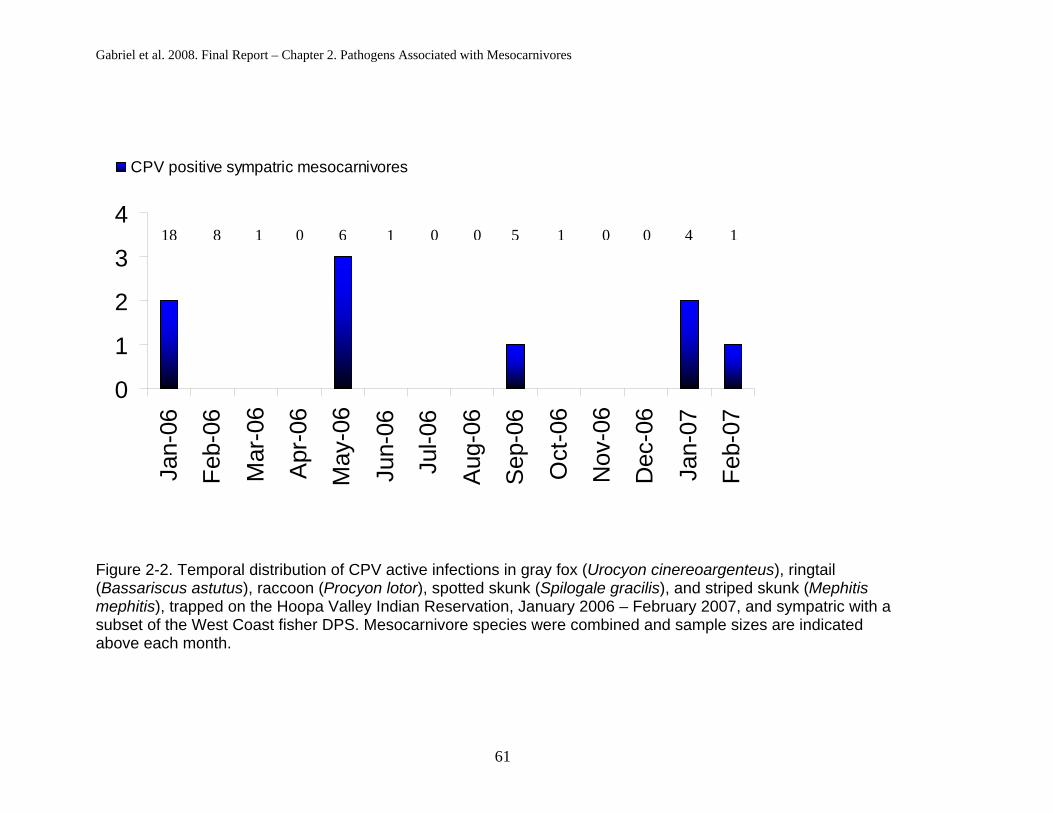
Gabriel et al. 2008. Final Report – Chapter 2. Pathogens Associated with Mesocarnivores
61
0
1
2
3
4
Jan-
06
Feb-
06
Mar
-06
Apr
-06
May
-06
Jun-
06
Jul-0
6
Aug
-06
Sep
-06
Oct
-06
Nov
-06
Dec
-06
Jan-
07
Feb-
07
CPV positive sympatric mesocarnivores
Figure 2-2. Temporal distribution of CPV active infections in gray fox (Urocyon cinereoargenteus), ringtail (Bassariscus astutus), raccoon (Procyon lotor), spotted skunk (Spilogale gracilis), and striped skunk (Mephitismephitis), trapped on the Hoopa Valley Indian Reservation, January 2006 – February 2007, and sympatric with a subset of the West Coast fisher DPS. Mesocarnivore species were combined and sample sizes are indicated above each month.
18 8 1 0 6 1 0 50 1 0 0 4 1

Gabriel et al. 2008. Final Report – Chapter 2. Pathogens Associated with Mesocarnivores
62
Comparison of Disease Exposures Between Fishers and Other Mesocarnivores
Comparison of pathogenic exposure between fishers and the group of
sympatric mesocarnivores yielded few significant differences (Table 2-2). Fisher
exposure to A. phagocytophilum was greater than in the other mesocarnivores
when all mesocarnivores were considered, and also when only backcountry
mesocarnivores were compared (Table 2-2). For all other pathogens, there was
no significant difference in prevalence between fishers and other mesocarnivores
(Table 2-2).
Fishers did not exhibit greater prevalence for active infections of either A.
phagocytophilum or CPV than other mesocarnivores; this was true when all
mesocarnivores were grouped together and also when only backcountry
mesocarnivores were considered (Table 2-3). Active infection with CPV varied
throughout the sampling period for both fishers and sympatric mesocarnivores
(Figure 2-3).
Spatial clustering was apparent for only two pathogen-host groupings.
Specifically, two individuals exposed to CDV, one fisher and one ringtail were
sampled within 500m of each other on the east side of the Trinity River
(Appendix A). All mesocarnivores other than fishers from which CPV DNA was
amplified were captured either in the valley, or in a localized area in the
southeast corner of HVIR; however, PCR-positive fishers were caught throughout
the HVIR (Appendix A).

Gabriel et al. 2008. Final Report – Chapter 2. Pathogens Associated with Mesocarnivores
63
Table 2-2. Comparison of pathogen exposure determined from plasma samples from fisher (Martes pennanti) and the group of sympatric mesocarnivores including gray fox (Urocyon cinereoargenteus), ringtail (Bassariscus astutus), raccoon (Procyon lotor), spotted skunk (Spilogale gracilis), and striped skunk (Mephitis mephitis), trapped on the Hoopa Valley Indian Reservation, February 2004 – February 2007. Pathogens evaluated include canine parvovirus (CPV), canine distemper virus (CDV), canine herpes virus (CHV), canine adenovirus (CAV2), West Nile virus (WNV), Anaplasma phagocytophilum (Ap) and Toxoplasma gondii (Tg). Comparisons were made between fishers and only the mesocarnivores trapped in the backcountry, and also between fishers and all mesocarnivores, regardless of capture location.
Fisher BackcountryMesocarnivores Fisher All
Mesocarnivores
Pathogen NNo. (%) Positive N
No. (%) Positive P N
No. (%) Positive N
No. (%) Positive P
CDV 98 5 (5%) 41 1 (2%) 0.67 98 5 (5%) 64 1 (2%) 0.40
CPV 90 28 (31%) 41 10 (24%) 0.53 90 28 (31%) 64 19 (30%) 0.99
CHV 96 5 (5%) 41 0 (0%) 0.32 96 5 (5%) 64 0 (0%) 0.16
CAV2 95 4 (4%) 41 3 (7%) 0.43 95 4 (4%) 64 6 (9%) 0.20
WNV 99 4 (4%) 41 4 (10%) 0.23 99 4 (4%) 64 4 (6%) 0.71
Ap 79 60 (76%) 41 21 (51%) 0.008* 60 79 (76%) 64 32 (50%) 0.002*
Tg 77 45 (58%) 11 4 (36%) 0.20 77 45 (58%) 20 8 (40%) 0.21
* indicates statistical significance

Gabriel et al. 2008. Final Report – Chapter 2. Pathogens Associated with Mesocarnivores
64
Table 2-3. Comparison of polymerase chain reaction amplification (PCR) denoting active infection of canine parvovirus (CPV) and Anaplasma phagocytophilum (AP) determined from fecal samples from fisher (Martes pennanti) and the group of sympatric mesocarnivores including gray fox (Urocyon cinereoargenteus), ringtail (Bassariscus astutus), raccoon (Procyon lotor), spotted skunk (Spilogale gracilis), and striped skunk (Mephitis mephitis), trapped on the Hoopa Valley Indian Reservation, February 2006 – February 2007. Comparisons were made between fishers and only the mesocarnivores trapped in the backcountry, and also between fishers and all mesocarnivores, regardless of capture location.
Fisher BackcountryMesocarnivores Fisher All
Mesocarnivores
Pathogen NNo. (%) Positive N
No. (%) Positive P N
No. (%) Positive N
No. (%) Positive P
CPV 98 18 (18%) 32 4 (13%) 0.59 98 18 (18%) 47 9 (19%) 0.99
AP 78 3 (4%) 11 0 (0%) 0.99 78 3 (4%) 20 3 (15%) 0.10

Gabriel et al. 2008. Final Report – Chapter 2. Pathogens Associated with Mesocarnivores
65
0
1
2
3
4
5
6
7
8Ja
n-06
Feb-
06
Mar
-06
Apr
-06
May
-06
Jun-
06
Jul-0
6
Aug
-06
Sep
-06
Oct
-06
Nov
-06
Dec
-06
Jan-
07
Feb-
07
Hoopa FishersSympatric Mesocarnivores
Figure 2-3. Temporal distribution of polymerase chain reaction amplification (PCR) denoting active infection of canine parvovirus (CPV) in fishers (Martes pennanti) versus sympatric mesocarnivores, gray fox (Urocyon cinereoargenteus),ringtail (Bassariscus astutus), raccoon (Procyon lotor), spotted skunk (Spilogale gracilis), and striped skunk (Mephitismephitis), trapped on the Hoopa Valley Indian Reservation, January 2006 – February 2007. Mesocarnivore species were combined and sample sizes are indicated above each month.
4 18 9 8
5 1 12 0
5 6
0 1 0 0 0 0 6 5
17 1
15 0
5 0
1 4
0 1

DISCUSSION
Canine Distemper
Canine distemper is a highly infectious disease, which affects all families of
carnivores (Deem et al. 2000, Williams 2001). The transmission of CDV is primarily via
aerosol or oral, respiratory, or oculonasal exudate fluids from either direct contact or
possibly fomites previously visited by an actively infected individual (Deem et al. 2000,
Williams 2001). CDV has variable incubation and clinical presentation periods that can
range from one week to more than a month (Deem et al. 2000, Williams 2001)
Clinical signs of CDV are dependent on various abiotic and biotic factors which
include strain type, environmental conditions, host species, immune status and age
(Deem et al. 2000, Williams 2001). CDV causes 30-50% mortality among domestic
canids, with 50%-70% of the individuals that survive serving as asymptomatic carriers
(Deem et al. 2000). Gray foxes have been shown to be highly susceptible to CDV and
develop clinical manifestations which include fever, vomiting, diarrhea, incoordination,
oculonasal exudates, lack of fear from predators, tremors, ataxia, and depression
(Deem et al. 2000, Williams 2001). It has been demonstrated that CDV is a significant
mortality factor among some gray fox populations (Davidson et al. 1992b, Kelly and
Sleeman 2003). The lack of antibody detection within this gray fox population could be
due to at least two factors: that the sample size was too small to demonstrate exposure
or, perhaps more likely, individual gray foxes rarely recover from CDV infections.
Among raccoons, natural CDV infections typically cause high mortality (Roscoe
1993, Deem et al. 2000, Williams 2001). Our sample size was small, but the lack of
antibody detection within this species leads us to suspect that the same factors
mentioned may be the reason. To date, there has been no documentation of the
66
Gabriel et al. 2008. Final Report – Chapter 2. Pathogens Associated with Mesocarnivores

Gabriel et al. 2008. Final Report – Chapter 2. Pathogens Associated with Mesocarnivores
67
exposure rates of CDV in spotted skunks, though striped skunks are suspected to be
relatively resistant to CDV (Diters and Nielsen 1978).
Six mesocarnivores sampled during this study (five fishers and one ringtail) had
antibodies to CDV, and this is the first documented report of CDV exposure in a ringtail.
Assuming that ringtails have similar clinical manifestations as other procyonids, CDV
may pose a threat to ringtail population health and other community species.
Highly susceptible mesocarnivores, including gray foxes, raccoons and possibly
ringtails, may pose a spillover threat to vulnerable fisher populations by increasing the
likelihood of transmission to fishers. Such risks are magnified if clinically ill animals
manifest neural disease that reduce fear of predators (potentially fishers) (Gulland et al.
1995). It is interesting to note that the only two individuals on the east side of the Trinity
River that were seropositive for CDV were trapped in close proximity to each other.
Though no conclusions can be drawn from this occurrence, it does indicate a possible
pattern to be investigated further for this directly transmitted pathogen.
Canine parvovirus
Canine parvovirus has been reported in numerous wild and domestic animals in
North America since its emergence in the late 1970’s (Kelly 1978, Steinel et al. 2001,
Mccaw and Hoskins 2006). Antigenically and genetically-related parvoviruses include
feline panleukopenia virus, canine parvovirus, mink enteritis virus, raccoon parvovirus,
raccoon dog parvovirus, and blue fox parvovirus; all of which have genomic
conservation of >98% (Berns et al. 2000).
Serology is inadequate to determine which parvovirus infected these animals
because strain-specific identification of the infecting parvovirus would require virus

Gabriel et al. 2008. Final Report – Chapter 2. Pathogens Associated with Mesocarnivores
68
isolation and or sequencing of viral DNA from infected hosts. However, sequencing of
four sympatric mesocarnivore CPV-positive fecal DNA samples from three different
species (gray fox, raccoon, spotted skunk) matched CPV2 via BLAST and GenBank
(Accession number AY742936). These sequences also matched the amplified canine
parvovirus DNA from six PCR-positive fisher scats confirming all amplified parvoviral
DNA was from CPV.
The disease caused by CPV manifests by lysis (cell rupture and death) of
multiplying cells (Parrish 1995, Steinel et al. 2001, Mccaw and Hoskins 2006). Disease
symptoms typically include fever, hemorrhagic diarrhea, vomiting, immunosuppression,
and the reduction of white blood cells (Parrish 1995, Barker and Parrish 2001, Steinel et
al. 2001, Mccaw and Hoskins 2006). Inflammation and hemorrhaging of the intestinal
epithelium of animals suffering from parvovirus enteritis often allows bacterial leakage
into the blood vascular system, leading to sepsis, and endotoxic shock. The
combination of weakness and dehydration from the diarrhea and endotoxic shock
causes some animals to die acutely before outward signs become apparent (Parrish
1995, Barker and Parrish 2001, Steinel et al. 2001, Mccaw and Hoskins 2006). Animals
that survive acute infections typically have long-lasting and possibly life-long immunity
to CPV (Steinel et al. 2001).
In our study, gray foxes, ringtails, and raccoons showed high seroprevalence for
exposure to a parvovirus. Exposure of gray foxes, ringtails and raccoons to
parvoviruses has been shown previously, and gray foxes and raccoons can develop
severe clinical signs that reduce the fitness of infected individuals (Barker et al. 1983,
Davidson et al. 1992a, Garcelon et al. 1992, Riley et al. 2004, Suzan and Ceballos
2005, Clifford et al. 2006). Spotted skunks also develop antibody titers to CPV (Suzan

Gabriel et al. 2008. Final Report – Chapter 2. Pathogens Associated with Mesocarnivores
69
and Ceballos 2005), suggesting that our negative serological results may result from
small sample sizes. Striped skunks have been shown to develop low antibody titers
without showing clinical signs, and infected striped skunks have not been shown to
shed CPV in their feces (Barker et al. 1983, Barker and Parrish 2001).
Parvoviruses are resilient and can persist in the environment for months or even
years under various conditions (Barker and Parrish 2001, Mccaw and Hoskins 2006).
Nineteen percent of sympatric mesocarnivores at Hoopa were actively shedding CPV in
their feces; suggesting that non-target captures in a fisher project may pose
contamination risk to other species via contaminated handling material (Brown et al.
2006). Additionally, species sympatric with threatened species may serve as possible
sources of spillover infections through feces left at latrines or marking sites, or even
when preyed upon and their contaminated intestinal contents are consumed (Woodroffe
1999, Barker and Parrish 2001) .
Canine Herpesvirus
CHV causes a number of ailments in juvenile canids such as respiratory disease,
inappetence, abdominal pain and possibly death, but within older domestic dogs the
symptoms tend to be mild (Gaskell and Willoughby 1999). This virus not only infects
canids but has been documented to infect skunks and mink (Mustela vison) (Diters and
Nielsen 1978, Mcdonald and Lariviere 2001). None of the mesocarnivores captured at
Hoopa had significant antibodies to CHV which again may result from small sample
sizes.

Gabriel et al. 2008. Final Report – Chapter 2. Pathogens Associated with Mesocarnivores
70
Canine Adenovirus-2
Adenovirises cause infectious hepatitis in domestic dogs, gray foxes, skunks
and raccoons (Garcelon et al. 1992, Woods 2001). The clinical signs of infection from
CAV2 include hepatitis, encephalitis, oculonasal discharge, vomiting, mucoid or bloody
diarrhea, paralysis, and death with a short clinical course or even sudden death (Woods
2001). This virus is highly contagious and can be shed via urine, feces, ocular, and
nasal discharges from relatively healthy animals and can be transmitted via direct
contact or fomites such as trapping and handling equipment (Woods 2001). Though no
skunks or raccoons were seropositive for CAV2, striped skunks and raccoons have
been shown to contract the virus and develop clinical manifestations to CAV2 infections,
often resulting in death (Jamison et al. 1973, Karstad et al. 1975, Hamir et al. 1995).
West Nile Virus
The hosts of WNV include a wide range of species of birds and mammals (Marra
et al. 2004). Clinical symptoms and signs of disease in people, horses, and birds
include fever, muscle aches, lethargy, inappetence, diarrhea, vomiting, muscle stiffness,
abdominal pain, blindness, head tremors, cardiac arrhythmias, coma, and death
(Centers-for-Disease-Control-and-Prevention 2000, Marra et al. 2004). No information
is available on the clinical manifestations in mammalian wildlife populations.
Striped skunks and raccoons are among the few species we tested for these
diseases that have previously been documented to show WNV disease or exposure
(Centers-for-Disease-Control-and-Prevention 2000, Dietrich et al. 2005, Bentler et al.
2007). This is the first report of exposure of gray foxes, ringtails and spotted skunks to
WNV. Exposure to WNV as well as clinical disease have been demonstrated in canine

Gabriel et al. 2008. Final Report – Chapter 2. Pathogens Associated with Mesocarnivores
71
species, including wolves (Canis lupus) (Lichtensteiger et al. 2003, Lanthier et al. 2004),
coyotes (Bischof and Rogers 2005) and domestic dogs (Blackburn et al. 1989,
Lichtensteiger et al. 2003).
WNV is a vector-borne disease that is transmitted in North America by many
species of mosquitoes (Marra et al. 2004). There is also evidence that carnivores can
be exposed to WNV through the consumption of infected prey (Austgen et al. 2004,
Kuno 2001). Therefore, prevention and management of WNV infections would be
difficult. Additionally, potentially synergistic effects of WNV infections simultaneous with
infections of other pathogens are unknown.
Anaplasma phagocytophilum
The area surrounding Hoopa has been shown to exhibit high levels of
seroprevalence for A. phagocytophilum in its wildlife and domestic animal populations
(Brown et al. 2003, Foley et al. 2004, Drazenovich et al. 2006, Gabriel 2006, Foley et al.
2007, Gabriel 2006). Domestic canids in nearby towns had a 50% seroprevalence
while 52% of 54 gray foxes on the HVIR were shown to have been exposed to A.
phagocytophilum in previous studies (Foley 2007, Gabriel et al. 2006) Clinical
manifestations of granulocytic anaplasmosis in horses, cats, and dogs include fever,
ataxia, ventral limb edema, icterus, and thrombocytopenia, all which facilitate
immunosuppresion (Madigan and Gribble 1987, Foley et al. 2001, Foley et al. 2004).
Anaplasma phagocytophilum is transmitted to domestic animals and wildlife by ticks of
the family Ixodidae (Richter et al. 1996, Kramer et al. 1999, Foley et al. 2004). Known
tick vectors include Ixodes pacificus (the western black-legged tick) in western North
America (Richter et al. 1996, Nicholson 1998, Brown et al. 2005).

Gabriel et al. 2008. Final Report – Chapter 2. Pathogens Associated with Mesocarnivores
72
Gray foxes had levels of exposure to A. phagocytophilum similar to levels found
during previous studies at Hoopa (Gabriel et al. 2006). This level of exposure indicates
that the risk of contracting this tick-borne disease is high within the region, and also
suggests that the related immunosuppression caused by A. phagocytophilum might
exacerbate risks associated with coinfecting pathogens. Fishers showed a higher
prevalence of A. phagocytophilum than the other mesocarnivores, even when only
backcountry individuals were considered. This trend is not surprising since Gabriel et al.
(2006) found backcountry foxes more likely to be A. phagocytophilum-positive than
valley foxes. Prevalence of A. phagocytophilum may be greater in fishers due to their
occurrence primarily within the backcountry.
Toxoplasma gondii
Toxoplasma gondii is a widespread zoonotic protozoan that parasitizes
mesocarnivores with a range of outcomes that can result in asymptomatic infections or
lameness, encephalitis with uncoordinated movements and head and body tremors,
hepatitis, pneumonia, anorexia, blindness and death; importantly, T. gondii is also
infamous for causing abortions, still births, and neonatal deaths of congenitally infected
mustelid offspring (Dubey and Odening 2001, Burns et al. 2003, Jones et al. 2006).
Oocysts of T. gondii are shed from infective felids, the only known definitive host for this
parasite (Dubey and Odening 2001). Intermediate hosts acquire infections by either
exposure to infective oocysts from felid feces or by consumption of prey items that
contain T. gondii cysts in their tissues (Dubey and Odening 2001, Burns et al. 2003). All
of the mesocarnivores sampled at Hoopa have been shown to serve as intermediate

Gabriel et al. 2008. Final Report – Chapter 2. Pathogens Associated with Mesocarnivores
73
hosts (Dubey and Odening 2001, Suzan and Ceballos 2005), but the development of
clinical disease within these species is not fully understood.
Pathogenicity of T. gondii has been linked to the four genotypes that have been
documented (Dubey and Odening 2001, Miller et al. 2004). Sea otters have
demonstrated high mortality when infected with both genotypes II and X of T. gondii
(Miller et al. 2004). To date it is not known which genotype of T. gondii is present in
Hoopa or whether the T. gondii infections have a negative impact on fisher populations
as they do in some other mustelid populations.
MANAGEMENT AND CONSERVATION IMPLICATIONS
Many of the pathogens assessed in this study are contracted via direct contact or
from contaminated inanimate sources (fomites). These fomites could include trapping
and handling equipment used by those of us studying and managing wildlife. Therefore,
we recommend that researchers and managers disinfect all handling and trapping
equipment between captures with an approved virocide and bacteriocide when
conducting research on these mesocarnivore species.
Although there have been repeated recommendations in the scientific literature
to monitor and gather baseline data of disease risk of threatened species or their
sympatric community before health crises occur (Woodroffe 1999, Mathews et al. 2006),
this is the first proactive demonstration to determine if sympatric species could possibly
be utilized as sentinels of pathogenic risk to a species of concern in California. Our
findings suggest that the sympatric species observed in this study have the same
pathogenic exposure rates as fishers with the exception of A. phagocytophilum.

Gabriel et al. 2008. Final Report – Chapter 2. Pathogens Associated with Mesocarnivores
74
This study documents potential disease risks from sympatric mesocarnivores that
may provide a reservoir for spillover and suppression of fisher populations. Wildlife
managers considering the option of reintroduction may opt to screen the community of
mesocarnivores at the release site in order to avoid releasing naïve animals into a high
disease risk area. This precaution may reduce the likelihood of disease outbreaks in the
newly released population, and increase the possibility of successful colonization and
establishment of the translocated population (Mathews et al. 2006). We emphasize the
potential benefits of monitoring carnivore species sympatric with fishers in Northern
California in all assessments of risks from disease.
ACKNOWLEDGEMENTS FROM AUTHORS OF BOTH CHAPTERS
The authors thank the USFWS for supporting this research through grant
#813335G021 to Integral Ecology Research Center. We thank MGW Biological for
providing additional funding and support. We thank the Hoopa Valley Tribe and the
Wildlife Conservation Society for facilitating all aspects of this work. We thank Steven
Self (Sierra Pacific Industries, Redding, California), Richard Callas and Pete Figura
(California Department of Fish and Game, Redding, California) for supplying samples
collaboratively from the fisher populations near Redding, and Mark Jordan (University
of California, Berkeley) for supplying fecal samples from fishers from the southern
Sierras. Thanks also to Nikki Drazenovich at the University of California, Davis,
Center for Vector-borne Diseases, for developing the appropriate assays.

LITERATURE CITED
Addison, E. M., I. K. Barker. and D. B. Hunter. 1999. Diseases and parasites of furbearers. In Wild furbearer management and conservation in North America (CD-ROM version), M. NOVAK, J. A. BAKER, M. E. OBBARD, AND B. MALLOCH, (eds.). Ministry of Natural Resources, Peterborough, Ontario, Canada. pp. 893-909
Altschul, S. F., W. Gish, W. Miller, E. W. Myers and D. J. Lipman. 1990. Basic local alignment search tool. Journal of Molecular Biology 215: 403-410.
Apperson, C. S., H. K. Hassan, B. A. Harrison, H. M. Savage, S. E. Aspen, A. Farajollahi, W. Crans, T. J. Daniels, R. C. Falco, M. Benedict, M. Anderson, L. Mcmillen. and T. R. Unnasch. 2004. Host feeding patterns of established and potential mosquito vectors of West Nile virus in the eastern United States. Vector-Borne and Zoonotic Diseases 4: 71-82.
Austgen, L. E., R. A. Bowen, M. L. Bunning, B. S. Davis, C. J. Mitchell. and G. J. Chang. 2004. Experimental infection of cats and dogs with West Nile virus. Emerging Infectious Diseases 10: 82-86.
Barker, I. K., R. C. Povey and D. R. Voigt. 1983. Response of mink, skunk, red fox and raccoon to inoculation with mink virus enteritis, feline panleukopenia and canine parvovirus and prevalence of antibody to parvovirus in wild carnivores in Ontario. Canadian Journal of Comparative Medicine 47: 188-197.
Barker, I. and C. R. Parrish. 2001. Parvovirus infections. In Infectious diseases of wild mammals, E. T. THORNE, E. S. WILLIAMS, AND I. K. BARKER, (eds.). Iowa State University Press, Ames, Iowa. pp. 131-147.
Beldomenico, P. M., B. B. Chomel, J. E. Foley, B. N. Sacks, C. J. Baldi, R. W. Kasten. and I. A. Gardner. 2005. Environmental factors associated with Bartonellavinsonii subsp berkhoffii seropositivity in free-ranging coyotes from northern California. Vector-Borne and Zoonotic Diseases 5: 110-119.
Bentler, K. T., J. S. Hall, J. J. Root, K. Klenk, B. Schmit, B. F. Blackwell, P. C. Ramey and L. Clark. 2007. Serologic evidence of West Nile virus exposure in North American mesopredators. American Journal of Tropical Medicine and Hygiene 76: 173-179.
Berns, K. I., M. Bergoin, M. Bloom, M. Lederman, N. Muzyczka, G. Siegl, J. Tal and P. Tattersall. 2000. Parvoviridae.In Virus taxonomy, classification and nomenclature of viruses Academic Press, Inc. , San Diego, Calif. pp. 311–323.
75

Literature Cited
76
Bischof, R. and D. G. Rogers. 2005. Serologic Survey of Select Infectious Diseases in Coyotes and Raccoons in Nebraska. Journal of Wildlife Diseases 41: 787-791.
Blackburn, N. K., F. Reyers, W. L. Berry and A. J. Shepherd. 1989. Susceptibility of dogs to West Nile virus: a survey and pathogenicity trial. Journal of Comparative pathology 100: 59-66.
Brown, R. N.. and E. C. Burgess. 2001. Lyme borreliosis.In Infectious diseases of wild mammals, E. T. THORNE, E. S. WILLIAMS, AND I. K. BARKER, (eds.). Iowa State Press, Ames. pp. 435-454.
Brown, R. N., J. L. Sajecki, J. M. Higley and J. E. Foley. 2003. Exposure of American black bears to infectious disease agents in a managed Douglas-fir forest. Northwestern Naturalist 84: 95-96.
Brown, R. N., R. S. Lane and D. T. Dennis. 2005. Geographic distributions of tick-borne diseases and their vectors. In Tick-Borne Diseases of Humans, J. GOODMAN, D. T. DENNIS, AND D. SONENSHINE, (eds.). ASM Press, Washington, D.C. pp. 363-391.
Brown, R.N., M.W. Gabriel, G.M. Wengert., S. Matthews, J.M. Higley, and J.E. Foley. 2006. Fecally Transmitted Viruses Associated with Pacific Fishers (Martespennanti) in Northwestern California. Transactions Western Section Wildlife Society.42: 40-46.
Brown, R. N., M. A. Peot. and R. S. Lane. 2006. Sylvatic maintenance of Borreliaburgdorferi (Spirochaetales) in northern California: untangling the web of transmission. Journal of Medical Entomology 43: 743-751.
Burridge, M. J., W. J. Bigler, D. J. Forrester. and J. M. Hennemann. 1979. Serologic survey for Toxoplasma gondii in wild animals in Florida. Journal of the American Veterinary Medical Association 175: 964-967.
Burns, R., E. S. Williams, D. O'toole and J. P. Dubey. 2003. Toxoplasma gondii infections in captive black-footed ferrets (Mustela nigripes), 1992-1998: clinical signs, serology, pathology, and prevention. Journal of Wildlife Diseases 39: 787-797.
Cabasso, V. J. 1981. Infectious canine hepatitis. In Infectious diseases of wild mammals, J. W. DAVIS, L. H. KARSTAD, AND D. O. TRAINER, (eds.). Iowa State University Press, Ames, Iowa. pp. 191-195
Centers for Disease Control and Prevention. 2000. West Nile virus activity--eastern United States, 2000. Journal of the American Medical Association 284: 3119-3120.

Literature Cited
77
Centers for Disease Control and Prevention. 2003. West Nile virus activity--United States, November 13-19, 2003. Morbidity and Mortality Weekly Report 52: 1132.
Centers for Disease Control and Prevention. 2005. West Nile virus activity--United States, January 1-December 1, 2005. Morbidity and Mortality Weekly Report 54: 1253-1256.
Chang, C., K. Yamamoto, B. B. Chomel, R. W. Kasten, D. C. Simpson, C. R. Smith. and V. L. Kramer. 1999. Seroepidemiology of Bartonella vinsonii subsp. berkhoffiiinfection in California coyotes, 1994-1998. Emerging Infectious Diseases 5: 711-715.
Chang, C. C., R. W. Kasten, B. B. Chomel, D. C. Simpson, C. M. Hew, D. L. Kordick, R. Heller, Y. Piemont. and E. B. Breitschwerdt. 2000. Coyotes (Canis latrans) as the reservoir for a human pathogenic Bartonella sp.: molecular epidemiology of Bartonella vinsonii subsp. berkhoffii infection in coyotes from central coastal California. Journal of Clinical Microbiology 38: 4193-4200.
Charlton, K. M., G. C. Dulac, F. C. Thomas. and H. K. Mitchell. 1977. Necrotizing encephalitis in skunks caused by Herpes simplex virus. Canadian Journal of Comparative Medicine 41: 460-465.
Chen, C. C., S. Louie, B. Mccormick, W. A. Walker. and H. N. Shi. 2005. Concurrent infection with an intestinal helminth parasite impairs host resistance to enteric Citrobacter rodentium and enhances Citrobacter-induced colitis in mice. Infection and Immunity 73: 5468-5481.
Cheung, R. C., S. M. Matsui and H. B. Greenberg. 1994. Rapid and sensitive method for detection of hepatitis C virus RNA by using silica particles. Journal of Clinical Microbiology 32: 2593-2597.
Cho, J. G., S. A. Dee, J. Deen, A. Guedes, C. Trincado, E. Fano, Y. Jiang, K. Faaberg, J. E. Collins, M. P. Murtaugh. and H. S. Joo. 2006. Evaluation of the effects of animal age, concurrent bacterial infection, and pathogenicity of porcine reproductive and respiratory syndrome virus on virus concentration in pigs. American Journal of Veterinary Research 67: 489-493.
Chomel, B. B., M. T. Jay, C. R. Smith, P. H. Kass, C. P. Ryan. and L. R. Barrett. 1994. Serological surveillance of plague in dogs and cats, California, 1979-1991. Comparative Immunology, Microbiology, and Infectious Diseases 17: 111-123.
Chomel, B. B., R. L. Zarnke, R. W. Kasten, P. H. Kass. and E. Mendes. 1995. Serologic survey of Toxoplasma gondii in grizzly bears (Ursus arctos) and black bears (Ursus americanus), from Alaska, 1988 to 1991. Journal of Wildlife Diseases 31: 472-479.

Literature Cited
78
Cleaveland, S., G. R. Hess, A. P. Dobson, M. K. Laurenson, H. I. Mccallum, M. G. Roberts and R. Woodroffe. 2002. The role of pathogens in biological conservation.In The ecology of wildlife diseases, P. J. HUDSON, A. RIZZOLI, B. T. GRENFELL, H. HEESTERBEEK, AND A. P. DOBSON, (eds.). Oxford University Press, New York, New York. pp. 139-150.
Clifford, D. L., J. A. K. Mazet, E. J. Dubovi, D. K. Garcelon, T. J. Coonan, P. A. Conrad and L. Munson. 2006. Pathogen exposure in endangered island fox (Urocyonlittoralis) populations: implications for conservation management. Biological Conservation 131: 230-243.
Clover, J. R., T. D. Hofstra, B. G. Kuluris, M. T. Schroeder, B. C. Nelson, A. M. Barnes. and R. G. Botzler. 1989. Serologic evidence of Yersinia pestis infection in small mammals and bears from a temperate rainforest of north coastal California. Journal of Wildlife Diseases 25: 52-60.
Cole, R. A., D. S. Lindsay, D. K. Howe, C. L. Roderick, J. P. Dubey, N. J. Thomas. and L. A. Baeten. 2000. Biological and molecular characterizations of Toxoplasma gondii strains obtained from southern sea otters (Enhydra lutris nereis). Journal of Parasitology 86: 526-530.
Conrad, P. A., M. A. Miller, C. Kreuder, E. R. James, J. Mazet, H. Dabritz, D. A. Jessup, F. Gulland. and M. E. Grigg. 2005. Transmission of Toxoplasma: clues from the study of sea otters as sentinels of Toxoplasma gondii flow into the marine environment. International Journal for Parasitology 35: 1155-1168.
Courtenay, O., R. J. Quinnell. and W. S. Chalmers. 2001. Contact rates between wild and domestic canids: no evidence of parvovirus or canine distemper virus in crab-eating foxes. Veterinary Microbiology 81: 9-19.
Cunningham, A. A. 1996. Disease risks of wildlife translocations. Conservation Biology 10: 349-353.
Davidar, P.. and E. S. Morton. 2006. Are multiple infections more severe for purple martins (Progne subis) than single infections? The Auk 123: 141–147.
Davis, A., M. L. Bunning, P. Gordy, N. A. Panella, B. Blitvich. and R. A. Bowen. 2005. Experimental and natural infection of North American bats with West Nile virus. American Journal of Tropical Medicine and Hygiene 73: 467-469.
Davidson, W. R., M. J. Appel, G. L. Doster, O. E. Baker and J. F. Brown. 1992a. Diseases and parasites of red foxes, gray foxes, and coyotes from commercial sources selling to fox-chasing enclosures. Journal of Wildlife Diseases 28: 581-589.
Davidson, W. R., V. F. Nettles, L. E. Hayes, E. W. Howerth and C. E. Couvillion. 1992b. Diseases diagnosed in gray foxes (Urocyon cinereoargenteus) from the

Literature Cited
79
southeastern United States. Journal of Wildlife Diseases 28: 28-33.
Dekrey, G. K.. and R. A. Adams. 2005. West Nile virus infection in wild bats of Colorado. In Annual Meeting of the American Association of Immunologists, San Diego, California. pp.
Deem, S. L., L. H. Spelman, R. A. Yates and R. J. Montali. 2000. Canine distemper in terrestrial carnivores: a review. Journal of Zoo and Wildlife Medicine 31: 441-451.
Deem, S. L., W. B. Karesh and W. Weisman. 2001. Putting theory into practice: wildlife health in conservation. Conservation Biology 15: 1224-1233.
Delahay, R.. and K. Frolich. 2000. Absence of antibodies against canine distemper virus in free-ranging populations of the Eurasian badger in Great Britain. Journal of Wildlife Diseases 36: 576-579.
Del Piero, F., D. W. Stremme, P. L. Habecker. and C. Cantile. 2006. West Nile Flavivirus Polioencephalomyelitis in a harbor seal (Phoca vitulina). Veterinary Pathology 43: 58-61.
Dick, T. A.. and R. D. Leonard. 1979. Helminth parasites of fisher Martes pennanti(Erxleben) from Manitoba, Canada. Journal of Wildlife Diseases 15: 409-412.
Dick, T. A., B. Kingscote, M. A. Strickland. and C. W. Douglas. 1986. Sylvatic trichinosis in Ontario, Canada. Journal of Wildlife Diseases 22: 42-47.
Dietrich, G., J. A. Montenieri, N. A. Panella, S. Langevin, S. E. Lasater, K. Klenk, J. C. Kile and N. Komar. 2005. Serologic evidence of West Nile virus infection in free-ranging mammals, Slidell, Louisiana, 2002. Vector-Borne and Zoonotic Diseases 5: 288-292.
Dietz, H. H., P. Henriksen, M. Lebech. and S. A. Henriksen. 1993. Experimental infection with Toxoplasma gondii in farmed mink (Mustela vison S.). Veterinary Parasitology 47: 1-7.
Diters, R. W. and S. W. Nielsen. 1978. Toxoplasmosis, distemper, and herpesvirus infection in a skunk (Mephitis mephitis). Journal of Wildlife Diseases 14: 132-133.
Douglas, C. W. and M. A. Strickland. 1999. Fisher. In Wild furbearer management and conservation in North America (CD-ROM version), M. NOVAK, J. A. BAKER, M. E. OBBARD, AND B. MALLOCH, (eds.). Ministry of Natural Resources, Peterborough, Ontario. pp. 510-529
Drazenovich, N., J. Foley and R. N. Brown. 2006. Use of real-time quantitative PCR targeting the msp2 protein gene to identify cryptic Anaplasma phagocytophiluminfections in wildlife and domestic animals. Vector Borne and Zoonotic Diseases 6: 83-90.

Literature Cited
80
Dubey, J. P., A. N. Hamir. and C. E. Rupprecht. 1990. Acute disseminated toxoplasmosis in a red fox (Vulpes vulpes). Journal of Wildlife Diseases 26: 286-290.
Dubey, J. P., A. N. Hamir, C. A. Hanlon. and C. E. Rupprecht. 1992. Prevalence of Toxoplasma gondii infection in raccoons. Journal of the American Veterinary Medical Association 200: 534-536.
Dubey, J. P. and K. Odening. 2001. Toxoplasmosis and related infections. In Parasitic Diseases of Wild Mammals, W. M. SAMUEL, M. J. PYBUS, AND A. A. KOCAN, (eds.). Iowa State University Press, Ames. pp. 478-519.
Farajollahi, A., N. A. Panella, P. Carr, W. Crans, K. Burguess. and N. Komar. 2003. Serologic evidence of West Nile virus infection in black bears (Ursus americanus)from New Jersey. Journal of Wildlife Diseases 39: 894-896.
Farajollahi, A., R. Gates, W. Crans. and N. Komar. 2004. Serologic evidence of West Nile virus and St. Louis encephalitis virus infections in white-tailed deer (Odocoileus virginianus) from New Jersey, 2001. Vector-Borne and Zoonotic Diseases 4: 379-383.
Foley, J. E., L. Crawford-Miksza, J. S. Dumler, C. Glaser, J. S. Chae, E. Yeh, D. Schnurr, R. Hood, W. Hunter. and J. E. Madigan. 1999. Human granulocytic ehrlichiosis in Northern California: two case descriptions with genetic analysis of the Ehrlichiae. Clinical Infectious Diseases 29: 388-392.
Foley, J. E., P. Foley and J. E. Madigan. 2001. The distribution of granulocytic ehrlichia seroreactive dogs in California. American Journal of Veterinary Research 62: 1599-1605.
Foley, J. E., P. Foley, R. N. Brown, R. S. Lane, J. S. Dumler. and J. E. Madigan. 2004. Ecology of Anaplasma phagocytophilum and Borrelia burgdorferi in the western United States. Journal of Vector Ecology 29: 41-50.
Foley, J. E., R.N. Brown, M. W. Gabriel, J. Henn, N. Drazenovich, R. Kasten, S. L. Green and B. Chomel. 2007. Spatial epidemiological analysis of tick-borne pathogen exposure in dogs in rural northern California coastal communities. The Veterinary Record 161: 653-657.
Fox, J. G. 1998. Biology and diseases of the ferret. Second edition. Williams and Wilkins, Baltimore, Maryland.
Frank, R. K. 2001. An outbreak of toxoplasmosis in farmed mink (Mustela vison S.). Journal of Veterinary Diagnostic Investigations 13: 245-249.

Literature Cited
81
Furze, R. C., T. Hussell. and M. E. Selkirk. 2006. Amelioration of influenza-induced pathology in mice by coinfection with Trichinella spiralis. Infection and Immunity 74: 1924-1932.
Gabriel, M. W., J. Henn, J. E. Foley, R. N. Brown, R. Kasten, P. Foley. and B. Chomel. In Preparation Zoonotic Bartonella species in fleas collected on gray foxes (Urocyon cinereoargenteus) in Northern California.
Gabriel, M. W. 2006. Exposure to Anaplasma phagocytophilum and ticks in gray foxes (Urocyon cinereoargenteus) in northern Humboldt County, California. M.S. thesis. Humboldt State University, Arcata, California.
Gabriel, M. W. and G. M. Wengert. 2005. Assessment of the modification of stress-causing environmental factors in an improved nest box design for mesocarnivores. In Poster presented at the Annual Meeting of the Western Section of The Wildlife Society, Sacramento, California.
Gage, K. L.. and M. Y. Kosoy. 2005. Natural history of plague: perspectives from more than a century of research. Annual Review of Entomology 50: 505-528.
Gale, K. R., G. Leatch, C. M. Dimmock. and M. G. Gartside. 1997. Increased resistance to Anaplasma marginale infection in cattle chronically infected with Theileria buffeli (syn. T. orientalis). Veterinary Parasitology 69: 187-196.
Garcelon, D. K., R. K. Wayne and B. J. Gonzales. 1992. A serologic survey of the island fox (Urocyon littoralis) on the Channel Islands, California. Journal of Wildlife Diseases 28: 223-229.
Gaskell, R. and K. Willoughby. 1999. Herpesviruses of carnivores. Veterinary Microbiology 69: 73-88.
Gasper, P. W.. and R. P. Watson. 2001. Plague and yersiniosis.In Infectious Diseases of Wild Mammals, I. K. B. E. S. WILLIAMS, (ed.). Iowa State University Press, Ames, Iowa. pp. 313-329
Gaydos, J. K., P. A. Conrad, K. V. Gilardi, G. M. Blundell. and M. Ben-David. 2007. Does human proximity affect antibody prevalence in marine-foraging river otters (Lontra canadensis)? Journal of Wildlife Diseases 43: 116-123.
Gibilisco, C. J. 1994. Distributional dynamics of modern Martes in North America. InMartens, sables, and fishers: biology and conservation, S. W. BUSKIRK, A. S. HARESTAD, M. G. RAPHAEL, AND R. A. POWELL, (eds.). Cornell University Press, Ithaca, New York, USA. pp. 59-71.
Graham, A. L. 2002. When T-helper cells don't help: immunopathology during concomitant infection. Quarterly Review of Biology 77: 409-434.
Literature Cited
82
Greenacre, C. B. 2003. Incidence of adverse events in ferrets vaccinated with distemper or rabies vaccine: 143 cases (1995-2001). Journal of the American Veterinary Medical Association 223: 663-665.
Gulland, F. M. D., B. T. Grenfell and A. P. Dobson. 1995. The impact of infectious diseases on wild animal populations-a review. In Ecology of Infectious Diseases in Natural Populations, B. T. GREENFELL, AND A. P. DOBSON, (eds.). Cambridge University Press, Cambridge. pp. 533.
Hamir, A. N., G. Moser, M. Kao, N. Raju and C. E. Rupprecht. 1995. Herpesvirus-like infection in a raccoon (Procyon lotor). Journal of Wildlife Diseases 31: 420-423.
Hanni, K. D., J. A. Mazet, F. M. Gulland, J. Estes, M. Staedler, M. J. Murray, M. Miller. and D. A. Jessup. 2003. Clinical pathology and assessment of pathogen exposure in southern and Alaskan sea otters. Journal of Wildlife Diseases 39: 837-850.
Heinz-Taheny, K. M., J. J. Andrews, M. J. Kinsel, A. P. Pessier, M. E. Pinkerton, K. Y. Lemberger, R. J. Novak, G. J. Dizikes, E. Edwards. and N. Komar. 2004. West Nile virus infection in free-ranging squirrels in Illinois. Journal of Veterinary Diagnotic Investigation 16: 186-190.
Henn, J. B., M. W. Gabriel, R. W. Kasten, R. N. Brown, J. H. Theis, J. E. Foley. and B. B. Chomel. 2007. Gray foxes (Urocyon cinereoargenteus) as a potential reservoir of a Bartonella clarridgeiae-like bacterium and domestic dogs as sentinels for zoonotic arthropod-borne pathogens in northern California. Journal of Clinical Microbiology In Press.
Henn, J. B., C.-H. Liu, R. W. Kasten, B. A. Vanhorn, L. A. Beckett, P. H. Kass. and B. B. Chomel. 2005. Seroprevalence of antibodies against Bartonella species and evaluation of risk factors and clinical signs associated with seropositivity in dogs. American Journal of Veterinary Research 66: 688-694.
Hill, R. E., Jr., J. J. Zimmerman, R. W. Wills, S. Patton. and W. R. Clark. 1998. Seroprevalence of antibodies against Toxoplasma gondii in free-ranging mammals in Iowa. Journal of Wildlife Diseases 34: 811-815.
Hirasawa, T., T. Kaneshige and K. Mikazuki. 1994. Sensitive detection of canine parvovirus DNA by the nested polymerase chain reaction. Veterinary Microbiology 41: 135-145.
Hoar, B. R., B. B. Chomel, D. L. Rolfe, C. C. Chang, C. L. Fritz, B. N. Sacks. and T. E. Carpenter. 2003. Spatial analysis of Yersinia pestis and Bartonella vinsoniisubsp. berkhoffii seroprevalence in California coyotes (Canis latrans). Preventive Veterinary Medicine 56: 299-311.

Literature Cited
83
Holden, K., J. T. Boothby, S. Anand. and R. F. Massung. 2003. Detection of Borreliaburgdoferi, Ehrlichia chaffeensis, and Anaplasma phagocytophilum in ticks (Acari: Ixodidae) from a coastal region of California. Journal of Clinical Microbiology 40: 534-539.
Holden, K., E. Hodzic, S. Feng, K. J. Freet, R. B. Lefebvre. and S. W. Barthold. 2005. Coinfection with Anaplasma phagocytophilum alters Borrelia burgdorferi population distribution in C3H/HeN mice. Infection and Immunity 73: 3440-3444.
Hofmann-Lehmann, R., M. L. Meli, U. M. Dreher, E. Gonczi, P. Deplazes, U. Braun, M. Engels, J. Schupbach, K. Jorger, R. Thoma, C. Griot, K. D. C. Stark, B. Willi, J. Schmidt, K. M. Kocan. and H. Lutz. 2004. Concurrent infections with vector-borne pathogens associated with fatal hemolytic anemia in a cattle herd in Switzerland. Journal of Clinical Microbiology 42: 3775–3780.
Hudson, P. and J. Greenman. 1998. Competition mediated by parasites: biological and theoretical progress. Trends in Ecology & Evolution 13: 387-390.
Hubalek, Z.. and J. Halouzka. 1999. West Nile fever--a reemerging mosquito-borne viral disease in Europe. Emerging Infectious Diseases 5: 643-650.
Hurkova, L.. and D. Modry. 2006. PCR detection of Neospora caninum, Toxoplasma gondii and Encephalitozoon cuniculi in brains of wild carnivores. Veterinary Parasitology 137: 150-154.
Hutcheson, H. J., C. H. Gorham, C. Machain-Williams, M. A. Lorono-Pino, A. M. James, N. L. Marlenee, B. Winn, B. J. Beaty. and C. D. Blair. 2005. Experimental transmission of West Nile virus (Flaviviridae : Flavivirus) by Carios capensis ticks from North America. Vector-Borne and Zoonotic Diseases 5: 293-295.
Jamison, R. K., E. C. Lazar, L. N. Binn and A. D. Alexander. 1973. Survey for antibodies to canine viruses in selected wild mammals. Journal of Wildlife Diseases 9: 2-3.
Jones, Y. L., S. D. Fitzgerald, J. G. Sikarske, A. Murphy, N. Grosjean and M. Kiupel. 2006. Toxoplasmosis in a Free-ranging Mink. Journal of Wildlife Diseases 42: 865-869.
Karstad, L., R. Ramsden, T. J. Berry and L. N. Binn. 1975. Hepatitis in skunks caused by the virus of infectious canine hepatitis. Journal of Wildlife Diseases 11: 494-496.
Kelly, W. R. 1978. An enteric disease of dogs resembling feline panleucopaenia. Australian Veterinarian Journal 54: 593.
Kelly, T. R. and J. M. Sleeman. 2003. Morbidity and mortality of red foxes (Vulpes vulpes) and gray foxes (Urocyon cinereoargenteus) admitted to the Wildlife Center of Virginia, 1993-2001. Journal of Wildlife Diseases 39: 467-469.

Literature Cited
84
Kidd, L., B. Hegarty, D. Sexton and E. Breitschwerdt. 2006. Molecular characterization of Rickettsia rickettsii infecting dogs and people in North Carolina. Annals of the New York Academy of Sciences 1078:400-409.
Kile, J. C., N. A. Panella, N. Komar, C. C. Chow, A. Macneil, B. Robbins. and M. L. Bunning. 2005. Serologic survey of cats and dogs during an epidemic of West Nile virus infection in humans. Journal of the American Veterinary Medical Association 226: 1349-1353.
Kimber, K. R., G. V. Kollias. and E. J. Dubovi. 2000. Serologic survey of selected viral agents in recently captured wild North American river otters (Lontra canadensis).Journal of Zoo and Wildlife Medicine 31: 168-175.
Komar, N., N. A. Panella. and E. Boyce. 2001. Exposure of domestic mammals to West Nile virus during an outbreak of human encephalitis, New York City, 1999. Emerging Infectious Diseases 7: 736-738.
Kramer, V. L., M. P. Randolph, L. T. Hui, W. E. Irwin, A. G. Gutierrez and D. J. Vugia. 1999. Detection of the agents of human ehrlichioses in ixodid ticks from California. American Journal of Tropical Medicine and Hygiene 60: 62-65.
Kuno, G. 2001. Transmission of arboviruses without involvement of arthropod vectors. Acta Virologica 45: 139-150.
Lane, R. S., R. W. Emmons, V. Devlin, D. V. Dondero. and B. C. Nelson. 1982. Survey for evidence of Colorado tick fever virus outside of the known endemic area in California. Am. J. Trop. Med. Hyg. 31: 837-843.
Lane, R. S., J. E. Foley, L. Eisen, E. T. Lennette. and M. A. Peot. 2001. Acarologic risk of exposure to emerging tick-borne bacterial pathogens in a semirural community in northern California. Vector-Borne and Zoonotic Diseases 1: 197-210.
Lane, R. S., D. B. Steinlein. and J. Mun. 2004. Human behaviors elevating exposure to Ixodes pacificus (Acari: Ixodidae) nymphs and their associated bacterial zoonotic agents in a hardwood forest. Journal of Medical Entomology 41: 239-248.
Langlois, I. 2005. Viral diseases of ferrets. Veterinary Clinics of North America: Exotic Animal Practice 8: 139-160.
Lanthier, I., M. Hebert, D. Tremblay, J. Harel, A. D. Dallaire and C. Girard. 2004. Natural West Nile virus infection in a captive juvenile Arctic wolf (Canis lupus). Journal of Veterinary Diagnostic Investigation 16: 326-329.
Lawrie, C. H., N. Y. Uzcategui, E. A. Gould. and P. A. Nuttall. 2004. Ixodid and argasid tick species and West Nile virus. Emerging Infectious Diseases 10: 653-657.
Literature Cited
85
Leighton, F. A. 2002. Health risk assessment of the translocation of wild animals. Review of Science and Technology 21: 187-195.
Lichtensteiger, C. A., K. Heinz-Taheny, T. S. Osborne, R. J. Novak, B. A. Lewis and M. L. Firth. 2003. West Nile virus encephalitis and myocarditis in wolf and dog. Emerging Infectious Diseases 9: 1303-1306.
Lively, C. M. 2005. Evolution of virulence: coinfection and propagule production in spore-producing parasites. Evolutionary Biology 5: 64.
Ludwig, G. V., P. P. Calle, J. A. Mangiafico, B. L. Raphael, D. K. Danner, J. A. Hile, T. L. Clippinger, J. F. Smith, R. A. Cook. and T. Mcnamara. 2002. An outbreak of West Nile virus in a New York City captive wildlife population. American Journal of Tropical Medicine and Hygiene 67: 67-75.
Macdonald, D. W., C. D. Buesching, P. Stopka, J. Henderson, S. A. Ellwood and S. E. Baker. 2004a. Encounters between two sympatric carnivores: red foxes (Vulpes vulpes) and European badgers (Meles meles). Journal of Zoology 263: 385-392.
Macdonald, K. A., B. B. Chomel, M. D. Kittleson, R. W. Kasten, W. P. Thomas. and P. Pesavento. 2004b. A prospective study of canine infective endocarditis in Northern California (1999-2001): emergence of Bartonella as a prevalent etiologic agent. Journal of Veterinary Internal Medicine 18: 56-64.
Madi , J., D. Huber. and B. Lugovic. 1993. Serologic survey for selected viral and rickettsial agents of brown bears (Ursus arctos) in Croatia. Journal of Wildlife Diseases 29: 572-576.
Madigan, J. E. and D. Gribble. 1987. Equine ehrlichiosis in northern California: 49 cases (1968-1981). Journal of the American Veterinary Medical Association 190: 445-448.
Marra, P. P., S. Griffing, C. Caffrey, A. M. Kilpatrick, R. G. Mclean, C. Brand, E. M. I. Saito, A. P. Dupuis, L. Kramer and R. J. Novak. 2004. West Nile virus and wildlife. BioScience 54: 393-402.
Mathews, F., D. Moro, R. Strachan, M. Gelling and N. Buller. 2006. Health surveillance in wildlife reintroductions. Biological Conservation 131: 338-347.
Mayer, K. E.. and W. F. Laudenslayer, (eds.) 1988. A guide to wildlife habitats of California. California Department of Forestry and Fire Protection, Sacramento, California. 166 pp.
Mccaw, D. L. and J. D. Hoskins. 2006. Canine viral enteritis. In Infectious diseases of the dog and cat, C. E. GREENE, (ed.). WB Saunders\ Elsevier Science, London,
Literature Cited
86
UK. pp. 1424.
Mcdonald, R. A., M. J. Day. and R. J. Birtles. 2001. Histological evidence of disease in wild stoats (Mustela erminea) in England. Veterinary Record 149: 671-675.
Mcdonald, R. A.. and S. Lariviere. 2001. Diseases and pathogens of Mustela spp., with special reference to the biological control of introduced stoat Mustela ermineapopulations in New Zealand. Journal of the Royal Society of New Zealand 31: 721-744.
Miller, M. A., I. A. Gardner, C. Kreuder, D. M. Paradies, K. R. Worcester, D. A. Jessup, E. Dodd, M. D. Harris, J. A. Ames, A. E. Packham. and P. A. Conrad. 2002. Coastal freshwater runoff is a risk factor for Toxoplasma gondii infection of southern sea otters (Enhydra lutris nereis). International Journal for Parasitology 32: 997-1006.
Miller, M. A., M. E. Grigg, C. Kreuder, E. R. James, A. C. Melli, P. R. Crosbie, D. A. Jessup, J. C. Boothroyd, D. Brownstein and P. A. Conrad. 2004. An unusual genotype of Toxoplasma gondii is common in California sea otters (Enhydra lutris nereis) and is a cause of mortality. International Journal for Parasitology 34: 275-284.
Mitchell, S. M., D. J. Richardson. and D. S. Lindsay. 2006. Prevalence of agglutinating antibodies to Toxoplasma gondii in striped skunks (Mephitis mephitis), opossums (Didelphis virginiana), and raccoons (Procyon lotor) from Connecticut. Journal of Parasitology 92: 664-665.
Mosquera, J.. and F. R. Adler. 1998. Evolution of virulence: a unified framework for coinfection and superinfection. Journal of Theoretical Biology 195: 293-313.
Munson, L. and W. B. Karesh. 2002. Disease monitoring for the conservation of terrestrial animals.In Conservation Medicine: ecological health in practice, R. S. O. A. AGUIRRE, GARY M. TABOR, CAROL HOUSE, MARY C. PEARL (ed.). Oxford University Press US, New York. pp. 95-103.
Murphy, F. A., E. P. Gibbs, M. C. Horzinek. and M. J. Studdert. 1999. Veterinary virology. Third edition. Academic Press, New York, New York.
Nettles, V. F., J. E. Pearson, G. A. Gustafson. and J. L. Blue. 1980. Parvovirus infection in translocated raccoons. Journal of the American Veterinary Medical Association 177: 787-789.
Nicholson, W. 1998. Epidemiology of human granulocytic ehrlichiosis, with special reference to the role of wild rodents and ixodid ticks as natural hosts of Ehrlichiaphagocytophila sensu lato. PhD dissertation. Johns Hopkins University, Baltimore, MD.

Literature Cited
87
Nutter, F. B., J. F. Levine, M. K. Stoskopf, H. R. Gamble. and J. P. Dubey. 1998. Seroprevalence of Toxoplasma gondii and Trichinella spiralis in North Carolina black bears (Ursus americanus). Journal of Parasitology 84: 1048-1050.
Parrish, C. R. 1995. Pathogenesis of feline panleukopenia virus and canine parvovirus. Baillieres Clinical Haematology 8: 57-71.
Petersen, L. R.. and J. T. Roehrig. 2001. West Nile virus: a reemerging global pathogen. Emerging Infectious Diseases 7: 611-614.
Philip, R. N., R. S. Lane. and E. A. Casper. 1981. Serotypes of tick-borne spotted fever group rickettsiae from western California. American Journal of Tropical Medicine and Hygiene 30: 722-727.
Philippa, J. D., F. A. Leighton, P. Y. Daoust, O. Nielsen, M. Pagliarulo, H. Schwantje, T. Shury, R. Van Herwijnen, B. E. Martina, T. Kuiken, M. W. Van De Bildt. and A. D. Osterhaus. 2004. Antibodies to selected pathogens in free-ranging terrestrial carnivores and marine mammals in Canada. Veterinary Record 155: 135-140.
Pilipski, J. D., L. M. Pilipski. and L. S. Risley. 2004. West Nile virus antibodies in bats from New Jersey and New York. Journal of Wildlife Diseases 40: 335-337.
Poole, K. G., G. M. Matson, S. A. Strickland, A. J. Magoun, R. P. Graf. and L. M. Dix. 1994. Age and sex determination for American martens and fishers. In Martens, sables, and fishers: biology and conservation, S. W. BUSKIRK, A. S. HARESTAD, M. G. RAPHAEL, AND R. A. POWELL, (eds.). Cornell University Press, Ithaca, New York. pp. 204-223.
Pusterla, N., C. C. Chang, B. B. Chomel, J. S. Chae, J. E. Foley, E. Derock, V. L. Kramer, H. Lutz. and J. E. Madigan. 2000. Serologic and molecular evidence ofEhrlichia spp. in coyotes in California. Journal of Wildlife Diseases 36: 494-499.
Quinn, P. J., R. O. Ramsden. and D. H. Johnston. 1976. Toxoplasmosis: a serological survey in Ontario wildlife. Journal of Wildlife Diseases 12: 504-510.
Richter, P. J., Jr., R. B. Kimsey, J. E. Madigan, J. E. Barlough, J. S. Dumler and D. L. Brooks. 1996. Ixodes pacificus (Acari: Ixodidae) as a vector of Ehrlichia equi (Rickettsiales: Ehrlichieae). Journal of Medical Entomology 33: 1-5.
Riley, S. P., J. Foley and B. Chomel. 2004. Exposure to feline and canine pathogens in bobcats and gray foxes in urban and rural zones of a national park in California. Journal of Wildlife Diseases 40: 11-22.
Root, J. J., J. S. Hall, R. G. Mclean, N. L. Marlenee, B. J. Beaty, J. Gansowski. and L. Clark. 2005. Serologic evidence of exposure of wild mammals to flaviviruses in

Literature Cited
88
the central and eastern United States. American Journal of Tropical Medicine and Hygiene 72: 622-630.
Roscoe, D. E. 1993. Epizootiology of canine distemper in New Jersey raccoons. Journal of Wildlife Diseases 29: 390-395.
Ruppanner, R., D. A. Jessup, I. Ohishi, D. E. Behymer. and C. E. Franti. 1982. Serologic survey for certain zoonotic diseases in black bears in California. Journal of the American Veterinary Medical Association 181: 1288-1291.
Ruprecht, C., K. Stöhr. and C. Meredith. 2001. Rabies. In Infectious diseases of wild mammals, THORNE, E. S. WILLIAMS, AND I. K. BARKER, (eds.). Iowa State University Press, Ames, Iowa. pp. 3-36.
Scott, M. E. 1988. The impact of infection and disease on animal populations: implications for conservation biology. Conservation Biology 2: 40-56.
Self, S.. and S. J. Kerns. 2001. Pacific fisher use of a managed forest landscape in northern California. Sierra Pacific Industries, Redding, California, 32 pp.
Singer, B. C.. and E. L. Begg. 1975. Soil Survey, Hoopa Valley, California. Agricultural Extension Service of the University of California, Davis, California pp.
Sonenshine, D. E. 1993. Biology of Ticks. Oxford University Press, New York.
Steinel, A., C. R. Parrish, M. E. Bloom and U. Truyen. 2001. Parvovirus infections in wild carnivores. Journal of Wildlife Diseases 37: 594-607.
Suzan, G. and G. Ceballos. 2005. The role of feral mammals on wildlife infectious disease prevalence in two nature reserves within Mexico City limits. Journal of Zoo and Wildlife Medicine 36: 479-484.
Thorne, E. T. and E. S. Williams. 1988. Disease and endangered species: the black-footed ferret as a recent example. Conservation Biology 2: 66-74.
Thornton, R. N. 1990. Toxoplasmosis in ferrets. New Zealand Veterinary Journal 38: 123.
Tizard, I. R., J. B. Billett. and R. O. Ramsden. 1976. The prevalence of antibodies against Toxoplasma gondii in some Ontario mammals. Journal of Wildlife Diseases 12: 322-325.
Tocidlowski, M. E., M. R. Lappin, P. W. Summer. and M. K. Stoskopf. 1997. Serologic survey for toxoplasmosis in river otters. Journal of Wildlife Diseases 33: 649-652.

Literature Cited
89
Turell, M. J., M. R. Sardelis, D. J. Dohm. and M. L. O'guinn. 2001. Potential North American vectors of West Nile virus. Annals of the New York Academy of Sciences 951: 317-324.
United-States Fish and Wildlife-Service. 2004. 50-CFR Part 17 Endangered and threatened wildlife and plants; 12 month findings for a petition to list the west coast distinct population segment of the fisher (Martes pennanti); proposed rule. Federal Register 69: 18770-18792.
Wilbert, J. C. 1992. Spatial scale and seasonality of habitat selections by martins in southeastern Wyoming. Master’s Thesis. University of Wyoming, Laramie, Wyoming.
Williams, E. S., E. T. Thorne, M. J. Appel. and D. W. Belitsky. 1988. Canine distemper in black-footed ferrets (Mustela nigripes) from Wyoming. Journal of Wildlife Diseases 24: 385-398.
Williams, E. S., E. T. Thorne, J. Quan. and S. L. Anderson. 1991. Experimental infection of domestic ferrets (Mustela putorius furo) and Siberian polecats (Mustelaeversmanni) with Yersinia pestis. Journal of Wildlife Diseases 27: 441-445.
Williams, E. S., K. Mills, D. R. Kwiatkowski, E. T. Thorne. and A. Boerger-Fields. 1994. Plague in a black-footed ferret (Mustela nigripes). Journal of Wildlife Diseases 30: 581-585.
Williams, E. S.. and E. T. Thorne. 1996. Infectious and parasitic diseases of captive carnivores, with special emphasis on the black-footed ferret (Mustela nigripes).Reviews of Science and Technology 15: 91-114.
Williams, E. S. 2001. Canine distemper. In Infectious diseases of wild mammals, E. T.THORNE, E. S. WILLIAMS, AND I. K. BARKER, (eds.). Iowa State University Press, Ames, Iowa. pp. 50-59.
Wobeser, G. 2002. Disease management strategies for wildlife. Review of Science and Technology 21: 159-178.
Wobeser, G. A. 2006. How disease is detected, described, and measured. InEssentials of Disease in Wild Animals, G. A. WOBESER, (ed.). Blackwell Publishing, Ames, Iowa. pp. 45-59..
Woodroffe, R. 1999. Managing disease threats to wild mammals. Animal Conservation 2: 185-193.
Woodford, M. H.. and P. B. Rossiter. 1993. Disease risks associated with wildlife translocation projects. Review of Science and Technology 12: 115-135.
Woods, L. W. 2001. Adenoviral diseases. In Infectious diseases of wild mammals, E. T.

Literature Cited
90
THORNE, E. S. WILLIAMS, AND I. K. BARKER, (eds.). Iowa State University Press, Ames, Iowa. pp. 202-212.
Yaeger, J. S. 2005. Habitat at fisher resting sites in the Klamath province of northern California. MS thesis. Humboldt State University, Arcata.
Yamamoto, K., B. B. Chomel, L. J. Lowenstine, Y. Kikuchi, L. G. Phillips, B. C. Barr, P. K. Swift, K. R. Jones, S. P. D. Riley, R. W. Kasten, J. E. Foley. and N. C. Pedersen. 1998. Bartonella henselae antibody prevalence in free-ranging and captive wild felids from California. Journal of Wildlife Diseases 34: 56-63.
Zarnke, R. L., J. P. Dubey, O. C. Kwok. and J. M. Ver Hoef. 2000. Serologic survey for Toxoplasma gondii in selected wildlife species from Alaska. Journal of Wildlife Diseases 36: 219-224.
Zielinski, W. J. 1984. Plague in pine martens and the fleas associated with its occurrence. Great Basin Naturalist 41: 170-175.
Zielinski, W. J., T. E. Kucera. and R. H. Barrett. 1995. Current distribution of the fisher, Martes pennanti, in California. California Fish and Game 81: 104-112.
Zielinski, W. J., N. P. Duncan, E. C. Farmer, R. L. Truex, A. P. Clevenger. and R. H. Barrett. 1999 Diet of fishers (Martes pennanti) at the southernmost extent of their range. Journal of Mammalogy 80: 961-971.
Appendix A
91
Appendix A. Spatial distribution of disease exposures among fishers (Martes pennanti)and sympatric mesocarnivores, gray foxes (Urocyon cinereoargenteus), ringtails (Bassariscus astutus), raccoons (Procyon lotor), striped skunks (Mephitis mephitis), and spotted skunks (Spilogale gracilis) trapped at Hoopa Valley Indian Reservation, February 2004 – February 2007.

Appendix A
92
A-1. Spatial orientation of fishers (Martes pennanti, red triangles) and sympatric mesocarnivores (green triangles) that were captured and sampled for exposure to and active infections of various pathogens at Hoopa Valley Indian Reservation, February 2004 – February 2007.

Appendix A
93
A-2. Spatial orientation of fishers (Martes pennanti, red triangles, n=5) and sympatric mesocarnivores (green triangles, n=1) that were seropositive for canine distemper virus (CDV) at Hoopa Valley Indian Reservation, February 2004 – February 2007.

Appendix A
94
A-3. Spatial orientation of fishers (Martes pennanti, red triangles, n=28) and sympatric mesocarnivores (green triangles, n=19) that were seropositive for canine parvovirus (CPV) at Hoopa Valley Indian Reservation, February 2004 – February 2007.

Appendix A
95
A-4. Spatial orientation of fishers (Martes pennanti, red triangles, n=5) and sympatric mesocarnivores (green triangles, n=0) that were seropositive for canine herpesvirus (CHV) at Hoopa Valley Indian Reservation, February 2004 – February 2007.

Appendix A
96
A-5. Spatial orientation of fishers (Martes pennanti, red triangles, n=4) and sympatric mesocarnivores (green triangles, n=6) that were seropositive for canine adenovirus (CAV2) at Hoopa Valley Indian Reservation, February 2004 – February 2007.

Appendix A
97
A-5. Spatial orientation of fishers (Martes pennanti, red triangles, n=4) and sympatric mesocarnivores (green triangles, n=4) that were seropositive for West Nile virus (WNV) at Hoopa Valley Indian Reservation, February 2004 – February 2007.

Appendix A
98
A-6. Spatial orientation of fishers (Martes pennanti, red triangles, n=78) and sympatric mesocarnivores (green triangles, n=32) that were seropositive for Anaplasma phagocytophilum at Hoopa Valley Indian Reservation, February 2004 – February 2007.

Appendix A
99
A-7. Spatial orientation of fishers (Martes pennanti, red triangles, n=45) and sympatric mesocarnivores (green triangles, n=8) that were seropositive for Toxoplasma gondii at Hoopa Valley Indian Reservation, February 2004 – February 2007.

Appendix A
100
A-8. Spatial orientation of fishers (Martes pennanti, red triangles, n=18) and sympatric mesocarnivores (green triangles, n=9) that showed nested polymerase chain reaction active infection of canine parvovirus (CPV) at Hoopa Valley Indian Reservation, February 2004 – February 2007.