Pathogenesis of Post-traumatic Ankylosis of the TMJ
5
British Journal of Oral and Maxillofacial Surgery 50 (2012) 8–12 Available online at www.sciencedirect.com Leading article Pathogenesis of post-traumatic ankylosis of the temporomandibular joint: a critical review Gururaj Arakeri a,∗ , Atul Kusanale b , Graeme A. Zaki c , Peter A. Brennan c a Department of Oral and Maxillofacial Surgery, Sri Sai College of Dental Surgery, Vikarabad, Andhra Pradesh, India b Oral and Maxillofacial Surgery Department, Queen Alexandra Hospital, Portsmouth PO6 3LY, UK c Queen Alexandra Hospital, Portsmouth PO6 3LY, UK Accepted 16 September 2010 Available online 20 October 2010 Abstract Many factors have been implicated in the development of bony ankylosis following trauma to the temporomandibular joint (TMJ) or ankylosis that recurs after surgical treatment for the condition. Although many reports have been published, to our knowledge very little has been written about the pathogenesis of the process and there are few scientific studies. Over the last 70 years various treatments have been described. Different methods have been used with perceived favourable outcomes although recurrence remains a problem in many cases, and ankylosis presents a major therapeutic challenge. We present a critical review of published papers and discuss the various hypotheses regarding the pathogenesis of the condition. © 2010 The British Association of Oral and Maxillofacial Surgeons. Published by Elsevier Ltd. All rights reserved. Keywords: Temporomandibular joint; Ankylosis; Review Introduction Temporomandibular disorder is a generic term used for any problem concerning the jaw joint. 1 Among these disorders ankylosis is one of the most debilitating and can have an adverse effect on quality of life. It is a challenging prob- lem, and often starts during the active growth stage of early childhood. 2–6 While there have been many publica- tions regarding the condition and its treatment, very little has been written about its pathogenesis. This review evaluates the role of various factors that are implicated in ankylosis of the temporomandibular joint (TMJ). Aetiology of ankylosis of the TMJ Ankylosis of the TMJ has various causes that include trauma (usually the most common), local or systemic infection, or ∗ Corresponding author at: Gangashri, Basaveshwara Nagar, Shahapur, Gulbarga 585223, Karnataka, India. Tel.: +91 9341428302/9347261640. E-mail address: [email protected] (G. Arakeri). systemic disease. 7–13 Trauma can result in an intra-articular haematoma leading to fibrosis, excessive bone formation, and ultimately to hypomobility of the joint (Fig. 1). 7 The region of the TMJ can also become infected from local sites such as oti- tis media and mastoiditis, or through haematogenous spread from diseases such as tuberculosis, gonorrhoea, and scarlet fever. 7 Systemic diseases that are implicated include anky- losing spondylitis, rheumatoid arthritis, and psoriasis. 7,13 Interinsical opening is an indicator of the severity of the ankylosis, and clinically, complete ankylosis is defined as a condition when opening is less than 5 mm. 14 In unilat- eral cases the mandible can be forced to open because of its elasticity and the minimal mobility of the cranial sutures. 14,15 The digastric and mylohyoid muscles produce marked notching in the lower border of the mandible in front of the insertion of the masseter and medial pterygoid muscles (Fig. 1). 14 The notching at the antegonion and the apparent distor- tion of the mandibular structure (which is pathognomonic of the condylar growth arrest) are thought to be caused by con- tinuous growth at the angle of the mandible as a result of 0266-4356/$ – see front matter © 2010 The British Association of Oral and Maxillofacial Surgeons. Published by Elsevier Ltd. All rights reserved. doi:10.1016/j.bjoms.2010.09.012
-
Upload
mr-ton-drg -
Category
Documents
-
view
95 -
download
1
description
jurnal
Transcript of Pathogenesis of Post-traumatic Ankylosis of the TMJ
L
PtGa
b
c
AA
A
MtaDpp©
K
I
Tpaaletbrt
A
A(
G
0
British Journal of Oral and Maxillofacial Surgery 50 (2012) 8–12
Available online at www.sciencedirect.com
eading article
athogenesis of post-traumatic ankylosis of theemporomandibular joint: a critical reviewururaj Arakeri a,∗, Atul Kusanale b, Graeme A. Zaki c, Peter A. Brennan c
Department of Oral and Maxillofacial Surgery, Sri Sai College of Dental Surgery, Vikarabad, Andhra Pradesh, IndiaOral and Maxillofacial Surgery Department, Queen Alexandra Hospital, Portsmouth PO6 3LY, UKQueen Alexandra Hospital, Portsmouth PO6 3LY, UK
ccepted 16 September 2010vailable online 20 October 2010
bstract
any factors have been implicated in the development of bony ankylosis following trauma to the temporomandibular joint (TMJ) or ankylosishat recurs after surgical treatment for the condition. Although many reports have been published, to our knowledge very little has been writtenbout the pathogenesis of the process and there are few scientific studies. Over the last 70 years various treatments have been described.ifferent methods have been used with perceived favourable outcomes although recurrence remains a problem in many cases, and ankylosis
resents a major therapeutic challenge. We present a critical review of published papers and discuss the various hypotheses regarding theathogenesis of the condition.2010 The British Association of Oral and Maxillofacial Surgeons. Published by Elsevier Ltd. All rights reserved.
shuttffl
aaee
eywords: Temporomandibular joint; Ankylosis; Review
ntroduction
emporomandibular disorder is a generic term used for anyroblem concerning the jaw joint.1 Among these disordersnkylosis is one of the most debilitating and can have andverse effect on quality of life. It is a challenging prob-em, and often starts during the active growth stage ofarly childhood.2–6 While there have been many publica-ions regarding the condition and its treatment, very little haseen written about its pathogenesis. This review evaluates theole of various factors that are implicated in ankylosis of theemporomandibular joint (TMJ).
etiology of ankylosis of the TMJ
nkylosis of the TMJ has various causes that include traumausually the most common), local or systemic infection, or
∗ Corresponding author at: Gangashri, Basaveshwara Nagar, Shahapur,ulbarga 585223, Karnataka, India. Tel.: +91 9341428302/9347261640.
E-mail address: [email protected] (G. Arakeri).
nt(
ttt
266-4356/$ – see front matter © 2010 The British Association of Oral and Maxillofaciadoi:10.1016/j.bjoms.2010.09.012
ystemic disease.7–13 Trauma can result in an intra-articularaematoma leading to fibrosis, excessive bone formation, andltimately to hypomobility of the joint (Fig. 1).7 The region ofhe TMJ can also become infected from local sites such as oti-is media and mastoiditis, or through haematogenous spreadrom diseases such as tuberculosis, gonorrhoea, and scarletever.7 Systemic diseases that are implicated include anky-osing spondylitis, rheumatoid arthritis, and psoriasis.7,13
Interinsical opening is an indicator of the severity of thenkylosis, and clinically, complete ankylosis is defined ascondition when opening is less than 5 mm.14 In unilat-
ral cases the mandible can be forced to open because of itslasticity and the minimal mobility of the cranial sutures.14,15
The digastric and mylohyoid muscles produce markedotching in the lower border of the mandible in front ofhe insertion of the masseter and medial pterygoid musclesFig. 1).14
The notching at the antegonion and the apparent distor-ion of the mandibular structure (which is pathognomonic ofhe condylar growth arrest) are thought to be caused by con-inuous growth at the angle of the mandible as a result of
l Surgeons. Published by Elsevier Ltd. All rights reserved.

G. Arakeri et al. / British Journal of Oral and Maxillofacial Surgery 50 (2012) 8–12 9
Fmt
stotpac
A
TstpmToiwpbp
M
Abamcgoer
F(
ltrpsoa
C
Stmccetdai(
ooltucumo
It
ig. 1. Notching at the antegonion and the apparent distortion of theandibular structure resulting in characteristic “warping” because of transar-
icular bony adhesion.
ubperiosteal apposition. Because of a failure of growth athe condyle, forward and downward movement of the bodyf the mandible does not occur, and a localised thickening ofhe bone at the angle accentuates the antegonion. This, cou-led with the obtuse angle formed between the cranial basend the lower border of the mandible, is responsible for theharacteristic “warping” (Fig. 1).16
etiology of recurrence of ankylosis
here are many reasons why ankylosis recurs after releaseurgery, but those most commonly implicated include failureo have aggressive physiotherapy, and poor compliance by theatient. Kaban et al. postulated that recurrent ankylosis is pri-arily caused by inadequate excision of the ankylotic mass.17
his results in failure to achieve passive mouth opening (with-ut the need for excessive force) in theatre. If excessive forces necessary to open the jaw intraoperatively then more forceill be required postoperatively. Under these circumstances,hysical therapy will be very painful, and the outcome wille poor regardless of how well the patient cooperates withhysiotherapy.
ethods of treatment
variety of techniques for the treatment of ankylosis haveeen described including gap arthroplasty, interpositionalrthroplasty, and osteotomy and excision of the ankyloticass within the TMJ. Reconstruction of the ramus and
ondyle with autogenous bone, such as costochondral grafts,rafts using fibula, clavicle, iliac crest, or metatarsal head,
r alloplastic material, have all been reported.8,9,17–24 How-ver, no single method has uniformly produced successfulesults.7,8–10,12,25–31 Furthermore, there is confusion and aSh
ig. 2. Intraoperative view of post-traumatic transarticular bony adhesionprimary ankylotic mass).
ack of consistency in published data regarding treatmentechniques.17 Limited range of movement and ankylosis thatecurs (usually within 6 months after operation) are the com-lications most often reported.7,32,33 A poor outcome afterurgery for ankylosis of the TMJ in children may be becausef a lack of compliance with physical therapy, which leads totendency for recurrence.17
oncept of ankylotic mass
alins described an ankylotic mass as being abnormal bonehat replaces the articulation and results in restriction of
andibular movement.34 Although it is not a neoplastic pro-ess, the bone is capable of continued growth, and it can beonsidered a reparative process similar to that found in anxuberant callus, typical of fractures in children or of frac-ures that have not been mobilised adequately. Remodellingoes not occur in ankylosis of the TMJ, and the joint is usu-lly surrounded by very dense fibrous tissue, particularly onts medial aspect, which further limits mandibular movementFigs. 2 and 3).34
Salins challenged the hypothesis about aggressive removalf the ankylotic mass.34 He proposed that radical removalf bone leaves opposing surfaces of healing bone that areikely to be bridged by dense fibrotic tissue that surroundshe mass and prevents unimpeded mandibular movement, andltimately results in recurrence of the ankylosis. He suggestedreating a pseudoarthrosis and leaving the ankylotic massndisturbed. Recently, Ko et al. osteotomised the ankyloticass instead of relieving or excising it to reduce the amount
f clot formation.35
s formation of an intra-articular haematoma alonehe primary cause of ankylosis and its recurrence?
everal authors have postulated that intra-articularaematoma alone may lead to ankylosis of the TMJ
10 G. Arakeri et al. / British Journal of Oral and
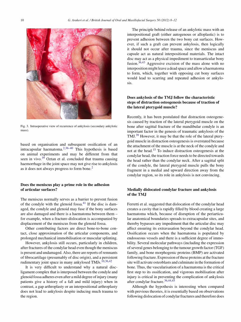
Fig. 3. Intraoperative view of recurrence of ankylosis (secondary ankyloticm
biosha
Do
Toaafd
tp
aior
lgpcdt
ipeicdfitws
Dst
RsbiTgtnctofc
Mo
FchltaOeboffsbfiiafter condylar fracture.
Although the hypothesis is interesting when compared
ass).
ased on organisation and subsequent ossification of anntracapsular haematoma.2,36–40 This hypothesis is basedn animal experiments and may be different from thateen in vivo.39 Oztan et al. concluded that trauma causingaemorrhage in the joint space may not give rise to ankylosiss it does not always progress to form bone.2
oes the meniscus play a prime role in the adhesionf articular surfaces?
he meniscus normally serves as a barrier to prevent fusionf the condyle with the glenoid fossa.41 If the disc is dam-ged, the condyle and glenoid may fuse if the bony surfacesre also damaged and there is a haematoma between them –or example, when a fracture-dislocation is accompanied byisplacement of the meniscus from the glenoid fossa.
Other contributing factors are direct bone-to-bone con-act, close approximation of the articular components, androlonged mechanical immobilisation or muscular splinting.
However, ankylosis still occurs, particularly in children,fter fractures of the condylar head even though the meniscuss present and undamaged. Also, there are reports of remnantsf fibrocartilage (presumably of disc origin), and a persistentudimentary joint space in many ankylosed TMJs.10,36,42
It is very difficult to understand why a natural disc-igament complex that is interposed between the condyle andlenoid fossa adheres even after a mild degree of injury (manyatients give a history of a fall and mild injury) when inontrast, a gap arthroplasty or an interpositional arthroplasty
oes not lead to ankylosis despite inducing much trauma tohe region.wf
Maxillofacial Surgery 50 (2012) 8–12
The principle behind release of an ankylotic mass with annterpositional graft (either autogenous or alloplastic) is torevent adhesion between the two bony cut surfaces. How-ver, if such a graft can prevent ankylosis, then logicallyt should not occur after trauma, since the meniscus andapsule act as natural interpositional materials. The intactisc may act as a physical impediment to transarticular bonyusion.36,43 Aggressive excision of the mass alone with nonterposition might leave a dead space and allow a haematomao form, which, together with opposing cut bony surfacesould lead to scarring and repeated adhesion or ankylo-
is.
oes ankylosis of the TMJ follow the characteristicteps of distraction osteogenesis because of traction ofhe lateral pterygoid muscle?
ecently, it has been postulated that distraction osteogene-is caused by traction of the lateral pterygoid muscle on theone after sagittal fracture of the mandibular condyle is anmportant factor in the genesis of traumatic ankylosis of theMJ.39 However, it may be that the role of the lateral ptery-oid muscle in distraction osteogenesis is overstated becausehe attachment of the muscle is at the neck of the condyle andot at the head.15 To induce distraction osteogenesis at theondylar head, the traction force needs to be directed towardshe head rather than the condylar neck. After a sagittal splitf the condyle, the lateral pterygoid muscle pulls the bonyragment in a medial and upward direction away from theondylar region, so its role in ankylosis is not convincing.
edially dislocated condylar fracture and ankylosisf the TMJ
erretti et al. suggested that dislocation of the condylar headreates a cavity that is rapidly filled by blood creating a largeaematoma which, because of disruption of the periarticu-ar anatomical boundaries spreads to extracapsular sites, andhereby bypasses any impediment that the articular disc mayffect ensuring its extravasation beyond the condylar head.ssification occurs when the haematoma is populated by
ndosseous vessels and there is a sufficient degree of immo-ility. Several molecular pathways (including the expressionf several genes belonging to the tumour growth factor (TGF)amily, and bone morphogenic proteins (BMP) are activatedollowing fracture. Expression of these proteins at the fractureite will activate osteoblasts and culminate in the formation ofone. Thus, the vascularisation of a haematoma is the criticalrst step to its ossification, and vigorous mobilisation after
njury is critical in preventing the complication of ankylosis36,44,45
ith previous theories, it is essentially based on observationsollowing dislocation of condylar fractures and therefore does

al and
nd
Dt
Ctbfttfsbmmbaa
Ra
Iamtar
sfmarswoibwaFfrf
If
Id
wocoC
igoRiiIpfipumFaa
C
Moablaits
C
N
R
G. Arakeri et al. / British Journal of Or
ot explain the sequence of events for ossification in non-islocated fractures of the condyle or condylar head.
oes the formation of ankylosis after trauma followhe characteristic events of fracture healing?
onceptually, ankylosis has been regarded as the fusion ofwo approximated and injured bony surfaces, and has alsoeen regarded as inappropriate tissue differentiation afterracture.40 Even so, for a process to be labelled as a fracture,here should firstly be discontinuity in a bone, and secondly,wo exposed bony edges. Ankylosis cannot be correlated withractures because it involves the fusion of two different bonyurfaces (condyle with cancellous, and glenoid with compactone); one static, and one constantly mobile. Furthermore, inost cases of trauma, the condyle (head or neck) is injuredore severely than the bony glenoid fossa, which is protected
y an interposed articular disc, so the sequence of events ofhealing fracture cannot be extrapolated to the process of
nkylosis.
ole of the sagittally split condyle in transarticulardhesion of the TMJ
n 1982, Rowe reported that ankylosis can be a result ofnteroposterior (sagittal) fracture of the condyle.46 Displace-ent of the lateral fragment upward over the outer rim of
he glenoid fossa, with associated displacement of the intra-rticular disc, and the accompanying loss of mobility, wereesponsible for the condition in these patients.
In 2008, He et al. suggested a strong relation betweenagittal split condylar fractures and simultaneous mandibularractures in the pathogenesis of ankylosis of the TMJ.47 Thisechanism increases the width between the condyles or rami
t the level of the lateral segment. The fractured surface of theamus or lateral condylar pole displaces laterally and possiblyuperiorly to the glenoid fossa and comes into close contactith the zygomatic arch, which causes a juxta-articular typef ankylosis (ankylosis that occurs lateral to the articulationtself36). A study of 206 patients with ankylosis of the TMJy Sarma and Dave showed that 93% were juxta-articularith a medially positioned non-ankylosed condylar head,48
lthough in their series there were no simultaneous fractures.erretti et al.36 reported only three simultaneous mandibularractures among 26 patients with ankylosis of the TMJ, so theole of such fractures in patients with sagittal split condylarracture remains controversial in the aetiology of ankylosis.
s protein-energy malnutrition a predisposing factor
or traumatic adhesion in the TMJ?t is interesting that the incidence of patients who go on toevelop ankylosis of the TMJ is high in some parts of the
Maxillofacial Surgery 50 (2012) 8–12 11
orld. The most striking finding is the perceived incidencef ankylosis in some countries, particularly in developingountries, and the relative scarcity of the disorder in devel-ped countries.47,49 Why would there be a higher incidence inhina, Africa, or India, than in the United States or Europe?47
Possible reasons why developing countries have anncreased number of such patients are larger populations,enetic predisposition, the lack of availability of proper care,r inadequate care because of poorly equipped hospitals.47
ecently, it has been shown in rats that malnutrition resultsn impaired callus formation, as well as fibrous ankylosisn the TMJ on healing of a displaced condylar process.49
n this study repair of the fracture was impaired, there wasroliferation of connective tissue, and atrophy of condylarbrocartilage that led to transarticular fibrous adhesion. Asrotein-energy malnutrition affects roughly half the pop-lation worldwide, particularly in developing countries,49
alnutrition could also be a predisposing cause of ankylosis.inally, one further explanation may be poor economic statuss possible neglect of minor facial trauma could progress tonkylosis of the TMJ.
onclusion
any factors have been suspected to affect the developmentf ankylosis of the TMJ after trauma, and its recurrencefter surgical treatment. While there exists a clear associationetween ankylosis and traumatic injuries of the mandibu-ar condyle, authors do not agree about its management andetiology. Further research is needed to understand the debil-tating condition more fully, and additional treatments needo be developed to reduce the incidence of recurrence afterurgery.
onflict of interest
one.
eferences
1. Ingawalé S, Goswami T. Temporomandibular joint: disorders, treat-ments, and biomechanics. Ann Biomed Eng 2009;37:976–96.
2. Oztan HY, Ulusal BG, Aytemiz C. The role of trauma on temporo-mandibular joint ankylosis and mandibular growth retardation: anexperimental study. J Craniofac Surg 2004;15:274–82.
3. Alexander RW. Improvement of facial symmetry after operative reliefof bony ankylosis of the jaw at the age of 10 years. Case report. PlastReconstr Surg 1978;62:896–901.
4. Wu XG, Hong M, Sun KH. Severe osteoarthrosis after fracture of themandibular condyle: a clinical and histologic study of seven patients. J
Oral Maxillofac Surg 1994;52:138–42.5. Rémi M, Christine MC, Gael P, Soizick P, Joseph-André J. Mandibularfractures in children: long term results. Int J Pediatr Otorhinolaryngol2003;67:25–30.
1 al and
1
1
1
1
1
1
1
1
1
1
2
2
2
2
2
2
2
2
2
2
3
3
3
3
3
3
3
3
3
3
4
4
4
4
4
4
4
4
4
2 G. Arakeri et al. / British Journal of Or
6. Strobl H, Emshoff R, Röthler G. Conservative treatment of unilat-eral condylar fractures in children: a long-term clinical and radiologicfollow-up of 55 patients. Int J Oral Maxillofac Surg 1999;28:95–8.
7. Kaban LB, Perrott DH, Fisher K. A protocol for managementof temporomandibular joint ankylosis. J Oral Maxillofac Surg1990;48:1145–52.
8. Guralnick WC, Kaban LB. Surgical treatment of mandibular hypomo-bility. J Oral Surg 1976;34:343–8.
9. Topazian RG. Etiology of ankylosis of the temporomandibularjoint: analysis of 44 cases. J Oral Surg Anesth Hosp Dent Serv1964;22:227–33.
0. Sawhney CP. Bony ankylosis of the temporomandibular joint: follow-upof 70 patients treated with arthroplasty and acrylic spacer interposition.Plast Reconstr Surg 1986;77:29–40.
1. Tideman H, Doddridge M. Temporomandibular joint ankylosis. AustDent J 1987;32:171–7.
2. Norman JE. Ankylosis of the temporomandibular joint. Aust Dent J1978;23:56–66.
3. Ware WH, Brown SL. Growth centre transplantation to replace mandibu-lar condyles. J Maxillofac Surg 1981;9:50–8.
4. Güven O. A clinical study on temporomandibular joint ankylosis inchildren. J Craniofac Surg 2008;19:1263–9.
5. Guyot L, Chossegros C, Cheynet F, Gola R, Lachard J, Blanc JL. Interpo-sition of a full-thickness skin graft in the surgery of temporomandibularjoint ankylosis. A study of 31 cases of which 20 had long-term followup. Rev Stommatol Chir Maxillofac 1995;96:372–8, in French.
6. Engel MB, Brodie AG. Condylar growth and mandibular deformities.Surgery 1947;22:976–92.
7. Kaban LB, Bouchard C, Troulis MJ. A protocol for management oftemporomandibular joint ankylosis in children. J Oral Maxillofac Surg2009;67:1966–78.
8. Phillips JH, Rechner B, Tompson BD. Mandibular growth followingreconstruction using a free fibula graft in the pediatric facial skeleton.Plast Reconstr Surg 2005;116:419–26.
9. Iconomou TG, Zuker RM, Phillips JH. Mandibular reconstructionin children using the vascularized fibula. J Reconstr Microsurg1999;15:83–90.
0. Wolford LM, Cottrell DA, Henry C. Sternoclavicular grafts fortemporomandibular joint reconstruction. J Oral Maxillofac Surg1994;52:119–29.
1. Kummoona R. Chondro-osseous iliac crest graft for one stage recon-struction of the ankylosed TMJ in children. J Maxillofac Surg1986;14:215–20.
2. Dattilo DJ, Granick MS, Soteranos GS. Free vascularized whole jointtransplant for reconstruction of the temporomandibular joint: a prelim-inary case report. J Oral Maxillofac Surg 1986;44:227–9.
3. Landa LE, Gordon C, Dahar N, Sotereanos GC. Evaluation of long-termstability in second metatarsal reconstruction of the temporomandibularjoint. J Oral Maxillofac Surg 2003;61:65–71.
4. Saeed N, Hensher R, McLeod N, Kent J. Reconstruction of the tem-poromandibular joint autogenous compared with alloplastic. Br J OralMaxillofac Surg 2002;40:296–9.
5. Moorthy AP, Finch LD. Interpositional arthroplasty for ankylosisof the temporomandibular joint. Oral Surg Oral Med Oral Pathol1983;55:545–52.
6. Lindqvist C, Pihakari A, Tasanen A, Hanpf G. Autogenous costochon-dral grafts in temporo-mandibular joint arthroplasty. A survey of 66
arthroplasties in 60 patients. J Maxillofac Surg 1986;14:143–9.7. Kent JN, Misiek DJ, Akin RK, Hinds EC, Homsy CA. Temporomandibu-lar joint condylar prosthesis: a ten-year report. J Oral Maxillofac Surg1983;41:245–54.
4
Maxillofacial Surgery 50 (2012) 8–12
8. Hibi G, Kaneda T, Oka T. Indication and appreciation of opera-tive procedures for mandibular ankylosis. Int J Oral Surg 1978;7:333–9.
9. Popescu V, Vasiliu D. Treatment of temporo-mandibular ankylosis withparticular reference to the interposition of full-thickness skin autotrans-plant. J Maxillofac Surg 1977;5:3–14.
0. Rajgopal A, Banerji PK, Batura V, Sural A. Temporomandibular anky-losis. A report of 15 cases. J Maxillofac Surg 1983;11:37–41.
1. Munro IR, Chen YR, Park BY. Simultaneous total correction of tem-poromandibular ankylosis and facial asymmetry. Plast Reconstr Surg1986;77:517–29.
2. Topazian RG. Comparison of gap and interposition arthroplasty inthe treatment of temporomandibular joint ankylosis. J Oral Surg1966;24:405–9.
3. Padgett GC, Robinson DW, Stephenson KL. Ankylosis of the temporo-mandibular joint. Surgery 1948;24:426–32.
4. Salins PC. New perspectives in the management of cranio-mandibularankylosis. Int J Oral Maxillofac Surg 2000;29:337–40.
5. Ko EC, Chen MY, Hsu M, Huang E, Lai S. Intraoral approach forarthroplasty for correction of TMJ ankylosis. Int J Oral Maxillofac Surg2009;38:1256–62.
6. Ferretti C, Bryant R, Becker P, Lawrence C. Temporomandibular jointmorphology following post-traumatic ankylosis in 26 patients. Int J OralMaxillofac Surg 2005;34:376–81.
7. Miller GA, Page Jr HL, Griffith CR. Temporomandibular joint ankylosis:review of the literature and report of two cases of bilateral involvement.J Oral Surg 1975;33:792–803.
8. el-Mofty S. Ankylosis of the temporomandibular joint. Oral Surg OralMed Oral Pathol 1972;33:650–60.
9. Meng FW, Zhao JL, Hu KJ, Liu YP. A new hypothesis of mechanismsof traumatic ankylosis of temporomandibular joint. Med Hypotheses2009;73:92–3.
0. Güven O. A clinical study on temporomandibular joint ankylosis. AurisNasus Larynx 2000;27:27–33.
1. Laskin DM. Role of the meniscus in the etiology of posttraumatic tem-poromandibular joint ankylosis. Int J Oral Surg 1978;7:340–5.
2. Goss AN, Bosanquet AG. The arthroscopic appearance of acute tem-poromandibular joint trauma. J Oral Maxillofac Surg 1990;48:780–4.
3. Miyamoto H, Kurita K, Ogi N, Ishimaru JI, Goss AN. Effect of limitedjaw motion on ankylosis of the temporomandibular joint in sheep. Br JOral Maxillofac Surg 2000;38:148–53;Miyamoto H, Kurita K, Ogi N, Ishimaru JI, Goss AN. Effect of lim-ited jaw motion on ankylosis of the temporomandibular joint in sheep.Erratum in: Br J Oral Maxillofac Surg 2000;38:575.
4. Deckers MM, van Bezooijen RL, van der Horst G, Hoogendam J, van DerBent C, Papapoulos SE, et al. Bone morphogenetic proteins stimulateangiogenesis through osteoblast-derived vascular endothelial growthfactor A. Endocrinology 2002;143:1545–53.
5. Trueta J. The roles of the vessels in osteogenesis. J Bone Joint Surg1963;45:402–18.
6. Rowe NL. Ankylosis of the temporomandibular joint. J R Coll SurgEdinb 1982;27:67–79.
7. He D, Ellis III E, Zhang Y. Etiology of temporomandibular joint ankylo-sis secondary to condylar fractures: the role of concomitant mandibularfractures. J Oral Maxillofac Surg 2008;66:77–84.
8. Sarma UC, Dave PK. Temporomandibular joint ankylosis: an Indianexperience. Oral Surg Oral Med Oral Pathol 1991;72:660–4.
9. Rodrigues L, Corrêa L, Luz JG. Healing of displaced condylar processfracture in rats submitted to protein undernutrition. J CraniomaxillofacSurg 2010, doi:10.1016/j.jcms.2010.03.006. April 22 (Epub ahead ofprint).