
Pathogen-Induced Defense Signaling and Signal Crosstalk...
72
Pathogen-Induced Defense Signaling and Signal Crosstalk in Arabidopsis Tarja Kariola Department of Biological and Environmental Sciences Division of Genetics Faculty of Biosciences and Viikki Graduate School in Biosciences University of Helsinki Academic dissertation To be presented for public criticism, with the permission of the Faculty of Biosciences of the University of Helsinki, in Auditorium 1041 of the Viikki Biocenter, Viikinkaari 5, Helsinki, on October 6 th , 2006, at 12 noon.
Transcript of Pathogen-Induced Defense Signaling and Signal Crosstalk...

Pathogen-Induced Defense Signaling and SignalCrosstalk in Arabidopsis
Tarja Kariola
Department of Biological and Environmental SciencesDivision of Genetics
Faculty of Biosciencesand
Viikki Graduate School in Biosciences
University of Helsinki
Academic dissertationTo be presented for public criticism, with the permission of the Faculty of Biosciences
of the University of Helsinki, in Auditorium 1041 of the Viikki Biocenter, Viikinkaari 5,Helsinki,
on October 6th, 2006, at 12 noon.

Supervisors: Professor Tapio PalvaDepartment of Biological and Environmental SciencesUniversity of Helsinki, Finland
Docent Günter BraderDepartment of Biological and Environmental SciencesUniversity of Helsinki, Finland
Reviewers: Professor Katri KärkkäinenFinnish Forest Research UnitVantaa Unit, Finland
Professor Teemu TeeriDepartment of Applied BiologyUniversity of Helsinki, Finland
Opponent: Professor Jean-Pierre MétrauxDepartment of Plant BiologyUniversity of Fribourg, Switzerland
ISSN 1795-7079ISBN 952-10-3333-9 (paperback)ISBN 952-10-3335-5 (PDF, online)
Edita Prima OyHelsinki 2006

Dedicated to the loving memory of my mother
Contents
LIST OF ORIGINAL PUBLICATIONS ............................................................................................... 6
ABBREVIATIONS .................................................................................................................................. 7
ABSTRACT.............................................................................................................................................. 8
INTRODUCTION.................................................................................................................................... 9
PLANT-PATHOGEN INTERACTION .........................................................................................................11PHYTOPATHOGENIC BACTERIA.............................................................................................................12PLANT DEFENSE ...................................................................................................................................13
Preformed defenses ........................................................................................................................13Inducible defenses ..........................................................................................................................14
Elicitation of plant defense........................................................................................................................... 15
General elicitors ...................................................................................................................................... 15
Race-specific elicitors ............................................................................................................................. 16
Elicitor perception ................................................................................................................................... 17
Defense signaling ......................................................................................................................................... 18
Role of reactive oxygen species in defense signaling............................................................................. 20
Role of nitric oxide in defense signaling ................................................................................................ 22
Salicylic acid-mediated defense signaling .............................................................................................. 22
Nature of the systemic signal............................................................................................................. 25
Jasmonic acid-mediated defense signaling ............................................................................................. 26
Jasmonic acid in systemic signaling .................................................................................................. 27
Ethylene-mediated defense signaling...................................................................................................... 28
Abscisic acid................................................................................................................................................. 30
Role of ABA in abiotic stress responses................................................................................................. 31
Role of ABA in biotic stress responses................................................................................................... 33
CROSSTALK BETWEEN SIGNALING PATHWAYS .....................................................................................35MODEL ORGANISM ERWINIA CAROTOVORA ............................................................................................37OPEN QUESTIONS .................................................................................................................................39
AIMS OF THE STUDY.........................................................................................................................40
MATERIALS AND METHODS...........................................................................................................41
RESULTS AND DISCUSSION ............................................................................................................ 42
ERWINIA CAROTOVORA AND ELICITORS HRPN AND PEHA TRIGGER DISTINCT BUT INTERACTING
DEFENSE RESPONSES AND CELL DEATH IN ARABIDOPSIS (I) ..................................................................42HrpN elicits lesion formation and induces resistance to E. carotovora in Arabidopsis .................42Establishment of HrpN-triggered resistance requires both SA and JA signaling ...........................43Synergistic action of elicitors HrpN and PehA triggers enhanced defense gene expression andcell death.........................................................................................................................................44
PLANT RESPONSES TO E. CAROTOVORA (II, III) .....................................................................................44CHLOROPHYLLASE 1 AFFECTS INDUCTION OF DEFENSE PATHWAYS IN PLANTS (II) .............................. 45
RNAi silencing of chlorophyllase 1 (AtCLH1) improves resistance to E. carotovora inArabidopsis .................................................................................................................................... 45Resistance of AtCLH1 RNAi-silenced plants is light-dependent and involves increasedaccumulation of ROS ..................................................................................................................... 46Silencing of AtCLH1 affects the balance between SA- and JA-dependent defenses inArabidopsis ................................................................................................................................. 47
ERD15 IS A NEGATIVE REGULATOR OF ABA RESPONSES IN ARABIDOPSIS (III) .................................... 49ERD15 is rapidly induced by both biotic and abiotic factors ......................................................... 49Modulation of ERD15 expression affects abiotic stress tolerance by influencing ABAsensitivity ...................................................................................................................................... 49Insensitivity to ABA enhances resistance of Arabidopsis to E. carotovora................................... 51ERD15 is a negative regulator of ABA responses in Arabidopsis ................................................. 52
CONCLUDING REMARKS AND FUTURE PERSPECTIVES ...................................................... 53
ACKNOWLEDGMENTS ..................................................................................................................... 56
REFERENCES ...................................................................................................................................... 58

6
LIST OF ORIGINAL PUBLICATIONS
This thesis is based on the following publications, referred to in the text by their Romannumerals.
I Kariola, T., Palomäki, T.A., Brader, G., and Palva, E.T. (2003). Erwiniacarotovora subsp. carotovora and Erwinia-derived elicitors HrpN and PehA triggerdistinct but interacting defense responses and cell death in Arabidopsis. Mol PlantMicrobe Interact 16, 179-187.
II Kariola, T., Brader, G., Li, J., and Palva E.T. (2005). Chlorophyllase 1, adamage control enzyme, affects the balance between defense pathways in plants. PlantCell 17, 282-294.
III Kariola, T., Brader, G., Helenius, E., Li, J., Heino, P., and Palva, E.T.(2006). EARLY RESPONSIVE TO DEHYDRATION 15 – a negative regulator of ABAresponses in Arabidopsis. Manuscript provisionally accepted (pending revision) to PlantPhysiology.
The publications have been reprinted with the kind permission of their copyright holders.
7
ABBREVIATIONS
ABA abscisic acidAPX ascorbate peroxidaseBABA -aminobutyric acidCAT catalaseCC coiled-coilCDPK calcium dependent protein kinaseCSN COP9 signalosomeDAB 3, 3´-diaminobenzidineEDS enhanced disease susceptibilityET ethyleneIR induced resistanceISR induced systemic resistanceJA jasmonic acidLPS lipopolysaccharideLRR leucine-rich repeatMAPK mitogen-activated protein kinaseMeJA methyl jasmonateMeSA methyl salicylateNBS nucleotide binding siteNDR nonrace-specific disease resistanceNO nitric oxideNOS NO synthaseOGA oligogalacturonic acidOPDA 12-oxo-phytodienoic acidPAD phytoalexin-deficientPAMP pathogen-associated molecular patternPCWDE plant cell wall-degrading enzymePHY phytochromePI proteinase inhibitorPR pathogenesis-relatedRES reactive electrolyte speciesRLK receptor-like kinaseROS reactive oxygen speciesSA salicylic acidSABP SA binding proteinSID SA induction-deficientTIR Toll and IL-1 receptorTTSS type III secretion systemWAK wall-associated kinase

8
ABSTRACT
Erwinia carotovora subsp. carotovora is a bacterial phytopathogen that causes soft rot invarious agronomically important crop plants. A genetically specified resistance to E.carotovora has not been defined, and plant resistance to this pathogen is establishedthrough nonspecific activation of basal defense responses. This, together with the broadhost range, makes this pathogen a good model for studying the activation of plantdefenses. Production and secretion of plant cell wall-degrading enzymes (PCWDE) arecentral to the virulence of E. carotovora. It also possesses the type III secretion system(TTSS) utilized by many Gram-negative bacteria to secrete virulence- promoting effectorproteins to plant cells. This study elucidated the role of E. carotovora HrpN (HrpNEcc), aneffector protein secreted through TTSS, and the contribution of this protein in thevirulence of E. carotovora. Treatment of plants with HrpNEcc was demonstrated to inducea hypersensitive response (HR) as well as resistance to E. carotovora. Resistance inducedby HrpNEcc required both salicylic acid (SA)- and jasmonate/ethylene (JA/ET)-dependentdefense signaling in Arabidopsis. Simultaneous treatment of Arabidopsis with HrpNEcc
and PCWDE polygalacturonase PehA elicited accelerated and enhanced induction ofdefense genes but also increased production of superoxide and lesion formation. Thisdemonstrates mutual amplification of defense signaling by these two virulence factors ofE. carotovora.
Identification of genes that are rapidly induced in response to a pathogen can providenovel information about the early events occurring in the plant defense response.CHLOROPHYLLASE 1 (AtCLH1) and EARLY RESPONSIVE TO DEHYDRATION 15(ERD15) are both rapidly triggered by E. carotovora in Arabidopsis. Characterization ofAtCLH1 encoding chlorophyll-degrading enzyme chlorophyllase indicated that it mighthave a role in chlorophyll degradation during plant tissue damage. Silencing of this generesulted in increased accumulation of reactive oxygen species (ROS) in response topathogen infection in a light-dependent manner. This led to enhanced SA-dependentdefenses and resistance to E. carotovora. Moreover, crosstalk between different defensesignaling pathways was observed; JA-dependent defenses and resistance to fungalpathogen Alternaria brassicicola were impaired, indicating antagonism between SA- andJA-dependent signaling.
Characterization of ERD15 suggested that it is a novel, negative regulator of abscisicacid (ABA) signaling in Arabidopsis. Overexpression of ERD15 resulted in insensitivityto ABA and reduced tolerance of the plants to dehydration stress. However,simultaneously, the resistance of the plants to E. carotovora was enhanced. Silencing ofERD15 improved freezing and drought tolerance of transgenic plants. This, together withthe reducing effect of ABA on seed germination, indicated hypersensitivity to thisphytohormone. ERD15 was hypothesized to act as a capacitor that controls the appropriateactivation of ABA responses in Arabidopsis.

9
INTRODUCTION
Plants provide, directly or indirectly, all the food upon which humans and animals depend.Therefore, throughout history, diseases affecting plants have been feared as much ashuman diseases and war. Since the early days of agriculture, plant diseases have beenresponsible for crop losses, resulting in devastating times of hunger and famine.Thousands of years ago, these were seen as punishments meted out by gods for sinscommitted by people. Thus, the plant protection methods used in those days mostlycomprised praying and sacrifices, which, understandably, were not very efficient. TheRomans even created a special god, Robigus, to protect grains from rust (Agrios 2005).Eventually, crop protection developed from being merely a matter of faith to a morepractical direction. Invention of the microscope in the late 17th century enabled scientiststo discover many previously invisible microorganisms in diseased plant tissue. However, afurther 100 years passed, before it was generally accepted that viruses, bacteria, fungi, andprotozoa were not spontaneously occurring natural products of diseases, but actually thecausative agents behind these diseases (Holub 2001; Agrios 2005).
The estimated total crop loss from diseases, insects, and weeds is about 36% ofpotential worldwide production, the proportion of diseases being 14% (Agrios 2005).However, added to this figure should be a further 6-10%, resulting from after-harvestdamages, a problem especially encountered in developing tropical countries. Fightingplant diseases is therefore a major challenge when striving for successful agriculture. Formillions of people who still cultivate their own food, plant diseases can make a significantdifference between a comfortable life and a life agonized by hunger. The Irish famine inthe 1840s that resulted from a potato (Solanum tuberosum) blight epidemic caused by thefungus Phytophtora infestans as well as part of the hunger in developing countries todayare examples of the devastating power of plant diseases (Agrios 2005). In more developedcountries, where food is plentiful, diseases manifest in economic losses to the farmers,followed by increases in consumer prices.
Diseases can affect plants in many ways, such as by reducing the quality and quantityof the crops. For example, infection by a pathogen can simply make the plants toxic toconsumers. Ergot is a disease of cereals and grasses caused by Claviceps purpurea andrelated fungi. These pathogens develop a fruiting structure that replaces the seed of theplant and produces toxic secondary metabolites (Keller et al. 2005). The harvested grain isthen contaminated and those who consume it may contract ergotism, a disease withsymptoms ranging from blistering of the skin to hallucinations, insanity, and even death(Agrios 2005). Plant diseases can also limit the kinds of plants that grow in certain areas.This is exemplified by the fate of American chestnut (Castanea dentata); before the turnof the century, this tree, which provided people with timber and chestnuts, dominated theeastern half of the United States. The invasion of the fungus Cryphonectria parasitica, thecausal agent of chestnut blight virtually eliminated this species of tree from North

10
America (Agrios 2005). Similarly, novel pests or pathogens or strains of pathogens couldgreatly reduce the area in which the major crop species wheat (Triticum sp.), rice (Oryzasativa), or maize (Zea mays) can be grown or eliminate these as vital crops, havingconsiderable effects on the nutritional status of the world.
Over the last 100 years, the control of plant diseases has increasingly depended on theuse of toxic chemicals. These chemicals are not only applied on plants and plant productsbut also to the soil. This leads to environmental pollution, eventually making the fieldsunsuitable for further cultivation. Also, traces of the chemicals may remain in the plantand make them harmful or even toxic for consumers. The use of pesticides also adds to thetotal costs of production; these chemicals as well as the machinery needed to spread themare expensive. Moreover, the human population continues to grow rapidly, resulting in anincreasing demand for cultivating land. Formerly rare plants, such as maize, have becomesome of the most abundant species on earth. This has also influenced the disease-causingmicrobes; potential pathogens that had never before encountered these species now do sofrequently, leading to an increased need for protection (Tillman 1999).
For the reasons presented above, much of the research today in the field of plant-pathogen interactions aims at finding both environmentally friendly and more efficientmeans of controlling plant diseases. Classical breeding for plant resistance has beenimportant since the early days of the 20th century, but these methods, i.e. seed selection,backcrossing, etc., can be quite complicated and time-consuming (Stascawicz 2001;Agrios 2005). Recently, such promising approaches as genetic engineering, includingRNA- and gene-silencing techniques, have started to emerge (Lindbo and Dougherty2005). However, often only one dominant or semi-dominant gene, such as the resistance(R) gene, has been employed in breeding crop resistance (Stascawicz 2001). If theresistance is directed against specific pathogens or pathogen races (as it is with R genes), itis not necessarily enduring. The appearance and spread of a mutation in the pathogenpopulation can adapt the pathogens to the presence of the R gene and break down theresistance (McDonald and Linde 2002).
The study of plant-pathogen interactions can provide tools for development of moredurable approaches. For example, knowledge of the kinds of molecular responses variouspathogens activate in the plant during the infection can have tremendous potential. Plantdefense responses are a result of a complex network of signaling events that involves theinterplay of kinases, hormones, and reactive oxygen species (ROS), leading toreprogramming of the plant transcriptome. These responses aim at the production ofdefensive compounds and, finally, resistance. Elucidation of the molecular componentsacting in these cascades provides useful tools for engineering more durable crops andresistance that is not so easily broken down. Modifications of signaling components canspeed up defense activation upon pathogen attack, thus improving the chances of the plantto successfully respond to current and future encounters with the invaders. In addition toengineering disease-resistant plants, plant resistance can also be improved by the use ofnontoxic chemical substances that elicit the activation of natural defense mechanisms, aprocess called “priming” (Conrath et al. 2002; Kohler et al. 2002). Knowledge aboutplant-pathogen interactions will hopefully lead to solutions for achieving broad-spectrumprotection, long-lasting effects, and reduced chemical input in modern-day agriculture.

11
PLANT-PATHOGEN INTERACTION
The causative agents of plant diseases belong to the same groups as those causing diseasein animals _ pathogenic microorganisms such as fungi, viruses, bacteria, protozoa, andnematodes (Agrios 2005). To be a successful pathogen, a microorganism needs to interferewith one or more essential functions of the plant, thereby causing disease. Regardless ofthe type of pathogen, a prerequisite for pathogenicity of a microorganism is the ability togain access to the plant interior. Pathogens force their way through plant surfaces bydifferent means; some take advantage of natural openings, such as stomata or lenticels, orenter the plant through wounds, while others simply penetrate the leaf surfaces. Fungi,such as powdery mildew, for instance, can grow a fine hypha directly into plant epidermalcells. Oomycetes and nematodes usually use the penetration method, while bacteria utilizewounds and natural openings. In most fungal diseases, the fungus penetrates not only thecuticle but also the cell wall _ the next obstacle for pathogens after reaching theintercellular spaces (apoplast). Some pathogens use chemical activities to overcome thisbarrier. For example, certain bacteria secrete cutin-degrading enzymes, cutinases, whileothers produce an arsenal of extracellular enzymes, including pectinases, cellulases, andpolygalacturonases, that degrade the cell wall (Toth et al. 2003; Agrios 2005).
The virulence mechanisms the pathogen uses to reach its final goal – to takeadvantage of the plant as a source of nutrients – include secretion of toxins, growthregulators, and other substances that disturb the metabolism of plant cells or interfere withthe plant defenses (Agrios 2005). The virulence strategy depends on how the pathogenintends to utilize the plant; biotrophs obtain nutrients from living plant tissue withoutkilling the cells, whereas necrotrophs kill the cells and make use of their contents duringinvasion (Glazebrook 2005). Some pathogens, called hemi-biotrophs, fill the requirementsof both biotrophs and necrotrophs, depending on the prevailing conditions they are in orthe stages of their life cycles (Glazebrook 2005). Most pathogens continue multiplyingindefinitely within the infected tissue, using it for nutrients until the plant is dead.
Indefinite multiplication would be true for every pathogen if plants were passive anddefenseless organisms, which is not the case. Plants are subject to attack by a wide varietyof microbial pathogens; nevertheless, the ability of a microorganism to cause disease inthe plant is usually an exception rather than the rule. Due to plants´ fortress-like cellstructure as well as their innate ability to recognize potential invading pathogens andactivate effective defenses, plants are generally resistant to most pathogens (Heath 2000;Nürnberger et al. 2004). Thus, in order to be successful, a pathogen also needs to evadethe plant surveillance system or suppress plant defenses. The ability to detect potentialpathogens has been essential to the development of modern plants (Chisholm et al. 2006).Perception of the pathogen is achieved through receptors, a surveillance system capable ofrecognizing both conserved molecular patterns and specific effector proteins, andactivation of the corresponding defenses (Montesano et al. 2003). Plants defendthemselves against invaders in two ways: with structural barriers that inhibit the pathogenfrom gaining entrance and spreading throughout the plant and with biochemical reactions
12
taking place in plant tissues. These reactions produce toxic substances that inhibit thegrowth of the pathogen. The combinations of these two defense types vary betweendifferent plant-pathogen interactions (Glazebrook 2005). If the plant fails to recognize thepathogen or an elicitor, appropriate defenses might not be mounted and disease results.Alternatively, if the plant responds with a rapid and well-aimed activation of defenses, theattempted infection is halted.
PHYTOPATHOGENIC BACTERIA
Approximately 1600 bacterial species are known, and of these, roughly 100 cause diseasesin plants (Agrios 2005). Bacteria are a significant group of phytopathogens and thediseases they cause are considerably difficult to control. This is due to the capability ofbacteria to multiply at an astonishing rate and produce enormous numbers of cells in ashort period of time. In addition, bacteria can alter their chemical environmentsignificantly by secreting toxins that contribute to their pathogenicity. In contrast to manybacterial pathogens of animals that enter the cells of their hosts, phytopathogenic bacteriamultiply in the apoplast of plant cells and remain extracellular (Staskawicz et al. 2001).Bacterial diseases affect all kinds of plants and occur in every place where it is reasonablymoist and warm. The spread of bacteria is aided by, for example, water, insects, animals,and humans. Insects wound the plant organs and thus facilitate the entry of the pathogeninto the plant. They also act as vectors for some bacteria; the corn flea beetle(Chaetocnema pulicaria) is the main vector of the bacterium Erwinia stewartii, whichcauses Stewart´s wilt on corn (Agrios 2005; Cook et al. 2005). Humans, on the other hand,help the long-distance spread of diseases by transporting infected plants to entirely newareas. Some of the most common plant pathogenic genera of bacteria includeAgrobacterium, Clavibacter, Erwinia, Pseudomonas, Xanthomonas, and Streptomyces(Agrios 2005). Infected plants show a variety of symptoms, such as leaf spots and blights,soft rots, wilts, and cancers. Of these, members of the genera Pseudomonas andXanthomonas are causative agents of almost all bacterial spots and blights of leaves,stems, and fruits, while Agrobacterium is the main cause of grown gall on many woodyplants (Agrios 2005).
One example of a damaging bacterial plant disease is soft rot. Pathogens causing themost common and destructive soft rots are found in the genus Erwinia (Toth et al. 2003;Agrios 2005). This disease predominantly occurs in the fleshy tissues of vegetables androot crops. Erwinias invade the plant through natural openings like stomata or takeadvantage of wounds in the plant tissue caused by, for example, feeding insects ormechanical damage. After invasion, these bacteria are able to reside in intercellular spacesof the plant until environmental conditions, such as temperature, oxygen availability, andwater content, become appropriate for disease development (Toth et al. 2003). Soft-rotsymptoms begin as a water-soaked lesion that enlarges further, culminating in slimymasses of bacteria and cellular debris oozing out of plant tissues (Perombelon and Kelman1980; Toth et al. 2003). In their noninfective phase, soft-rot Erwinias undergo endophytic,

13
epiphytic, and saprophytic lifestyles on plants and are also found in the soil and groundwater (Perombelon and Kelman 1980; Toth et al. 2003; Agrios 2005).
PLANT DEFENSE
Even if plants live a sessile life, they are dynamic organisms that fight the pressure ofpathogens with advanced defense strategies, including both preformed and inducibledefense systems. Resistance of an entire plant species to all strains of a pathogen is callednonhost resistance, the most common type of resistance expressed by plants (Heath 2000;da Cunha et al. 2006). Simply put, this means that, for example, the pathogens of tomato(Lycopersicon esculentum) do not infect spruce (Picea sp.), and vice versa. Significantcomponents of nonhost resistance are the preformed or constitutive defenses associatedwith plant structures and chemical compounds already present in the plant. These includestructural barriers, such as the plant cell wall, as well as inhibitory compounds, e.g.phenolics and tannins (Heath 2000; Nürnberger et al. 2004; Agrios 2005).
Inducible defenses are triggered by the recognition of the pathogen. Basal defense, aconstituent of both nonhost and host resistance, provides basal-level resistance (also calledinnate immunity or local induced resistance) that prevents infection by a wide range ofmicrobes (Heath 2000; Thordal-Christensen 2003; Nürnberger et al. 2004; Oh andCollmer 2005). Some pathogens have acquired the ability to suppress basal defenseresponses and enhance their virulence by delivering specific effector proteins to the plantcells that interfere with plant defense. Gene-for-gene or race-cultivar-specific resistanceoccurs when specific members of a plant species, but not the species as a whole, haveacquired resistance to a particular pathogen. This type of resistance is usually restricted toa particular pathogen species, being expressed against specific genotypes of that pathogen(Dangl and Jones 2001; Bonas and Lahaye 2002; Hammond-Kosack and Parker 2003;Chisholm et al. 2006).
PREFORMED DEFENSES
Noninducible, preformed structural defenses, such as a dense epidermal layer and wax andcuticle coverings on leaves, are the first line of plant defense to invading pathogens.Structures found on surfaces, such as spiky hairs called trichomes, can prevent feeding byinsects or insect larvae. Hairs on leaves can also have a water repelling effect, hencemaking it more difficult especially for bacteria to establish contact with the plant. Rigidcell walls composed of fibrils of cellulose embedded in a matrix of several other kinds ofpolymers, such as pectin and lignin, also serve as an efficient barrier to the invasion ofpathogens (Agrios 2005).
Chemical defenses include various antimicrobial peptides, proteins, andnonproteinaceous secondary metabolites present in plant cells that can prevent ingress ofthe invader (Heath 2000; Nürnberger et al. 2004). Toxic secondary metabolites stored in

14
specialized plant compartments can be activated or released upon tissue damage. Besidesbeing directly harmful to the invader, they can operate by inactivating the extracellularenzymes secreted by the pathogen (Zhao et al. 2005). Secondary metabolites are oftenrestricted in their distribution to particular plant families, genera, or species. For example,avenacin A-1 is a triterpenoid saponin found in the roots of oat plants. It is highly effectiveagainst the fungus Gaeumannomyces graminis var tritici, a major pathogen of wheat andbarley roots, but not oats due to the presence of this secondary metabolite (Buchanan et al.2000). The glucosinolate-myrosinase system is an example of a sophisticated chemicaldefense system characteristic of Arabidopsis and other Brassicaceae species (Halkier andGershenzon 2006). Glucosinolates, preformed amino acid-derived secondary metabolitesand myrosinase, an endogenous -thioglucosidase, are stored in separate compartments inplant cells. Upon tissue disruption, such as wounding, myrosinase cleaves nontoxicglucosinolates, resulting in the release of such products as isothiocyanates, which can beharmful to a wide range of plant enemies, including mammals, insects, and bacteria(Halkier and Gershenzon 2006).
INDUCIBLE DEFENSES
Inducible plant defenses are triggered by the perception of a pathogen or pathogen-derivedmolecules called elicitors. The elicitors can be either general, common to a group ofmicrobes, or specific to certain pathogen strains. In addition, pathogens can releasepolysaccharide oligomers from the plant surface, which can induce defenses (Montesanoet al. 2003; Nürnberger et al. 2004; Chisholm et al. 2006). Perception of elicitors takesplace in receptors located either at the cell surface or inside the cell (Dardick and Ronald2006). According to current knowledge, recognition of general and specific elicitorstriggers overlapping signaling responses in the plant (Espinosa and Alfano 2004; Kim etal. 2005). Interestingly, by comparing changes in plant mRNA profiles in response toavirulent and virulent P. syringae, Tao et al. (2003) demonstrated that the induction ofdefense genes was more rapid and enhanced in response to specific elicitors (i.e. theavirulent strain). This indicates a difference in the speed rather than the quality of responsetriggered by the two elicitor types (Espinosa and Alfano 2004; Kim et al. 2005).
Recognition of the elicitor induces several early responses (Figure 1): phosphorylationand dephosphorylation of plasma membrane proteins, increase of cytosolic Ca2+, ionfluxes, and alkalization of the apoplast. Synthesis and deposition of callose in the form ofpapillae can be initiated rapidly at the site of pathogen invasion. Mitogen-activated proteinkinases (MAPK) and NADPH oxidase are activated, and ROS is produced within minutesof contact with the elicitor (Zhao et al. 2005). Activation of transcription factors and earlyexpression of defense genes also occurs. The activated kinase cascades and ROS furtheramplify the defense signal to downstream reactions (Dardick and Ronald 2006).
A series of alarm signals are triggered that are transmitted intracellularly and also toadjacent cells. These are sequentially followed by late defense gene activation andphytoalexin accumulation. Phytoalexins are toxic antimicrobial substances and can, forexample, be flavonoids, alkaloids, and terpenoids produced in healthy cells in response to

15
signals from damaged cells adjacent to them (Hammerschmidt 1999; Zhao et al. 2005).Production of various defense-related proteins, such as pathogenesis-related (PR) proteins,which have antimicrobial activity and thus serve to contain the infection, is also activated(Wojstaszek 1997; Hammerschmidt 1999; Van Loon and Van Strien 1999). The formationof a hypersensitive response (HR), a rapid, localized cell death that restricts the growth ofthe pathogen, is more frequently associated with the recognition of a specific than ageneral elicitor (Greenberg 1997; Espinosa and Alfano 2004; Greenberg and Yao 2004)(Figure 1). The signals originating from the local infection site can then evolve into asystemic defense response involving distal, undamaged parts of the plant and conferringresistance to future pathogen infections.
Figure 1. Plant responses induced by the recognition of a pathogen (adapted from Buchanan et al.2002).
Elicitation of plant defense
General elicitors
Evolutionary ancient innate immunity, the ability to discriminate between self and nonself,is a quality of both animals and plants (Medzhitov and Janeway 2002; Parker 2003). Itrelies on the detection of pathogen-associated molecular patterns (PAMPs) characteristicof a whole class of potentially harmful microbial organisms (Heath 2000; Nürnberger andBrunner 2002; Nürnberger et al. 2004). Pathogen-derived molecules of diverse nature,

16
including lipopolysaccharides (LPS), flagellins, glucans, and chitins, serve as generalelicitors that trigger basal defense responses independently of the genotype of theindividual pathogen (Figure 2). For example, flagellin, the protein subunit of the bacterialsurface structure flagellum, acts as a PAMP in both animals and plants (Felix et al. 1999;Gomez-Gomez and Boller 2002; Smith et al. 2003). General elicitors are usuallymolecules that are indispensable in the lifestyle of microbes and thus provide a fitnesspenalty for the pathogen if recognized by the plant surveillance system (Nürnberger andBrunner 2002; Nürnberger et al. 2004). Endogenous plant cell wall-derived structuresreleased by the hydrolytic enzyme activities of invading microbes can also act as generalelicitors (Benhamou 1996; Nürnberger et al. 2004) (Figure 2).
Race-specific elicitors
During evolution some pathogens have developed to overcome the PAMP-triggered basalresistance by acquiring the ability to deliver effector proteins into plant cells (Espinosaand Alfano 2004; Chisholm et al. 2006) (Figure 2). These effector proteins interfere with,manipulate, or suppress disease signaling, thereby enhancing pathogen growth and diseasedevelopment (Oh and Collmer 2005; Chisholm et al. 2006; da Cunha et al. 2006; Trumanet al. 2006). In response, during co-evolution plants have adapted to detect these specificpathogen-derived molecules. This cultivar-specific, gene-for-gene disease resistancesystem is determined by pathogen-encoded effector proteins and the corresponding plant-derived R proteins (Hammond-Kosack and Jones 1997; Bonas and Lahaye 2002).
Many Gram-negative bacterial pathogens possess the hypersensitive response andpathogenicity (hrp) gene cluster that encodes the type III secretion system (TTSS). TTSSis utilized by the bacteria for injection of the effector proteins into plant cells (Feys andParker 2000; Lahaye and Bonas 2001; Alfano and Collmer 2004) (Figure 2). Chisholm etal. (2006) speculated that the effectors have developed to interfere with the components ofPAMP-triggered defense or to promote the pathogenicity of the microorganism byaffecting a variety of host proteins (Figure 2). Kim et al. (2005) demonstrated that P.syringae effector proteins AvrRpt2 and AvrRpm1 suppress PAMP-triggered defenseresponses in Arabidopsis by inhibiting flagellin-induced accumulation of callose.Moreover, another P. syringae effector, AvtPto, suppressed Arabidopsis genes encodingsecreted cell wall and defense proteins (Hauck et al. 2003). Some avirulence factors act bysuppressing HR response (Jamir et al. 2004), which is central in activating certain plantdefense responses. Although many effector proteins have been cloned, the biochemicalfunction of most remains unknown. AvrPtoB has been shown to have ubiquitin ligaseactivity in vivo (Janjusevic et al. 2005; Abramovitch et al. 2006). Deletion of key residuesfrom this protein eliminated ubiquitin ligase activity and the capability of AvrPtoB toinhibit cell death. Thus, this effector was suggested to act by targeting proteins responsiblefor regulation of programmed cell death to degradation mimiking ubiquitin ligase of thehost (Janjusevic et al. 2005; Chisholm et al. 2006).
However, if the effector protein meets a matching R gene in the plant, it becomes aspecific elicitor and the plant defense system is activated by the R protein (Figure 2).Several R genes confer specific resistance to fungal, viral, or bacterial pathogens carrying

17
the matching effector gene (Staskawicz et al. 2001; Bonas and Lahaye 2002). Resistanceis manifested by HR response, one of the most prominent features of gene-for-generesistance, and inhibition of pathogen growth (Feys and Parker 2000; Bonas and Lahaye2002). The oxidative bursts in the tissues undergoing HR response also appear to beimportant in propagating systemic defense signals (Oh and Collmer 2005; Truman et al.2006).
Figure 2. General and specific elicitors of plant defense (modified from Abramovitch et al. 2004and da Cunha et al. 2006).
Elicitor perception
Receptors functioning in pathogen surveillance are located either at the plant cell surfaceor inside the cell, and they rapidly activate defense signaling pathways following infection(Dardick and Ronald 2006). Given the vast array of different elicitors, the identification ofreceptors is a major challenge. Several types of putative receptors have been identified inplants, including receptor-like kinases (RLKs), which form a large family of over 400members in Arabidopsis (Johnson and Ingram 2005). RLKs are implicated in all aspectsof plant biology, from early embryogenesis to disease resistance. They are composed of anextracellular domain, a single transmembrane-spanning region, and a cytoplasmic partcontaining a conserved kinase domain, as well as other more variable segments (Johnsonand Ingram 2005). The expression patterns exhibited by certain RLKs in response toelicitor, pathogen, or signal molecule treatment have also been associated with pathogenresponses (Montesano et al. 2003). The best-characterized example of a RLK interactingwith a microbial elicitor is the PAMP receptor, the leucine-rich repeat (LRR)-containingFLAGELLIN SENSITIVE 2 (FLS2), which is essential for flagellin perception inArabidopsis (Gómez-Gómez et al. 1999; Gómez-Gómez and Boller 2002; Robatzek et al.2006). Other RLKs implicated in plant defense responses to pathogens include members
18
of the WALL-ASSOCIATED KINASE (WAK) family of RLKs (He et al. 1998).Interestingly, some RLKs have been identified as R gene products, e.g. Xa21 (an LRR-type of RLK) that confers resistance against strains of the bacterial pathogen X. oryzae pv.oryzae that carry AvrXa21 activity (Song et al. 1995).
The classical receptor-ligand model for gene-for-gene resistance suggests that effectorproteins act as ligands to bind and activate a matching R gene-encoded receptor, whichthen results in resistance (Bonas and Lahaye 2002; Hammond-Kosack and Parker 2003).Despite the wide array of pathogens, isolation of R genes has revealed that most of themare structurally related. Depending on structure and function, R genes have been dividedinto five classes that encode both cytoplasmic and transmembrane proteins (Agrios 2005).Many R proteins contain a series of LRRs, a nucleotide-binding site (NBS), and an amino-terminal TIR (Toll and IL-1 receptor) or CC (coiled-coil) structure (Feys and Parker 2000;Ellis J. et al. 2002; Holt et al. 2003). For example, among the TIR-NBS-LRR class ofcytoplasmic R proteins are RPP5 and RPS4, which confer resistance to oomycetePeronospora parasitica and bacterium P. syringae, respectively, in Arabidopsis(Gassmann et al. 1999; Nöel et al. 1999). RPM1 and RPS2 are of the CC-NBS-LRR-typeand render plant a resistant to different P. syringae strains that express the correspondingeffector genes (Holub 2001). In the elicitation of defense response, different R genesemploy common downstream elements, such as NDR1 (NONRACE-SPECIFIC DISEASERESISTANCE 1) and EDS1 (ENHANCED DISEASE SUSCEPTIBILITY 1) ofArabidopsis (Aarts et al. 1998).
However, experimental data that support the receptor–ligand model in gene-for-generesistance are rare, most probably indicating a more complicated interaction requiringadditional proteins. This has inspired the guard hypothesis (Dangl and Jones 2001; Bonasand Lahaye 2002; Hammond-Kosack and Parker 2003). This view suggests that the Rprotein does not interact directly with a pathogen effector but rather with another plantprotein (the guardee). The attempt of the pathogen to modify the guardee activates the Rprotein, and plant resistance is triggered (Dangl and Jones 2001). Arabidopsis RIN4 is anexample of a guarded protein. Two P. syringae effector proteins, AvrRpm1 and AvRpt2,manipulate RIN4, a regulator of PAMP signaling, and thus, interfere with the activation ofbasal defenses. The RIN4-associated perturbations are sensed by R proteins RPM1 andRPS2 and transduced into defense responses (Mackey et al. 2002; Kim et al. 2005).
Defense signaling
Recognition of a pathogen triggers diverse cellular events in plants (Figure 1). Asdiscussed earlier, several immediate and local responses take place in cells, includingchanges in ion fluxes and alkalization of the cytoplasm (Wojstaszek 1997; Peck 2003).Many of these events are activated within minutes of pathogen perception. Kinasecascades involving MAPKs and CDPKs (calcium-dependent protein kinases) undergorapid activation and amplify early responses (Peck 2003; Ludwig et al. 2005). Moreover,pathogen recognition and the early events trigger the production of the endogenous
19
signaling hormones salicylic acid (SA), jasmonic acid (JA), and ethylene (ET). Theyoperate in two major defense pathways in plants: one dependent on SA and the otherdependent on JA and ET, conferring resistance to different pathogens (Thomma et al.1998). ROS and nitric oxide (NO) also contribute to the transmission of defense signals(Karpinski et al. 2003; Crawford and Guo 2005). In addition, reactive electrophilic species(RES), lipid oxidation products containing , -unsaturated carbonyl groups, accumulateduring pathogen attack. Together with ROS and NO, these have been suggested to playimportant roles in the activation of defense genes (Farmer 2001; Alméras et al. 2003).
Upon appropriate stimulation, resistance can also be induced systemically in thenoninfected tissues of the plant. Pathogens, soilborne microorganisms, various chemicals,and several forms of stress can enhance the tolerance of the plant to future pathogenattacks. The induced resistance (IR) phenomena are often associated with an enhancedcapacity to mobilize cellular defense responses – i.e. the plants expressing IR are “primed”for potentiated induction of defense responses when they encounter a pathogen attack(Conrath et al. 2002; Van Hulten et al. 2006).
The classic form of IR is systemic acquired resistance (SAR) controlled by a signalingpathway that depends on endogenous accumulation of SA (Malamy et al. 1990; Métrauxet al. 1990; Uknes et al. 1992; Durrant and Dong 2004). SAR is associated with theaccumulation of defense compounds, such as PR proteins, in the uninfected parts of theplant, and it is mainly effective against biotrophic pathogens (Glazebrook 2005).Depending on the stimulus, also JA/ET-dependent IR can be triggered, and it has adifferent spectrum of effectiveness than SAR. For example, treatment of plants withelicitors of JA/ET signaling induces the systemic accumulation of defense-related proteinsand enhances the resistance of the plant against attacks by necrotrophic pathogens (Vidalet al. 1998). In addition, certain strains of nonpathogenic rhizobacteria induce JA/ET-dependent IR, and this is referred to as induced systemic resistance (ISR) (Pieterse et al.1998). ISR is effective against infection by different types of pathogens such as P.syringae pv tomato and fungal pathogens Fusarium oxysporum and P. parasitica.Interestingly, similar to SAR, rhizobacteria-induced ISR is dependent on the regulatoryprotein NPR1 (NONEXPRESSOR OF PR1) (Pieterse et al. 1998). Moreover, another typeof systemic resistance is induced upon application of -aminobutyric acid (BABA) (Jakabet al. 2001; Ton et al. 2005). BABA-induced resistance (BABA-IR) is effective againstbiotrophic and necrotrophic pathogens as well as abiotic stress. Depending on the stress, itinvolves either SA or ABA signaling (Ton and Mauch-Mani 2004; Ton et al. 2005).Rhizobacteria-triggered ISR and BABA-IR are not associated with direct activation ofdefense-related genes (Pieterse et al. 1998; Jakab et al. 2001), and therefore, they involveconsiderably lower fitness costs (i.e. reduction of growth or reproduction) than directlyinduced defense (Van Hulten et al. 2006).
Induction of systemic resistance seems to be a fairly common phenomenon in plants.Besides the types mentioned above, enhanced resistance to pathogens can also be inducedby treatment with many natural compounds or chemicals as well as by wounding causedby insects or certain pathogens (Fought and Kuc 1996; Schilmiller and Howe 2005).

20
Role of reactive oxygen species in defense signaling
In plants, normal, unstressed photosynthetic and respiratory metabolism taking place inchloroplasts and mitochondria results in endogenous generation of such ROS assuperoxide radical (O2
-.), hydroxyl radical (OH-), and hydrogen peroxide (H2O2) (Grene2002). ROS is also generated by cytoplasmic, membrane-bound, or exocellular enzymesinvolved in redox reactions (Foyer et al. 1994; Wojtaszek 1997). To avoid potentialdamage, plant cells contain several enzymatic and nonenzymatic antioxidant scavengingsystems that take care of ROS detoxification. These include ascorbate peroxidases(APXs), superoxide dismutases (SODs) and catalases (CATs) as well as such antioxidantsas ascorbic acid and glutathione (Noctor and Foyer 1998; Mittler 2002). Under unstressedconditions, the formation and scavenging of ROS are in balance. However, several formsof biotic and abiotic stress, such as pathogen invasion, excess light energy, dehydration,and low temperature, increase the generation of ROS. This can result in cellular damage,manifested in inactivation of enzymes or cell death, if the amount of ROS generatedexceeds the capacity of the scavenging systems (Foyer et al. 1994; Bartosz 1997; Dat et al.2000; Grene 2002).
Although potentially damaging, ROS has been shown to promote plant resistance topathogens in several ways. During defense responses, ROS is produced by plasmamembrane-bound NADPH oxidases and cell wall-bound peroxidases and amine oxidasesin the apoplast (Mahalingam and Fedoroff 2003; Laloi et al. 2004) (Figure 3). One of theearliest pathogen-induced defense responses is the oxidative burst, a rapid and transientproduction of large amounts of ROS at the site of attempted invasion (Doke 1983;Wojtaszek 1997). A likely source for this apoplastic O . generation is a NADPH oxidasehomologous to that of activated mammalian phagocytes and neutrophils (gp91phox)(Keller et al. 1998; Overmyer et al. 2003; Laloi et al. 2004) (Figure 3). AtRBOHD andAtRBOHF genes encoding NADPH oxidase in Arabidopsis are required for full ROSgeneration during bacterial and fungal challenge (Torres et al. 2002). Hydrogen peroxideis also produced in vitro by some peroxidase isoforms at an alkaline pH. Since theapoplast is alkaline following pathogen recognition, peroxidases have been suggested tocontribute to the oxidative burst (Bolwell et al. 1995; Wojtaszek 1997; Grene 2002). Theaccumulation of extracellular hydrogen peroxide induced by pathogen challenge has beenproposed to crosslink the cell wall proteins, thus strengthening the wall (Neill et al. 2001).The oxidative burst can be directly harmful to invading pathogens but it also contributes tocell death: ROS generated via the oxidative burst play a central role in the development ofhost cell death during the HR reaction (Lamb and Dixon 1997; Grant and Loake 2000).
Importantly, ROS is thought to have potential for being a signal in plant defenseresponses (Mullineaux et al. 2000; Neill et al. 2001). Hydrogen peroxide is a relativelystable form of ROS and has the ability to diffuse across membranes and reach locations farfrom the site of its original generation (Wojtaszek 1997). Increased ROS generationenhances the accumulation of SA as well as the transcripts of PR genes (Chen et al. 1995;Van Camp et al. 1998; Maleck and Dietrich 1999). Furthermore, SA has been shown tohave inhibitory effects on CAT and APX activities, which may lead to accumulation ofhydrogen peroxide, free radicals, and other ROS (Chen et al. 1993; Durner and Klessig

21
1995). SA has also been suggested to potentiate the production of NADPH oxidase-dependent O2
-. via a positive feedback loop (Van Camp et al. 1998).Photo-produced hydrogen peroxide and other ROS in the cell also participate in
controlling biotic and abiotic stress responses (Karpinski et al. 2003), and recently,mechanisms for plant defense against pathogens were linked to the light-sensing network.For example, induction of PR1 by SA and its functional analogs was found to correlatestrictly with the activity of the signaling pathway controlled by PHYA and PHYBphotoreceptors (Genoud et al. 2002). Moreover, the growth of avirulent P. syringae pv.tomato was enhanced in Arabidopsis phyA and phyB mutants (Genoud et al. 2002).
Plant responses to pathogens seem to share common elements with responses to excesslight (Karpinski et al. 2003). A rapid increase in ROS concentration, depletion ofantioxidant pools, chlorosis and necrosis of leaves, local and systemic defense responses,and induction of defense gene expression are markers of both responses (Karpinski et al.2003). However, while the ROS burst during pathogen infection is considered to originatemainly from cytoplasmic NADPH oxidase, during excess light stress ROS is produced inthe chloroplast and peroxisome (Karpinski et al. 2003) (Figure 3). High light also inducesthe accumulation of SA, a central hormone in pathogen defense; Karpinski and coworkers(2003) demonstrated that high-light-acclimated plants had several-fold greater foliar SAthan plants cultivated in low light.
Figure 3. Stress-triggered formation of reactive oxygen species (ROS) in the plant cell (adaptedfrom Mahalingam and Fedoroff 2003).

22
Role of nitric oxide in defense signaling
Nitric oxide (NO) was first identified as an important messenger in animal cells (Mayr andHemmes 1997). However, it is becoming increasingly clear that it has diverse signalingfunctions in plants as well (Wendehenne et al. 2004; Mur et al. 2006). Besidesdevelopmental regulation and promotion of germination, NO is an important mediator inplant defense signaling (Wendehenne et al. 2004; Delledonne 2005). In animals, the NOburst is a hallmark of innate immunity response, and also in Arabidopsis recognition ofbacterial LPS induces a rapid burst of NO (Zeidler et al. 2004). LPS from animal and plantpathogens were shown to induce NO synthase AtNOS1 as well as activate several defensegenes (Zeidler et al. 2004). Zeidler et al. (2004) also demonstrated the essential role of NOin basal resistance; AtNOS1 mutants were more susceptible to virulent P. syringae pv.tomato than wild-type plants. Besides contributing to the local and systemic induction ofdefense genes, NO can also trigger cell death, and thus, it has been suggested to play animportant role as an intercellular signal contributing to spread of HR (Romero-Puertas etal. 2004; Tada et al. 2004; Zeidler et al. 2004).
Salicylic acid-mediated defense signaling
The phytohormone salicylic acid (SA) has long been known to play a central role in plantdefense signaling. SA levels increase in response to pathogen attack at the site ofinfection, and this is essential in resistance against various pathogens (Glazebrook 2005).Moreover, exogenous application of SA protects plants against pathogens and induces theexpression of defense-related genes (Van Loon et al. 1997). SA is required also in theestablishment of systemic acquired resistance (SAR). SAR is an induced state ofresistance that is manifested throughout the plant in response to pathogen-triggeredlocalized necrosis (Malamy et al. 1990; Métraux et al. 1990; Uknes et al. 1993; Durrantand Dong 2004). It can last from weeks to even months and is effective against a widevariety of normally virulent pathogens, including viruses, bacteria, fungi, and oomycetes(Thomma et al. 2001; Durrant and Dong 2004). The induction of SA signaling and SAR isassociated with accumulation of such PR proteins as beta-1,3-glucanases, thaumatin-likeproteins, chitinases, and PR1, which are thought to contribute to resistance (Van Loon1997). Many of the PR proteins have antimicrobial activity in vitro, but their roles in theestablishment of SAR are unclear. Nevertheless, they serve as molecular markers for theonset of the defense response (Van Loon 1997; Durrant and Dong 2004).
SA-mediated defense signaling and SAR are often induced by infection with avirulentpathogens that trigger gene-for-gene resistance and HR, but also in response to necrotizingcell death-causing pathogens (Glazebrook et al. 1997; Durrant and Dong 2004;Glazebrook 2005). However, while virulent pathogens do not usually trigger HR, they caninduce SA signaling as part of the basal defense response to contain their growth(Glazebrook et al. 1997). SA-dependent defense responses are considered effective mainlyagainst biotrophic pathogens that feed on living tissues, such as the oomycete P.parasitica, the fungus Erysiphe orontii, and the bacterium P. syringae (Glazebrook 2005).Accordingly, impaired SA production leads to increased susceptibility to variouspathogens. For example, SA production is significantly reduced in sid2 (SA induction-

23
deficient) plants, resulting in increased susceptibility to both virulent and avirulent formsof P. syringae and P. parasitica (Nawrath and Métraux 1999). SID2 encodesisochorismate synthase (ICS1), and the drastic reduction in the accumulation of SA in thesid2 mutant indicates that the majority of this hormone in Arabidopsis is produced viaisochorismate (Wildermuth et al. 2001) rather than via the shikimate-phenylalaninepathway, as earlier presumed (Lee et al. 1995).
EDS1 and PHYTOALEXIN-DEFICIENT 4 (PAD4) are important activators of SAsignaling (Aarts et al. 1998; Wiemer et al. 2005). These proteins are essential for basalresistance against virulent pathogens, but they are also needed in mediating cultivar-specific resistance activated by R proteins (Feys et al. 2001). For instance, eds1 mutanthas defects in basal defense to virulent isolates of Erysiphe sp. and P. syringae, and it isalso impaired in specific resistance to certain strains of P. parasitica (Parker et al. 1996;Glazebrook et al. 1997). EDS1 and PAD4 interact in vivo and are induced by bothpathogen infection and SA application, suggesting that they act upstream of SAproduction (Aarts et al. 1998; Feys et al. 2001). SA also contributes to the expression ofboth EDS1 and PAD4 as part of a positive feedback loop that seems to be important indefense signal amplification (Feys et al. 2001; Wiemer et al. 2005) (Figure 4). Several Rgene products require the NDR1 gene in establishing resistance after inoculation withcertain avirulent pathogens (Century et al. 1995; Aarts et al. 1998). EDS5, a member ofthe multidrug and toxin extrusion (MATE) transporter family, is also required forpathogen-induced SA accumulation downstream of EDS1 and PAD4 (Nawrath et al.2002). In addition, ROS forms an amplification loop with SA; it enhances the SA signal(Shirasu et al. 1997; Durrant and Dong 2004) and SA then inhibits hydrogen peroxide-scavenging enzymes CAT and APX, enhancing ROS accumulation (Durrant and Dong2004) (Figure 4).
The first studies highlighting the importance of SA in defense signaling employedtransgenic Arabidopsis plants expressing the bacterial SA-degrading enzyme salicylatehydroxylase (NahG), which converts SA to catechol (Gaffney et al. 1993; Delaney et al.1994). NahG plants display enhanced susceptibility to several fungal, bacterial, oomyceteand viral pathogens, interpreted to result from the lack of SA (Gaffney et al. 1993;Delaney et al. 1994). However, recent studies comparing NahG plants with SA-deficientmutants indicate that the observed disease susceptibility phenotype might partly arise fromthe SA degradation product catechol rather than the lack of SA itself (Heck et al. 2003;Van Wees and Glazebrook 2003). Treatment of NahG plants with catalase seems toreverse the susceptibility to P. syringae pv. phaseolicola. This suggests that theaccumulation of catechol might trigger increased production of hydrogen peroxide,interfering with the true effects of the lack of SA (Van Wees and Glazebrook 2003).
Mutant screens aimed at finding components involved in SA signal transductionidentified multiple alleles of a single gene: NPR1/NIM1/SAI1 (NONEXPRESSOR OF PR1,Cao et al. 1994; NON-INDUCIBLE IMMUNITY 1, Delaney et al. 1995; SA-INSENSITIVE1, Shah et al. 1997) (Figure 4). NPR1 encodes an ankyrin repeat-containing protein thatplays a central role in SA signal transduction. In mammalian systems, the NPR1 homolog
B is involved in the repression of immune and inflammatory responses (Cao et al. 1997;Ryals et al. 1997). npr1 mutant plants accumulate SA in response to pathogen challenge,

24
but are unable to induce SAR-marker genes. They also display enhanced susceptibility tovirulent pathogens and are impaired in some R gene-mediated resistance responses (Cao etal. 1994; Delaney et al. 1995; Glazebrook et al. 1996). Overexpression of NPR1 does notresult in constitutive PR gene expression, but does enhance resistance to P. parasitica, P.syringae, and E. cichoracearum (Cao et al. 1998; Friedrich et al. 2001). This indicates thatNPR1 needs to be activated for SAR induction even if it is expressed at high levels (Cao etal. 1998; Durrant and Dong 2004). Indeed, in an uninduced state, NPR1 resides in thecytosol as an oligomer. Accumulation of SA induces a redox change, reducing NPR1 to amonomeric, active form that is localized to the nucleus. There it activates the expressionof PR genes through interaction with TGA transcription factors (Després et al. 2003; Mouet al. 2003; Spoel et al. 2003) (Figure 4).
Figure 4. Sequence of events from pathogen recognition to gene induction in defense signalinginvolving salicylic acid (modified from Durrant and Dong 2004).

25
Characterization of various lesion mimic mutants from Arabidopsis highlights the roleof cell death in the induction of SA-dependent defenses and SAR. Mutants such asaccelerated cell death (acd2) and lesions stimulating disease resistance (lsd1-7) developlesions due to light-induced (acd2) and spontaneous cell death (Weymann et al. 1995;Dietrich et al. 1997; Mach et al. 2001; Yao and Greenberg 2006). The common phenotypeof these lesion mimic mutants includes an elevated level of SA, constitutive expression ofPR genes, and enhanced resistance to virulent pathogens (Durrant and Dong 2004).
Nature of the systemic signal
SA has long been recognized as essential to the establishment of SAR; it accumulates ininfected tissues in concert with the induction of PR genes and resistance (Malamy et al.1990; Métraux et al. 1990; Uknes et al. 1993; Durrant and Dong 2004). SA was originallyproposed as the putative signaling molecule mediating the induction of SAR based on theresults obtained with cucumber (Cucumis sativus) (Métraux et al. 1990) and tobacco(Nicotiana tabacum) (Malamy et al. 1990; Malamy and Klessig 1992). Using Arabidopsisplants, Shulaev and coworkers (1995) showed that 18O-labeled SA is transported frompathogen-inoculated leaves of tobacco to systemic, noninoculated leaves, indicating thatSA itself is the signal. SA was also suggested to be converted to volatile methyl salicylate(MeSA), which could induce resistance not only in distal tissues of the infected plant butalso in neighboring plants (Shulaev et al. 1997).
Evidence arguing against SA as the mobile signal also exists. When a scion from wild-type tobacco was grafted to a pathogen-inoculated rootstock of a plant expressing the SAhydroxylase NahG gene, and hence, unable to accumulate SA, the SAR signal was stilltransmitted to the wild-type plant (Vernooij et al. 1994). However, the authors showedthat SA was needed in receiving the SAR signal since NahG scions grafted to wild-typerootstock were unable to establish SAR after the inoculation of the rootstock (Vernooij etal. 1994). Also, detachment of leaves from P. syringae-infected plants before SA levelsrose did not block SAR development (Rasmussen et al. 1991). In addition, high SAconcentrations have been detected in other plant species, such as potato and rice, undernoninducing conditions (Coquoz et al. 1995; Silverman et al. 1995).
Recent work suggests that the mobile SAR signal may be a lipid-based molecule. DIR1encodes a putative apoplastic lipid transfer protein, and dir1-1 (defective in inducedresistance 1-1) plants exhibit wild-type local resistance to virulent P. syringae, but fail todevelop SAR in systemic, uninoculated tissues (Maldonado et al. 2002). The phloem sapfrom dir1 is deficient in the mobile signal, but the plants were able to establish SAR inresponse to sap of wild-type plants. This indicates that DIR1 might function in thetransmission of the signal (Maldonado et al. 2002). Tobacco SA-BINDING PROTEIN 2(SABP2) is also a lipase (Du and Klessig 1997; Kumar and Klessig 2003), and silencingof this gene diminished both local resistance and SAR (Kumar and Klessig 2003). In

26
addition, both EDS1 and PAD4 have homology to lipase-like proteins (Wiemer et al.2005).
Jasmonic acid-mediated defense signaling
Certain oxygenated fatty acids, oxylipins, have key roles as regulators of different plantresponses (Farmer et al. 2003). Interestingly, these lipid-derived molecules have biologicalactivities that resemble some of the roles of well-known mediators in animals, mostnotably, prostaglandins, which are involved in inflammatory responses (Thoma et al.2004). Jasmonates, especially phytohormone jasmonic acid (JA) and its methyl ester,methyl jasmonate (MeJA), regulate developmental processes, including embryogenesis,pollen and seed development, and root growth (Creelman and Mullet 1997; Farmer et al.2003; Liechti et al. 2006). Moreover, JAs also mediate resistance to insects, microbialpathogens, and abiotic stress responses to wounding and ozone (Creelman and Mullet1997; Reymond and Farmer 1998; Norman-Setterblad et al. 2000; Overmyer et al. 2000).However, while JA is a terminal product of the octadecanoid pathway, it is not the onlyone with biological activity. Recent studies suggest that a cyclopentenone precursor of JA,12-oxo-phytodienoic acid (OPDA), can also induce defense gene expression (Farmer et al.2003).
Arabidopsis mutants impaired in the synthesis (fad3/7/8) or perception (coi1) of JAexhibit enhanced susceptibility to a variety of pathogens, including the fungi Alternariabrassicicola, Botrytis cinerea, and Pythium sp., and the bacterium E. carotovora(Thomma et al. 1998, 2001; Norman-Setterblad et al. 2000). These pathogens have acommon virulence strategy; they kill plant cells to obtain nutrients. Although JA responsesare generally considered effective in defense against necrotrophic pathogens (Turner et al.2002; Farmer et al. 2003), in some cases JA seems to contribute to plant resistance againstbiotrophs as well. For example, Arabidopsis constitutive expression of vsp1 (cev1) mutantexhibits constitutive JA signaling and enhanced defenses against fungus E. cichoracearumand bacterium P. syringae pv. maculicola (Ellis C et al. 2002).
JA can be metabolized by a variety of routes, including methylation to MeJA andconjugation to amino acids (Liechti et al. 2006). A recent study demonstrates thatJASMONIC ACID RESISTANT 1 (JAR1) is a JA-amino acid synthetase conjugating JAto isoleucine (Ile) (Staswick and Tiryaki 2004). jar1 plants exhibit decreased sensitivity toexogenous JA, are susceptible to certain pathogens, and are unable to exhibitrhizobacteria-induced ISR (Pieterse et al. 1998; Staswick et al. 1998). They also havealtered response to ozone (Overmyer et al. 2000). However, jar1 plants are not male-sterile, suggesting that the activity of JAR1 is required for optimal JA signaling in somebut not all responses in Arabidopsis (Staswick and Tiryaki 2004).
The perception and subsequent signal transduction of JA remain unclear. A receptorfor JA has not yet been characterized (Liechti et al. 2006). However, a central element ofthe JA signaling pathway seems to be the COI1 (CORONATINE INSENSITIVE 1)protein (Feys et al. 1994; Xie et al. 1998). coi1 mutants of Arabidopsis are male-sterile,fail to express JA-regulated genes, and are susceptible to pathogens (Thomma et al. 1998).COI1 is an F-box protein that forms an active SCFCOI1 complex, which together with the

27
COP9 signalosome (CSN) plays an essential role in JA signaling (Devoto et al. 2002; Xuet al. 2002) (Figure 5). This machinery functions in vivo as an ubiquitin ligase complexthat removes repression from JA-responsive defense genes. It is thought to targetregulatory proteins, including transcriptional repressors, to ubiquitin-proteasome-mediatedprotein-degradation (Devoto et al. 2002; Xu et al. 2002; Feng et al. 2003). Feng et al.(2003) demonstrated that, like the coi1 mutant, plants with reduced CSN function exhibit aJA-insensitive root elongation phenotype and an absence of specific JA-induced geneexpression. Interestingly, the recently characterized auxin receptor TIR1 is an F-boxprotein that, like COI1, forms an ubiquitin protein ligase SCFTIR complex (Dharmasiri etal. 2005). Thus, it is tempting to speculate that, similarly to TIR1, COI1 could act as areceptor for JA.
The production of JA eventually leads to the induction of many genes, includingVEGETATIVE STORAGE PROTEIN (VSP) and THIONIN 2.1 (THI2.1), used as markersfor JA-dependent defense responses (Berger et al. 1995; Epple et al. 1995; Penninckx etal. 1998; Devoto and Turner 2003). Moreover, transcription of genes that regulate JAsynthesis, e.g. DAD1, LOX2, AOS, and OPR3, is induced by JA (Devoto and Turner2003). Some defense-related genes, such as PLANT DEFENSIN 1.2 (PDF1.2), HEVEIN-LIKE PROTEIN (HEL), and BASIC CHITINASE (CHIB), are induced cooperatively byJA and ET in Arabidopsis (Penninckx et al. 1998; Norman-Setterblad et al. 2000) (Figure5). Conserved MYC transcription factors are involved in JA signaling in both Arabidopsisand tomato (Boter et al. 2004; Lorenzo et al. 2004). JASMONATE INSENSITIVE 1 (JIN1)encodes AtMYC2, a nuclear-localized basic helix-loop-helix-leucine zipper transcriptionfactor whose expression is rapidly upregulated by JA in a COI1–dependent manner(Lorenzo et al. 2004) (Figure 5). AtMYC2 seems to differentially regulate the expressionof two groups of JA-induced genes. Mutation in this locus prevents the activation of VSP,which is involved in JA-mediated plant responses to insects, herbivores, and mechanicaldamage. At the same time, the expression of JA-induced genes involved in pathogendefense is enhanced, and accordingly, jin1/AtMYC2 mutant plants show enhancedresistance to the necrotrophic fungi B. cinerea and Plectosphaerella cucumerina (Lorenzoet al. 2004).
Jasmonic acid in systemic signaling
Plants have evolved to respond with sophisticated mechanisms to attack by herbivores andcertain pathogens that rapidly destroy plant tissues. Wounding induces the expression ofdefensive foliar compounds that have toxic effects on the invader. In addition, plantsunder attack can also emit volatile substances that act indirectly by attracting predators ofthe herbivore (Schilmiller and Howe 2005; Wasternack et al. 2006). Importantly, signalingoriginating from the initial wound site induces systemic resistance in undamaged leaveslocated considerable distances away and protects the plant against a broad spectrum offuture attackers (Howe 2004). Wound response has most been studied in tomato and otherSolanaceae species, where it results in both local and systemic expression of defensiveproteinase inhibitors (PIs) that act by blocking digestive proteases in the herbivore gut(Pearce et al. 1991). Many structurally different molecules play regulatory roles in wound

28
signaling and PI induction. These include cell wall-derived oligogalacturonides (OGAs),the oligopeptide systemin, and molecules with hormonal activity such as JA, ET, andABA (León et al. 2001).
Gaps still exist in understanding the transmission of the systemic wound signal. Theearly events acting upstream of the octadecanoid pathway that couple tissue damage to theproduction of a primary wound signal are unknown. Nor is it clear how the woundresponse is transmitted from local to systemic tissues. JA with its volatile derivative MeJAand the oligopeptide systemin are considered central in mediating the long-distance signal(Bostock 2005; Ryan and Moura 2002; Schilmiller and Howe 2005). Recent studiessuggest a central role for JAs; using different mutants of tomato, Li et al. (2002)demonstrated that mutations affecting either JA biosynthesis or JA signaling abolish thesystemic expression of PI genes. Moreover, the requirement of JA biosynthesis at the siteof wounding and the ability to perceive JA at remote tissues was shown in graftingexperiments conducted with various tomato mutants (Li et al. 2002, 2005; Ryan andMoura 2002). Possible gene products involved in the transport of JA have not beencharacterized to date. Alternatively, JA could regulate the production of the actual signal(Li et al. 2005; Schilmiller and Howe 2005).
Wounding induces the production of systemin, which regulates the activation of over20 defensive genes in response to herbivore and pathogen attack (Pearce et al. 1991; Ryan2000). This 18-amino acid (aa) peptide is derived by proteolytic cleavage from a larger,200-aa precursor protein called prosystemin (Ryan and Moura 2002). Systemin releasedfrom the primary wound site promotes PI gene expression and contributes to the long-distance defense response by activating and amplifying JA production in vascular tissues(Schilmiller and Howe 2005). Systemin binds to SR160, a cell-surface receptorhomologous to brassinolide receptor BRI1 from Arabidopsis (Li and Chory 1997; Scheeret al. 2002). Interestingly, the existence and function of systemin or a related peptide havethus far been documented only in Solanaceae species (Ryan and Moura 2002).
Ethylene-mediated defense signaling
Ethylene (ET) is a gaseous plant hormone involved in various physiological processes,including seed germination, organ senescence, leaf abscission, fruit ripening, andmorphological responses of organs (Bleecker and Kende 2000). ET also regulates plantresponses to abiotic stresses, including those induced by flooding or drought, and to bioticstresses, such as pathogen attack (Penninckx et al. 1998; O'Donnell et al. 2003).
The production of ET is one of the earliest plant responses to pathogens. Diverse viral,bacterial, and fungal microbes trigger accumulation of ET, leading to induction of defensegenes, such as basic PR1, basic -1,3-GLUCANASE, and CHIB, which can also beinduced by ET-independent pathways (Deikman 1997; Thomma et al. 1998). ETcontributes to resistance in some interactions but can promote disease development inothers (Thomma et al. 1998, 1999; Hoffman et al. 1999; Norman-Setterblad et al. 2000).Arabidopsis ethylene-insensitive 2 (ein2) plants display enhanced susceptibility to B.cinerea and E. carotovora (Thomma et al. 1999; Norman-Setterblad et al. 2000). On theother hand, infection of ein2 with virulent P. syringae and X. campestris resulted in

29
reduced disease symptoms (Bent et al. 1992). Insensitivity to ET has also been shown toreduce foliar disease development in tomato (Lund et al. 1998). The never ripe (Nr)tomato mutant impaired in ET perception displayed decreased disease symptoms incomparison with the wild-type after inoculations with Pseudomonas, Xanthomonas, andFusarium pathogens (Lund et al. 1998). ET-insensitive tobacco was susceptible to thefungus Pythium sylvaticum, which normally is not pathogenic to this species (Knoester etal. 1998). The inability of ET response mutant etr1 (ethylene-resistant 1) to developpathogen resistance in response to nonpathogenic rhizobacteria demonstrated therequirement of ET in the establishment of ISR (Pieterse et al. 1998).
Characterization of ET-response mutants in Arabidopsis has identified components ofthe ET signal transduction pathway (Figure 5). One class of mutations, exemplified byetr1, led to the identification of ET receptors (Hua et al. 1998; Bleecker 1999). CTR1,which acts directly downstream of the ET receptors, is similar to the mitogen-activatedprotein kinase kinase kinases (MAPKKKs). This suggests that this signaling pathwaymight contain a MAP kinase cascade, but providing evidence supporting this possibilityhas proven tricky (Chang 2003; Ecker 2004). A role for MPK6 was thought to exist in ETsignaling (Ouaked et al. 2003), altough recent evidence indicates that it instead functionsas a key regulator of stress-responsive ET biosynthesis (Liu and Zhang 2004). EIN2 is atransmembrane protein required for ET signaling. While the role of this gene remainsunclear, genetic studies locate it between CTR1 and EIN3 (Bleecker and Kende 2000).Some of the mutations affecting ET signal transduction have identified transcriptionfactors such as the ERF1 protein. It is induced by the EIN3/EIL transcription factors,indicating that ethylene signaling involves a transcriptional cascade (Solano et al. 1998).ERF1 belongs to a family of ET response element binding factor (ERF) proteins (alsoknown as ethylene response element binding proteins (EREBPs)) that are transcriptionfactors unique to plants (Fujimoto et al. 2000). ERFs bind to a GCC box found in thepromoters of several pathogenesis-related genes, including -1,3-GLUCANASE, CHIB,and PDF1.2 (Ohme-Takagi and Shinshi 1995; Solano et al. 1998; Wang et al. 2002)(Figure 5).

30
Figure 5. JA and ET act together to activate production of defensive proteins such as PDF1.2. andCHIB (adapted from Glazebrook 2001 and Guo and Ecker 2004).
Abscisic acid
The role of abscisic acid (ABA) in abiotic stress signaling has long been recognized, but,interestingly, the importance of this phytohormone in biotic stress responses is onlybeginning to be acknowledged (Mauch-Mani and Mauch 2005). Various studieselucidating ABA-mediated signal transduction have identified many of the cellularcomponents that regulate or modulate ABA responses (Yamaguchi-Shinozaki andShinozaki 2005, 2006). However, an unanticipated complexity has also been revealed(Finkelstein and Rock 2002; Himmelbach et al. 2003; Shinozaki et al. 2003; Yamaguchi-Shinozaki and Shinozaki 2005, 2006). Transcriptome analyses have identified more thanone thousand genes differentially regulated by ABA. Recently, the role ofposttranscriptional regulation in ABA signal transduction has clearly emerged(Himmelbach et al. 2003; Kuhn and Schroeder 2003).

31
Role of ABA in abiotic stress responses
ABA is a carotenoid-derived hormone that has a wide range of essential functions in plantgrowth and development, including promotion of seed maturation and dormancy as wellas inhibition of seed germination (Finkelstein and Gibson 2002). Several studies haveshown that ABA is the key mediator in initiating adaptive responses to various abioticstresses (Verslues and Zhu 2005). The increase of endogenous ABA in plants challengedby osmotic stresses, such as desiccation, acts as a signal to initiate defense responses,including limitation of transpirational water loss by the regulation of stomatal aperture(Zhu 2002). Moreover, ABA controls the expression of many genes leading to productionof proteins that act as desiccation protectants (Shinozaki et al. 2003; Verslues and Zhu2005). Endogenous ABA levels also rise in response to low temperature, which is neededfor the establishment of plant freezing tolerance (Heino et al. 1990; Thomashow 1999).Furthermore, the decreased stress tolerance of mutants deficient in ABA synthesis orsensing has demonstrated the central role of ABA in abiotic stress responses (Finkelsteinand Rock 2002).
Several components of the ABA signaling pathway have been identified, but thesignaling network has proven very complex. Little is known of how the signal istransduced; however, one class of important components seems to be the type 2C proteinphosphatases ABI1 and ABI2, which act as negative regulators of ABA responses (Leunget al. 1997; Gosti et al. 1999). Mutant lines abi1-1 (abscisic acid insensitive 1-1) and abi2-1, which display considerable hormone resistance, carry dominant mutations and haveconstant phosphatase activity. These mutants demonstrate insensitivity to ABA in bothgermination and vegetative growth (Leung et al. 1997). Interestingly, loss of function forthese genes results in only mild hypersensitivity to ABA (Gosti et al. 1999). Protein-protein analyses indicate that ABI1 and ABI2 interact with several cellular targets. Bothhave been shown to interact with PKS3, a Ser/Thr protein kinase and a global negativeregulator of ABA responses (Guo et al. 2002). Other negative regulators of ABAresponses include the farnesyl transferase ERA1 (Cutler et al. 1996) and inositolphosphatase FRY1 (Xiong et al. 2001).
Transcriptome analyses have revealed that ABA has a considerable impact on geneexpression (Hoth et al. 2002). Promoters of many ABA-responsive, abiotic stressinducible genes, such as RAB18 (Lång and Palva 1992) and LTI78 (Nordin et al. 1993)contain an ABRE (ABA-responsive element) sequence that serves as a binding site fortranscription factors AREBs (ABRE binding proteins) and ABFs (ABRE binding factors)(reviewed by Yamaguchi-Shinozaki and Shinozaki 2005, 2006) (Figure 6). ABA-dependent phosphorylation is required to activate these transcription factors andconsequently the expression of ABRE-containing genes (Shinozaki et al. 2003;Yamaguchi-Shinozaki and Shinozaki 2005). Other cis-acting regulatory elements involvedin ABA-dependent gene expression include MYB and MYC recognition sites found in, forexample, the promoter of the drought-inducible RD22 gene (Abe et al. 2003) (Figure 6).AtMYC2 and AtMYB2 transcription factors have been shown to activate the expressionof RD22 by binding the cis-acting elements in its promoter. Interestingly, AtMYC2 wasalso reported to function in JA-regulated defense responses in Arabidopsis (Abe et al.2003), indicating that this transcription factor could mediate the crosstalk between ABA-

32
and JA-induced gene expression. Despite the central role of ABA in triggering abioticstress responses, ABA-independent systems are also involved in the regulation of drought-and/or cold-inducible genes (Yamaguchi-Shinozaki and Shinozaki 2005) (Figure 6).
Many recent studies characterizing mutations behind altered ABA sensitivity suggest alink between posttranscriptional mRNA processing and ABA signal transduction as ameans to influence transcript abundance (Himmelbach et al. 2003; Kuhn and Schroeder2003). The abh1 (ABA-hypersensitive 1) mutant is hypersensitive to ABA in seedgermination and stomatal closure and has reduced wilting symptoms in response todrought stress (Hugovieux et al. 2001). ABH1 encodes the large subunit of the Arabidopsisnuclear cap binding protein complex (CBC), which has been shown to participate inmRNA processing and nuclear export in human HeLa cells and yeast (Lewis andIzaurralde 1997; Ishigaki et al. 2001). Hugovieux and coworkers (2001) suggest a role forABH1 in regulation of the strength of ABA response in Arabidopsis. Arabidopsis mutantshyl1 (hyponastic leaves 1) and sad1 (supersensitive to aba and drought 1) are bothhypersensitive to ABA in germination and root growth. HYL1 is a double-stranded RNAbinding protein shown to play a role in microRNA-mediated gene regulation (Lu andFedoroff 2000; Han et al. 2004), and SAD1 is similar to multifunctional Sm-like snRNP(small nuclear ribonucleoprotein) proteins that participate in mRNA splicing, export, anddegradation (Xiong et al. 2001).
However, a recently characterized receptor controlling ABA-dependent flowering,FCA (Razem et al. 2006), establishes the most direct link between ABA signaling andRNA processing. FCA is an RNA-binding protein that binds ABA with high affinity andtogether with mRNA processing factor FY negatively regulates the expression offlowering repressor FLC (Razem et al. 2006). Interestingly, ABA inhibition of floweringwas not affected in abi1-1 and abi2-1 plants impaired in most stress-related ABAresponses (Razem et al. 2006). Thus, FCA apparently does not operate through thesepathways, indicating that additional receptors must exist. The diversity of ABA-mediatedplant responses suggests that proteins completely unrelated to FCA might function asABA receptors for responses other than flowering. This means that the hunt for a stress-associated ABA receptor continues.

33
Figure 6. Regulatory network of gene expression in response to drought and low temperature (LT)stress (modified from Yamaguchi-Shinozaki and Shinozaki 2005, 2006).
Role of ABA in biotic stress responses
An endogenous increase of ABA has been observed in response to infection with viruses,bacteria, and fungi (Steadman and Sequeira 1970; Whenham et al. 1986; Kettner andDörfling 1995), and increasing evidence suggests that ABA is significantly involved in theinteractions between plants and pathogens (Audenaert et al. 2002; Anderson et al. 2004;Thaler and Bostock 2004; Ton and Mauch-Mani 2004). ABA seems to influence bioticstress responses by interfering with defense signaling regulated by SA, JA, and ET, butalso through shared components of stress signaling (Mauch-Mani and Mauch 2005).However, recent research shows that ABA can also enhance plant resistance towardspathogens via its positive effect on callose deposition (Ton and Mauch-Mani 2004)(Figure 7).
Exogenous application of ABA prior to inoculation with the pathogen increasessusceptibility of barley (Hordeum sp.), tomato, soybean (Glycine max), potato, andArabidopsis (Edwards et al. 1983; Ward et al. 1989; Audenaert et al. 2002; Mohr andCahill 2003). Accordingly, plant resistance against many pathogens is improved by ABAdeficiency or by the use of the ABA biosynthesis inhibitor norflurazon (Kettner andDörffling 1995; Audenaert et al. 2002; Mohr and Cahill 2003; Anderson et al. 2004).
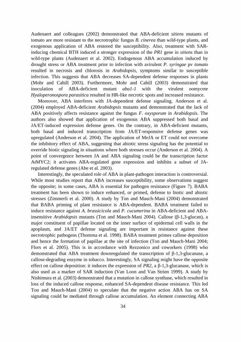
34
Audenaert and colleagues (2002) demonstrated that ABA-deficient sitiens mutants oftomato are more resistant to the necrotrophic fungus B. cinerea than wild-type plants, andexogenous application of ABA restored the susceptibility. Also, treatment with SAR-inducing chemical BTH induced a stronger expression of the PR1 gene in sitiens than inwild-type plants (Audenaert et al. 2002). Endogenous ABA accumulation induced bydrought stress or ABA treatment prior to infection with avirulent P. syringae pv tomatoresulted in necrosis and chlorosis in Arabidopsis, symptoms similar to susceptibleinfection. This suggests that ABA decreases SA-dependent defense responses in plants(Mohr and Cahill 2003). Furthermore, Mohr and Cahill (2003) demonstrated thatinoculation of ABA-deficient mutant aba1-1 with the virulent oomyceteHyaloperonospora parasitica resulted in HR-like necrotic spots and increased resistance.
Moreover, ABA interferes with JA-dependent defense signaling. Anderson et al.(2004) employed ABA-deficient Arabidopsis mutants and demonstrated that the lack ofABA positively affects resistance against the fungus F. oxysporum in Arabidopsis. Theauthors also showed that application of exogenous ABA suppressed both basal andJA/ET-induced expression defense genes. On the contrary, in ABA-deficient mutants,both basal and induced transcription from JA/ET-responsive defense genes wasupregulated (Anderson et al. 2004). The application of MeJA or ET could not overcomethe inhibitory effect of ABA, suggesting that abiotic stress signaling has the potential tooverride biotic signaling in situations where both stresses occur (Anderson et al. 2004). Apoint of convergence between JA and ABA signaling could be the transcription factorAtMYC2; it activates ABA-regulated gene expression and inhibits a subset of JA-regulated defense genes (Abe et al. 2003).
Interestingly, the speculated role of ABA in plant-pathogen interaction is controversial.While most studies report that ABA increases susceptibility, some observations suggestthe opposite; in some cases, ABA is essential for pathogen resistance (Figure 7). BABAtreatment has been shown to induce enhanced, or primed, defense to biotic and abioticstresses (Zimmerli et al. 2000). A study by Ton and Mauch-Mani (2004) demonstratedthat BABA priming of plant resistance is ABA-dependent. BABA treatment failed toinduce resistance against A. brassicicola and P. cucumerina in ABA-deficient and ABA-insensitive Arabidopsis mutants (Ton and Mauch-Mani 2004). Callose ( -1,3-glucan), amajor constituent of papillae located on the inner surface of epidermal cell walls in theapoplasm, and JA/ET defense signaling are important in resistance against thesenecrotrophic pathogens (Thomma et al. 1998). BABA treatment primes callose depositionand hence the formation of papillae at the site of infection (Ton and Mauch-Mani 2004;Flors et al. 2005). This is in accordance with Rezzonico and coworkers (1998) whodemonstrated that ABA treatment downregulated the transcription of -1,3-glucanase, acallose-degrading enzyme in tobacco. Interestingly, SA signaling might have the oppositeeffect on callose deposition: it induces the expression of PR2, a -1,3-glucanase, which isalso used as a marker of SAR induction (Van Loon and Van Strien 1999). A study byNishimura et al. (2003) demonstrated that a mutation in callose synthase, which resulted inloss of the induced callose response, enhanced SA-dependent disease resistance. This ledTon and Mauch-Mani (2004) to speculate that the negative action ABA has on SAsignaling could be mediated through callose accumulation. An element connecting ABA

35
and biotic stress responses might be the recently described poly (A)-specific endonucleaseAtPARN from Arabidopsis (Nishimura et al. 2005). The regulation of gene expression bythe mRNA-destabilizing activity of this protein is crucial for proper ABA and SAresponses (Nishimura et al. 2005).
Figure 7. The role of abscisic acid (ABA) in biotic stress responses (adapted from Mauch-Maniand Mauch 2005).
CROSSTALK BETWEEN SIGNALING PATHWAYS
Plants respond to a variety of abiotic and biotic stimuli from the environment. Followingperception of stress, several signal transduction pathways are switched on, resulting inphysiological and molecular changes in the plant. When pathways operating in defensesignaling are investigated, they are sometimes considered as independent units in order tosimplify the interpretation. However, it would be naive to think that signal transduction ismediated through isolated, linear pathways. Defense pathways influence each otherthrough a network of regulatory interactions, and thus, plant responses to various stressstimuli are a result of this complex interplay (Kunkel and Brooks 2002; Bostock 2005).
The term crosstalk is often used when discussing interactions in defense signaling.However, a good definition of what constitutes crosstalk does not exist, and differingopinions have been introduced concerning when it is appropriate to use this term todescribe plant defense signaling (Taylor and McAinsh 2004; Mundy et al. 2006).Uncertainty results partly because not all the components operating in the defensepathways are known. Nevertheless, crosstalk is usually described as including a networkof signal interactions in which functional outcomes can be positive, negative, or neutral

36
(Bostock 2005). In addition to different biotic stress signaling pathways, also biotic andabiotic pathways can “crosstalk”. This is exemplified by the effect of ABA on pathogendefense (Mauch-Mani and Mauch 2005), as discussed earlier. In conclusion, in most cases,ABA seems to have a negative effect on SA or JA signaling, impeding pathogen defense(Figures 7 and 8). However, on some occasions, ABA can have a positive impact onpathogen resistance; it enhances the accumulation of callose, and thus, increases resistanceto certain necrotrophic pathogens (Ton and Mauch-Mani 2004).
Several studies have described crosstalk among SA, JA, and ET signaling pathways(Kunkel and Brooks 2003; Bostock 2005). SA and JA signaling interact on many levels,and in most cases, this relationship seems to be mutually antagonistic (Kunkel and Brooks2002) (Figure 8). SA can inhibit the synthesis of JA and prevent the accumulation of PIsin response to JA, wounding, systemin, and oligosaccharides (Pena-Cortés et al. 1993;Doares et al. 1995). SA and its functional analogs have also been shown to prevent theexpression of JA-dependent defense genes on several occasions (Pena-Cortés et al. 1993;Gupta et al. 2000; Kunkel and Brooks 2002). Moreover, Petersen et al. (2000)demonstrated that MAP KINASE 4 (MPK4) regulates negative crosstalk between JA andSA in the activation of defenses. Gene induction triggered by JA is blocked in mpk4mutants, indicating the importance of this gene for mediation of the JA signal (Figures 5and 8). Simultaneously, this mutant constitutively expresses SA-regulated defense genes,probably as a result of the elevated SA levels. This suggests that a MAP kinase cascadeinvolving MPK4 represses SA biosynthesis and promotes either JA perception or response(Petersen et al. 2000). A node of convergence between SA and JA signaling seems to bethe plant-specific transcription factor WRKY70 (Li et al. 2004) (Figure 8). Plantsoverexpressing WRKY70 showed decreased JA- but enhanced SA-dependent defenseactivation, hence improving resistance to E. carotovora and P. syringae (Li et al. 2004).This indicates that WRKY70 integrates defense signals, and thus, affects pathwayactivation (Li et al. 2004).
Some evidence also supports synergism between SA and JA defenses. Simultaneousactivation of both SAR and rhizobacteria-triggered ISR resulted in an additive effect oninduced protection against P. syringae (VanWees et al. 2000). Moreover, ROS has beenshown to stimulate accumulation of SA and induction of SAR. At the same time, SAinduces the production of ROS such as hydrogen peroxide and NO (Van Camp et al.1998) (Figures 4 and 8). This synergism is thought to promote such defense responses asHR and killing of the pathogen.
Reported crosstalk between JA and ET signaling is mostly positive. Transcriptionfactors AtMYC2/JIN1 and ERF1 are important regulators of these interactions inArabidopsis (Lorenzo et al. 2004) (Figure 5). The expression of ERF1 (and its targetgenes) is synergistically activated by ET and JA, and ERF1 integrates these signals intothe activation of plant defenses (Lorenzo et al. 2003) (Figures 5 and 8). ET seems tomediate the interaction between MAPK and CDPK, both of which are triggered by abioticand biotic stress responses in Arabidopsis. Ludwig et al. (2005) demonstrated that CDPKsignaling triggers high ET levels, leading to inhibition of stimulus-dependent MAPKactivation. In rare cases, JA and ET have the opposite effects; in tobacco nicotinebiosynthesis, a direct defense against some herbivores is stimulated by JA and inhibited by
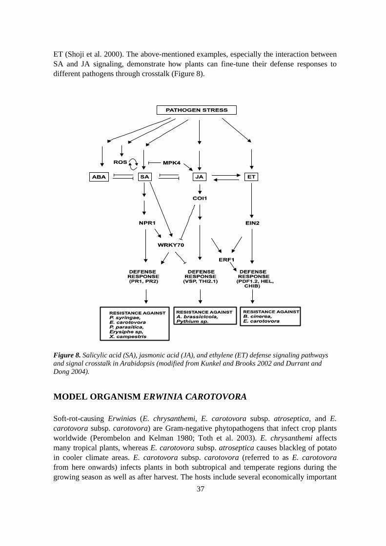
37
ET (Shoji et al. 2000). The above-mentioned examples, especially the interaction betweenSA and JA signaling, demonstrate how plants can fine-tune their defense responses todifferent pathogens through crosstalk (Figure 8).
Figure 8. Salicylic acid (SA), jasmonic acid (JA), and ethylene (ET) defense signaling pathwaysand signal crosstalk in Arabidopsis (modified from Kunkel and Brooks 2002 and Durrant andDong 2004).
MODEL ORGANISM ERWINIA CAROTOVORA
Soft-rot-causing Erwinias (E. chrysanthemi, E. carotovora subsp. atroseptica, and E.carotovora subsp. carotovora) are Gram-negative phytopathogens that infect crop plantsworldwide (Perombelon and Kelman 1980; Toth et al. 2003). E. chrysanthemi affectsmany tropical plants, whereas E. carotovora subsp. atroseptica causes blackleg of potatoin cooler climate areas. E. carotovora subsp. carotovora (referred to as E. carotovorafrom here onwards) infects plants in both subtropical and temperate regions during thegrowing season as well as after harvest. The hosts include several economically important

38
crop species such as potato, carrot (Daucus carota), turnip (Brassica rapa), and celery(Apium graveolens) (Perombelon and Kelman 1980; Toth et al. 2003).
The main weapon of E. carotovora in causing disease is wide a variety of plant cellwall-degrading enzymes (PCWDE) including polygalacturonases, pectinases, cellulases,and proteases (Perombelon and Kelman 1980). These enzymes macerate plant cell wallsand release nutrients for bacterial growth, hence the soft-rot phenotype of the infection.Besides extracellular enzymes, E. carotovora also secretes effector proteins to enhance itspathogenicity, and the hrp gene cluster encoding the TTSS has been characterized fromthis pathogen (Rantakari et al. 2001). One group of effector proteins secreted throughTTSS are the harpins, acidic, glycine-rich, heat-stable proteins including HrpZ from P.syringae and HrpN from E. chrysanthemi, E. amylovora, and E. carotovora (Bauer et al.1995; Cui et al. 1996; Rantakari et al. 2001). The role of harpins in virulence iscontroversial; the hrpN mutants of E. amylovora (Wei et al. 1992; Barny 1995) and E.chrysanthemi (Bauer et al. 1995) were less virulent than the wild-type form, whereas E.carotovora lacking hrpN remained fully pathogenic on tobacco plants (Cui et al. 1996).Harpins cause various effects on plants, including the HR, which is often associated withgene-for-gene interactions (Lahaye and Bonas 2001), despite no R-effector protein type ofinteraction involving harpin having been characterized thus far. The mechanismunderlying induction of HR and plant defenses by harpin is unclear, but they seem tointerfere with the ion balance of challenged cells by activating ion fluxes across theplasmalemma (Baker et al. 1993). Harpin from P. syringae binds lipid bilayers in vitro,resulting in the formation of ion-conducting pores, and induces formation of ROS (El-Maarouf et al. 2001; Lee et al. 2001; Samuel et al. 2005). Harpin can also induceresistance in plants; Jang et al. (2006) demonstrated that transgenic tobacco plantsoverproducing E. amylovora harpin are more resistant to the fungal pathogen B. cinerea.
A central factor influencing the success of the pathogen is the coordinate production ofvirulence determinants at the appropriate stage of the infection; not before the number ofbacteria is sufficient for efficient attack, but before the plant becomes aware of theinvasion and activates defenses. A sophisticated way to ensure correct timing is the use ofa system called quorum sensing. E. carotovora secretes and detects small diffusibleacylhomoserine lactone-based signal molecules called autoinducers (Pirhonen et al. 1993;Brader et al. 2005). The amount of secreted autoinducer increases as the bacterialpopulation multiplies in the plant. It finally reaches a level sufficient to activate theproduction of PCWDEs, and hence, attack on the plant. Interestingly, autoinducers areunstable in alkaline pH, and one of the early counterattacks of the plant may takeadvantage of this by alkalinizing the site of bacterial invasion to a pH of >8.2 (Byers et al.2002).
The virulence of E. carotovora is controlled by a complex regulatory network that caninfluence one or several virulence determinants. Many components that play a significantrole in this complex system have been identified (Marits et al. 2002; Hyytiäinen et al.2003). For example, inactivation of regulatory gene expA was shown to decrease theproduction of extracellular enzymes and result in an avirulent phenotype (Eriksson et al.1998). Saarilahti et al. (1992) also demonstrated that mutation in polygalacturonase

39
(PehA) alone decreases virulence. Studies by Saarilahti et al. (1992) and Eriksson et al.(1998) highlight the central role of PCWDEs in the virulence of E. carotovora.
E. carotovora is a nonspecific pathogen, i.e. a race-cultivar-specific resistance to thispathogen has not been characterized. The broad host range, including the model plantArabidopsis, makes this pathogen a good model for studying nonspecific plant-pathogeninteractions. E. carotovora triggers basal plant defense responses presumably through therecognition of pathogen-delivered general elicitors, such as LPS, or OGAs released fromplant cell walls by the action of PCWDEs. E. carotovora or E. carotovora elicitors triggerlocal and systemic induction of SA- and JA/ET-dependent defense genes in plants (Palvaet al. 1993; Vidal et al. 1997, 1998; Norman-Setterblad et al. 2000). Interestingly,resistance against this pathogen can be generated by the induction of either JA/ET-mediated (Vidal et al. 1998; Norman-Setterblad et al. 2000) or SA-mediated (Palva et al.1994; Li et al. 2004) defenses.
OPEN QUESTIONS
The study of defense mechanisms against pathogens is one of the faster moving fieldswithin plant science. Interactions between plants and the different microorganismsattacking them have been elucidated, revealing some of the molecular componentsinvolved. Moreover, the pathogen-triggered plant defense signaling networks engaged inthe induction of resistance are beginning to be pieced together. Plant defense responsesagainst pathogens are regulated through pathways currently known to involve three centralendogenous phytohormones: SA, JA, and ET. ABA has recently also been included in thisteam of hormones influencing the establishment of plant defense. Furthermore, ROS andNO add to the complexity of defense signal transduction.
Although progress and research producing novel data are impressive, many gaps inknowledge need to be filled before the big picture of plant defense signaling network canbe drawn or the information applied to the development of durable crops. The defensepathways interact significantly with each other, and therefore, the characterization ofpotential convergence points where pathway crosstalk occurs is particularly important.Due to the abundance of the signals and the myriad of interactions therein, delineating theroles of these signals in integrated networks is challenging. Nevertheless, this informationis critical for complete understanding of signaling events and outcomes in diverse stresssituations.
What can the study of the interaction between the pathogen E. carotovora and plantscontribute to the knowledge of the plant defense signaling network? Firstly, novelinformation would be yielded concerning the induction of basal defense responses inplants. Due to the nonspecific nature of the interaction between E. carotovora and plants,this knowledge can also be applied to interactions with other pathogens. Secondly, theresistance strategies developed to control E. carotovora are likely to be effective against abroad range of different plant pathogens. This can greatly benefit the development ofefficient and durable applications for plant protection _ the ultimate objective of plant-pathogen interaction studies.

40
AIMS OF THE STUDY
This work aimed at achieving novel information about the plant-pathogen interactionbetween the phytopathogen E. carotovora and the model plant Arabidopsis. This includedcharacterization of both the plant defense triggered by pathogen elicitors and the plantgenes involved in early responses to the pathogen.
Studies focused on the following topics:
- Characterization of plant defense responses triggered by the effector proteinHrpN of E. carotovora, and the plant responses synergistically elicited byeffector HrpN and PCWDE PehA
- Characterization of the specific roles of two Arabidopsis genes, AtCLH1 andERD15, involved in early plant defense responses triggered by E. carotovora
- Elucidation of how transgenic expression of these early pathogen responsivegenes alters plant defense responses against E. carotovora

41
MATERIALS AND METHODS
BIOLOGICAL MATERIAL
The biological material used in this work is described in detail in Studies I-III. In brief, weused Arabidopsis thaliana and Nicotiana tabacum wild-type species and transgenicArabidopsis plants. In addition, several different Arabidopsis mutants were employed.Erwinia carotovora subsp. carotovora strain SCC1 and Alternaria brassicicola strainCBSnr 567.77 were used as pathogen models. Escherichia coli strains JM109 and M15(pREP4) were applied for protein expression.
Method Used and described in study
Assay for bacterial virulence and growth in planta I-IIIAssay for freezing survival IIIAssay for drought tolerance IIIChlorophyll extraction IIDetermination of bacterial growth in planta I-IIIDiaminobenzidine staining of H2O2 II, IIIElectrolyte leakage assay for ABA-induced freezing tolerance IIIElicitor treatments of plants II, IIIExtraction of chlorophyll IIGeneration and characterization of transgenic plants II, IIIHigh-light stress treatment IIInfection of Arabidopsis with A. brassicicola IIInfection of Arabidopsis with E. carotovora I-IIIIsolation of plant RNA I-IIIIsolation of plant proteins II, IIINorthern blot analysis I-IIIPhytohormone extraction and quantification II, IIIPlant growth conditions I-IIIProtein expression in E. coli IPurification of 6xHis-tagged proteins IQuantitation of fungal biomass by PCR IISeed germination assay for ABA sensitivity IIIWestern blot analysis I, III

42
RESULTS AND DISCUSSION
Erwinia carotovora and elicitors HrpN and PehA trigger distinct butinteracting defense responses and cell death in Arabidopsis (I)
Induced defense responses in plants can be activated upon recognition of a pathogen, by apathogen-derived elicitor molecule, or by elicitors released from the plant by the action ofthe pathogen (Montesano et al. 2003; Nürnberger et al. 2004). E. carotovora is a broadhost range pathogen that uses an array of plant cell wall-degrading enzymes (PCWDE) asprimary virulence factors (Pérombelon and Kelman 1980; Kotoujansky 1987). Earlierstudies have demonstrated that PCWDEs release oligogalacturonide elicitors from plantcell walls (Palva et al. 1993) that elicit SA-independent defenses (Vidal et al. 1997, 1998).In addition, some E. carotovora strains have been shown to possess hrp genes thatcontribute to virulence (Rantakari et al. 2001). These include proteins required for type IIIsecretion (TTSS) and delivery of effector proteins to plant cells (Rantakari et al. 2001).Study I characterizes plant defense responses triggered by effector protein HrpN of E.carotovora (HrpNEcc). Plant responses synergistically induced by HrpNEcc and a PCWDE,the polygalacturonase PehA, were also investigated.
HrpN elicits lesion formation and induces resistance to E. carotovora in Arabidopsis
Harpins are acidic, glycine-rich, heat-stable effector proteins secreted through the TTSS.These proteins have been characterized from several phytopathogens, including HrpZfrom P. syringae and HrpN from E. carotovora (Bauer et al. 1995; Cui et al. 1996;Rantakari et al. 2001). The impact of harpin on virulence is somewhat inconsistent: it isessential for pathogenicity in E. amylovora and E. chrysanthemi (Wei et al. 1992; Barny1995), but dispensable in E. carotovora (Cui et al. 1996). Harpin has been demonstrated tocause various effects on plants, including the HR response. The role of HrpN in thevirulence of E. carotovora is particularly difficult to assess due to the massive productionof PCWDEs and maceration of plant tissue, which can mask any HR response. Toovercome this problem, we treated tobacco and Arabidopsis plants with purified HrpNprotein. The hrpN gene of E. carotovora strain SCC1 was isolated from a genomic lambdalibrary, cloned to an expression vector, and the N-terminally His6-tagged protein waspurified. Infiltration of purified HrpN protein caused visible, HR-like symptoms in bothtobacco and Arabidopsis, whereas no lesion formation was detected when plants weretreated with controls, MES buffer, or acetylated bovine serum albumin (I, Fig. 1B). HrpNtreatment was accompanied by increased ion leakage three to four days after infiltration (I,Fig. 1C), indicating cell death.
Earlier studies have shown that harpin treatment can induce resistance against E.amylovora in Arabidopsis (Dong et al. 1999) and P. syringae in cucumber (Strobel et al.
43
1996). This prompted us to examine whether E. carotovora HrpN could induce resistancein Arabidopsis. Leaves of wild-type Arabidopsis were pretreated with HrpN 24 h prior toinoculation with E. carotovora. After pathogen inoculation, a clear difference wasobserved in the maceration of the plants; HrpN-pretreated plants were significantly lessmacerated than MES-pretreated control plants (I, Fig. 2A). The bacterial numbers ininoculated plants also showed that after the initial multiplication the growth of E.carotovora was halted in HrpN-pretreated plants (I, Fig. 2B). This indicates that, similar toother harpins characterized, HrpNEcc is a potent inducer of lesion formation as well asdisease resistance in Arabidopsis.
Establishment of HrpN-triggered resistance requires both SA and JA signaling
Earlier studies have shown that resistance to E. carotovora can be generated by inductionof either JA/ET-mediated (Vidal et al. 1998; Norman-Setterblad et al. 2000) or SA-mediated (Palva et al. 1994) defenses triggered by distinct E. carotovora elicitors (Palva etal. 1993; Norman-Setterblad et al. 2000). It was therefore of great interest to elucidatewhich defense pathways HrpNEcc induced in Arabidopsis. Earlier work by Dong et al.(1999) and Strobel et al. (1996) demonstrates that E. amylovora and P. syringae harpinsinduce SAR in Arabidopsis and cucumber. To determine the contribution of HrpNEcc, wecharacterized the expression of two defense pathway specific marker genes: PR1responsive to SA and PDF1.2 responsive to JA/ET after treatment with this protein. Theexpression of these genes in response to HrpNEcc was compared with that induced byanother E. carotovora-derived elicitor, the polygalacturonase PehA, a known inducer ofJA/ET-dependent defenses (Vidal et al. 1998; Norman-Setterblad et al. 2000) and thepathogen itself. Inoculation of Arabidopsis with E. carotovora induced both PR1 andPDF1.2, while PehA triggered only PDF1.2 (I, Fig. 3A). Similar to harpins from E.amylovora and P. syringae, HrpNEcc triggered accumulation of PR1 transcripts (I, Fig.3A). However, in addition to PR1, HrpNEcc induced the expression of PDF1.2, which is incontrast to the earlier studies by Stroebel et al. (1996) and Dong et al. (1999)demonstrating that HrpNEcc is capable of triggering both SA- and JA/ET-dependentdefense signaling in Arabidopsis.
The involvement of both SA- and JA/ET-dependent pathways in the HrpNEcc-induceddefense response was further elucidated by the use of Arabidopsis mutants and transgenicplants impaired in defense signaling. HrpN did not induce resistance in transgenic NahGplants unable to accumulate SA (I, Fig. 4). Nor was resistance induced in JA-insensitivecoi1 (Feys et al. 1994) or JA amino acid synthetase mutant jar1 (Staswick and Tiryaki2004) in response to HrpN treatment. The double mutant NahG coi1, where both SA- andJA-dependent defense pathways were affected, was most severely macerated, and thebacteria grew to the highest density (I, Fig. 4). These results demonstrate that HrpN-induced resistance requires functional SA-dependent as well as JA-dependent signalpathways in Arabidopsis.

44
Synergistic action of elicitors HrpN and PehA triggers enhanced defense geneexpression and cell death
The enhanced expression of plant defense genes in response to inoculation with E.carotovora compared with either elicitor alone (I, Fig. 3A) prompted us to assess theeffect of simultaneous treatment with HrpN and PehA on wild-type Arabidopsis. Thecombined effect of these two elicitors led to both faster and stronger induction of PR1 andPDF1.2 (I, Fig. 5A). Moreover, the systemic induction of these genes was clearlyenhanced (I, Fig. 5B), indicating that HrpN and PehA synergistically trigger amplifiedplant defense response.
The role of ROS is central to the formation of the HR response, which is also inducedby HrpNEcc in Arabidopsis (I, Fig. 1B). The harpin of P. syringae has been suggested toamplify ROS production through the activation of AtMPK6 (Desikan et al. 2001).Moreover, simultaneous application of different elicitors has been shown to amplify theoxidative burst that follows (Chandra et al. 2000). Synergism of HrpN and PehA ininducing defense gene expression prompted us to test whether lesion formation andsuperoxide production were also enhanced. Indeed, simultaneous application of HrpN andPehA did result in a response that was clearly stronger than with either elicitor alone; theproduction of rapidly appearing HR-like lesions was accompanied by increased productionof superoxide and enhanced ion leakage, indicating accelerated cell death (I, Fig. 6A-C).
How might E. carotovora benefit from triggering multiple responses in the host,including cell death, which can be an effective plant defense response limiting pathogengrowth? Interestingly, responses, such as HR, that are effective against one pathogen canbe ineffective against another (Kunkel and Brooks 2002; Glazebrook 2005). EnhancedROS production can interfere with the plant metabolism, and the dying cells could providenutrients for the growing bacterial population. Cell death responses of the plant arepossibly also utilized by E. carotovora to spread infection. For example, in the case of thenecrotrophic fungus B. cinerea, the HR response has been demonstrated to facilitatepathogenesis (Govrin and Levine 2000). Alternatively, perhaps E. carotovora does notaim at triggering cell death or benefit from it. Both elicitors might act as PAMPs andtrigger plant defense, simultaneously being indispensable for the pathogenicity of E.carotovora. Saarilahti et al. (1992) have demonstrated that a mutation in the pehA genereduces the virulence of this pathogen. Whether the same holds true for HrpN remains tobe elucidated.
PLANT RESPONSES TO E. CAROTOVORA (II, III)
Early defense responses of the plant are crucial in determining the outcome of the plant-pathogen interaction, and thus, characterization of the molecular responses occurring earlyin the E. carotovora-Arabidopsis interaction was of great interest. To isolate the rapidlyinduced plant genes, Arabidopsis was treated with a preparation of E. carotovora elicitors.A cDNA library enriched for sequences preferentially expressed in elicitor-treated leaves

45
was established using a suppressive subtractive hybridization method (Brader et al. 2001).Approximately 200 differentially expressed genes were identified, and several of thesewere subjected to further analysis. Studies II and III elucidate the roles of two genes,CHLOROPHYLLASE 1 (AtCLH1) (II) and EARLY RESPONSIVE TO DEHYDRATION 15(ERD15) (III), in plant stress responses.
Studies II and III demonstrated how transgenic modification of two early pathogen-induced genes can improve the basal, SA-dependent defense response and lead toenhanced resistance against E. carotovora. Both studies also implicated pathway crosstalkin modulating defense responses. Study II demonstrated antagonism between SA- and JA-dependent signaling pathways, whereas Study III revealed the occurrence of crosstalkbetween biotic (SA) and abiotic (ABA) defense pathways.
CHLOROPHYLLASE 1 AFFECTS INDUCTION OF DEFENSEPATHWAYS IN PLANTS (II)
RNAi silencing of CHLOROPHYLLASE 1 (AtCLH1) improves resistance to E.carotovora in Arabidopsis
Degradation of chlorophyll is initiated by the dissociation of chlorophyll and its phytoltail, resulting in the formation of chlorophyllide. This reaction is catalyzed by the firstenzyme in the degradation pathway, chlorophyllase (Matile et al. 1999).CHLOROPHYLLASE 1 (AtCLH1) is one of the two chlorophyllases characterized fromArabidopsis, and it is rapidly induced in response to wounding and methyl jasmonate(MeJA) (Benedetti et al. 1998; Tsuchiya et al. 1999; Benedetti and Arruda 2002).Interestingly, our studies showed a rapid induction of this gene also in response to thepathogen E. carotovora, while no induction was observed after treatment with SA (II, Fig.1A). The expression pattern of this gene suggests that it could be a component of JA-dependent defense in Arabidopsis.
To elucidate the contribution of AtCLH1 in plant defense, it was either overexpressedor silenced with RNAi in Arabidopsis. The effect of the transgenes on AtCLH1 transcriptaccumulation was evaluated by Northern blot analysis using a gene-specific RNA probefor AtCLH1. Lines with enhanced and silenced expression of AtCLH1 were chosen forfurther studies (II, Fig. 1B). In addition, the transgenic lines were evaluated by measuringchlorophyll breakdown in plants. Overexpression of AtCLH1 resulted in increasedaccumulation of chlorophyllide, suggesting more efficient chlorophyll degradation in theseplants, whereas RNAi silencing of AtCLH1 led to a higher chlorophyll-chlorophyllideratio in transgenic plants (II, Fig. 1D).
To elucidate the effect of altered AtCLH1 expression on plant defense response, thetransgenic plants were infected with E. carotovora. After 24 h, it was clear that silencingof this gene made the plants more resistant to the pathogen (II, Fig. 2A). No or only minorsymptom development was observed in AtCLH1 RNAi-silenced plants, while in thecontrol and in AtCLH1 overexpression plants the maceration had already spread
46
considerably (II, Fig. 2A), suggesting that AtCLH1, and thus, chlorophyll degradationmight have a role in plant defense responses and that downregulation of this gene benefitsthe resistance of Arabidopsis against E. carotovora.
Resistance of AtCLH1 RNAi-silenced plants is light-dependent and involves increasedaccumulation of ROS
Due to their capability to harvest light, chlorophylls and their porphyrin ring-containingderivatives are photoactive (Matile et al. 1999). If the resistance of AtCLH1 RNAi plantsresulted from impaired chlorophyll degradation and phototoxicity of the chlorophyllmolecules, light conditions should make a difference in the outcome of the infection. Toexplore this, transgenic plants were inoculated with E. carotovora in low light conditions(~ 50 µmol min-2 sec-1 photons). The resistance of AtCLH1 RNAi plants observed innormal light conditions was abolished, and maceration proceeded similarly to vectorcontrol and AtCLH1 overexpression plants (II, Fig. 2C), indicating that the resistancearising from silencing of AtCLH1 could indeed be due to the phototoxic effects of thechlorophylls.
Why would downregulated chlorophyll degradation have an impact on pathogendefense? E. carotovora infection causes damage to plant tissues, resulting in the release ofchlorophyll molecules from the thylakoids. If not efficiently removed, these light-reactivemolecules can increase ROS formation in the cells (Takamiya et al. 2000). To avoid suchsituations, chlorophyll degradation needs to be initiated effectively and evidently;decreased AtCLH1 activity can hamper this process. Since ROS has an impact on cellularmetabolism and can also affect defense responses, it is not far-fetched that alteredchlorophyllase activity could influence the plant resistance response to E. carotovora. Toinvestigate this, we monitored ROS formation in response to E. carotovora inoculation innormal light conditions. Hydrogen peroxide accumulation was visualized by 3, 3´-diaminobenzidine (DAB) staining. Already 2 h after inoculation with the pathogen, theAtCLH1 RNAi-silenced plants had increased amounts of hydrogen peroxide incomparison with vector control plants (II, Fig. 3A). This also correlated with enhancedinduction of GLUTATHIONE-S-TRANSFERASE I (GSTI), a marker gene indicating theactivation of antioxidant defenses needed for ROS detoxification in plant cells (Conklinand Last 1995) ( II, Fig 3B).
The response of AtCLH1 RNAi plants to pathogen stress in combination with the rapidinducibility of this gene in response to wounding and defense elicitors strongly indicatethat AtCLH1 could act as a rapidly activated “emergency chlorophyllase” in initiatingchlorophyll degradation after tissue damage. A role for AtCLH1 in tissue repair was alsosuggested by Benedetti et al. (2001). High light stress can also cause tissue damage, andindeed, it seems that AtCLH1 is essential in protection against this type of stress as well –an observation that further suggests an essential role for this chlorophyllase in situationswhere rapid chlorophyll degradation is needed. Similarly to the pathogen response ofAtCLH1 RNAi-silenced plants, high light stress resulted in increased accumulation ofhydrogen peroxide and GSTI transcripts (II, Fig. 4A and B). Accordingly, previous studiesshow that transferring plants to high light conditions induces the degradation of the major

47
light-harvesting complex (LHCII) of photosystem II (PSII) (Yang et al. 1998). Thisprobably leads to chlorophyll release from LHCII, creating a need for rapid degradation ofthese light-reactive molecules.
AtCLH1 RNAi plants do not develop lesions, which is unexpected when comparedwith, for example, acd2 plants, which have a mutation in the RED CHLOROPHYLLCATABOLITE REDUCTASE (RCCR) gene and are compromised in chlorophylldegradation (Mach et al. 2001; Yao and Greenberg 2006). However, AtCLH1 is onlyinduced in response to tissue damage or injury, whereas ACD2 is constitutively expressedand presumably participates in all chlorophyll degradation occurring in the plant cell(Mach et al. 2001). The absence of ACD2 results in constant accumulation of photoactivechlorophyll breakdown products, and hence, lesions (Yao and Greenberg 2006). Benedettiand Arruda (2002) also showed that the expression of the second Arabidopsischlorophyllase gene, AtCLH2, did not vary in AtCLH1 antisense and overexpressionplants, and this could hypothetically mean that it can replace the missing AtCLH1 activityin case of injury. The free chlorophyll molecules would therefore eventually be degraded,albeit slowly, and ROS detoxified by the activated antioxidant defenses.
Silencing of AtCLH1 affects the balance between SA- and JA-dependent defenses inArabidopsis
What could be the mechanism through which altered chlorophyll metabolism affects plantdefense responses? ROS can be directly harmful to invading pathogens, and it has alsobeen implicated as a signal for activation of plant defense responses (Chen et al., 1995;Lamb and Dixon, 1997; Wojtaszek 1997; Bolwell 1999; Mullineaux et al., 2000). ROScan, for example, potentiate SA-dependent defense signaling (Durrant and Dong 2004). Toelucidate the role of different defense pathways in the observed resistance of AtCLH1RNAi-silenced plants to E. carotovora, we characterized the expression of marker genesfor these pathways. Moreover, to clarify the impact of light on pathway induction,expression analysis was done with plants infected in both normal and low light conditions.In the resistant AtCLH1 RNAi-silenced plants, expression of SAR marker genes PR1 andPR2 was clearly enhanced compared with vector control or overexpression plants (II, Fig.6A). Interestingly, this induction was abolished in low light conditions, where theexpression of PR1 and PR2 was reduced to the level of vector control plants (II, Fig. 6B),underlining the importance of light and phototoxic chlorophyll molecules in the observedresistance. The antagonism between SA- and JA-dependent defenses is well-characterized (Kunkeland Brooks 2002; Bostock 2005), and the enhanced SA defense prompted us to investigatewhether the JA-dependent defense response of the transgenic plants was altered.Compared with vector control plants, the induction of PDF1.2, a marker gene for JA-dependent defense, was clearly suppressed (II, Fig. 6D), suggesting that the silencing ofAtCLH1 upregulates SA-dependent but downregulates JA-dependent defense inArabidopsis. For further proof of the downregulation of JA-dependent defenses, westudied the response of AtCLH1 RNAi-silenced plants to the necrotrophic fungus A.brassicicola. Resistance to this pathogen is dependent on JA defenses: A. brassicicola is

48
unable to infect wild-type Arabidopsis, but the coi1 mutant is susceptible to it (Thomma etal. 1999, 1998). AtCLH1 RNAi-silenced and vector control plants were infected with A.brassicicola under normal light conditions, and disease development was followedvisually. After seven days, no disease symptoms were observed in the vector controlplants. However, approximately 70% of the A. brassicicola-infected leaves of AtCLH1RNAi plants showed disease development (II, Fig. 7), indicating downregulation of JA-dependent defenses in these plants. This could be a result of enhanced SA-dependentdefense that inhibits the activation of JA-dependent defenses.
Depending on the invading pathogen, plants employ different defense strategies. Fine-tuning the activation of defense genes and the balance between different signalingpathways is essential to achieve the best possible result in a battle against the invader.However, for a pathogen, ability to interfere with the defense response of the plant can bean asset when aiming at successful infection. How can E. carotovora benefit from theinduction of chlorophyllase? This pathogen seems to take advantage of the activation ofchlorophyll degradation (II, Fig 8); induction of AtCLH1 decreases the accumulation ofROS in response to tissue damage at an early stage of infection. Increased ROSaccumulation appears to enhance the basal SA-dependent defenses induced in response toE. carotovora, and without this effect, these defenses might not contain the infection andlead to resistance against this pathogen.
Activation of one defense signaling pathway can have an antagonistic effect on another(II, Fig. 8). For example, SA-dependent defenses are effective against P. syringaeinfection. However, this pathogen seems to exploit plant defense by producing the JA-mimicking toxin coronatine, which contributes to its virulence by inducing JA signalingand thereby repressing SA-dependent defense mechanisms (Bender et al. 1999; Greenberg2005). Early induction of AtCLH1, and hence, JA-dependent signaling, could have anantagonistic effect on SA-dependent defenses effective against E. carotovora, thus givingan advantage to this pathogen in the plant-pathogen battle.
Interestingly, silencing of AtCLH1 resulted in enhanced SA- but decreased JA/ET-defenses and resistance to E. carotovora. This is somewhat contradictory to the results ofStudy I, where HrpN-triggered resistance to E. carotovora required both SA- and JA/ET-dependent defense signaling. What makes it even more difficult to reach conclusionsconcerning plant resistance to E. carotovora is that earlier studies have shown that eitherJA/ET- or SA-dependent defenses alone triggered by different elicitors can be efficientagainst this pathogen (Palva et al. 1994; Norman-Setterblad et al. 2000; Li et al. 2004).Thus, resistance to the nonspecific pathogen E. carotovora can apparently be triggeredthrough different defense pathways. Both the timing and degree of the active defensecomponent might be critical to the outcome of the E. carotovora-plant interaction. Onecan hypothesize that in the case of silenced AtCLH1 the modulation of one component ofplant defense leads to induction of significantly enhanced SA responses that alone areefficient against E. carotovora. This response simultaneously antagonizes JA/ETsignaling, which is not needed in addition to the strong SA response. HrpN-inducedactivation of these pathways could be different _ less vigorous _ and thus not lead toantagonism. The synergism between the activation of SA and JA/ET defenses thenestablishes the resistance to E. carotovora. The difference between study systems should,

49
however, be taken into account. The role of AtCLH1 was investigated in transgenic plantswith an overexpression or RNAi-silencing construct of the gene, whereas the effect ofHrpN was monitored from plants infiltrated with a preparation containing this protein. Theactivation of defense responses is likely to be affected differently depending on the studysystem used.
ERD15 IS A NEGATIVE REGULATOR OF ABA RESPONSES INARABIDOPSIS (III)
ERD15 is rapidly induced by both biotic and abiotic factors
EARLY RESPONSIVE TO DEHYDRATION 15 (ERD15), a gene originally identified asinducible by drought (Kiyosue et al. 1994), was rapidly induced after treatment with E.carotovora elicitors. Moreover, ABA, SA, E. carotovora infection, and wounding inducedthis gene rapidly, suggesting that it could be a component of both biotic and abiotic stressresponses in Arabidopsis (III, Fig. 1A). Interestingly, while both SA- and JA-mediateddefenses are effective against E. carotovora in Arabidopsis, SA but not MeJA induced theexpression of ERD15 (III, Fig. 1A). The rapid inducibility of ERD15 indicates that it couldmediate early steps in the activation of plant stress signaling.
Modulation of ERD15 expression affects abiotic stress tolerance by influencing ABAsensitivity
The role of ERD15 in plant responses has been an enigma since it was first characterized(Kiyosue et al., 1994). Expression analysis of this gene (see above) did not shed any lighton this issue, and thus, transgenic plants carrying overexpression and RNAi-silencingconstructs of ERD15 were created. The effect of the transgenes was verified bycharacterizing the expression levels in Northern blot analysis, and accordingly, three lineswith increased and three lines with decreased expression of ERD15 were chosen forfurther studies (III, Fig. 1B). Surprisingly, of approximately 50 different lines, very fewwith clearly decreased ERD15 expression were found. The lines chosen were furtherevaluated with ERD15-specific antisera, demonstrating the effect of the transgenes also atthe protein level (III, Fig. 1C).
The inducibility of ERD15 by dehydration stress (Kiyuosue et al. 1994; III, Fig. 1D)prompted us to investigate the possible role of ERD15 in abiotic stress responses. To thisend, the transgenic ERD15 plants were challenged with two related stresses, drought andfreezing, both of which result in cellular dehydration (Thomashow 1999). The plants weredrought-stressed for two weeks in low humidity conditions, and their survival wasassessed visually. The difference between the transgenic lines was striking after twoweeks; most of the plants overexpressing ERD15 were dead, whereas the control plantsremained alive, although they did not appear very healthy and showed a strong

50
accumulation of anthocyanin (III, Fig. 2A). However, the majority (83%) of the ERD15RNAi-silenced plants were still alive and, unlike the control plants, had maintained theirgreen color (III, Fig. 2A, B). Applying freezing stress to the ERD15 transgenic plantssupported the observations from the drought experiment. Freezing stress brought about thegreatest damage to the plants overexpressing ERD15, while the plants with the RNAi-silenced gene clearly survived better than the control plants (III, Fig. 2C and D).
The phytohormone ABA is central in plant responses to dehydration stress; it inducesstomatal closure and production of desiccation protectants (Finkelstein and Gibson 2002;Zhu 2002; Zhu and Verslues 2005). The dramatic changes in the drought and freezingtolerance phenotypes of the transgenic ERD15 plants suggested that this protein might beinvolved in ABA-mediated stress responses in Arabidopsis. The application of ABA caninduce the development of freezing tolerance in higher plants such as wild potato (Chenand Gusta 1983) and Arabidopsis (Lång et al. 1989; Mäntylä et al. 1995). Interestingly,plants overexpressing ERD15 were impaired in their capacity to increase freezingtolerance in response to ABA treatment. This was observed as increased electrolyteleakage compared with wild-type plants after the ABA-treated plants were exposed tofreezing stress (III, Fig. 3A). This supports the argument that the changes in ERD15expression alter the sensitivity of the plants to ABA.
Expression of numerous plant genes involved in plant responses to abioticenvironmental stresses, especially those involved in drought response, is regulated byABA (Yamaguchi-Shinozaki and Shinozaki 2005). The expression of two ABA-responsive genes, RAB18 (Lång and Palva 1992) and LTI78 (Nordin et al. 1993), waselucidated in transgenic ERD15 plants (III, Fig. 4A). In plants overexpressing ERD15, thedrought-induced expression of these genes was reduced, indicating that ERD15 interfereswith ABA signaling in Arabidopsis (III, Fig. 4A).
Studies with Arabidopsis mutant abscisic acid-insensitive 1-1 (abi1-1) plantsdemonstrate that insensitivity to ABA can lead to an increase in the basal and stress-induced endogenous levels of ABA (Lång et al. 1989; Mäntylä et al. 1995). The same wasobserved in plants overexpressing ERD15 after the extraction of phytohormones (Schmelzet al. 2004); both basal and drought stress-induced ABA concentrations were higher thanthose of the wild-type plants (III, Fig. 4B). This supports the hypothesis that ERD15overexpression makes plants insensitive to ABA and that the observed increase in theendogenous hormone concentration is due to feedback regulation. The question thenarises: if overexpression of ERD15 decreases the sensing of ABA in these plants, couldsilencing of this gene do the opposite, result in hypersensitivity? The ABA germinationassays suggested exactly this; ERD15 RNAi-silenced plants germinated poorly and only avery small number of them had green cotyledons (III, Fig. 3B and C). One can alsohypothesize that the improved drought (III, Fig. 2A and B) and freezing (III, Fig. 2C andD) tolerance of ERD15-silenced plants could result from a more rapid response to thestress-induced ABA signal due to hypersensitivity of the plants. In conclusion, thedrought-tolerance phenotype, inhibition of seed germination by ABA, and the gene-expression data of transgenic plants all indicate that ERD15 has an impact on thecapability of Arabidopsis to sense ABA.

51
Insensitivity to ABA enhances resistance of Arabidopsis to E. carotovora
Interestingly, ERD15 is also induced by E. carotovora and SA in Arabidopsis (III, Fig.1A). Therefore, it was of interest to elucidate whether resistance to pathogens was alteredin the transgenic ERD15 plants. According to several recent studies, ABA is also involvedin plant defense responses to pathogens (Audenaert et al. 2002; Mohr and Cahill 2003;Ton and Mauch-Mani 2004; Anderson et al. 2005; Mauch-Mani and Mauch 2005). ABAhas been shown to have an antagonistic effect on SA (Audenaert et al. 2002; Mohr andCahill 2003) and JA (Anderson et al. 2005) -dependent defense responses, but it seems tobe essential in the BABA-induced callose accumulation needed to defend against certainnecrotrophic pathogens (Ton and Mauch-Mani 2004). Since our results strongly indicatedthat modulation of ERD15 expression in Arabidopsis decreased the sensitivity of theplants to ABA, we investigated whether this had an impact on the pathogen resistance oftransgenic plants. Transgenic ERD15 plants were inoculated with E. carotovora, and thedisease symptoms were followed. The symptom development of plants overexpressingERD15 was clearly reduced compared with that of control plants or plants with RNAi-silenced ERD15 (III, Fig. 6A). The enhanced resistance was also reflected in the lowbacterial numbers in inoculated plants (III, Fig. 6B), indicating that overexpression ofERD15, besides reducing ABA sensitivity, also promotes resistance to E. carotovora.
Earlier studies (Palva et al. 1993, 1994; Vidal et al. 1997, 1998; Norman-Setterblad etal. 2000; Li et al. 2004; I, II) have shown that resistance to E. carotovora can be generatedby either JA/ET- or SA-dependent mechanisms. The potential contribution of thesepathways to the observed resistance in ERD15 transgenics was elucidated bycharacterizing the induction of pathway-specific marker genes by Northern blot analysis.The expression pattern of these genes suggested that the resistance of ERD15overexpression plants was due to enhanced SA defense (III, Fig. 7B). This was seen afterinfection with E. carotovora but also after plant treatment with pathway elicitor SA. Thistreatment was done to eliminate possible difficulties in interpretation of gene expression inresponse to the pathogen due to variation in progress of the infection. In ERD15overexpression plants, the expression of PR2, a marker gene for SA signaling, was morerapidly induced in response to E. carotovora and enhanced after SA treatment (III, Fig.7B). This suggests that insensitivity to ABA enhances the induction of SA-dependentdefense responses. However, the induction of PDF1.2, a marker gene of the JA/ETpathway, was decreased in the more resistant ERD15 overexpression plants (III, Fig. 7A).This might be due to the enhanced SA-dependent defense and the well-characterizedantagonism between SA- and JA-dependent defense responses (Kunkel and Brooks 2002;Li et al. 2004; Bostock 2005).
The phenotype of ERD15 overexpression plants indicating that ABA insensitivitycould alter the resistance of Arabidopsis to E. carotovora by enhancing SA-dependentdefense signaling prompted us to study the impact of ABA insensitivity on pathogenresponses further. While earlier studies have characterized enhanced SA signaling only inABA-deficient and not in ABA-insensitive mutants, the BABA-primed accumulation of

52
callose required in the resistance against certain necrotrophic fungi is impaired in bothmutants (Ton and Mauch-Mani 2004), indicating that insensitivity can also affect defenseresponses. We employed the ABA-insensitive mutants abi1-1 and abi1-2 and determinedwhether the resistance of these plants was altered in response to E. carotovora.Interestingly, similar to ERD15 overexpression plants, both of these mutants displayedimproved resistance to this pathogen (III, Fig. 8A). This was accompanied by enhancedinduction of PR1, a marker for SA-dependent defenses (III, Fig. 8C). These resultsdemonstrate that not only ABA deficiency but also ABA insensitivity can have beneficialeffects on resistance to certain pathogens, such as E. carotovora, by enhancing SA-dependent defense responses.
ERD15 is a negative regulator of ABA responses in Arabidopsis
Our results suggest that ERD15 is a novel, negative regulator of ABA responses inArabidopsis (III, Fig. 9). Overexpression of ERD15 results in insensitivity to ABA,whereas RNAi silencing leads to a hypersensitive seed germination phenotype in thepresence of this phytohormone. The improved drought and freezing tolerance of theERD15-silenced plants can also be explained by ABA hypersensitivity; the response isactivated more rapidly, leading to enhanced tolerance. Moreover, in the case of ERD15overexpression, the insensitivity to ABA strengthens SA-dependent signaling (III, Fig. 9).This suggests that ERD15 might also be a factor influencing the selection of activation ofeither ABA- or SA-dependent stress responses. We hypothesized that ERD15 acts as a“capacitor” that prevents plants from responding too quickly after the onset of stress. Itprevents stress signaling until it is certain that the stress prevails and the plant´s survivaldepends on defense activation. Simply put, the role of the capacitor is to ensure that thelimited assets are invested in stress adaptation only when it is absolutely essential to do so.Activation of a large-scale defense response consumes plant resources that wouldotherwise be allocated to such targets as growth or reproduction. It is therefore beneficialfor the plant to avoid these fitness costs before it is certain that the stress will prevail andadaptation is necessary. Heil (2002) introduced this concept for biotic stress.
The actual mechanism underlying the influence of ERD15 on ABA signaling can onlybe speculated without further biochemical experimentation. ERD15 has a PAM2 motif,indicating an interaction with the C-terminus of poly-A-binding proteins (PABP)(Albrecht and Lengauer 2004). Indeed, Wang and Grumet (2004) demonstrated thebinding of ERD15 to PABP in a yeast two-hybrid screen. Thus, ERD15 may act byinhibiting translation of certain ABA-regulated transcripts in Arabidopsis. Accumulatingevidence also suggests a central role for posttranscriptional regulation in ABA signaling(Hugovieux et al. 2001; Himmelbach et al. 2003; Kuhn and Schroder 2003). The recentlycharacterized poly-A-specific ribonuclease AtPARN has been demonstrated to have poly(A) degradation activity in vitro (Chiba et al. 2004). This gene is induced by ABA and SA,and is presumed to be crucial for proper abiotic and biotic stress responses in Arabidopsis(Nishimura et al. 2005). ERD15, which also has an impact on both SA and ABAresponses, could be involved in the same processes; it has been demonstrated to interactwith PABPs that bind poly (A) tails of mRNAs, tails which AtPARN possibly degrades.

53
However, despite evidence of translational regulation in ABA signaling, a model puttingthese pieces together has thus far not been constructed. The role of ERD15 also remainselusive.
CONCLUDING REMARKS AND FUTURE PERSPECTIVES
The studies presented in this thesis provide novel data about the interaction betweenArabidopsis and E. carotovora. Roles of harpins in the virulence of several plantpathogens, including P. syringae and E. chrysanthemi, have been investigated, but the roleof E. carotovora HrpN as a virulence factor has remained uncharacterized until now. Thisis partly due to the massive maceration of plant tissue brought about by the action ofPCWDEs, which potentially masks plant responses induced by effector proteins. Usingpurified HrpN, we showed that this protein elicits an HR response and triggers resistanceto E. carotovora infection. The input of HrpN in E. carotovora-induced plant defense wasfurther clarified; it induces both SA- and JA/ET-dependent defense signaling inArabidopsis. Thus, HrpN might contribute to the capability of E. carotovora to triggerSA-dependent plant defense signaling. However, most probably other, currently unknowneffector proteins exist that have an impact on elicitation of plant defense.
Also demonstrated was that the power and efficiency of E. carotovora resides notonly in the massive production of PCWDEs but also in the parallel use of an effectorprotein. HrpN and the PCWDE PehA synergistically triggered enhanced cell death butalso enhanced plant defense signaling. Future studies will reveal whether the enhancedcell death actually benefits the pathogenicity of E. carotovora; as a necrotroph, it doesfeed from dead plant tissue. Alternatively, the pathogen might not necessarily aim atproducing cell death, with cell death instead resulting from the activation of plant defensesin response to pathogen elicitors. E. carotovora is nevertheless capable of overcoming thisresponse and infecting the plant. The question of whether or not HrpN is indispensable asan effector for the pathogenicity of E. carotovora will be addressed in future studiescharacterizing the virulence of the hrpN mutant. Finally, this work emphasizes thatclassification of pathogens to certain categories according to their lifestyle does notnecessarily mean that members of these groups trigger the same defense pathways. Whileinduction of HR and SA signaling is usually considered to occur in response to biotrophicpathogens (Glazebrook 2005), the necrotroph E. carotovora is also capable of inducingthis type of defense.
Studies II and III of this thesis characterized the plant defense-related roles of twogenes, AtCLH1 and ERD15, both rapidly induced in response to E. carotovora. Novel datademonstrating how the transgenic manipulation of these early stress-induced componentscan benefit (or weaken) plant stress tolerance were also uncovered. In addition, thesestudies demonstrated signal crosstalk between the defense pathways. First, silencing of

54
AtCLH1 suggested that the amount of ROS formed in plant cells increases after tissuedamage if rapid chlorophyll degradation is impaired. This was shown to be light-dependent. ROS accumulation enhanced SA-dependent signaling and resistance of theplants to E. carotovora. Therefore, we speculated that AtCLH1 acts as a damage controlenzyme that degrades free chlorophyll molecules in plant injury caused by, for example,pathogen attack.
Second, until now the function of ERD15 in plants was unknown, but our studiesplaced this gene in the signaling network mediating ABA responses in plants. Wehypothesized, that ERD15 is a novel, negative regulator that affects the capability ofplants to respond to a stress-related ABA signal. Transgenic modulation of ERD15expression influenced the ability of plants to tolerate abiotic as well as biotic stress.Improved pathogen resistance, presumably due to altered ABA sensing, was a furtherindication of the emerging theme that ABA influences biotic stress responses (reviewed byMauch-Mani and Mauch 2005).
Third, we were able to demonstrate crosstalk between the plant defense signalingpathways and show that it influences the outcome of defense responses. Silencing ofAtCLH1 enhanced SA-dependent defenses and resistance to E. carotovora, but also had anantagonistic effect on JA/ET signaling and resistance to the fungus A. brassicicola.Moreover, the modulation of ERD15 expression introduced crosstalk; it decreased ABAsensitivity of the plants, while SA-dependent defenses and resistance to E. carotovorawere enhanced.
The long-term objective of the studies presented in this thesis is to enhance thedevelopment of stress-resistant crop plants. To this end, consideration of the strengths andweaknesses of AtCLH1 and ERD15 studies in the context of applying them to cropprotection would be useful. However, certain facts should be borne in mind. Unlike in thelaboratory, in the field plants are challenged with more than one stress at a time, witheither combinations of different biotic stresses or situations where simultaneous biotic andabiotic stresses occur. Rendering the situation even more complex is the crosstalk betweenstress signaling pathways. Moreover, in the case of induced defenses, one must considerdifferent fitness costs (Heil and Baldwin 2002). Studies characterizing the role of AtCLH1and ERD15 in plant stress responses highlight different aspects that influence the totaleffectiveness of induced resistance. Often there is only a fine line between overall cost andbenefit; improving one characteristic easily impairs another. Silenced AtCLH1 activityexemplifies a tradeoff between defenses; resistance against one group of pathogens mightbe detrimental if the plant is attacked by another group. For AtCLH1-silenced plants, theenhanced SA defense and resistance to E. carotovora also had a reverse side. Wild-typeArabidopsis cannot be infected by the fungus A. brassicicola since it has functional JAdefenses to resist this pathogen. However, in plants with silenced AtCLH1, the resistanceto A. brassicicola was compromised, indicating antagonistic crosstalk between SA- andJA-dependent defenses. Thus, in the situation where plants are under threat from differentpathogens, improving only SA-dependent defenses would be insufficient. An interestingtopic for future research would be determining whether the modulation of AtCLH1 activityinfluences abiotic stress tolerance of transgenic plants.

55
Autotoxicity costs can be brought about by a continuously expressed resistance trait(Heil and Baldwin 2002); these are costs that establish a metabolic burden on the plant andhave physiological effects that manifest in the plant morphology. For example,Arabidopsis mutants expressing constant SA (cpr1) or JA (cev1) defenses often havestunted growth phenotypes (Bowling et al. 1994; Ellis and Turner 2001), indicating thatsuppression of defenses in enemy-free conditions is beneficial. Continuous accumulationof ROS can be toxic to the plants and could result in physiological effects, includinglesion formation. This has been observed in chlorophyll biosynthesis mutant acd2 plants(Greenberg and Yao 2004). However, in AtCLH1-silenced plants, the increase in oxidativestress and the subsequent activation of defense genes only occur when needed – afterpathogen invasion. Once the stress is over, ROS levels return to normal and the plant doesnot suffer from continuous metabolic imbalance. Thus, the phenotype of AtCLH1-silencedplants is normal, and they grow and produce seeds normally. In conclusion, while themanipulation of chlorophyll catabolism by silencing AtCLH1 has certain costs for theplant (reduction in JA defenses), it may offer an elegant means of enhancing the toleranceof agronomically important plant species to pathogens, like E. carotovora, that can becontrolled by SA-dependent responses.
Studies of ERD15 reveal crosstalk between abiotic and biotic stress signaling. ERD15overexpression resulted in diverse outcomes with regard to tolerance to drought andpathogen stress. SA-dependent defense signaling, and hence, resistance to E. carotovorawas improved. However, at the same time, the plants paid a considerable price for this inthe form of severely impaired tolerance to two abiotic stresses, drought and freezing,which arose from decreased ABA sensitivity. Silencing of ERD15 clearly improved planttolerance to these abiotic stresses, while the susceptibility of these plants to E. carotovorawas only moderately increased. This raises the question of how moderate abiotic stresses,such as mild drought, commonly encountered in crops during the growing seasoninfluence the capability of plants to induce pathogen resistance, or vice versa.
Allocation costs can occur if large quantities of fitness-limiting resources are allocatedto resistance (Heil and Baldwin 2002). Thus, induction of defenses is always a tradeoffwith some alternative target for energy, such as growth and reproduction. Activatingdefense responses only when it is absolutely necessary for the survival of the plant istherefore critical. In this context, ERD15 is hypothesized to act as a capacitor that guardsthe use of plant resources; it delays the ABA signal, and thus, activation of defenses, untilthe stress has continued for a certain period of time and exceeds the negative regulation.Only then is the inhibition relieved and the stress signaling aimed at initiating adjustiveresponses induced. Further studies are needed to characterize the specific gene products“guarded” by ERD15 and to figure out how this is executed at a molecular level.
A topic for future research would be to determine whether pathogen resistance ordrought tolerance of agronomically important species, such as potato could be altered bymodulating the expression of AtCLH1 or ERD15. Also awaiting elucidation is whether theeffects observed on stress tolerance under laboratory conditions hold true in the field.

56
ACKNOWLEDGMENTS
This study was carried out at the Department of Biological and Environmental Sciences,Division of Genetics, University of Helsinki, in the Viikki Biocenter. Funding wasprovided by the Academy of Finland and the Viikki Graduate School of Biosciences.
I am grateful to my supervisors, Professor Tapio Palva and Docent Günter Brader. I thankTapio for giving me the opportunity to do my Master´s work in his group and for thenenabling me to continue with a PhD thesis. His guidance, ideas, and encouragement overthese years have been invaluable. I am also indebted to Günter; without his guidance andhis brilliant scientific mind finishing this thesis would have been much more difficult.
I am thankful to Professors Katri Kärkkäinen and Teemu Teeri for kindly reviewing thisthesis. Carol Ann Pelli is acknowledged for editing the English language of themanuscript.
I thank all present and former members of Tapio Palva´s, Pekka Heino´s, HannuSaarilahti´s, and Jaakko Kangasjärvi´s groups. My warmest thanks are due to my friendsand colleagues Elina Helenius, Günter Brader, Solveig Sjöblom, Roosa Laitinen, HanneMikkonen, and Jorma Vahala with whom I have shared good moments during these years.I am also grateful to all plant-pathogen interaction people, especially Jing Li, Karen Sims-Huopaniemi, and Leila Miettinen, as well as the personnel on 5th floor; thank you for allthe support and help over the years. I’ve enjoyed working with all of you! Pekka Heinoand Hannu Saarilahti are also thanked for advising me about many practical mattersrelated to this thesis as well as for giving sound scientific advice. I am grateful to ArjaIkävalko and Arja Välimäki for all generous help with all manner of things.
My friends Reetta Kariola, Heikki Vilen, Mari Kaunisto, Hanna Kivelä, and TuulaSiljander are warmly thanked for friendship and the good times that we have shared.
I am particularly indebted to all of my friends outside science, especially Petra and Juuso,Ninni and Heka, Kikka and Kari, Pasi, Timo and Minna, and Jarkko and Minna for theirfriendship. Thank you also for not asking too often that when will I finally finish mythesisJ.
My heartfelt thanks go to my father Reijo, my late mother Anna-Liisa, and my brotherRisto and his family. Their endless support and faith in me as well as my father’scontinuous advice “Ota rauhallisesti” have helped me greatly particularly in moments ofdoubt.

57
Most of all, I thank my husband Kimmo, whose love, encouragement, and sense of humorare by far the most important things in my life.
Kotalahti, July 2006
Tarja Kariola
58
REFERENCES
Aarts, N., Metzdagger, M., Holub, E., Staskawicz, B.J., Daniels, M.J., and Parker, J.E. (1998).Different requirements for EDS1 and NDR1 by disease resistance genes define at least two R gene-mediated signaling pathways in Arabidopsis. PNAS 95, 10306-10311.
Abe, H., T. Urao, T. Ito, M. Seki, K. Shinozaki and K. Yamaguchi-Shinozaki (2003). ArabidopsisAtMYC2 (bHLH) and AtMYB2 (MYB) function as transcriptional activators in abscisic acid signaling,Plant Cell 15, 63–78.
Abramovitch, R.B. and Martin, G.B. (2004) Strategies used by bacterial pathogens to suppress plantdefenses. Current Opinion in Plant Biology 7, 356-364.
Abramovitch, R.B., Janjusevic, R., Stebbins, C.E., and Martin, G.B. (2006). Type III effector AvrPtoBrequires intrinsic E3 ubiquitin ligase activity to suppress plant cell death and immunity. Proc Natl AcadSci U S A. Feb 13, Epub ahead of print.
Agrios, G.N. (2005). Plant Pathology 5 th ed. Academic Press, San Diego, CA.Albrecht M, and Lengauer, T. (2004). Survey on the PABC recognition motif PAM2. Biochem Biophys
Res Commun. 26, 129-38.Alfano, J.R., and Collmer, A. (2004). TYPE III SECRETION SYSTEM EFFECTOR PROTEINS: Double
Agents in Bacterial Disease and Plant Defense. Annu Rev Phytopathol. 42, 385-414.Alméras, E., Stolz, S., Vollenweider, S., Reymond, P., Mène-Saffrané, L., and Farmer, E.E. (2003).
Reactive electrophile species activate defense gene expression in Arabidopsis. Plant J. 34, 205-216.Anderson, J.P., Badruzsaufari, E., Schenk, P.M., Manners, J.M., Desmond, O.J., Ehlert, C., Maclean,
D.J., Ebert, P.R., and Kazan, K. (2004). Antagonistic interaction between abscisic acid andjasmonate-ethylene signaling pathways modulates defense gene expression and disease resistance inArabidopsis, Plant Cell 16, 3460–3479.
Audenaert, K., De Meyer, G.B., and Höfte, M.M. (2002). Abscisic acid determines basal susceptibility oftomato to B. cinerea and suppresses salicylic acid dependent signaling mechanisms. Plant Physiol. 128,491-501.
Baker, C.J., Orlandi, E.W., and Mock, N.M. (1993). Harpin, An elicitor of the hypersensitive response intobacco caused by Erwinia amylovora, elicits active oxygen production in suspension cells. PlantPhysiol 102, 1341-1344.
Bartosz, G. (1997). Oxidative stress in plants. Acta Physiologia Plantarum 19, 47–64.Barny, M.A. (1995). Erwinia amylovora hrpN mutants, blocked in harpin synthesis, express a reduced
virulence on host plants and elicit variable hypersensitive reactions on tobacco. Eur J Plant Pathol 101,333-340.
Bauer, D.W., Wei, Z.M., Beer, S.V. and Collmer, A. (1995) Erwinia chrysanthemi harpinEch: an elicitorof the hypersensitive response that contributes to soft-rot pathogenesis. Mol. Plant Microbe Interact. 8,484–491
Bender, C.L., Alarcon-Chaidez, F., and Gross, D.C. (1999). Pseudomonas syringae phytotoxins: mode ofaction, regulation, and biosynthesis by peptide and polyketide synthetases. Microbiol. Mol. Biol. Rev.63, 266–292.
Benedetti, C.E., Costa, C.L., Turcinelli, S.R., and Arruda, P. (1998) Differential expression of a novelgene in response to coronatine, methyl jasmonate, and wounding in the Coi1 mutant of Arabidopsis.Plant Physiol. 116, 1037-1042.
59
Benedetti, C.E., and Arruda, P. (2002) Altering the expression of the chlorophyllase gene ATHCOR1 intransgenic Arabidopsis caused changes in the chlorophyll-to-chlorophyllide ratio. Plant Physiol. 128,1255-1263.
Benhamou, N. (1996). Elicitor-induced plant defense pathways. Trends Plant Sci. 1, 233-240.Bent, A.F., Inners, R.W., Ecker, J.R., and Staskawicz, B.J. (1992). Disease development in ethylene-
insensitive Arabidopsis thaliana infected with virulent and avirulent Pseudomonas and Xanthomonaspathogens. Mpl Plant Microbe Interact 5, 372-378.
Berger, S., Bell, E., Sadka, A., and Mullet, J.E. (1995). Arabidopsis thaliana Atvsp is homologous tosoybean VspA and VspB, genes encoding vegetative storage protein acid phosphatases, and is regulatedsimilarly by methyl jasmonate, wounding, sugars, light and phosphate. Plant Mol. Biol. 27, 933–942.
Bleecker, A.B. (1999). Ethylene perception and signalling: an evolutionary perspective. Trends in PlantScience 4, 269–274.
Bleecker, A.B., and Kende, H. (2000). Ethylene: A gaseous signal molecule in plants. Annu. Rev. CellDev. Biol. 16, 1–18.
Bolwell GP, Butt VS, Davies DR, and Zimmerlin A. (1995). The origin of the oxidative burst in plants.Free Radic Res. 23, 517-532.
Bolwell, G.P. (1999). Role of active oxygen species and NO in plant defence responses. Curr Opin PlantBiol. 2, 287-294.
Bonas, U., and Lahaye, T. (2002). Plant disease resistance triggered by pathogen-derived molecules:refined models of specific recognition. Curr Opin Microbiol. 5, 44-50.
Bostock, R.M. (2005). Signal crosstalk and induced resistance: straddling the line between cost and benefit.Annu Rev Phytopathol. 43, 545-580.
Boter, M., Rúiz-Rivero, O., Abdeen, A., and Prat, S. (2004). Conserved MYC transcription factors play akey role in jasmonate signaling both in tomato and Arabidopsis. Genes Dev. 18, 1577-1591.
Bowling, S.A., Guo, A., Cao, H., Gordon, A.S., Klessig, D.F., and Dong, X. (1994). A mutation inArabidopsis that leads to constitutive expression of systemic acquired resistance. Plant Cell 6, 1845-1857.
Brader, G., Tás, E., and Palva, E.T. (2001). Jasmonate-Dependent Induction of Indole Glucosinolates inArabidopsis by Culture Filtrates of the Nonspecific Pathogen Erwinia carotovora. Plant Physiol. 126,849-860.
Brader, G., Sjöblom, S., Hyytiäinen, H., Sims-Huopaniemi, K., and Palva, E.T. (2005). Alteringsubstrate chain length specificity of an acylhomoserine lactone synthase in bacterial communication. JBiol Chem. 280, 10403-10409.
Buchanan, B., Gruissem, W., and Jones, R. (eds). Biochemistry & Molecular Biology of Plants. AmericanSociety of Plant Physiologists 2000.
Byers, J. T., Lucas, C., Salmond, G. P. and Welch, M. (2002). Nonenzymatic turnover of an Erwiniacarotovora quorum-sensing signalling molecule. J Bacteriol 184, 1163–1171.
Cao, H., Bowling, S.A., Gordon, S., and Dong, X. (1994). Characterization of an Arabidopsis mutant thatis nonresponsive to inducers of systemic acquired resistance. Plant Cell 6, 1583–1592.
Cao, H., Glazebrook, J., Clarke, J.D., Volko, S., and Dong, X. (1997). The Arabidopsis NPR1 gene thatcontrols systemic acquired resistance encodes a novel protein containing ankyrin repeats. Cell 10, 57-63.
Cao, H., Li, X., and Dong, X. (1998). Generation of broad-spectrum disease resistance by overexpressionof an essential regulatory gene in systemic acquired resistance Proc Natl Acad Sci U S A 95, 6531-6536.
Century, K.S., Holub, E.B., and Staskawicz, B.J. (1995). NDR1, a locus of Arabidopsis thaliana that isrequired for disease resistance to both bacterial and fungal pathogens. Proc. Natl. Acad. Sci. USA 92,6597–6601.
Chandra, S., Cessna, S.G., Yahraus, T., Devine, R., and Low, P.S. (2000). Homologous and heterologousdesensitization and synergy in pathways leading to the soybean oxidative burst. Planta 211, 736-742.
Chang, C. (2003) Ethylene signalling: the MAPK module has finally landed. Trends Plant Sci. 8, 365–368.
60
Chen, T., and Gusta, L. (1983) Abscisic acid-induced freezing resistance in cultured plant cells. PlantPhysiol 73, 71–75.
Chen, Z., Silva, H., and Klessig, D.F. (1993). Active oxygen species in the induction of plant systemicacquired resistance by salicylic acid. Science. 17, 1883-1886.
Chen, Z., Malamy, J., Henning, J., Conrath, U., Sanchez-Casas, P., Silva, H., Ricigliano, J., andKlessig, D.K. (1995). Induction, modification, and transduction of the salicylic acid signal in plantdefense responses. Proc. Natl. Acad. Sci. U S A 92, 4134-4137.
Chiba, Y., Johnson, M.A., Lidder, P., Vogel, J.T., van Erp, H., and Green, P.J. (2004). AtPARN is anessential poly(A) ribonuclease in Arabidopsis. Gene 328, 95-102.
Chisholm, S., Coaker, G., Day, B., and Staskawicz, B. (2006). Host-Microbe Interactions: Shaping theEvolution of the Plant Immune Response. Cell 124, 803-814.
Coquoz, J.L., Buchala, A.J., Meuwly, P., and Métraux, J.P. (1995). Arachidonic acid induces local butnot systemic synthesis of salicylic acid and confers systemic resistance in potato plants to Phytophthorainfestans and Alternaria solani. Phytopathology. 85, 1219-1224.
Conklin, P.L., and Last, R.L. (1995) Differential accumulation of antioxidant mRNAs in Arabidopsisthaliana exposed to ozone. Plant Physiol. 109, 203-212.
Conlon, I., and M. Raff. (1999). Size control in animal development. Cell 96, 235-244.Conrath, U., Pieterse, C.M.J., and Mauch-Mani, B. (2002). Priming in plant–pathogen interactions.
Trends Plant Sci 7, 210–216.Cook, K.A., Weinzierl, R.A., Pataky, J.K., Esker, P.D., and Nutter, F.W. Jr. (2005). Population
densities of corn flea beetle (Coleoptera: Chrysomelidae) and incidence of Stewart's wilt in sweet corn. JEcon Entomol. 98, 673-682.
Crawford, N.M., and Guo, F.M. (2005). New insights into nitric oxide metabolism and regulatoryfunctions. Trends Plant Sci. 4, 195-200.
Creelman, R.A., and Mullet, J.E. (1997). Biosynthesis and action of jasmonates in plants. Ann. Rev. PlantPhysiol. Plant Mol. Biol. 48, 355–381.
Cui, Y., Madi, L., Mukherjee, A., Dumenyo, C.K., and Chatterjee, A. (1996). The RsmA-mutants ofErwinia carotovora subsp. carotovora strain Ecc71 overexpress hrpNEcc and elicit a hypersensitivereaction-like response in tobacco leaves. Mol Plant-Microbe Interact 9, 565-573.
da Cunha, L., McFall, A.J., and Mackey, D. (2006). Innate immunity in plants: A continuum of layereddefenses. Microbes and Infection 1-10.
Cutler, S., Ghassemian, M., Bonetta, D., Cooney, S., and McCourt, P. (1996). A protein farnesyltransferase involved in abscisic acid signal transduction in Arabidopsis. Science 273, 1239–1241
Dangl, J.L., and Jones, J.D. (2001). Plant pathogens and integrated defence responses to infection. Nature411, 826-833.
Dardick, C., and Ronald, P. (2006). Plant and animal pathogen recognition receptors signal through non-RD kinases. PLoS Pathog 2, e2.
Dat, J., Vandenbeele, S., Vranova, E., Van Montagu, M., Inzé, D., and Van Breusegem, F. (2000). Dualaction of the active oxygen species during plant stress responses. Cellular and Molecular Life Sciences57, 779–795.
Deikman, J. (1997). Molecular mechanism of ethylene regulation of gene transcription. Physiol. Plant. 100,561–566.
Delaney, T. P., Uknes, S., Vernooij, B., Friedrich, L., Weymann, K., Negrotto, D., Gaffney, T., Gut-Rella, M., Kessmann, H., Ward, E. and Ryals, J. (1994). A central role of salicylic acid in plantdisease resistance. Science 266, 1247-1250.
Delaney, T.P., Friedrich, L., and Ryals, J.A. (1995). Arabidopsis signal transduction mutant defective inchemically and biologically induced disease resistance. Proc. Natl. Acad. Sci. USA 92, 6602–6606.
Delledonne, M. (2005). NO news is good news for plants. Curr Opin Plant Biol. 8, 390-396.

61
Desikan, R., Hancock, J.T., Ichimura, K., Shinozaki, K., and Neill, S.J. (2001). Harpin inducesactivation of the Arabidopsis mitogen-activated protein kinases AtMPK4 and AtMPK6. Plant Physiol.126, 1579-1587.
Déspres, C., Chubak, C., Rochon, A., Clark, R., Bethune, T., Desveaux, D., and Fobert, P.R. (2003).The Arabidopsis NPR1 disease resistance protein is a novel cofactor that confers redox regulation ofDNA binding activity to the basic domain/leucine zipper transcription factor TGA1. Plant Cell 15, 2181-2191.
Devoto, A., Nieto-Rostro, M., Xie, D., Ellis, C., Harmston, R., Patrick, E., Davis, J., Sherratt, L.,Coleman, M., and Turner, J.G. (2002). COI1 links jasmonate signalling and fertility to the SCFubiquitin-ligase complex in Arabidopsis. Plant J. 32, 457-466.
Devoto, A., and Turner, J.G. (2003). Regulation of jasmonate-mediated plant responses in Arabidopsis.Ann Bot (Lond). 92, 329-337.
Dharmasiri, N., Dharmasiri, S., and Estelle, M. (2005). The F-box protein TIR1 is an auxin receptor.Nature 435, 441-445.
Dietrich, R.A., Delaney, T.P., Uknes, S.J., Ward, E.R., Ryals, J.A., and Dangl, J.L. (1994). Arabidopsismutants simulating disease resistance response. Cell 77, 565-577.
Doares, S.H., Narvez-Vasques, J., Conconi, A., and Ryan, C.A. (1995). Salicylic acid inhibits synthesisof proteinase inhibitors in tomato leaves induced by systemin and jasmonic acid. Plant Physiol. 108,1741-1746.
Doke, N. (1983). Generation of superoxide anion by potato tuber protoplasts during the hypersensitiveresponse to hyphal wall components of Phytopthora infestans and specific inhibition of the reaction bysuppressors of hypersensitivity. Physiol. Plant Pathol. 23, 359-367.
Dong, H., Delaney, T. P., Bauer, D.W. and Beer, S. V. (1999). Harpin induces disease resistance inArabidopsis through the systemic acquired resistance pathway mediated by salicylic acid and the NIM1gene. Plant J 20, 207-215.
Du, H., and Klessig, D.F. (1997). Identification of a soluble, high-affinity salicylic acid-binding protein intobacco. Plant Physiol. 113, 1319–1327.
Durner, J., and Klessig, D.F. (1995). Inhibition of ascorbate peroxidase by salicylic acid and 2,6-dichloroisonicotinic acid, two inducers of plant defense responses. Proc Natl Acad Sci U S A. 92,11312-11316.
Durrant, W.E., and Dong, X. (2004). Systemic acquired resistance. Annu Rev Phytopathol. 42, 185-209.Ecker, J.R. (2004). Reentry of the ethylene MPK6 module. Plant Cell 16, 3169–3173.Edwards, H.H. (1983). Effect of kinetin, abscisic acid and cations on host–parasite relation of barley
inoculated with Erysiphe graminis f. sp. hordei. J Phytopath 107, 22–30.El-Maarouf, H., Barny, M.A., Rona, J.P., and Bouteau, F. (2001). Harpin, a hypersensitive response
elicitor from Erwinia amylovora, regulates ion channel activities in Arabidopsis thaliana suspensioncells. FEBS Lett 25, 82–84.
Ellis, J., Dodds, P., and Pryor, D. (2002). Structure, function and evolution of plant disease resistancegenes. Curr Opin Plant Biol 4, 278-284.
Ellis, C., and Turner, J.G. (2001). The Arabidopsis Mutant cev1 has constitutively active jasmonate andethylene signal pathways and enhanced resistance to pathogens. Plant Cell 13, 1025-1033.
Ellis, C., Karafyllidis, I., and Turner, J.G. (2002). Constitutive activation of jasmonate signaling in anArabidopsis mutant correlates with enhanced resistance to Erysiphe cichoracearum, Pseudomonassyringae, and Myzus persicae. Molecular Plant Microbe Interact. 15, 1025-1030.
Epple, P., Apel, K., and Bohlmann, H. (1995). An Arabidopsis thaliana thionin gene is inducible via asignal transduction pathway different from that for pathogenesis-related proteins. Plant Physiol. 109,813-820.
Eriksson, A. R. B., Andersson, R. A., Pirhonen, M. & Palva, E. T. (1998). Two-component regulatorsinvolved in the global control of virulence in Erwinia carotovora subsp. carotovora. Mol Plant–MicrobeInteract 11, 743-752.

62
Espinosa, A., and Alfano, J.R. (2004). Disabling surveillance: bacterial type III secretion system effectorsthat suppress innate immunity. Cell Microbiol 6, 1027-1040.
Farmer, E. E. (2001). Surface-to-air signals. Nature 411, 854-856.Farmer, E.E., Almeras, E., and Krishnamurthy, V. (2003). Jasmonates and related oxylipins in plant
responses to pathogenesis and herbivory. Curr Opin Plant Biol. 6, 372-378.Flors, V., Ton, J,. Jakab, G., and Mauch-Mani, B. (2005). Abscisic acid and callose: team players in
defense against pathogens?, J Phytopathol 153, 1–7.Felix, G., Duran, J.D., Volko, S., and Boller, T. (1999). Plants have a sensitive perception system for the
most conserved domain of bacterial flagellin. Plant J. 8, 265-276.Feng, S., Ma, L., Wang, X., Xie, D., Dinesh-Kumar, S.P., Wei, N., and Deng, X.W. (2003). The COP9
signalosome interacts physically with SCF COI1 and modulates jasmonate responses. Plant Cell 15,1083-1094.
Feys, B., Benedetti, C.E., Penfold, C.N., and Turner, J.G. (1994). Arabidopsis mutants selected forresistance to the phytotoxin coronatine are male sterile, insensitive to methyl jasmonate, and resistant toa bacterial pathogen. Plant Cell 6, 751-759.
Feys, B.J., and Parker, J.E. (2000). Interplay of signaling pathways in plant disease resistance. TrendsGenet 16, 449–455.
Feys, B.J., Moisan, L.J., Newman, M.A., and Parker, J.E. (2001). Direct interaction between theArabidopsis disease resistance signaling proteins, EDS1 and PAD4. EMBO J. 20, 5400-5411.
Finkelstein, R.R., and Gibson, S.I. (2002). ABA and sugar interactions regulating development: 'cross-talk'or 'voices in a crowd'? Curr. Opin. Plant Biol. 5, 26-32.
Finkelstein, R.R., and Rock, C.D. (2002). Abscisic acid biosynthesis and response. The Arabidopsis Book.Editors CR Somerville and EM Meyerowitz. American Society of Plant Biologists, Rockville MD USA.
Foyer, C.H., Lelandais, M., and Kunert, K.J. (1994). Photo-oxidative stress in plants. Physiol Plant. 92,696-717.
Fought, L., and Kuc, J.A. (1996). Lack of specificity in plant extracts and chemicals as inducers ofsystemic resistance in cucumber plants to anthracnose. Journal of Phytopathology (Berlin) 144, 1-6.
Friedrich, L., Lawton, K., Dietrich, R., Willits, M., Cade, R., and Ryals, J. (2001). NIM1 overexpressionin Arabidopsis potentiates plant disease resistance and results in enhanced effectiveness of fungicides.Mol Plant Microbe Interact. 14, 1114-1124.
Fujimoto, S.Y., Ohta, M., Usui, A., Shinshi, H., and Ohme-Takagi, M. (2000). Arabidopsis ethylene-responsive element binding factors act as transcriptional activators or repressors of GCC box-mediatedgene expression. Plant Cell. 12, 393–404.
Gaffney, T., Friedrich, L., Vernooij, B., Negrotto, D., Nye, G., Uknes, S., Ward, E., Kessman, H., andRyals, J. (1993). Requirement of salicylic acid for the induction of systemic acquired resistance.Science 261, 754-756.
Gassmann, W., Hinsch, M.E., and Staskawicz, B.J. (1999). The Arabidopsis RPS4 bacterial-resistancegene is a member of the TIR-NBS-LRR family of disease-resistance genes. Plant J. 20, 265-277.
Genoud, T., Buchala, A., J., Chua, N.-H., and Métraux, J.-P. (2002). Phytochrome signalling modulatesthe SA-perceptive pathway in Arabidopsis. Plant J. 31, 87–95.
Glazebrook, J. (2001). Genes controlling expression of defense responses in Arabidopsis--2001 status. CurrOpin Plant Biol. 4, 301-308.
Glazebrook, J. (2005). Contrasting mechanisms of defense against biotrophic and necrotrophic pathogens.Annu Rev Phytopathol. 43, 205-227.
Glazebrook, J., Zook, M., Mert, F., Kagan, I., Rogers, E.E., Crute, I.R., Holub, E.B., Hammerschmidt,R., and Ausubel, F.M. (1997). Phytoalexin-deficient mutants of Arabidopsis reveal that PAD4 encodesa regulatory factor and that four PAD genes contribute to downy mildew resistance. Genetics 146, 381-392.
Glazebrook J, Rogers EE, Ausubel FM. (1996). Isolation of Arabidopsis mutants with enhanced diseasesusceptibility by direct screening. Genetics 143, 973–982.

63
Gómez-Gómez, L., Felix, G. and Boller, T. (1999) A single locus determines sensitivity to bacterialflagellin in Arabidopsis thaliana. Plant J. 18, 277–284.
Gómez-Gómez, L. and Boller, T. (2002) Flagellin perception: a paradigm for innate immunity. TrendsPlant Sci. 7, 251–256.
Gosti, F., Beaudoin, N., Serizet, C., Webb, A.A., Vartanian, N., and Giraudat, J. (1999). ABI1 proteinphosphatase 2C is a negative regulator of abscisic acid signaling. Plant Cell. 11, 1897-1910.
Govrin, E., and Levine, A. (2000). The hypersensitive response facilitates plant infection by thenecrotrophic pathogen Botrytis cinerea, Curr. Biol. 10, 751–757.
Grant J., and Loake, G.J. (2000). Redox Signalling during the Establishment of Plant Disease Resistance.Plant Physiology 123, 411-419.
Greenberg, J. (1997). Programmed cell death in plant-pathogen interactions. Annu Rev Plant Physiol PlantMol Biol. 48, 525-545.
Greenberg, J., and Yao, N. (2004). The role and regulation of programmed cell death in plant–pathogeninteractions. Cel Microbiol 6, 201-211.
Grene, R. (2002). Oxidative stress and acclimation mechanisms in plants. The Arabidopsis Book. eds. C.R.Somerville and E.M. Meyerowitz, American Society of Plant Biologists, Rockville, MD,doi/10.1199/tab.0036.
Guo, H., and Ecker, JR (2004). The ethylene signaling pathway: New insights. Curr. Opin. Plant Biol. 7,40–49.
Guo, Y., Xiong, L., Song, C.-P., Gong, D., Halfter, U., and Zhu, J.-K. (2002). A calcium sensor and itsinteracting protein kinase are global regulators of abscisic acid signaling in Arabidopsis. Dev. Cell 3,233–244.
Gupta, V., Willits, M.G., and Glazebrook, J. (2000) Arabidopsis thaliana EDS4 contributes to salicylicacid (SA)-dependent defense responses: Evidence for inhibition of jasmonic acid signaling by SA. Mol.Plant-Microbe Interact. 13, 503-511.
Halkier, B.A., and Gershenson, J. (2006). Biology and Biochemistry of Glucosinolates. Annu Rev PlantBiol. Jan 30; [Epub ahead of print].
Hammerschmidt, R. (1999). Phytoalexins: what have we learned after 60 years? Annu Rev Phytopathol.34, 387-412.
Hammond-Kosack, K.E., and Parker, J.E. (2003). Deciphering plant–pathogen communication: freshperspectives for molecular resistance breeding. Curr Opin Biotech 14, 177-193.
Han, M-H., Goud, S., Song, L., and Fedoroff, N. (2004). The Arabidopsis double-stranded RNA-bindingprotein HYL1 plays a role in microRNA-mediated gene regulation. PNAS 101, 1093-1098.
Hauck, P., Thilmony, R. and He, S.Y. (2003). A Pseudomonas syringae type III effector suppresses cellwall-based extracellular defense in susceptible Arabidopsis plants. PNAS USA 100, 8577–8582.
He, Z.-H., He, D., and Kohorn, B. (1998). Requirement for the induced expression of a cell wall associatedreceptor kinase for survival during the pathogen response. Plant J 14, 55–63.
Heath, M.C. (2000). Nonhost resistance and nonspecific plant defenses. Curr Opin Plant Biol. 3, 315-319.Heck ,S., Grau, T., Buchala, A., Métraux, J.P., and Nawrath, C. (2003). Genetic evidence that
expression of NahG modifies defence pathways independent of salicylic acid biosynthesis in theArabidopsis-Pseudomonas syringae pv. tomato interaction. Plant J. 36, 342-352.
Heil, M. (2002). Ecological costs of induced resistance. Curr Opin Plant Biol. 5, 1-6.Heil, M., and Baldwin, I.T. (2002).Fitness costs of induced resistance: emerging experimental support for a
slippery concept. Trends Plant Sci. 7, 61-67.Heino, P., Sandman, G., Lång, V., Nordin, K., and Palva, E.T. (1990). Abscisic acid deficiency prevents
development of freezing tolerance in Arabidopsis thaliana L. Heynh. Theoret Appl Genet 79, 801-806.Himmelbach, A., Yang, Y., and Grill, E. (2003). Relay and control of abscisic acid signaling. Curr Opin
Plant Biol.6, 470-479.

64
Hoffman, T., Schmidt, J.S., Zheng, X., and Bent, A.F. (1999). Isolation of ethylene-insensitive soybeanmutants that are altered in pathogen susceptibility and gene-for-gene disease resistance Plant Physiol119, 935-950.
Holt, B.F. 3rd, Hubert, D.A., and Dangl, J.L. (2003). Resistance gene signaling in plants--complexsimilarities to animal innate immunity. Curr Opin Immunol. 15, 20-25.
Holub, E.B. (2001). The arms race is ancient history in Arabidopsis, the wildflower. Nature Rev Gen 2,516-527.
Hoth, S., Morgante, M., Sanchez, J.P., Hanafey, M.K., Tingey, S.V., and Chua, N.H. (2002). Genome-wide gene expression profiling in Arabidopsis thaliana reveals new targets of abscisic acid and largelyimpaired gene regulation in the abi1-1 mutant. J Cell Sci 115, 4891–4900.
Howe, G. (2004). Jasmonates as signals in the wound response. J. Plant Growth Regul. 23, 223-237.Hua, J., Sakai, H., Nourizadeh, S., Chen, Q.G., Bleecker, A.B., Ecker, J.R., and Meyerowitz, E.M.
(1998). Ein4 and ERS2 are members of the putative ethylene receptor family in Arabidopsis. Plant Cell10, 1321–1332.
Hugouvieux, V., Kwak, J.M., and Schroeder, J.I. (2001). An mRNA cap binding protein, ABH1,modulates early abscisic acid signal transduction in Arabidopsis. Cell 106, 477-487.
Hyytiäinen, H., Sjöblom, S., Palomäki, T., Tuikkala, A., and Palva, E.T. (2003). The PmrA-PmrB two-component system responding to acidic pH and iron controls virulence in the plant pathogen Erwiniacarotovora ssp. carotovora. Mol Microbiol. 50, 795-807.
Ishigaki, Y., Li, X., Serin, G., and Maquat, L.E. (2001). Evidence for a pioneer round of mRNAtranslation: mRNAs subject to nonsense-mediated decay in mammalian cells are bound by CBP80 andCBP20. Cell 106, 607–617.
Jakab, G., Cottier, V, Toquin, V., Rigoli, G., Zimmerli, L., Métraux, J.-P. and Mauch-Mani, B. (2001).-Aminobutyric Acid-induced resistance in plants. Eur J Plant Pathol 107, 29-37.
Jamir, Y., Guo, M., Oh, H.S., Petnicki-Ocwieja, T., Chen, S., Tang, X., Dickman, M.B., Collmer, A.,and Alfano, J.R. (2004). Identification of Pseudomonas syringae type III effectors that can suppressprogrammed cell death in plants and yeast. Plant J. 37, 554-65.
Jang, Y-S., Sohn, S-I., and Wang, M-H. (2006). The hrpN gene of Erwinia amylovora stimulates tobaccogrowth and enhances resistance to Botrytis cinerea. Planta 223, 449-456.
Janjusevic, R., Abramovitch, R., Martin, G.B., and Stebbins, E.C. (2005). A bacterial inhibitor of hostprogrammed cell death defenses is an E3 ubiquitin ligase. Science 311, 222 – 226.
Johnson, K.L., and Ingram, G.C. (2005). Sending the right signals: regulating receptor kinase activity.Curr Opin Plant Biol 8, 648-656.
Karpinski, S., Gabrys, H., Mateo, A., Karpinska, B., and Mullineaux, P.M. (2003). Light perception inplant disease defense signalling. Curr. Opin. Plant Biol. 6, 390-396.
Keen, N.T. (1990). Gene-for-gene complementarity in plant–pathogen interactions. Annu Rev Genet 24,447–463
Keller T, Damude HG, Werner D, Doerner P, Dixon RA, and Lamb, C. (1998) A plant homologue ofthe neutrophil NADPH oxidase gp91phox subunit gene encodes a plasma membrane protein with Ca2+binding motifs. Plant Cell 10, 255-266.
Keller, N.P., Turner, G., and Bennett, J.W. (2005). Fungal secondary metabolism — from biochemistryto genomics. Nature Reviews Microbiology 3, 937-947.
Kettner, J., and Dörfling, K. (1995). Biosynthesis and metabolism of abscisic acid in tomato leavesinfected with Botrytis cinerea. Planta 196, 627-634.
Kim, L., da Cunha, A.J., McFall, Y., Belkhadir, S., DebRoy, J.L. Dangl and Mackey, D. (2005). TwoPseudomonas syringae type III effectors inhibit RIN4-regulated basal defense in Arabidopsis. Cell 121,749–759.
Kiyosue, T., Yamaguchi-Shinozaki, K., and Shinozaki, K. (1994). ERD15, a cDNA for a dehydration-induced gene from Arabidopsis thaliana. Plant Physiol. 106, 1707.

65
Knoester, M., van Loon, L.C., van den Heuvel, J., Hennig, J., Bol, J.F., and Linthorst, H.J.M. (1998).Ethylene-insensitive tobacco lacks nonhost resistance against soil-borne fungi. PNAS 95, 1933-1937.
Kohler, A., Schwindling, S., and Conrath, U. (2002). Benzothiadiazole-induced priming for potentiatedresponses to pathogen infection, wounding, and infiltration of water into leaves requires theNPR1/NIM1 gene in Arabidopsis. Plant Physiol 128, 1046–1056.
Kotoujansky, A. (1987). Molecular genetics of pathogenesis by soft-rot Erwinias. Annu Rev Phytopathol25, 405-430.
Kuhn, J.M., and Schroeder, J.I. (2003). Impacts of altered RNA metabolism on abscisic acid signaling.Curr Opin Plant Biol. 6, 463-469.
Kumar, D., and Klessig, D.F. (2003). High-affinity salicylic acid-binding protein 2 is required for plantinnate immunity and has salicylic acid-stimulated lipase activity. Proc Natl Acad Sci U S A. 23, 16101-16106.
Kunkel, B.N., and Brooks, D.M. (2002). Cross talk between signalling pathways in pathogen defense. CurrOpin Plant Biol. 5, 325-331.
Lahaye, T., and Bonas, U. (2001). Molecular secrets of bacterial type III effector proteins. Trends PlantSci. 6, 479-485.
Laloi, C., Apel, K., and Danon, A. (2004). Reactive oxygen signalling: the latest news. Curr Opin PlantBiol. 7, 323-328.
Lamb, C., and Dixon, R.A. (1997) The oxidative burst in plant disease resistance. Annu. Rev. PlantPhysiol. Plant Mol. Biol. 48, 251–275.
Lee, H.-I., León, J., and Raskin, I. (1995). Biosynthesis and metabolism of salicylic acid. Proc. Natl. Acad.Sci. USA 92, 4076–79
Lee, J., Klusener, B., Tsiamis, G., Stevens, C., Neyt, C., Tampakaki, A.P., Panopoulos, N.J., Noller, J.,Weiler, E.W., Cornelis, G.R., Mansfield, J.W., Nürnberger, T. (2001). HrpZ(Psph) from the plantpathogen Pseudomonas syringae pv. phaseolicola binds to lipid bilayers and forms an ion-conductingpore in vitro. Proc. Natl Acad. Sci. USA 98, 289–294.
Leon, J., Rojo, E., and Sanchez-Serrano, J.J. (2001). Wound signalling in plants. J Exp Bot. 52, 1-9.Leung, J., Merlot, S., and Giraudat, J. (1997). The Arabidopsis ABSCISIC ACID-INSENSITIVE2
(ABI2) and ABI1 genes encode homologous protein phosphatases 2C involved in abscisic acid signaltransduction. Plant Cell 9, 759-771.
Lewis, J.D. and Izaurralde, E. (1997). The cap structure in RNA processing and nuclear export. Euro. J.Biochem. 274, 461-469.
Li, J., Brader, G., and Palva, E.T. (2004). The WRKY70 transcription factor: a node of convergence forjasmonate-mediated and salicylate-mediated signals in plant defense. Plant Cell 16, 319-331.
Li, J., and Chory, J. (1997). A putative leucine-rich repeat receptor kinase involved in brassinosteroidsignal transduction. Cell 90, 926-938.
Li, C. Li, G.I. Lee and Howe, G.A. (2002). Distinct roles for jasmonate synthesis and action in thesystemic wound response of tomato, Proc Natl Acad Sci USA 99, 6416–6421.
Li, C., Schilmiller, A.L., Liu, G., Lee, G.I., Jayanty, S., Sageman, C., Vrebalov, J., Giovannoni, J.J.,Yagi, K., Kobayashi, Y., and Howe, G.A. (2005). Role of beta-oxidation in jasmonate biosynthesisand systemic wound signaling in tomato. Plant Cell 17, 971–986.
Liechti, R., Gfeller, A., and Farmer, E.E. (2006) Jasmonate Signaling Pathway. Sci. STKE 2006, cm2.Lindbo, J.A., and Dougherty, W.G. (2005). Plant pathology and RNAi: a brief history. Annu Rev
Phytopathol. 43, 191-204.Liu, Y., and Zhang, S. (2004). Phosphorylation of 1-aminocyclopropane-1-carboxylic acid synthase by
MPK6, a stress-responsive mitogen- activated protein kinase, induces ethylene biosynthesis inArabidopsis. Plant Cell 16, 3386–3399.
Lorenzo, O., Chico, J.M., Sanchez-Serrano, J.J, and Solano, R. (2004). JASMONATE-INSENSITIVE1encodes a MYC transcription factor essential to discriminate between different jasmonate-regulateddefense responses in Arabidopsis. Plant Cell 16, 1938-1950.

66
Lorenzo, O., Piqueras, R., Sanchez-Serrano, J.J., and Solano, R. (2003). ETHYLENE RESPONSEFACTOR1 integrates signals from ethylene and jasmonate pathways in plant defense, Plant Cell 15,165–178.
Lu, C., and Fedoroff, N. (2000). A mutation in the Arabidopsis HYL1 gene encoding a dsRNA bindingprotein affects responses to abscisic acid, auxin and cytokinin. Plant Cell 12, 2351-2366.
Ludwig, A.A., Saitoh, H., Felix, G., Freymark, G., Miersch, O., Wasternack, C., Boller T, Jones, J.D.,and Romeis, T. (2005). Ethylene-mediated cross-talk between calcium-dependent protein kinase andMAPK signaling controls stress responses in plants. Proc Natl Acad Sci U S A. 102, 10736-10741.
Lund, A.T., Stall, R.E., and Klee, H.J. (1998). Ethylene regulates the susceptible response to pathogeninfection in tomato. Plant Cell 10, 371-382.
Lång, V., Heino, P., and Palva, E.T. (1989) Low temperature acclimation and treatment with exogenousabscisic acid induce common polypeptides in Arabidopsis thaliana (L.) Heynh. Theor Appl Genet 77,729-734.
Lång, V., and Palva, E.T. (1992). The expression of a rab-related gene, rab18, is induced by abscisic acidduring the cold acclimation process of Arabidopsis thaliana (L.) Heynh. Plant Mol Biol. 5, 951-962.
Mach, J., Castillo, A.R., Hoogstraten, R., and Greenberg, J.T. (2001) The Arabidopsis-accelerated celldeath gene ACD2 encodes red chlorophyll catabolite reductase and suppresses the spread of diseasesymptoms. Proc. Natl. Acad. Sci 98, 771-776.
Mackey, D., Holt III, B.F., Wiig, A., and Dangl, J.L. (2002). RIN4 interacts with Pseudomonas syringaetype III effector molecules and is required for RPM1-mediated disease resistance in Arabidopsis. Cell108, 743–754.
Mahalingam, R., and Fedoroff, N (2003). Stress response, cell death and signaling: the many faces ofROS. Physiologia Plantarum 119, 56-68.
Malamy, J., Carr, J.P., Klessig, D.F., and Raskin, I. (1990). Salicylic acid: a likely endogenous signal inthe resistance response of tobacco to viral infection. Science 250, 1002-1004.
Malamy, J., and Klessig, D.F. (1992). Salicylic acid and plant disease resistance. Plant J 2, 643-654.Maldonado, A.M., Doerner, P., Dixon, R.A., Lamb, C.J., and Cameron, R.K. (2002). A putative lipid
transfer protein involved in systemic resistance signalling in Arabidopsis. Nature 26, 399-403.Maleck, K., and Dietrich, R.A. (1999) Defense in multiple fronts: how do plants cope with diverse
enemies? Trends Plant Sci. 4, 215-219.Marits, R., Tshuikina, M., Pirhonen, M., Laasik, E., and Mae, A. (2002). Regulation of the expression of
prtW::gusA fusions in Erwinia carotovora subsp. carotovora. Microbiology 148, 835-842.Matile, P., and Hörtensteiner, S. (1999) Chlorophyll degradation. Annu. Rev. Plant Physiol. Plant Mol.
Biol. 50, 67-95.Mauch-Mani, B. and Mauch, F. (2005). The role of abscisic acid in plant-pathogen interactions. Curr Opin
Plant Biol. 8, 1-6.Mayer and B. Hemmens (1997). Biosynthesis and action of nitric oxide in mammalian cells. Trends
Biochem Sci 22, 477–481.McDonald, B.A., and Linde, C. (2002). Pathogen population genetics, evolutionary potential, and durable
resistance. Annu Rev Phytopathol. 40, 349-379.Medzhitov, R., and Janeway, C.A. (2002). Decoding the Patterns of Self and Nonself by the Innate
Immune System. 296, 298 - 300.Métraux, J.P., Signer, H., Ryals, J., Ward, E., Wyss-Benz, M. Gaudin, J., Raschdorf, K., Schmid, E.,
Blum, W., and Inverardi, B. (1990). Increase in salicylic acid at the onset of systemic acquiredresistance in cucumber. Science 250, 1004-1006.
Mittler, R. (2002). Oxidative stress, antioxidants and stress tolerance. Trends Plant Sci 7, 405–410.Mohr, P.G., and Cahill, D.M. (2003). Abscisic acid influences the susceptibility of Arabidopsis thaliana to
Pseudomonas syringae pv. tomato and Peronospora parasitica. Funct. Plant Biol. 30, 461–469.Montesano, M., Brader, G., and Palva, E.T. (2003). Pathogen derived elicitors: searching for receptors in
plants. MolPlant Pathol 4, 73-79.

67
Mou, Z., Fan, W., and Dong, X. (2003). Inducers of plant systemic acquired resistance regulate npr1function through redox changes, Cell 113, 935–944.
Mullineaux, P., Ball, L., Escobar, C., Karpinska, B., Creissen, G., and Karpinski, S. (2000) Are diversesignalling pathways integrated in the regulation of Arabidopsis antioxidant defense gene expression inresponse to excess excitation energy?
Philos. Trans. R. Soc. Lond. B. Biol. Sci. 355, 1531-1540.Mundy, J., Nielsen, H.B., and Brodersen, P. (2006). Crosstalk. Trends Plant Sci 11, 63-64.Mur, L.A.J., Carver, T.L.W., and Prats, E. (2006). NO way to live; the various roles of nitric oxide in
plant-pathogen interactions. J Exp Botany 57, 489-505.Mäntyla, E., Lång, V., and Palva, E.T. (1995) Role of abscisic acid in drought-induced freezing tolerance,
cold acclimation, and accumulation of LT178 and RAB18 proteins in Arabidopsis thaliana . PlantPhysiol. 107, 141-148.
Nawrath, C., and Métraux, J.P. (1999). Salicylic acid induction-deficient mutants of Arabidopsis expressPR-2 and PR-5 and accumulate high levels of camalexin after pathogen inoculation. Plant Cell 11, 1393-1404.
Nawrath, C., Heck, S., Parinthawong, N., and Métraux, J.P. (2002). EDS5, an essential component ofsalicylic acid-dependent signaling for disease resistance in Arabidopsis, is a member of the MATEtransporter family. Plant Cell 14, 275-286.
Neill, S.J., Desikan, R., Clarke, A., Hurst, R.D., and Hancock, J.T. (2001). Hydrogen peroxide and nitricoxide as signalling molecules in plants. Journal of Experimental Botany 372, 1237-1247.
Neill, S., Desikan, R., and Hancock, J. (2002). Hydrogen peroxide signalling. Curr Opin Plant Biol 5, 388-395.
Nishimura, N., Kitahata, N., Seki, M., Narusaka, Y., Narusaka, M., Kuromori, T., Asami, T.,Shinozaki, K., and Hirayama, T. (2005). Analysis of ABA Hypersensitive Germination2 revealed thepivotal functions of PARN in stress response in Arabidopsis. Plant J. 44, 972-984.
Nishimura, M. T., Stein, M., Hou, B-H., Vogel, J.P., Edwards, H., and Somerville, S.C. (2003). Loss ofa Callose Synthase Results in Salicylic Acid-Dependent Disease Resistance. Science 301, 969 – 972.
Noctor, G., and Foyer, C.H. (1998). ASCORBATE AND GLUTATHIONE: Keeping Active OxygenUnder Control. Annu Rev Plant Physiol Plant Mol Biol. 49, 249-279.
Noël, L., Moores, T.L., Van der Biezen, E.A., Parniske, M., Daniels, M.J., Parker, J.E., and Jones,J.D.G. (1999) Pronounced intra specific haplotype divergence at the RPP5 complex disease resistancelocus of Arabidopsis. Plant Cell 11, 2099–2111.
Nordin, K., Vahala, T., and Palva, E.T. (1993). Differential expression of two related low-temperature-induced genes in Arabidopsis thaliana L. Heynh. Plant Mol Biol 21, 641-653.
Norman-Setterblad, C., Vidal, S., and Palva, E.T. (2000). Interacting signal pathways control defensegene expression in Arabidopsis in response to cell wall-degrading enzymes from Erwinia carotovora.Mol Plant Microbe Interact. 13, 430-438.
Nürnberger, T., and Brunner, F. (2002). Innate immunity in plants and animals: emerging parallelsbetween the recognition of general elicitors and pathogen-associated molecular patterns. Curr OpinPlant Biol 5, 318–324.
Nürnberger, T., Brunner, F., Kemmerling, B. and Piater, L. (2004) Innate immunity in plants andanimals: striking similarities and obvious differences. Immunol. Rev. 198, 249–266.
O'Donnell PJ, Schmelz E, Block A, Miersch O, Wasternack C, Jones JB, Klee, H.J. (2003). Multiplehormones act sequentially to mediate a susceptible tomato pathogen defense response. Plant Physiology133, 1181–1189.
Oh, H.S., and Collmer, A. (2005). Basal resistance against bacteria in Nicotiana benthamiana leaves isaccompanied by reduced vascular staining and suppressed by multiple Pseudomonas syringae type IIIsecretion system effector proteins. Plant J. 44, 348-359.
Ohme-Takagi M., and Shinshi H. (1995). Ethylene-inducible DNA binding proteins that interact with anethylene-responsive element. Plant Cell. 7, 173–182.

68
Ouaked, F., Rozhon, W., Lecourieux, D. and Hirt, H.A. (2003) MAPK pathway mediates ethylenesignaling in plants. EMBO J. 22, 1282–1288.
Overmyer, K., Brosche, M., and Kangasjarvi, J. (2003). Reactive oxygen species and hormonal control ofcell death. Trends Plant Sci. 8, 335-342.
Overmyer, K., Tuominen, H., Kettunen, R., Betz, C., Langebartels, C., Sandermann, H., Jr., andKangasjärvi, J. (2000). Ozone-sensitive Arabidopsis rcd1 mutant reveals opposite roles for ethyleneand jasmonate signaling pathways in regulating superoxide-dependent cell death. Plant Cell 12, 1849-1862.
Palva, T.K., Holmstrom, K.-O, Heino, P., and Palva, E.T. (1993). Induction of plant defense response byexoenzymes of Erwinia carotovora subsp. carotovora. Mol Plant-microbe Interact 6, 190-196.
Palva, T,K., Hurtig, M., Saindrenan, P., and Palva, E.T. (1994). Salicylic acid induced resistance toErwinia carotovora subsp. carotovora in tobacco. Molecular Plant-Microbe Interactions 7, 356-63.
Parker, J.E., Holub, E.B., Frost, L.N., Falk, A., Gunn, N.D., and Daniels, M.J. (1996). Characterizationof eds1, a mutation in Arabidopsis suppressing resistance to Peronospora parasitica specified by severaldifferent RPP genes. Plant Cell 8, 2033-2046.
Parker, J.E. (2003). Plant recognition of microbial patterns. Trends Plant Sci. 8, 245-247.Pearce, G., Strydom, D., Johnson, S. and Ryan, C. A. (1991). A polypeptide from tomato leaves induces
wound-inducible proteinase inhibitor proteins. Science 253, 895-989.Peck, S. C. (2003). Early phosphorylation events in biotic stress. Curr. Opin. Plant Biol. 6, 334-338.Peña-Cortés, H., Albrecht, T., Prat, S., Weiler, E.W., and Willmitzer, L. (1993). Aspirin prevents
wound-induced gene expression in tomato leaves by blocking jasmonic acid biosynthesis. Planta191,123-128.
Penninckx IA, Thomma BP, Buchala A, Metraux JP, Broekaert WF. (1998). Concomitant activation ofjasmonate and ethylene response pathways is required for induction of a plant defensin gene inArabidopsis. Plant Cell 10, 2103–2113.
Pérombelon, M.C.M., and Kelman, A. (1980) Ecology of the soft rot Erwinias. Annu. Rev. Phytopathol.18, 361-387.
Petersen, M., Brodersen, P., Naested, H., Andreasson, E., Lindhart, U., Johansen, B., Nielsen, H.B.,Lacy, M., Austin, M.J., Parker, J.E., Sharma, S.B., Klessig, D.F., Martienssen, R., Mattsson, O.,Jensen, A.B., and Mundy, J. (2000). Arabidopsis map kinase 4 negatively regulates systemic acquiredresistance. Cell 115, 679-689.
Pieterse, C.M.J., van Wees, S.C.M., van Pelt, J.A., Knoester, M., Laan, R., Gerrits, H., Weisbeek, P.J.,van Loon, L.C. (1998). A novel signaling pathway controlling induced systemic resistance inArabidopsis. Plant Cell 10, 1571–1580.
Pirhonen, M., Flego, D., Heikinheimo, R., and Palva, E.T. (1993). A small diffusible signal molecule isresponsible for the global control of virulence and exoenzyme production in the plant pathogen Erwiniacarotovora. EMBO J. 12, 2467-2476.
Rantakari, A., Virtaharju, O., Vahamiko, S., Taira, S., Palva, E.T., Saarilahti, H.T., andRomantschuk, M. (2001) Type III secretion contributes to the pathogenesis of the soft-rot pathogenErwinia carotovora: partial characterization of the hrp gene cluster. Mol. Plant-Microbe Interact. 14,962-968.
Rasmussen, J.B., Hammerschmidt, R., and Zook, M.N. (1991). Systemic induction of salicylic acidaccumulation in cucumber after inoculation with Pseudomonas syringae pv. syringae. Plant Physiol.97,1342–1347.
Razem, F.A., El-Kereamy, A., Abrams, S.R., and Hill, R.D. (2006). The RNA-binding protein FCA is anabscisic acid receptor. Nature 439, 290-294.
Reymond, P., and Farmer, E.E. (1998). Jasmonate and salicylate as global signals for defense geneexpression. Curr Opin Plant Biol 1, 404–411.

69
Rezzonico, E., Flury, N., Meins, Jr., F., and Beffa, R. (1998). Transcriptional down-regulation by abscisicacid of pathogenesis-related beta-1,3-glucanase genes in tobacco cell cultures, Plant Physiol 117, 585–592.
Robatzek, S., Chinchilla, D., and Boller, T. (2006). Ligand-induced endocytosis of the pattern recognitionreceptor FLS2 in Arabidopsis. Genes Dev 20, 537-542.
Romero-Puertas, M.C., Perazzolli, M., Zago, E.D., and Delledonne, M. (2004). Nitric oxide signallingfunctions in plant–pathogen interactions. Cell Microbiol. 6, 795-.
Ryals, J., Weymann, K., Lawton, K., Friedrich, L., Ellis, D., Steiner, H.Y., Johnson, J., Delaney, T.P.,Jesse, T., Vos, P., and Uknes, S. (1997). The Arabidopsis NIM1 protein shows homology to themammalian transcription factor inhibitor I kappa B. Plant Cell 9, 425-439.
Ryan, C.A. (2000). The systemin signaling pathway: differential activation of plant defensive genes.Biochem Biophys Acta 477, 112-121.
Ryan, C.A., and Moura, D.S. (2002). Systemic wound signaling in plants: a new perception. Proc NatlAcad Sci U S A. 99, 6519-6520.
Saarilahti, H.T., Pirhonen, M., Karlsson, M.-B., Flego, D., and Palva, E.T. (1992). Expression of pehA-bla gene fusions in Erwinia carotovora subsp. carotovora and isolation of regulatory mutants affectingpolygalacturonase production. Mol Gen Genet 43, 81–88.
Samuel, M. A., Hall, H., Krzymowska, M, Drzewiecka, K, Hennig, J and Ellis, B. E. (2005). SIPKsignaling controls multiple components of harpin-induced cell death in tobacco. Plant J 42, 406-416.
Scheer, J.M., and Ryan, C.A. (2002). The systemin receptor SR160 from Lycopersicon peruvianum is amember of the LRR receptor kinase family. PNAS 99, 9585-9590.
Schilmiller, A.L., and Howe, G.A. (2005). Systemic signaling in the wound response. Curr Opin PlantBiol. 8, 369-377.
Schmelz, E.A., Engelberth, J., Tumlinson, J.H., Block, A., and Alborn, H.T. (2004). The use of vaporphase extraction in metabolic profiling of phytohormones and other metabolites. Plant J. 39, 790-808.
Shah, J., Tsui, F., and Klessig, D.F. (1997). Characterization of a salicylic acid-insensitive mutant (sai1) ofArabidopsis thaliana, identified in a selective screen utilizing the SA-inducible expression of the tms2gene. Mol Plant-Microbe Interact. 10, 69-78.
Shinozaki, K., Yamaguchi-Shinozaki, K., and Seki, M. (2003). Regulatory network of gene expression inthe drought and cold stress responses. Curr Opin Plant Biol 6, 410-417.
Shirasu, K., Nakajima, H., Rajasekhar, V.K., Dixon, R.A., and Lamb, C. (1997) Salicylic acidpotentiates an agonist-dependent gain control that amplifies pathogen signals in the activation ofdefence mechanisms. Plant Cell 9, 261-270.
Shoji T, Nakajima K, Hashimoto T. (2000). Ethylene suppresses jasmonate-induced gene expression innicotine biosynthesis. Plant Cell Physiol. 41, 1072–1076.
Shulaev, V., Leon, J., and Raskin, I. (1995). Is Salicylic Acid a Translocated Signal of Systemic AcquiredResistance in Tobacco? Plant Cell 7,1691-1701.
Shulaev, V., Silverman, P., and Raskin, I. (1997) Airborne signaling by methyl salicylate in plantpathogen resistance. Nature 385, 718-721.
Silverman, P., Seskar, M., Kanter, D., Schweizer, P., Métraux, J.P., and Raskin, I. (1995). SalicylicAcid in Rice (Biosynthesis, Conjugation, and Possible Role). Plant Physiol 108, 633-639.
Smith, K.D., Andersen-Nissen, E., Hayashi, F., Strobe, K., Bergman, M.A., Rassoulian Barrett, S.L.,Cookson, B.T., and Aderem, A. (2003). Toll-like receptor 5 recognizes a conserved site on flagellinrequired for protofilament formation and bacterial motility. Nature Immunol 4, 1247 – 1253.
Solano R., Stepanova A., Chao Q., and Ecker J.R. (1998). Nuclear events in ethylene signaling: atranscriptional cascade mediated by ETHYLENE-INSENSITIVE3 and ETHYLENE-RESPONSE-FACTOR1. Genes Dev. 12, 3703–3714.
Song, W.Y., Wang, G.L., Chen, L.L., Kim, H.S., Pi, L.Y., Holsten, T., Gardner, J., Wang, B., Zhai,W.X., Zhu, L.H., Fauquet, C., and Ronald, P. (1995). A receptor kinase-like protein encoded by therice disease resistance gene, Xa21. Science. 270, 1804-1806.

70
Spoel, S.H., Koornneef, A., Claessens, S.M., Korzelius, J.P., Van Pelt, J.A., Mueller, M.J., Buchala,A.J., Metraux, J.P., Brown, R., Kazan, K., Van Loon, L.C., Dong, X., and Pieterse, C.M. (2003).NPR1 modulates cross-talk between salicylate- and jasmonate-dependent defense pathways through anovel function in the cytosol. Plant Cell 15, 760–770.
Staskawicz, B.J. (2001). Genetics of plant-pathogen interactions specifying plant disease resistance. PlantPhysiol. 125, 73-76.
Staskawicz, B.J., Mudgett, M.J., Dangl, J.L., and Galan, J.E. (2001). Common and Contrasting Themesof Plant and Animal Diseases. Science 292, 2285 – 2289.
Staswick, P.E., Yuen, G.Y., and Lehman, C.C. (1998). Jasmonate signaling mutants of Arabidopsis aresusceptible to the soil fungus Pythium irregulare. Plant J. 15, 747-754.
Staswick, P., and Tiryaki, I. (2004). The oxylipin signal jasmonic acid is activated by an enzyme thatconjugates it to isoleucine in Arabidopsis. Plant Cell 16, 2117-2127.
Steadman, J.R, and Sequeira, L. (1970). Abscisic Acid in Tobacco Plants: Tentative Identification and itsRelation to Stunting Induced by Pseudomonas solanaccarum1. Plant Physiol. 45, 691–697.
Strobel, R.N., Gopalan, J.S., Kuc, J.A., and He, S.Y. (1996). Induction of systemic acquired resistance incucumber by Pseudomonas syringae pv. syringae 61 HrpZPss protein. Plant J 9, 431–439
Tada, Y., Mori, T., Shinogi, T., Yao, N., Takahashi, S., Betsuyaku, S., et al. (2004) Nitric oxide andreactive oxygen species do not elicit hypersensitive cell death but induce apoptosis in the adjacent cellsduring the defense response of oat. Mol Plant Microbe Interact 17, 245–253.
Takamiya, K.I., Tsuchiya, T., and Ohta, H. (2000). Degradation pathway(s) of chlorophyll: what has genecloning revealed? Trends Plant Sci. 5, 426-431.
Tao, Y., Xie, Z., Chen, W., Glazebrook, J., Chang, H.S., Han, B., Zhu, T., Zou, G., and Katagiri, F.(2003). Quantitative nature of Arabidopsis responses during compatible and incompatible interactionswith the bacterial pathogen Pseudomonas syringae. Plant Cell 15, 317–330.
Taylor, J.E., and McAinsh, M.R. (2004). Signalling crosstalk in plants: emerging issues. J Exp Bot. 55,147-149.
Thaler, S.J. and Bostock, R.M. (2004). Interactions between abscisic-acid-mediated responses and plantresistance to pathogens and insects. Ecology 85, 48–58.
Thoma, I., Krischke, M., Loeffler, C., and Mueller, M.J. (2004). The isoprostanoid pathway in plants.Chem. Phys. Lipids 128, 135–148.
Thomashow, M.F. (1999). PLANT COLD ACCLIMATION: Freezing Tolerance Genes and RegulatoryMechanisms. Annu Rev Plant Physiol Plant Mol Biol. 50, 571-599.
Thomma, B.P.H.J., Eggermont, K., Penninckx, I.A.M.A., Mauch-Mani, B., Vogelsang, R., Cammue,B.P.A., and Broekaert, W.F. (1998). Separate jasmonate-dependent and salicylate-dependent defense-response pathways in Arabidopsis are essential for resistance to distinct microbial pathogens. Proc NatlAcad Sci USA 95, 15107–15111
Thomma BP, Eggermont K, Tierens KF, Broekaert WF. (1999). Requirement of functional ethylene-insensitive 2 gene for efficient resistance of Arabidopsis to infection by Botrytis cinerea. Plant Physiol.121, 1093–1102.
Thomma, B.P., Penninckx, I.A., Broekaert, W.F., and Cammue, B.P. (2001). The complexity of diseasesignaling in Arabidopsis. Curr Opin Immunol. 13, 63-68.
Thordal-Christensen, H. (2003). Fresh insights into processes of nonhost resistance. Curr Opin Plant Biol.6, 351-357.
Tillman, D. (1999). Global environmental impacts of agricultural expansion: the need for sustainable andefficient practices. PNAS 96, 5995-6000.
Ton, J. and Mauch-Mani, B. (2004) -amino-butyric acid-induced resistance against necrothropicpathogens is based on ABA-dependent priming for callose. Plant J. 38, 119-130.
Ton, J., Jakab, G., Toquin, V., Flors, V., Iavicoli, A., Maeder, M.N., Metraux, J.P., and Mauch-Mani,B. (2005). Dissecting the beta-aminobutyric acid-induced priming phenomenon in Arabidopsis. PlantCell.17, 987-999.

71
Torres, M.A., Dangl, J.L., and Jones, J.D. (2002). Arabidopsis gp91phox homologues AtrbohD andAtrbohF are required for accumulation of reactive oxygen intermediates in the plant defense response.Proc Natl Acad Sci U S A. 8, 517-522.
Toth, I.K., Bell, K.S., Holeva, M.C., and Birch, P.R.J. (2003). Soft rot erwiniae: from genes to genomes.Mol Plant Pathol. 4, 17–30.
Truman, W., Torres de Zabala and Grant, M. (2006). Type III effectors orchestrate a complex interplaybetween transcriptional networks to modify basal defence responses during pathogenesis and resistance.Plant J. 46, 14-33.
Tsuchiya, T., Ohta, H., Okawa, K., Iwamatsu, A., Shimada, H., Masuda, T., and Takamiya, K. (1999)Cloning of chlorophyllase, the key enzyme in chlorophyll degradation: finding of a lipase motif and theinduction by methyl jasmonate. Proc Natl Acad Sci U S A. 96, 15362-15367.
Turner, J.G., Ellis, C., and Devoto, A. (2002). The jasmonate signal pathway. Plant Cell 14, 153-164.Uknes, S., Mauch-Mani, B., Moyer, M., Potter, S., Williams, S., Dincher, S., Chandler, D., Slusarenko,
A., Ward, E., and Ryals, J. (1992) Acquired resistance in Arabidopsis. Plant Cell 4, 645-656.Van Camp, W., Van Montagu, M., and Inze, D. (1998). H2O2 and NO: redox signals in disease resistance.
Trends Plant Sci 3, 330-334.Van Hulten, M., Pelser, M., Van Loon, L.C., Pieterse, C.M.J., and Ton, J. (2006). Costs and benefits of
priming for defense in Arabidopsis. PNAS 103, 5602-5607.Van Loon, L.C. (1997). Induced resistance in plants and the role of pathogenesis-related proteins. Eur. J.
Plant Pathol. 103, 753–765.Van Loon, L.C., and Van Strien, E.A. (1999). The families of pathogenesis-related proteins, their
activities, and comparative analysis of PR-1 type proteins. Physiol. Mol. Plant Pathol. 55, 85-97.Van Wees, S.C.M., and Glazebrook, J. (2003). Loss of non-host resistance of Arabidopsis NahG to
Pseudomonas syringae pv. phaseolicola is due to degradation products of salicylic acid. Plant J. 33,733-742.
Van Wees, S.C., de Swart, E.A., van Pelt, J.A., van Loon, L.C., and Pieterse, C.M. (2000).Enhancement of induced disease resistance by simultaneous activation of salicylate- and jasmonate-dependent defense pathways in Arabidopsis thaliana. Proc. Natl. Acad. Sci. USA 97, 8711–8716.
Vernooij, B., Friedrich, L., Morse, A., Reist, R., Kolditz-Jawhar, R., Ward, E., Uknes, S., Kessmann,H., and Ryals, J. (1994). Salicylic acid is not the translocated signal responsible for inducing systemicacquired resistance but is required in signal transduction. Plant Cell 6, 959–965.
Verslues, P.E., and Zhu, J.K. (2005). Before and beyond ABA: upstream sensing and internal signals thatdetermine ABA accumulation and response under abiotic stress. Biochemical Society Transactions. 33,375-379.
Vidal, S., Ponce de Leon, I., Denecke, J., and Palva, E.T. (1997) Salicylic acid and the plant pathogenErwinia carotovora induce plant defense genes by antagonistic pathways. Plant J. 11, 115-123.
Vidal, S., Eriksson, A., Montesano, M., Denecke, J., and Palva, E.T. (1998) Cell wall degrading enzymesfrom Erwinia carotovora cooperate in the salicylic acid-independent induction of plant defenseresponse. Molec. Plant-Microbe Interact. 11, 23-32.
Wang, X., and Grumet, R. (2004). Identification and characterization of proteins that interact with thecarboxy terminus of poly(A)-binding protein and inhibit translation in vitro. Plant Mol Biol. 54, 85-98.
Wang, K., Li, H., and Ecker, J.R. (2002). Ethylene biosynthesis and signaling networks. Plant Cell 14,s131-s151.
Ward, E.W.B., Cahill, D.M., and Bhattacharyya, M.K. (1989). Abscisic acid suppression ofphenylalanine ammonia-lyase activity and mRNA, and resistance of soybeans to Phytophthoramegasperma f. sp. glycinea. Plant Physiol. 91, 23–27.
Wasternack, C., Stenzel, I., Hause, B., Hause, G., Kutter, C., Maucher, H., Neumerkel, J., Feussner, I.,and Miersch, O. (2006). The wound response in tomato – Role of jasmonic acid. J Plant Physiol 163,297–306.

72
Wendehenne, D., Durner, J., Klessig, D.F. (2004). Nitric oxide: a new player in plant signalling anddefence responses. Curr Opin Plant Biol. 7, 449-455.
Weymann, K., Hunt, M., Uknes, S., Neuenschwander, U., Lawton, K., Steiner, H.Y., and Ryals, J.(1995). Suppression and Restoration of Lesion Formation in Arabidopsis lsd Mutants. Plant Cell 7,2013-2022.
Whenham, R.J., R.S.S. Fraser, L.P. Brown and J.A. Payne. (1986). Tobacco-mosaic-virus-inducedincrease in abscisic-acid concentration in tobacco leaves: intracellular location in light and dark-greenareas, and relationship to symptom development. Planta 168, 592-598.
Wei, Z.M., Laby, R.J., Zumoff, C.H., Bauer, D.W., He, S.Y., Collmer, A., and Beer, S.V. (1992).Harpin, elicitor of the hypersensitive response produced by the plant pathogen Erwinia amylovora.Science 3, 85-88.
Wiermer, M., Feys, B.J., and Parker, J.E. (2005). Plant immunity: the EDS1 regulatory node. Curr OpinPlant Biol 8, 383-389.
Wildermuth, M.C., Dewdney, J., Wu, G., and Ausubel, F.M. (2001). Isochorismate synthase is requiredto synthesize salicylic acid for plant defence. Nature 414, 562-565.
Wojtaszek, P. (1997). Oxidative burst: an early plant response to pathogen infection. Biochem J. 322, 681-692.
Xie, D.X., Feys, B.F., James, S., Nieto-Rostro, M., and Turner, J,G. (1998). COI1: an Arabidopsis generequired for jasmonate-regulated defense and fertility. Science 280, 1091-1094.
Xiong, L., Lee, B.-h., Ishitani, M., Lee, H., Zhang, C., and Zhu, J.-K. (2001). FIERY1 encoding aninositol polyphosphate 1-phosphatase is a negative regulator of abscisic acid and stress signaling inArabidopsis. Genes Dev 15, 1971-1984.
Xu, L., Liu, F., Lechner, E., Genschik, P., Crosby, W.L., Ma, H., Peng, W., Huang, D., and Xie, D.(2002). The SCFCOI1 Ubiquitin-Ligase Complexes Are Required for Jasmonate Response inArabidopsis. Plant Cell 14, 1919-1935.
Yamaguchi-Shinozaki, K., and Shinozaki, K. (2005). Organization of cis-acting regulatory elements inosmotic- and cold-stress-responsive promoters. Trends in Plant Science 10, 88-94.
Yamaguchi-Shinozaki, K., and Shinozaki, K. (2006). Transcriptional Regulatory Networks in CellularResponses and Tolerance to Dehydration and Cold Stresses. Annu Rev Plant Biol. Mar 3; [Epub aheadof print].
Yang, D-H., Webster, J., Adam, Z., Lindahl, M., and Andersson, B. (1998). Induction of acclimativeproteolysis of the light-harvesting chlorophyll a/b protein of photosystem II in response to elevated lightintensities. Plant Phys. 118, 827-834.
Yao, N. and Greenberg, J.T. (2006). Arabidopsis ACCELERATED CELL DEATH2 ModulatesProgrammed Cell Death. Plant Cell. 18, 397-411.
Zeidler, D., Zahringer, U., Gerber, I., Dubery, I., Hartung, T., Bors, W., Hutzler, P., and Durner, J.(2004). Innate immunity in Arabidopsis thaliana: lipopolysaccharides activate nitric oxide synthase(NOS) and induce defense genes. PNAS USA 101, 15811-15816.
Zhao, J., Davis, L.C., and Verpoorte, R. (2005). Elicitor signal transduction leading to production of plantsecondary metabolites. Biotechnol Adv. 23, 283-333.
Zhu, J.K. (2002). Salt and drought stress signal transduction in plants. Annu Rev Plant Biol 53, 247-273.Zimmerli, L., Jakab, G., Metraux, J.P., and Mauch-Mani, B. (2000). Potentiation of pathogen-specific
defense mechanisms in Arabidopsis by beta -aminobutyric acid. Proc Natl Acad Sci U S A. 97, 12920-12925.


![Research Paper TGF-β and NF-κB signaling pathway crosstalk ... · expression via a Smad3-mediated signaling pathway [22]. Increased levels of TGF-β have been reported in several](https://static.fdocuments.net/doc/165x107/5dd0c05cd6be591ccb6284f7/research-paper-tgf-and-nf-b-signaling-pathway-crosstalk-expression-via.jpg)


