Partial purification and characterization of Anti-MRSA ... et al.pdf · Partial purification and...
10
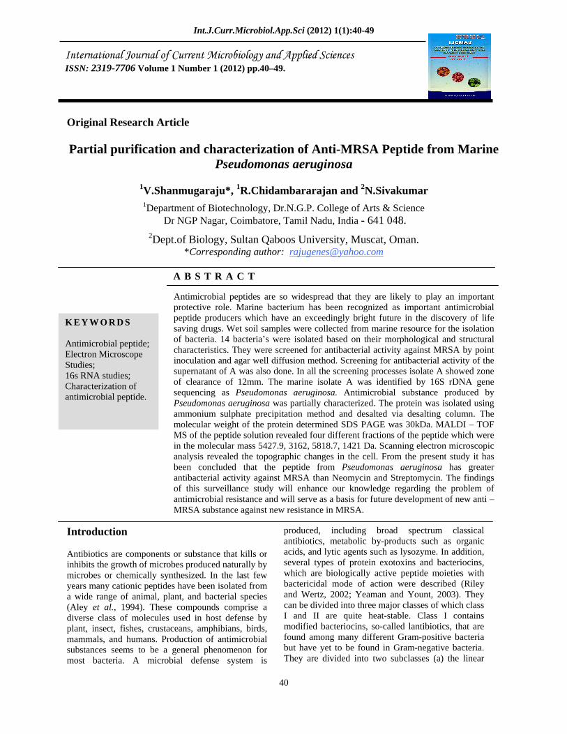
Int.J.Curr.Microbiol.App.Sci (2012) 1(1):40-49 40 Original Research Article Partial purification and characterization of Anti-MRSA Peptide from Marine Pseudomonas aeruginosa 1 V.Shanmugaraju*, 1 R.Chidambararajan and 2 N.Sivakumar 1 Department of Biotechnology, Dr.N.G.P. College of Arts & Science Dr NGP Nagar, Coimbatore, Tamil Nadu, India - 641 048. 2 Dept.of Biology, Sultan Qaboos University, Muscat, Oman. *Corresponding author: [email protected] ABSTRACT Introduction Antibiotics are components or substance that kills or inhibits the growth of microbes produced naturally by microbes or chemically synthesized. In the last few years many cationic peptides have been isolated from a wide range of animal, plant, and bacterial species (Aley et al., 1994). These compounds comprise a diverse class of molecules used in host defense by plant, insect, fishes, crustaceans, amphibians, birds, mammals, and humans. Production of antimicrobial substances seems to be a general phenomenon for most bacteria. A microbial defense system is produced, including broad spectrum classical antibiotics, metabolic by-products such as organic acids, and lytic agents such as lysozyme. In addition, several types of protein exotoxins and bacteriocins, which are biologically active peptide moieties with bactericidal mode of action were described (Riley and Wertz, 2002; Yeaman and Yount, 2003). They can be divided into three major classes of which class I and II are quite heat-stable. Class I contains modified bacteriocins, so-called lantibiotics, that are found among many different Gram-positive bacteria but have yet to be found in Gram-negative bacteria. They are divided into two subclasses (a) the linear ISSN: 2319-7706 Volume 1 Number 1 (2012) pp.40 49. KEYWORDS Antimicrobial peptide; Electron Microscope Studies; 16s RNA studies; Characterization of antimicrobial peptide. Antimicrobial peptides are so widespread that they are likely to play an important protective role. Marine bacterium has been recognized as important antimicrobial peptide producers which have an exceedingly bright future in the discovery of life saving drugs. Wet soil samples were collected from marine resource for the isolation of bacteria. 14 bacteria s were isolated based on their morphological and structural characteristics. They were screened for antibacterial activity against MRSA by point inoculation and agar well diffusion method. Screening for antibacterial activity of the supernatant of A was also done. In all the screening processes isolate A showed zone of clearance of 12mm. The marine isolate A was identified by 16S rDNA gene sequencing as Pseudomonas aeruginosa. Antimicrobial substance produced by Pseudomonas aeruginosa was partially characterized. The protein was isolated using ammonium sulphate precipitation method and desalted via desalting column. The molecular weight of the protein determined SDS PAGE was 30kDa. MALDI TOF MS of the peptide solution revealed four different fractions of the peptide which were in the molecular mass 5427.9, 3162, 5818.7, 1421 Da. Scanning electron microscopic analysis revealed the topographic changes in the cell. From the present study it has been concluded that the peptide from Pseudomonas aeruginosa has greater antibacterial activity against MRSA than Neomycin and Streptomycin. The findings of this surveillance study will enhance our knowledge regarding the problem of antimicrobial resistance and will serve as a basis for future development of new anti MRSA substance against new resistance in MRSA.
Transcript of Partial purification and characterization of Anti-MRSA ... et al.pdf · Partial purification and...
Int.J.Curr.Microbiol.App.Sci (2012) 1(1):40-49
40
Original Research Article
Partial purification and characterization of Anti-MRSA Peptide from Marine Pseudomonas aeruginosa
1V.Shanmugaraju*, 1R.Chidambararajan and 2N.Sivakumar 1Department of Biotechnology, Dr.N.G.P. College of Arts & Science
Dr NGP Nagar, Coimbatore, Tamil Nadu, India - 641 048. 2Dept.of Biology, Sultan Qaboos University, Muscat, Oman.
*Corresponding author: [email protected]
A B S T R A C T
Introduction
Antibiotics are components or substance that kills or inhibits the growth of microbes produced naturally by microbes or chemically synthesized. In the last few years many cationic peptides have been isolated from a wide range of animal, plant, and bacterial species (Aley et al., 1994). These compounds comprise a diverse class of molecules used in host defense by plant, insect, fishes, crustaceans, amphibians, birds, mammals, and humans. Production of antimicrobial substances seems to be a general phenomenon for most bacteria. A microbial defense system is
produced, including broad spectrum classical antibiotics, metabolic by-products such as organic acids, and lytic agents such as lysozyme. In addition, several types of protein exotoxins and bacteriocins, which are biologically active peptide moieties with bactericidal mode of action were described (Riley and Wertz, 2002; Yeaman and Yount, 2003). They can be divided into three major classes of which class I and II are quite heat-stable. Class I contains modified bacteriocins, so-called lantibiotics, that are found among many different Gram-positive bacteria but have yet to be found in Gram-negative bacteria. They are divided into two subclasses (a) the linear
ISSN: 2319-7706 Volume 1 Number 1 (2012) pp.40 49.
K E Y W O R D S
Antimicrobial peptide; Electron Microscope Studies; 16s RNA studies; Characterization of antimicrobial peptide.
Antimicrobial peptides are so widespread that they are likely to play an important protective role. Marine bacterium has been recognized as important antimicrobial peptide producers which have an exceedingly bright future in the discovery of life saving drugs. Wet soil samples were collected from marine resource for the isolation of bacteria. 14 bacteria s were isolated based on their morphological and structural characteristics. They were screened for antibacterial activity against MRSA by point inoculation and agar well diffusion method. Screening for antibacterial activity of the supernatant of A was also done. In all the screening processes isolate A showed zone of clearance of 12mm. The marine isolate A was identified by 16S rDNA gene sequencing as Pseudomonas aeruginosa. Antimicrobial substance produced by Pseudomonas aeruginosa was partially characterized. The protein was isolated using ammonium sulphate precipitation method and desalted via desalting column. The molecular weight of the protein determined SDS PAGE was 30kDa. MALDI
TOF MS of the peptide solution revealed four different fractions of the peptide which were in the molecular mass 5427.9, 3162, 5818.7, 1421 Da. Scanning electron microscopic analysis revealed the topographic changes in the cell. From the present study it has been concluded that the peptide from Pseudomonas aeruginosa has greater antibacterial activity against MRSA than Neomycin and Streptomycin. The findings of this surveillance study will enhance our knowledge regarding the problem of antimicrobial resistance and will serve as a basis for future development of new anti
MRSA substance against new resistance in MRSA.

Int.J.Curr.Microbiol.App.Sci (2012) 1(1):40-49
41
and cationic peptide and (b) the globular peptides; the latter normally are hydrophobic but not cationic. The second major class (II) contains small heat-stable bacteriocins that lack post translational modifications as found in lantibiotics, and are presently clustered into atleast two groups: pediocin-like bacteriocins and two-peptide bacteriocins (Diep et al., 2002). A third class (III) of bacteriocins has been also defined. They are normally larger in size and are easily subjected to heat inactivation (Klaenhammer et al., 1993).
Hundreds of peptide antibiotics have been described in the past half-century (Honcock et al., 1995). These falls into two classes, non ribosomally synthesized peptides, such as the gramicidins, polymyxins, bacitracins, glycopeptides, etc., and ribosomally synthesized peptides. Nonribosomally synthesized peptides can be described as peptides elaborated in bacteria, fungi and actinomycete that contain two or more moieties derived from amino acids (Kleinkauf et al., 1988). Antimicrobial, ribosomally synthesized, cationic peptides have been recognized only recently as an important part of innate immunity (Bevins, 1994). As current antibiotic therapy options are becoming limited for staphylococcal infections, there is an urgent need for new antimicrobial agents to combact these resistant pathogens. Antimicrobial peptides are now promising class of antimicrobial agents derived from naturally occurring peptides (Hanoc, 1997).
The marine surface environment is a site of intense competition for living space by a wide variety of organisms. Bacteria are generally recognized as primary colonizers of this habitat (Bryers, 1982). The microbial diversity in the sea is yet to be revealed. In the last few years marine microorganisms emerged as a new field for the discovery of novel biologically active compounds (Fenical, 1997). Marine natural products have an exceedingly bright future in the discovery of life saving drugs. The first antibiotic from marine bacterium was identified and characterized (Burkholder et al., 1996). The potential role in the production of active metabolites from marine bacteria against pathogens is a promising new topic for research; the present study reveals the isolation and partial characterization of anti
MRSA peptide producing bacteria from marine soil
Materials and Methods
Sample Collection Wet soil samples were collected from Chennai, Pondicherry, Muthupettai, Muthukuda, and Manora
(Thanjavur). The five samples collected were used for the isolation of bacteria which have antibacterial activity.
Isolation of bacteria
Serial dilution agar plating method (or viable plate counting method) was followed to isolate bacteria from the collected samples.
Collection of MRSA
The clinical isolate of Methicillin Resistant Staphylococcus aureus was collected from clinical laboratory of K.M.C.H., Coimbatore, and Tamil Nadu. MRSA was preserved in agar slants for further use and stored at 4 C. Then Kirby
Bauer method (agar diffusion method) was followed to determine the susceptibility of MRSA to antibiotics.
Screening of marine isolates for its antibacterial activity
Point Inoculation Method:
Muller Hinton Agar plates were prepared .The overnight culture of MRSA in nutrient broth was uniformly swabbed on the surface of Muller Hinton Agar plates using sterile cotton swab, making a lawn culture. The marine isolates from the agar slants were spotted on to Muller Hinton Agar plates seeded with actively growing cells of the clinically isolated MRSA. The plates were incubated at 37 C for 24 hours. After the incubation period, the plates were observed for the zone of inhibition and the results were recorded.
Agar Well Diffusion Method
After screening by point inoculation method, the antimicrobial substance production of the marine isolates was tested by agar- well diffusion method. Muller Hinton agar plate was prepared. The overnight culture of MRSA in nutrient broth was uniformly swabbed on the surface of Muller Hinton agar plates using sterile cotton swab. Two wells of 3mm size were made with sterile cork borer on the seeded plate. Around 50 l of overnight broth culture of marine isolate was added to the well aseptically. The plates were incubated without inverting for 24hrs at 30°C and the zone of inhibition was observed and recorded.

Int.J.Curr.Microbiol.App.Sci (2012) 1(1):40-49
42
Screening of marine isolate s supernatant for its antibacterial activity
The overnight grown culture of marine isolate in nutrient broth was centrifuged at 6000 rpm for 10 minutes and the supernatant was collected. The overnight culture of MRSA in nutrient broth was uniformly swabbed on the surface of sterile Muller Hinton agar plate using cotton swab. Two wells of 3mm size were made with sterile cork borer on the seeded plate. Around 50 l of overnight broth culture of marine isolate was added to the well aseptically. The plates were incubated without inverting for 24hrs at 30°C and the zone of inhibition was observed and recorded.
Protein precipitation by ammonium sulphate method
An overnight broth culture of Sample was centrifuged at 6000 rpm for 10 minutes at 4°C.To the cell free supernatant, ammonium sulphate was added to achieve 30% saturation. It was stirred well and incubated at 4°C overnight. The following day, the precipitates were collected by centrifugation at 6000rpm for 25 minutes at 4°C. The precipitates were dissolved in 500 l of sterile distilled water. Antibacterial activity of both the supernatant and the precipitate were tested using agar well diffusion assay and the results were recorded.
Desalting of ammonium sulphate precipitated protein
The column and buffer was brought to room temperature before use. The top cap of the column was removed followed by the bottom cap. Care should be taken to prevent the air bubbles in the column. The column was washed with 10ml of buffer and was allowed to flow under gravitational force. 2ml of the sample was loaded gently and allowed to drain completely. 0.5ml of buffer was passed to it and 1.5ml of high molecular weight substance were eluted and collected. Antibacterial activity of the desalted protein was tested by agar well diffusion assay and the result was recorded.
Identification of marine isolate by 16S rDNA gene sequencing
The marine isolate which showed antibacterial activity against MRSA was identified by 16s rDNA gene sequencing. Then the closest homologue organisms and phylogenetic relationship were studied.
Determination of molecular weight by SDS-PAGE
Glass plates, spacers and comb were cleaned thoroughly and dried. Glass plates were sealed using Vaseline and assembled using clips. 12% separating gel was prepared and poured inside a chamber between the glass plates without any air bubbles and allowed to polymerize for 30-60 minutes. After polymerization, 3% stacking gel was prepared and poured over the resolving gel. Comb was inserted between the glass plate and the gel was allowed for polymerization. After polymerization of stacking gel, comb was removed gently without destroying the shape of the wells. The spacer at the bottom of the glass plate was removed and the plate was fixed in the electrophoretic apparatus. Running buffer was poured into the electrophoretic tank without any air bubbles. The sample to be loaded was prepared by mixing sample and loading dye in 1:2 concentrations. Marker was also prepared in the same way. It was then denatured for 3 minutes and loaded in the wells. Marker was added in the first well and power supply was on. Once the Bromophenol blue dye reaches 1cm above the bottom of the gel, current was put off. The gel was then carefully removed for staining.
Protein visualization by silver staining method
The gel was kept in fixation solution for 1 hour with gentle shaking. It was then rinsed with 50% methanol for 12 minutes. The gel was rinsed with double distilled water twice for 5 minutes. The gel was then immersed in 0.02% sodium thiosulphate solution for 60 seconds exactly with gently shaking. It was rinsed with double distilled water twice for a minute. The gel was immersed in chilled 0.1% silver nitrate solution. It was covered with foil paper and incubated for 25 minutes with gentle shaking in the cold room. The gel was rinsed with double distilled water twice for 20 seconds. It was then immersed in developing solution and was shaken in room temperature until bands appear. The reaction was stopped by removing developer and swirling in fix solution for 5 minutes. Finally, the gel was rinsed with double distilled water thrice for 1 minute.
Separation and identification of Amino Acids by Thin Layer Chromatography (TLC)
TLC plate used is cut into 5 cm x 20 cm sheets. Using pencil, a line across the plate at the bottom of 1.0 cm was marked. Sample was spotted onto the line using fine capillary tube. Developing solvent was poured into the beaker to a depth of less than 0.5 cm. The prepared TLC plate was placed into the developing solution. It was allowed to run until the

Int.J.Curr.Microbiol.App.Sci (2012) 1(1):40-49
43
solvent is about half a centimeter below the top of the plate. Then the plate is removed from the developing solution. A line across the plate was marked at the solvent front. To visualize the spot, the plate is sprayed with ninhydrin reagent. The plate is dried in oven at 110 °C for 5 minutes. The spots were marked and the Rf values were calculated.
Determination of molecular mass by MALDI-TOF MS
The molecular mass of the desalted peptide solution was analyzed by MALDI-TOF MS using -cyano 4 hydroxy cinnamic acid as matrix. Scanning Electron Microscopy (SEM) analysis of antibacterial peptide treated MRSA:
An overnight culture of MRSA was centrifuged at 6000rpm for 10 minutes and the pellet was collected. 2ml of purified peptide was added to the pellet and incubated at 30°C for 24 hours. It was then centrifuged at 5000rpm for 10 minutes at 4°C and the pellet was collected. It was then lyophilized and served as test sample. The pellet of MRSA was lyophilized and served as control. These were viewed under scanning electron microscope to observe the morphological changes of the bacteria after treated with the peptide.
Results and Discussion Isolation of Bacteria
Marine bacteria were isolated from five wet soil samples from Chennai, Pondicherry, Muthupettai, Muthukuda and Manora (Thanjavur) using Marine agar 2216 by serial dilution spread plate method. A total of 14 bacteria were isolated based on their morphological and structural characteristics.
Antimicrobial Susceptibility of MRSA to antibiotics
Antimicrobial susceptibility of MRSA to bacteria, Kanamycin, Vancomycin, Metronidazole, Neomycin, Gentamicin, Erythromycin, Chloramphenicol, Streptomycin, Tetracycline and Methicillin was carried out by Kirby Bauer disc diffusion assay. The results were tabulated (Table 1). The antimicrobial activity of the peptide produced by Pseudomonas sp. Strain 4B was tested against a panel of bacteria. Antimicrobial activity was observed against Staphylococcus aureus and the zone of inhibition was found to be 15 mm (Roberta et al., 2008). The present study revealed the zone of inhibition of about 12 mm against MRSA. Plant pathogenic bacterial
strains were used in both the agar-diffusion method and the agar-spot assay to evaluate the antagonistic activity of P. syringae ciccaronei and the spectrum of antimicrobial activity of the bacteriocin. Growth of Ps. Syringae persicae NCPPB2761 was inhibited by a P. syringae ciccaronei colony and its culture filtrate with the zone of inhibition 7mm (Lavermicocca et al., 1999). Similarly the culture filtrate of Pseudomonas aeruginosa inhibited the growth of MRSA with the zone of inhibition 12mm.
Table 1: Antibiotic Sensitivity Pattern of MRSA
Screening of marine isolates for its antibacterial activity
All of the marine isolates were tested for their ability to produce antibacterial substance against MRSA. Marine isolates from agar slants were spotted onto the Muller Hinton agar. Out of the 14 isolates, only one isolates (A) showed inhibitory activity against MRSA. Zone of clearance of about 12mm was observed after 24 hours of incubation. After screening by point inoculation method, the antibacterial substance production was confirmed by agar well diffusion method. The overnight culture of isolate A was added to the wells in Muller Hinton agar (MHA) plate. A clear zone of inhibition of 12mm was observed against MRSA (Table 2).
Table 2 Zone of inhibition of marine isolate against MRSA by point inoculation and agar well diffusion method
S.No. Marine isolate A
Zone of inhibition against MRSA
(mm) 1
2
Spotted Marine isolate
Culture broth
12
12
Antibiotic disc
Zone of inhibition
(mm) Remarks
Chloramphenicol Tetracycline Neomycin Gentamycin Meteonidazole Kanamycin Erythromycin Vancomycin Streptomycin Methicillin
20 Nil 4 12 Nil Nil Nil 14 8
Nil
Sensitive Resistant Sensitive Sensitive Resistant Resistant Resistant Sensitive Sensitive Resistant
Int.J.Curr.Microbiol.App.Sci (2012) 1(1):40-49
44
Screening of marine isolate s supernatant for its antibacterial activity
To check whether the antibacterial substance produced by isolate A is an extracellular compound, the supernatant of A was tested for its antibacterial activity by agar well diffusion method. Zone of clearance of 12mm against MRSA was detected. The active metabolites produced by Lactobacillus sp NRRL B-227 exhibited various degrees of activities against Staphylococcus aureus with 21mm zone of inhibition (Atta et al., 2009). Similarly the active metabolite of Pseudomonas aeruginosa inhibited the growth of MRSA with the zone of inhibition 12mm.
Antibacterial activity of the protein precipitated by ammonium sulphate method
The protein precipitated by ammonium sulphate method was dissolved in sterile distilled water and was desalted. Antibacterial activity of the precipitate protein and the supernatant was checked. A clear zone of 12mm was noted around the well loaded with the protein sample. No zone of clearance was seen around the well loaded with the supernatant (Table 3). The peptide antibiotic produced by L. plantarum NRRL B- 227 was treated with Ammonium sulphate 60% saturation. The mixture were stirred for 2 hrs at 4°C and later centrifuged at 10,000 rpm for 50 min at 4°C. The pellet was resuspended in 25 ml of 0.05 M potassium phosphate buffer pH 7.0. Dialysis was carried out against the same buffer for 18 hrs in spectrapor dialysis tubing. Assay of the antibacterial activity was carried out increase of antibacterial activity comparing with culture supernatant (Atta et al., 2009). In the present study the peptide antibiotic produced by Pseudomonas aeruginosa was treated with ammonium sulphate 30% saturation and the precipitate was desalted with desalting column. The fractions collected showed antibacterial activity against MRSA.
Table 3: Zone of inhibition of ammonium sulphate precipitates against MRSA by Agar well difusion method
S.No. Percentage (%) of ammonium
sulphate
Zone of inhibition against MRSA
(mm) 1 2 3
20% 30% 40%
6 12 8
Pseudomonas syringae ciccaronei bacteriocin was purified by Ammonium sulphate precipitation, and the molecular weight of bacteriocin was found to be approximately 100 kDa; this was confirmed by the
data from gel filtration through Sephadex G150. However, a sample of bacteriocin purified using preparative gel electrophoresis revealed three bands on SDS-PAGE with molecular weights of 76, 63 and 45 kDa, respectively. (Lavermicocca et al., 1999). The molecular mass of cerein 8A is quite distinct of other bacteriocins or BLS produced by B. cereus (Naclerio et al.. 1993; Oscariz et al., 1999; Torak and Matijasic 2003). Also, these characteristics are contrasting with the antibiotics, which are cationic peptides with low- MW when estimated by SDS-PAGE (Montville and Chen, 1998).
Identification of Marine Isolate (A) by 16S rDNA gene sequencing:
The marine isolate which showed antibacterial peptide activity against MRSA was identified by 16S rDNA gene sequencing. Based on nucleotide homology and phylogenetic analysis, the microbe was detected as Pseudomonas aeruginosa and the nearest homolog species was found to be Pseudomonas aeruginosa . Using 16S r DNA sequencing the phylogenetic analysis revealed that all bioactive isolates belonged to the genus Bacillus. The isolates A184, A190 and A202 showed greater than 98.0% sequence similarity to Bacillus subtilis strain 168, while the isolate A 586 showed greater than 98.9% sequence similarity to Bacillus pumilus strain OM-F6. (Christian et al., 2003).
Phylogenetic analysis using 16S rDNA sequences and the neighbour-joining method showed the X153 strain is a member of Pseudoalteromonas close to P. piscicida, P. peptidysin, and Pseudoalteromonas sp. named Y. this indicates that X153 strain is very close to a strain Pseudoalteromonas sp. Y, which displayed a algicidal effect against harmful micro-algae (Lovejoy et al., 1998). In this present study the organism was identified to be Pseudomonas aeruginosa by 16S rDNA gene sequencing. The information about the Phylogenetic relationship of these microbes with other bacteria were found using combination of NCBI Gene Bank and RDB database. The sequence description revealed that the marine isolate A was phylogenetically related to Pseudomonas aeruginosa.
Detremination of molecular weight by SDS-PAGE:
The molecular weight of the peptide was determined by SDS-PAGE and was found to be 30 kDa (Fig.1). The antimicrobial peptide produced by Pseudomonas sp. 4B was purified by sequential precipitation, gel

Int.J.Curr.Microbiol.App.Sci (2012) 1(1):40-49
45
filtration, and ion-exchange chromatography process. A major peptide band of about 30 kDa was observed by SDS-PAGE analysis, coinciding with antimicrobial activity. This molecular mass is close to the lectin-like bacteriocin LlpA from a Pseudomonas sp. isolated from banana rhizosphere (Parret et al., 2004). Similarly in the present work the molecular weight of the antimicrobial peptide (protein) from Pseudomonas aeruginosa was identified as 30 kDa..
Figure.1: Determination of Molecular weight by SDS-PAGE
Separation and identification of amino acids by Thin Layer Chromatography (TLC):
The amino acids present in the purified peptide sample were identified by TLC and was assumed to be Cystine, Histidine and Proline (Table 4& Fig.2).Thin layer chromatography profiling was done for the haemolymph samples of crab (Thalamita crenata) in two different solvent systems of A and B. The solvent system A consisted of methanol:chloroform (1:9) and B was the combination of butanol : acetic acid : water in proportions of (5:1:4). The plate developed in both the solvent system showed light pink spots when sprayed in ninhydrin indicates the presence of aminoacids and peptides (Rameshkumar et al., 2009). In the present study, the solvent Butanol: Acetic acid: Water in proportions of (4:1:1) was used and the plate showed pink and yellow spots.
Table 4: Separation and identification of Amino acids by Thin Layer Chromatography (TLC):
S. No. Amino Acids Rf Value
1 2 3 4 5 6 7 8 9 10
11
Lysine Arginine Proline Serine
Alanine Glycine
Glutamine Histidine Cystine
Column Precipitate (i) Spot 1 Culture Supernatant (i) Spot 1
(ii) Spot 2 (iii) Spot 3
0.18 0.56 0.42 0.26 0.38 0.26 0.27 0.33 0.30 0.30
0.30 0.32 0.41
The peptide produced by the Ergot Fungus Verticillium kibiense, was hydrolyzed with acid and separated by TLC. The substance was a polypeptide and showed positive signals which are specific for the guanidino group (arginine) and the imidazole ring (histidine) or phenol group (tyrosine), respectively (Nishikawa and Ogawa, 2004). Extracts from the two thermophilic strains showed relatively high antibacterial activity against Staphylococcus aureus. Partial purification of antimicrobial agents by thin layer chromatography gave two inhibitory bands with Rf values of 0.22 for extract from Yersinia sp.1 and 0.25 for extract from Aeromonas hydrophila (Amjad et al., 2006). Likewise, in the present study the Rf values of 0.30, 0.32 and 0.41 for P. aeruginosa culture supernatant and 0.30 for column precipitate were compared with the standard amino acids and it has been assumed that the antibacterial peptide contains the amino acid cystine.
Figure 2: separation and identification of amino acids by Thin Layer Chromatography ( TLC)
LY=Lysine ; AR=Arginine ; PR=Proline ; SE=Serine ; L=Alanine ; GL=Glycine ;G=Glutamine; HI=Histidine; C=Cystine; S1=Column Sample
L1 L2 L3 L4 Marker

Int.J.Curr.Microbiol.App.Sci (2012) 1(1):40-49
46
Scanning electron microscopy of antibacterial peptide treated MRSA:
To understand the mechanism of action of antibacterial peptide activity of Pseudomonas aeruginosa against MRSA, the sample was visualized under scanning electron microscope. Changes in the morphology of MRSA were observed (Plate 3a, 3b) Electron microscope showed the effect of phenazine-1-carboxamide (zag1) on the ultra structure of Aspergillus niger, cell wall, plasma membrane, transfer septum, mitochondria, nucleus, nucleolus, Vacuole, electron dense body, germ tube, Cytoplasm, ribosome and Lysed cell (Hassanein et al., 2009)
An in vitro model to investigate Pseudomonas aeruginosa adherence to human type IV basement collagen was developed by using scanning electron microscopy (SEM). There were significantly less Pseudomonas aeruginosa bacilli when cultured in 5 mg/mL of Erythromycin compared with control (absence of Erythromycin). Adherence density for Pseudomonas aeruginosa obtained from no Erythromycin was significantly higher than that obtained from 5 mg/mL erythromycin. By using SEM it was found that Pseudomonas aeruginosa, when incubated in Erythromycin had a significant reduction in its diagonal length, radius, height, volume and surface area. (Tsang et al., 2003).
Figure 3(A): Scanning Electron Microscopy of Antibacterial Peptide Treated MRSA

Int.J.Curr.Microbiol.App.Sci (2012) 1(1):40-49
47
Transmission and scanning electron micrographs of methicillin-resistant Staphylococcus aureus (MRSA) and E. coli that had been treated with Styelin D and clavaspirin suggested that both peptides induced osmotic disregulation. Treated bacteria manifested cytoplasmic swelling and extrusion of cytoplasmic contents through their peptidoglycan cell wall. (Robert et al., 2003).
In this present study, the MRSA was treated with antibacterial peptide of Pseudomonas aeruginosa. The nature and effects of the peptide interaction with MRSA created rupture in the cell wall while the untreated MRSA remained same. Their morphological changes were observed under the SEM.
Figure 3(B): Scanning Electron Microscopy of UnTreated MRSA

Int.J.Curr.Microbiol.App.Sci (2012) 1(1):40-49
48
References
Aley, S.B., M. Zimmirman, E. Hetsko, Selsted and Gillin, F.D. 1994. Killing of Giardia lamblia by cryptdins and cationic neutrophil peptides. Infect Immu. 62: 5397-5403.
Atta, H.M., B.M. Refaat and A.A. El-Waseif, 2009. Application of Biotechnology for Production, Purification and Characterization of Peptide from Ps.aeruginosa. World. J. Microbiol Biotechnol. 35: 208-215.
Bevins, C., 1994. Antimicrobial peptides as agents of mucosal immunity. Ciba Found Symp. 186:250 269.
Bryers, J.D., and Characklis, W.G. 1982. Processes governing primary biofilm formation. Biotechnology Bioeng. 24:24512476.
Burkholder, P.R.,, P. Fisher and Leitz, R.M. 1996. Production of pyrrole antibiotic by a marine bacterium. App Microbiol. 14:649-653.
Christian, T., Pabel, Joachim vater, Christopher wilde, Peter franke, Jurgen hofemeister, Barbara adlar, Gerhard bringmann, Jorg hacker and Ute hentschel. 2003. Antimicrobial activities and MALDI MS of Bacillus isolates from the marine sponge Aplysina aerophoba. Marine biotechnol. 5: 424 -434.
Diep, D.B., and I Nes,I.F. 2002. Ribossomally synthesized antibacterial peptides in Gram-positive bacteria. Curr Drugs Target. 3:107122.
Fenical, W., 1997. Chemical studies of marine bacteria: Developing a new resource. Chem Rev.4:1673-1683.
Hancock, R.E.W., T. Falla and Brown,M.H. 1995. Cationic bactericidal peptides. Adv Microb Physiol. 37:135 175.
Hanoc, R.E., 1997. Peptide antibiotics. Lancet.349:418-422.
Hassanein, W.A., N.M. Awny, A.A. El-Mougith and El-Dien, S.H. 2009, Characterization and Antagonistic Activities of Metabolite Produced by Pseudomonas aeruginosa Sha8. J Appl Sci Res. 5(4): 392-403.
Klaenhammer, T.R. , 1993. Genetics of bacteriocins produced by lactic acid bacteria. FEMS Microbiol Rev. 12:39 86.
Kleinkauf, H., and Von Dohren, H. 1988. Peptide antibiotics, -lactams and related compounds. Crit Rev Biotechnol. 8:1 32.
Lavermicocca, P., S.L. Lonigro, A. Evident and Andolfi, A. 1999. Bacteriocin production by Pseudomonas syringae pv. ciccaronei NCPPB2355. Isolation and partial characterization of the antimicrobial
compound. J.App. Microbiol. 86: 257 265. Lovejoy, C., J.P. Bowman and Hallegraeff, G.M.,
1998. Algicidal effects of a novel marine Antibiotic proteins of polymorphonuclear leukocytes. Eur J Haematol. 56:263 277.
Monica, B., S. Marco, Sabrina pacor, Alessandro tossi, Donatella nobili, B. Giancarlo, Marina busetti and Renato Gennaro, 2006. Fungicidal activity of five cathelicidin peptides against clinically isolated yeasts. J Antimicrobial Chem. 58: 950 959.
Naclerio, G., E. Ricca, M. Sacco and Felice, M. 1993. Antimicrobial activity of a newly identified bacteriocin of Bacillus cereus. Appl Environ Microbiol. 59: 4313 4316.
Nishikawa, M., and Ogawa, K. 2002. Distribution of Microbes Producing Antimicrobial -Poly-L- Lysine Polymers in Soil Micro flora Determined by a Novel Method. Appl Environm Microbiol. 68 (7): 3575 3581.
Oscariz, J.C., I. Lasa and Pisabarro, A.G. 1999, Detection and characterization of cerein 7, a new bacteriocin produced by Bacillus cereus with a broad spectrum of activity. FEMS Microbiol Lett. 178: 337 341.
Ping, F., W. Jianwei and Guo, G. 2009. Purification and Molecular Identification of an Antifungal Peptide from the Hemolymph of Musca domestica (housefly). Cell Molecular Immunol. 6(4):245-251.
Rameshkumar, G., S. Ravichandran., G. Kaliyavaradhan and Ajithkumar,T.T. 2009. Antimicrobial peptide from the crab, Thalamita crenata. World J Fish Marine Sci. 1(2): 74-79.
Riley, M.A., and Wertz, J.E. 2002. Bacteriocins: evolution, ecology, and application. Annu Rev Microbiol. 56:117 137.
Robert, L., A.Tincu., S.W. Taylor., L.P.Menzel and Waring, A.J. 2003. Natural Peptide Antibiotics from Tunicates: Structures, Functions and Potential Uses. Integr Comp Biol. 43:313 322
Roberta Fontoura., Jordana Corralo, Spada, Terra Silveira, Siu Mui Tsai., Adriano Brandelli, 2008. Purification and characterization of an antimicrobial peptide produced by Pseudomonas sp. strain 4B. World J .Microbiol Biotechnol 25:205 213.
Torak, K.G., and Matijasic,B.B. 2003. Partial characterisation of bacteriocins produced by Bacillus cereus isolates from milk and milk products. Food Technol Biotechnol. 41: 121129.
Tsang, K.W., P.L. Ho., S. Chan.., G. Tipoe., R. Leung., J. Sun., J.C. Ho and Lam, W.K. 2003,
Int.J.Curr.Microbiol.App.Sci (2012) 1(1):40-49
49
Effects of erythromycin on Pseudomonas aeruginosa adherence to collagen and morphology in vitro. Eur Respir J. 21: 401406.
Wellham, K.J., M.A. Domin., D.E. Scannell., E. Cohen, and Asthon, D.S. 1998.The characterization of microorganisms by Matrix-assisted laser Desorption/ Ionization Mass Spectrometry, Time-Of-Flight (MALDI
TOF). Rapid commun Mass Spect. 12(4): 176-180.
Yeaman, M.R., and Yount, N.Y. 2003, Mechanisms of Antimicrobial peptides Action and Resistence. Pharmacol Rev. 55:27-55.