Part VII: Modeling Neurotransmission – A GABAergic...
4
MSOE Center for BioMolecular Modeling Synapse Kit: Section 7-8 | 1 Part VII: Modeling Neurotransmission – A GABAergic Synapse The cholinergic and dopaminergic synapses previously modeled are examples of excitatory synapses because a depolarization occurs in the postsynaptic neuron. Inhibitory synapses reduce the postsynaptic neuron’s ability to generate an action potential. You will next model the events of neurotransmission at a GABAergic synapse. GABAergic synapses utilize gamma- amino butyric acid (GABA) as the chemical of neurotransmission. 7a. Model the GABAergic synapse shown below. Be sure to label all of the following: voltage- gated sodium channel, voltage-gated potassium channel, GABA, synaptic vesicle, presynaptic cell, postsynaptic cell, potassium leak channel, sodium-potassium pump, synaptic cleft, GABA receptor, and calcium channel.
Transcript of Part VII: Modeling Neurotransmission – A GABAergic...

MSOE Center for BioMolecular Modeling Synapse Kit: Section 7-8 | 1
Part VII: Modeling Neurotransmission – A GABAergic Synapse The cholinergic and dopaminergic synapses previously modeled are examples of excitatory synapses because a depolarization occurs in the postsynaptic neuron. Inhibitory synapses reduce the postsynaptic neuron’s ability to generate an action potential. You will next model the events of neurotransmission at a GABAergic synapse. GABAergic synapses utilize gamma-amino butyric acid (GABA) as the chemical of neurotransmission. 7a. Model the GABAergic synapse shown below. Be sure to label all of the following: voltage-
gated sodium channel, voltage-gated potassium channel, GABA, synaptic vesicle, presynaptic cell, postsynaptic cell, potassium leak channel, sodium-potassium pump, synaptic cleft, GABA receptor, and calcium channel.
MSOE Center for BioMolecular Modeling Synapse Kit: Section 7-8 | 2
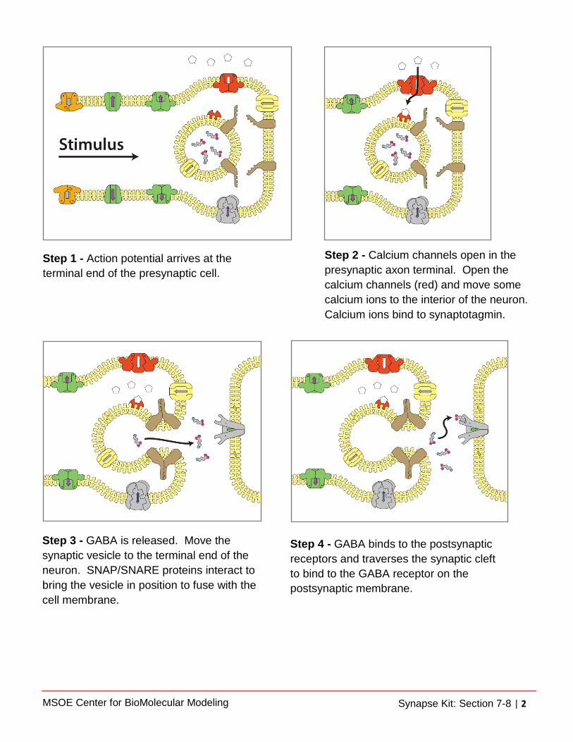
Step 1 - Action potential arrives at the terminal end of the presynaptic cell.
Step 2 - Calcium channels open in the presynaptic axon terminal. Open the calcium channels (red) and move some calcium ions to the interior of the neuron. Calcium ions bind to synaptotagmin.
Step 3 - GABA is released. Move the synaptic vesicle to the terminal end of the neuron. SNAP/SNARE proteins interact to bring the vesicle in position to fuse with the cell membrane.
Step 4 - GABA binds to the postsynaptic receptors and traverses the synaptic cleft to bind to the GABA receptor on the postsynaptic membrane.

MSOE Center for BioMolecular Modeling Synapse Kit: Section 7-8 | 3
7b. Identify the GABAergic synapse as ionotropic or metabotropic. Explain your choice. 7c. What molecule is released into the synaptic cleft in this model? 7d. Why is the GABAergic synapse considered to be inhibitory?
Step 5 - Ion channels open in the postsynaptic membrane. The GABA receptor opens to allow chloride ions (Cl-) to follow their concentration gradient into the postsynaptic cell. Hyperpolarization of the postsynaptic cell occurs inhibiting the generation of an action potential.
Step 6 - High-affinity transporters terminate the action of GABA by returning it to the presynaptic neuron for reuse.

MSOE Center for BioMolecular Modeling Synapse Kit: Section 7-8 | 4
Part VIII: Modeling a Perturbance in the GABAergic Synapse Next you will introduce a substance that perturbs neural transmission in the GABAergic synapse. Propofol binds to the GABA receptor to hold the channel open.
8a. How might over stimulation of the GABA receptor impact nervous transmission? 8b. How might under stimulation of the GABA receptor impact nervous transmission? 8c. Explain how propofol perturbs the GABAergic synapse.