Parallel Transport of Surface Deformations from Pole ...
10
HAL Id: hal-01860274 https://hal.inria.fr/hal-01860274 Submitted on 23 Aug 2018 HAL is a multi-disciplinary open access archive for the deposit and dissemination of sci- entific research documents, whether they are pub- lished or not. The documents may come from teaching and research institutions in France or abroad, or from public or private research centers. L’archive ouverte pluridisciplinaire HAL, est destinée au dépôt et à la diffusion de documents scientifiques de niveau recherche, publiés ou non, émanant des établissements d’enseignement et de recherche français ou étrangers, des laboratoires publics ou privés. Parallel Transport of Surface Deformations from Pole Ladder to Symmetrical Extension Shuman Jia, Nicolas Duchateau, Pamela Moceri, Maxime Sermesant, Xavier Pennec To cite this version: Shuman Jia, Nicolas Duchateau, Pamela Moceri, Maxime Sermesant, Xavier Pennec. Parallel Trans- port of Surface Deformations from Pole Ladder to Symmetrical Extension. Shape in Medical Imag- ing. ShapeMI 2018., Sep 2018, Granada, Spain. pp.116-124, 10.1007/978-3-030-04747-4_11. hal- 01860274
Transcript of Parallel Transport of Surface Deformations from Pole ...

HAL Id: hal-01860274https://hal.inria.fr/hal-01860274
Submitted on 23 Aug 2018
HAL is a multi-disciplinary open accessarchive for the deposit and dissemination of sci-entific research documents, whether they are pub-lished or not. The documents may come fromteaching and research institutions in France orabroad, or from public or private research centers.
L’archive ouverte pluridisciplinaire HAL, estdestinée au dépôt et à la diffusion de documentsscientifiques de niveau recherche, publiés ou non,émanant des établissements d’enseignement et derecherche français ou étrangers, des laboratoirespublics ou privés.
Parallel Transport of Surface Deformations from PoleLadder to Symmetrical Extension
Shuman Jia, Nicolas Duchateau, Pamela Moceri, Maxime Sermesant, XavierPennec
To cite this version:Shuman Jia, Nicolas Duchateau, Pamela Moceri, Maxime Sermesant, Xavier Pennec. Parallel Trans-port of Surface Deformations from Pole Ladder to Symmetrical Extension. Shape in Medical Imag-ing. ShapeMI 2018., Sep 2018, Granada, Spain. pp.116-124, �10.1007/978-3-030-04747-4_11�. �hal-01860274�

Parallel Transport of Surface Deformationsfrom Pole Ladder to Symmetrical Extension
Shuman Jia1, Nicolas Duchateau2, Pamela Moceri1,3, Maxime Sermesant1 andXavier Pennec1
1 Universite Cote d’Azur, Epione Project, Inria Sophia Antipolis, France2 Creatis, CNRS UMR5220, INSERM U1206, Universite Lyon 1, France
3 Hopital Pasteur, CHU de Nice, France
Abstract. Cardiac motion contains information underlying disease de-velopment, and complements the anatomical information extracted foreach subject. However, normalization of temporal trajectories is neces-sary due to anatomical differences between subjects. In this study, weencode inter-subject shape variations and temporal deformations in acommon space of diffeomorphic registration. They are parameterized bystationary velocity fields. Previous normalization algorithms applied inmedical imaging were first order approximations of parallel transport. Incontrast, pole ladder was recently shown to be a third order scheme ingeneral affine connection spaces and exact in one step in affine symmetricspaces. We further improve this procedure with a more symmetric map-ping scheme, which relies on geodesic symmetries around mid-points. Weapply the method to analyze cardiac motion among pulmonary hyperten-sion populations. Evaluation is performed on a 4D cardiac database, withmeshes of the right-ventricle obtained by commercial speckle-trackingfrom echo-cardiogram. We assess the stability of the algorithms by com-puting their numerical inverse error. Our method turns out to be veryaccurate and efficient in terms of compactness for subspace representa-tion.
1 Introduction
Analyzing cardiac function, notably myocardial deformation along cardiac cy-cle, can reveal subtle changes underlying disease development, and thereforeimprove risk stratification for patients. However, temporal deformations (cycli-cal or longitudinal) are complex due to subject-specific anatomy. Normalizationof these trajectories into a common reference frame is an essential prerequisitein statistical analysis [4, 1].
Nevertheless, it is a challenging task to construct a robust method for thenormalization of temporal deformations, both theoretically and numerically. Incardiac imaging, the first step often includes segmenting the region of interestfrom the images, which generates its surface or volume as results. Then, to modelthe deformation between two shapes, depending on properties of the shapes,different non-rigid registration algorithms may be suitable in specific scenario.

2 S. Jia, N. Duchateau, P. Moceri, M. Sermesant, and X. Pennec
Fig. 1. Illustration of parallel transport of vectors a and b along a curve (left) and itsapplication to cardiac imaging (right) with a focus on surfaces.
We choose to place our method in a space of diffeomorphic transformations.In particular, diffeomorphism provides invertible and folding-free registrationresults, which enables a symmetric encoding of deformation trajectories.
Given that diffeomorphisms are parameterized via flow of velocity fields, herestationary velocity field (SVF) [13], their normalization requires vector-basedtransport. In differential geometry, parallel transport specifies how to realize aninfinitesimal transformation (a tangent vector) along a curve from one point toanother of a manifold, as illustrated in Fig. 1(a). The concept was introduced inmedical imaging by [14] and used for neuroimaging studies in [11]. To apply itto cardiac sequences, we need to:
– Parameterize the temporal deformations Φti from the subject-specific shapeS at baseline to its shape S′ti at time ti by SVF vti ; this step amounts tocomputing the logarithm of the deformations such that Φti = exp(vti), or toregister S to S′ti .
– Transfer these SVFs along the inter-subject deformation geodesic ΦS fromthe space of S to a common geometry T , often called a template.
These steps lead to a representation of all the subject-specific deformations asinfinitesimal deformations of the same anatomy. We can thus perform properpopulation comparison in terms of shape evolution along the cardiac cycle, asshown in Fig. 1(b).
Despite the fact that the definition of logarithm may be well developed intheory, implementations of its computation could be unstable due to numericalinconsistency. While computing registration/geodesics, the logarithms of surfacedeformations are only sparsely defined in the space (on the shape), which differsfrom image deformations. Careful numerical implementation of parallel transportalgorithm is also needed.
Parallel transport methods in medical image analysis are currently based onladders (like Schild’s and pole ladders [6, 7]) or on variations of Jacobi Fields[14, 8]. These methods are first order approximation schemes which need to be

Parallel Transport of Surface Deformations 3
Algorithm 1 Pole ladder transport of the vector u along the geodesic [S, T ]
- Compute the midpoint M = expS( 12
logS(T )) on the intersubject geodesic;- Compute the geodesic form the endpoint S′ = expS(u) of the temporal geodesicsegment to the midpoint M , and shoot twice to get the inverse endpoint: T ′′ =expS′(2 logS′(M));- Return the vector trans(u) = − logT (T ′′) and then apply it to T : T ′ =expT (trans(u)).
iterated along the curve. In contrast, pole ladder was recently discovered to be ofthird order in general affine connection spaces [10], which makes it a very attrac-tive method to implement. In order to further improve the symmetry and thusthe numerical stability with respect to the implementation of [7], our methodrelies on the definition of middle point and geodesic symmetry.
2 Methodology
2.1 Pole Ladder
Pole ladder is based on using the curve as the diagonal of the geodesic par-allelogram to realize parallel transport (see Fig 2 and Algo. 1). Let u be theinitial tangent vector to the geodesic segment that encodes the temporal de-formation expS(tu) (t ∈ [0, 1]). Originally, pole ladder computes a midpoint onthe geodesic [S, T ] and expands twice the geodesic form the endpoint S′ to themidpoint to obtain the point T ′′. In this paper, we propose to reformulate thedoubling of the geodesics with a mid-point geodesic symmetry (Algo. 2). Themid-point M of a geodesic segment [S, T ] is defined as M = γ[S,T ](
12 ). The map-
pings from M towards the two sides meet the definition of a geodesic symmetry:γ[M,S](t) = −γ[M,T ](t). Although this novel version of pole ladder is theoreticallyequivalent to the previous one, it is numerically more stable in our experiments.This may be emphasized by the fact that in medical imaging we are encodingtangent vectors with infinitesimal deformations and that changing the objectwhich is deformed may have a drastic numerical impact.
2.2 Numerical Accuracy
Recent mathematical developments have enabled the numerical analysis of poleladder in affine connection spaces with symmetric connections [10]. In Lie groups,
Algorithm 2 Mid-point symmetric pole ladder transport of the geodesic seg-ment [S, S′] along the geodesic [S, T ]
- Compute the midpoint M = γ[S,T ](12) on the intersubject geodesic;
- Compute the symmetric point T ′′ = expM (− logM (S′)) of S′ with respect to M ;- Compute the symmetric point T ′ = expT (− logT (T ′′)) of T ′′ with respect to T , andreturn the geodesic segment [T, T ′].

4 S. Jia, N. Duchateau, P. Moceri, M. Sermesant, and X. Pennec
Fig. 2. The symmetry-based pole ladder structure.
the Baker-Campbell-Hausdorff (BCH) formula provides an expansion of the com-position of two group exponentials in the Lie algebra: BCH(v, u) = log(exp(v)◦exp(u)). This formula has been thoroughly used in registration algorithms [2].In general affine connection manifolds, a somewhat similar formula can be es-tablished based on the curvature instead of the Lie bracket [5]. The doubleexponential expx(v, u) = expy(Πy
xu) corresponds to a first geodesic shootingfrom the point x along the vector v, followed by a second geodesic shooting fromy = expx(v) along the parallel transport Πy
xu of the vector u. [5] has shown thatthe Taylor expansion of the log of this endpoint hx(v, u) = logx(expx(v, u)) is:
hx(v, u) =v + u+1
6R(u, v)v +
1
3R(v, u)u+
1
12∇vR(u, v)v +
1
24(∇uR)(u, v)v
+5
24(∇vR)(u, v)u+
1
12(∇uR)(u, v)u+O(‖u+ v‖5).
When applied to our reformulation of pole ladder using geodesic symmetry,we find that the error on one step of pole ladder to transport the vector u along ageodesic segment of tangent vector [S, T ] = [expM (−v), expM (v)] (all quantitiesbeing parallel translated at the mid-point M) is:
ΠMT trans(u)−ΠM
S u =1
12((∇vR)(u, v)(5u− 2v) + (∇uR)(u, v)(v − 2u))+O(‖v+u‖5).
We see here that one single step of pole ladder is of order three, thus muchmore accurate than the other first order parallel transport schemes. Moreover,the fourth order error term vanishes in affine symmetric spaces since the cur-vature is covariantly constant in these spaces. In fact, one can actually prove

Parallel Transport of Surface Deformations 5
that all error terms vanish in a convex normal neighborhood of an affine connec-tion space: one step of pole ladder realizes an exact parallel transport (providedthat geodesics and mid-points are computed exactly of course) [10]. This resultmakes pole ladder a very attractive scheme. In particular, Lie groups have acanonical symmetric space structure thanks to the symmetry sg(h) = gh−1g forany elements g, h of the Lie group. This is an affine structure which is generallynot metric. The symmetry generates a canonical connection which is exactly thesymmetric Cartan-Schouten connection. Since the geodesics of this connectiongoing through identity are parameterized the flow of SVFs, we can conclude thatpole ladder on SVFs is exact, at least theoretically.
2.3 SVF-Based Transport
In practice, we consider sparse point-to-point correspondences between two shapes(established here with the commercial speckle-tracking software) to compute dif-feomorphic transformation. Thus, here the solution for the log is not unique andneeds to be spatially regularized. Following [12], the sparse displacement fieldon the mesh vertices is interpolated to a dense image grid using with a standardthin-plate spline (TPS) kernel (stiffness of zero) [3]. The TPS fits the smoothsurfaces and resistance in myocardial contraction. Then, a symmetric iterativeprocedure computes explicitly a SVF from the displacement field using BCHformula. The result gives a smooth SVF that linearly extends outside the gridborders.
The SVFs for temporal and intrasubject transformations are then used tocompute the transported SVF to the template space using our symmetric poleladder transport. In this context, we hypothesize that cardiac shape variationsand temporal deformations can be efficiently modeled in a space of diffeomorphictransformations. The detailed shape variability that is lost during registrationprocess will be transported. That is the reason why we report fiducial localisationerror due to shape registration in Sec 3.
2.4 Benchmark Transport Algorithm
Simple transport, conjugate action, which has been tested for cardiac motionanalysis [4, 1], serves as a benchmark. The conjugate action of two transforma-tions is defined as ConjΦs
(Φt) = ΦsΦtΦ−1s . Numerically, it can be computed
as the solution of exp(trans(vt)) = exp(vs) exp(vt) exp(−vs) in the SVF settingusing iterated BCH formulas.
3 Experiments
3.1 Materials
We assess our methods on a 4D cardiac database of right-ventricular (RV)meshes. The population includes 34 healthy subjects, and 104 subjects with
6 S. Jia, N. Duchateau, P. Moceri, M. Sermesant, and X. Pennec
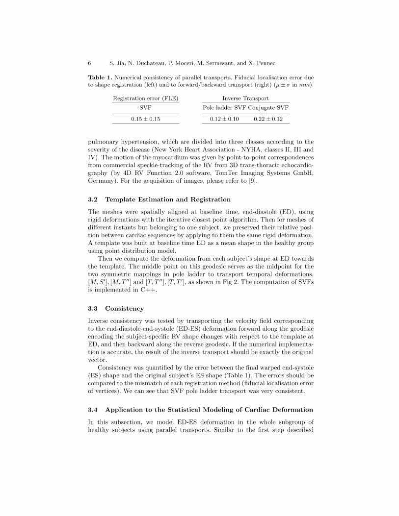
Table 1. Numerical consistency of parallel transports. Fiducial localisation error dueto shape registration (left) and to forward/backward transport (right) (µ± σ in mm).
Registration error (FLE)
SVF
0.15 ± 0.15
Inverse Transport
Pole ladder SVF Conjugate SVF
0.12 ± 0.10 0.22 ± 0.12
pulmonary hypertension, which are divided into three classes according to theseverity of the disease (New York Heart Association - NYHA, classes II, III andIV). The motion of the myocardium was given by point-to-point correspondencesfrom commercial speckle-tracking of the RV from 3D trans-thoracic echocardio-graphy (by 4D RV Function 2.0 software, TomTec Imaging Systems GmbH,Germany). For the acquisition of images, please refer to [9].
3.2 Template Estimation and Registration
The meshes were spatially aligned at baseline time, end-diastole (ED), usingrigid deformations with the iterative closest point algorithm. Then for meshes ofdifferent instants but belonging to one subject, we preserved their relative posi-tion between cardiac sequences by applying to them the same rigid deformation.A template was built at baseline time ED as a mean shape in the healthy groupusing point distribution model.
Then we compute the deformation from each subject’s shape at ED towardsthe template. The middle point on this geodesic serves as the midpoint for thetwo symmetric mappings in pole ladder to transport temporal deformations,[M,S′], [M,T ′′] and [T, T ′′], [T, T ′], as shown in Fig 2. The computation of SVFsis implemented in C++.
3.3 Consistency
Inverse consistency was tested by transporting the velocity field correspondingto the end-diastole-end-systole (ED-ES) deformation forward along the geodesicencoding the subject-specific RV shape changes with respect to the template atED, and then backward along the reverse geodesic. If the numerical implementa-tion is accurate, the result of the inverse transport should be exactly the originalvector.
Consistency was quantified by the error between the final warped end-systole(ES) shape and the original subject’s ES shape (Table 1). The errors should becompared to the mismatch of each registration method (fiducial localisation errorof vertices). We can see that SVF pole ladder transport was very consistent.
3.4 Application to the Statistical Modeling of Cardiac Deformation
In this subsection, we model ED-ES deformation in the whole subgroup ofhealthy subjects using parallel transports. Similar to the first step described

Parallel Transport of Surface Deformations 7
Fig. 3. Fist two modes of variation after applying PCA on the transported ED-ESchanges, for the healthy subgroup. Comparison of point-to-point (top) and pole ladderwith SVF (bottom). Surface folds appear at the arrow.
in the previous experiments, we transported the velocity fields associated toeach subject’s ED-ES changes to the template at ED. Then, we applied princi-pal component analysis (PCA) on the transported velocity fields, and warpedthe template with resulting principal components to show the main modes ofdeformation among this subgroup. We also compared this with the PCA modesobtained from the point-to-point ED-ES displacement.
Table 2. Compactness of the representations
Explained variations 85% 95% 98%
Displacement 6 11 16
Pole ladder SVF 7 10 14
The first mode from point-to-point displacement accounts for theelongation (no relative displacementof the two valves), the second ac-counts for the vertical displacementand a slight orientation of the tri-cuspid valve. The first two modesfrom the pole ladder with SVF aremainly related to the orientationand the relative position of the tri-cuspid valve and the pulmonary valve. Figure 3 summarizes these results forthe point-to-point displacement and pole ladder with SVF. We compared thecompactness of the representations (Table 2) and found pole ladder with SVF avery good explanation.

8 S. Jia, N. Duchateau, P. Moceri, M. Sermesant, and X. Pennec
3.5 Limitations
Cardiac motion tracking establishes point-to-point correspondences between meshvertices, available here to benchmark our method. For databases that do nothave such correspondence, one option is to register the segmented surfaces tothe template, with the framework of currents or varifold-based deformation,which provide elegant mathematical approaches to match surfaces and estab-lish point-to-point correspondence. The limitation of the method notably laysin registration accuracy, but this is compensated by the stability and one-steptransport structure of the algorithm.
4 Conclusion
We proposed a general scheme to perform statistical modeling of the temporaldeformation of the heart, directly based on meshes. The extensions of the poleladder for parallel transport to mesh deformations were stable and accurate,of importance for the assessment of pathological changes. They also provideda more compact and interpretable representation that is useful for statisticalmodeling, and prevented surface folds in the range of population variability.
The method is adaptable to other anatomies with temporal or longitudinaldata. Perspectives notably include synthetic experiments; the possibility to sim-ulate physiologically-realistic motion on a given subject’s anatomy, of relevanceto better understand shape and motion interactions, or inversely to generatetemplate sequences derived from populations, to be used for physiological mod-eling.
Acknowledgments. Part of the research was funded by the Agence Nationalede la Recherche (ANR)/ERA CoSysMed SysAFib project. The authors wouldlike to thank fellows from Hopital Pasteur, CHU de Nice, France for preparingthe data.
References
1. Bai, W., Shi, W., de Marvao, A., Dawes, T.J., ORegan, D.P., Cook, S.A., Rueckert,D.: A bi-ventricular cardiac atlas built from 1000+ high resolution mr images ofhealthy subjects and an analysis of shape and motion. Medical image analysis26(1), 133–145 (2015)
2. Bossa, M., Hernandez, M., Olmos, S.: Contributions to 3d diffeomorphic atlas esti-mation: application to brain images. In: International Conference on Medical ImageComputing and Computer-Assisted Intervention. pp. 667–674. Springer (2007)
3. Davis, M.H., Khotanzad, A., Flamig, D.P., Harms, S.E.: A physics-based coordi-nate transformation for 3-d image matching. IEEE transactions on medical imaging16(3), 317–328 (1997)
4. Duchateau, N., De Craene, M., Piella, G., Silva, E., Doltra, A., Sitges, M., Bij-nens, B.H., Frangi, A.F.: A spatiotemporal statistical atlas of motion for the quan-tification of abnormal myocardial tissue velocities. Medical image analysis 15(3),316–328 (2011)

Parallel Transport of Surface Deformations 9
5. Gavrilov, A.V.: Algebraic properties of covariant derivative and composition ofexponential maps. Matematicheskie Trudy 9(1), 3–20 (2006)
6. Lorenzi, M., Ayache, N., Pennec, X.: Schilds ladder for the parallel transport ofdeformations in time series of images. In: Biennial International Conference onInformation Processing in Medical Imaging. pp. 463–474. Springer (2011)
7. Lorenzi, M., Pennec, X.: Efficient parallel transport of deformations in time seriesof images: from schilds to pole ladder. Journal of Mathematical Imaging and Vision50(1-2), 5–17 (2014)
8. Louis, M., Bone, A., Charlier, B., Durrleman, S., Initiative, A.D.N., et al.: Par-allel transport in shape analysis: a scalable numerical scheme. In: InternationalConference on Geometric Science of Information. pp. 29–37. Springer (2017)
9. Moceri, P., Duchateau, N., Baudouy, D., Schouver, E.D., Leroy, S., Squara, F., Fer-rari, E., Sermesant, M.: Three-dimensional right-ventricular regional deformationand survival in pulmonary hypertension. European Heart Journal-CardiovascularImaging (2017)
10. Pennec, X.: Parallel transport with pole ladder: a third order scheme in affineconnection spaces which is exact in affine symmetric spaces. arXiv preprintarXiv:1805.11436 (2018)
11. Qiu, A., Younes, L., Miller, M.I., Csernansky, J.G.: Parallel transport in diffeomor-phisms distinguishes the time-dependent pattern of hippocampal surface deforma-tion due to healthy aging and the dementia of the alzheimer’s type. NeuroImage40(1), 68–76 (2008)
12. Rohe, M.M., Datar, M., Heimann, T., Sermesant, M., Pennec, X.: Svf-net: Learningdeformable image registration using shape matching. In: International Conferenceon Medical Image Computing and Computer-Assisted Intervention. pp. 266–274.Springer (2017)
13. Vercauteren, T., Pennec, X., Perchant, A., Ayache, N.: Symmetric log-domain dif-feomorphic registration: A demons-based approach. In: International conferenceon medical image computing and computer-assisted intervention. pp. 754–761.Springer (2008)
14. Younes, L.: Jacobi fields in groups of diffeomorphisms and applications. Quarterlyof applied mathematics pp. 113–134 (2007)