Liver Gallbladder Common bile duct Pancreas Pancreatic duct Duodenum Biliary and Pancreatic Anatomy.
Upload
duonghuongCategory
view
218download
4
Pancreatic Pseudocysts
Eric Rellinger
October 3, 2012 Vanderbilt General Surgery

Overview
- Pancreatitis (Acute, Necrotizing, Chronic) - Pancreatic Pseudocysts
- Clinical Presentation - Diagnostic Evaluation - Therapeutic options - Management

Acute pancreatitis - 200,000 cases of acute
pancreatitis per year - Management focuses on
maximizing tissue oxygen perfusion and pain control
- Scoring systems are available
- Severe acute pancreatitis- Ransom score >3 or Apache II score > 8
- Acute pancreatitis < Severe acute pancreatitis < Necrotizing
acute pancreatitis < Infected necrotizing pancreatitis

Necrotizing Pancreatitis - Diagnosis based on opacification
of pancreatic tissue during a CT scan with IV contrast
- Necrotic tissue indistinguishable from inflamed tissue for first 72-96h
- More necrosis correlates with likelihood of infection
- Infected necrosis diagnosed by the presence of gas or FNA cultures
- Clinical condition guides intervention
- Attempt to delay operative intervention to permit demarcation of necrosis and compartmentalization of infection

Chronic pancreatitis - The histologic hallmark of chronic
pancreatitis is the persistent inflammation and irreversible fibrosis associated with atrophy of the pancreatic parenchyma
- Associated with chronic pain and endocrine (HgA1C) and exocrine insufficiency (fecal elastase); may be associated with jaundice, persistent N/V
- CT useful for diagnosis; ERCP- gold standard for defining ductal anatomy- should be considered a therapeutic modality for ductal complications
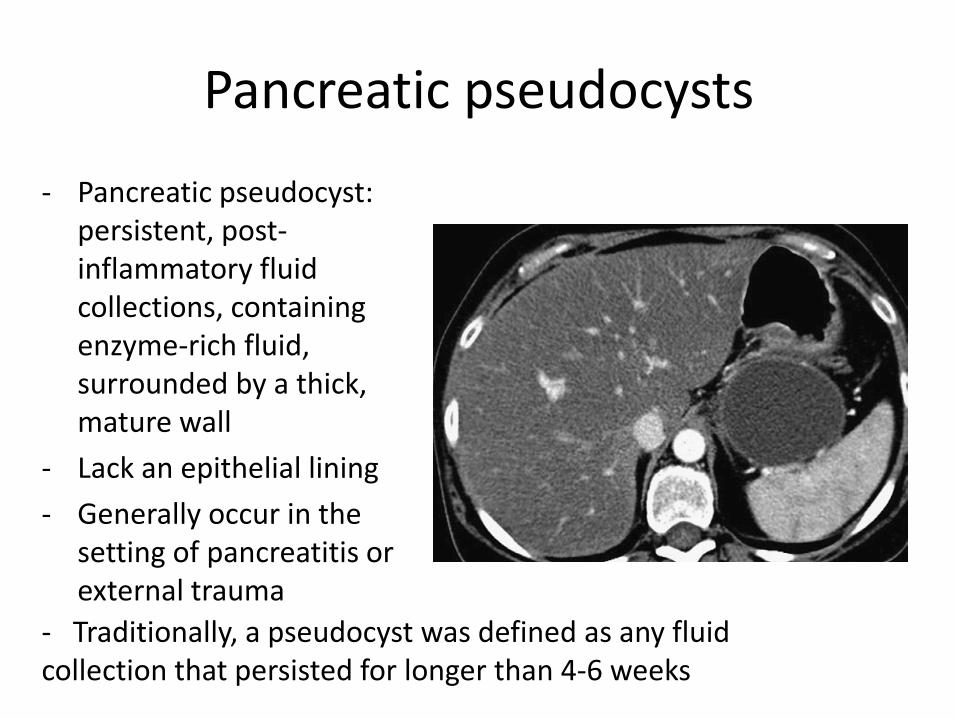
Pancreatic pseudocysts
- Pancreatic pseudocyst: persistent, post-inflammatory fluid collections, containing enzyme-rich fluid, surrounded by a thick, mature wall
- Lack an epithelial lining - Generally occur in the
setting of pancreatitis or external trauma
- Traditionally, a pseudocyst was defined as any fluid collection that persisted for longer than 4-6 weeks

Clinical Presentation of Pseudocysts
- Intraparenchymal vs. peripancreatic - Asymptomatic vs. symptomatic - Symptomatology:
1) failure to clinically progress (5-7 days) 2) abdominal pain, 3) GI dysmotility (early satiety, nausea, vomitting, obstruction) 4) biliary obstruction 5) pancreatic ascites/effusion 6) hemorrhage

Pathophysiology
- Most common etiology: EtOH, gallstones, trauma, iatrogenic
- Acute episode: normal pancreatic duct, ductal disruption heals spontaneously
- Pancreatic necrosis: necrosis, organizaition, liquification; may also connect with main duct
- Chronic pancreatitis: pancreatic parenchymal fibrosis, irregularity leads to elevated intraductal pressure leading to disruption of an obstructed duct
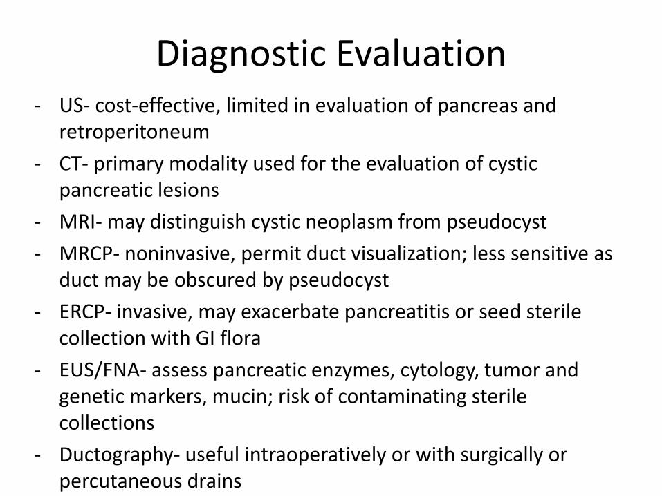
Diagnostic Evaluation - US- cost-effective, limited in evaluation of pancreas and
retroperitoneum - CT- primary modality used for the evaluation of cystic
pancreatic lesions - MRI- may distinguish cystic neoplasm from pseudocyst - MRCP- noninvasive, permit duct visualization; less sensitive as
duct may be obscured by pseudocyst - ERCP- invasive, may exacerbate pancreatitis or seed sterile
collection with GI flora - EUS/FNA- assess pancreatic enzymes, cytology, tumor and
genetic markers, mucin; risk of contaminating sterile collections
- Ductography- useful intraoperatively or with surgically or percutaneous drains

Therapeutic Options - Open vs. laparoscopic procedures: a. Cystic drainage procedures: 1) Cystogastrostomy, 2) Cystoduodenostomy, 3) Roux-en-Y cystojejunostomy b. Ductal drainage procedures: 1) Peustow, 2) Frey, 3) Beger c. Resection: 1) distal pancreatectomy +/- splenectomy, 2) cyst resection - Endoscopic: 1) endoscopic cystogastrostomy, 2) endoscopic cystoduodenostomy 3) transpapillary stent placement - Percutaneous drainage- less invasive, risks prolonged drainage,
bacterial colonization

Cystogastrostomy
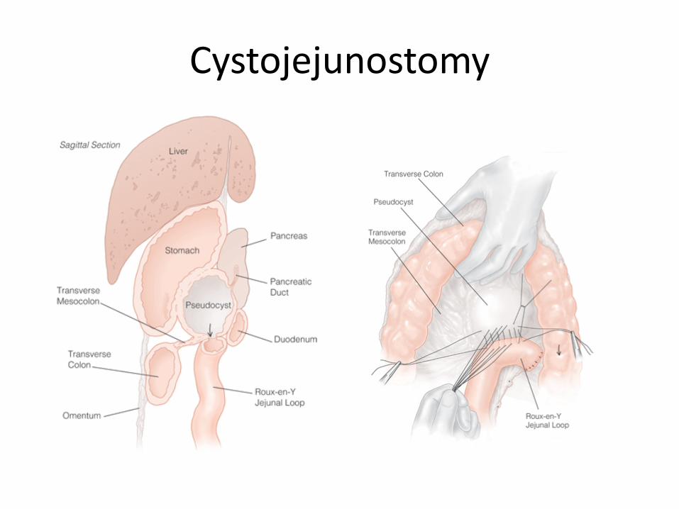
Cystojejunostomy

Puestow (longitudinal pancreaticojejunostomy)

Beger/Frey Procedures
Beger Procedure Frey Procedure

Management
- Treatment is driven by symptomatology - Traditionally, pseudocysts were treated if persisting beyond 6wks citing 50% risk of complications - Pseudocysts continue to spontaneously resolve for up to a year (complication rate=10%) - Larger pseudocysts (>6cm), less likely to resolve but
may be safely watched in the absence of symptoms - Little data exists on the appropriate interval for repeat
imaging (most reimage in 3-6mos)

Management- Important Considerations
1) Etiology of pancreatitis 2) Maturity of pseudocyst wall 3) Relationship to solid organs/vasculature 4) Pancreatic duct anatomy 5) Clinical stability

Etiology of Pancreatitis
- Acute Pancreatitis- first-line therapy endoscopic intervention; isolated cysts may be amenable to percutaneous drainage
- Pancreatic necrosis- attempt to delay operative management to permit areas of necrosis to organize; internal drainage seldom appropriate in context of infection
- Chronic pancreatitis- management of the pseudocyst alone is seldom effective; tailor therapy to ductal anatomy/drainage
**infected pseudocysts are classically drained externally
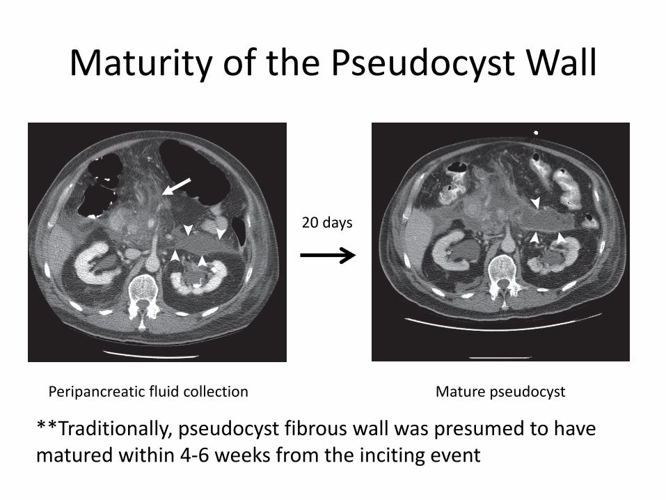
Maturity of the Pseudocyst Wall
Peripancreatic fluid collection
20 days
Mature pseudocyst
**Traditionally, pseudocyst fibrous wall was presumed to have matured within 4-6 weeks from the inciting event

Relationship to solid organs/vasculature

Pancreatic duct communication

- Single center retrospective case series 563 pts with pseudocysts ( 1985 to 2006) - Goal to utilize pancreatic ductal anatomy to predict course and direct therapies - Policy of routine evaluation of pancreatic duct anatomy (ERCP, MRCP, ductogram)
was used pts with acute or chronic pancreatitis and pseudocysts - ERCP was performed 24h prior to intervention; MRCP limited by sensitivity - Operative intervention indicated for persistent symptomatic cysts, hemorrhage,
infection, rupture, or obstruction - Subgroup analysis of severe acute pancreatitis were included (Ranson >5,
evidence of necrosis, Balthazaar grade III or IV, or organ failure) - Attempt to delay intervention 4 weeks in severe or necrotizing pancreatitis;
utilized large bore percutaneous drains for temporization/treatment - Success of intervention- no recurrence of pseudocyst on follow-up imaging - Outcomes- relief of pain, resumption of PO intake, LOH, readmission, need for
critical care, need for operative debridement, success of percutaneous drainage

Nealon’s Classification

* Ductal anatomy predicts persistence of pseudocysts

* Ductal anatomy predicts persistence of pseudocysts

**Normal ductal anatomy predicts success with percutaneous pseudocyst drainage

**Operative debridement was required in 70% of patients with severe pancreatitis, more than twice as frequently in type II and III (83-85%) vs. type I (39%)

** Ductal narrowing/ disconnection predicts recurrent pancreatitis and sepsis

When to evaluate ductal anatomy?
- Ductal anatomy is not utilized in the setting of hemorrhage and plummeting clinical status - Most pseudocysts resolve in the first 4-6wks - ERCP- invasive, risk of recurrent pancreatitis and
infecting sterile pancreatic fluid collections - ERCP should only be completed with intervention
planned within the subsequent 48h - MRCP- noninvasive, less sensitive, improved
ductal visualization s/p cyst drainage

Therapy directed by ductal anatomy
Nonoperative (percutaneous vs. endoscopic)
Transpapillary stent vs. operative resection or drainage (duct vs. cyst)
Operative resection or drainage (duct vs. cyst)
Endotherapy (stent, stone extraction, lithotripsy) vs.operative ductal drainage/pancreatic head resection

Summary - Pancreatic pseudocysts are persistent, post-
inflammatory fluid collections, containing enzyme-rich fluid, surrounded by a thick, mature wall
- Intervention is driven by symptomatology - Multidisciplinary approach to management (Surgery,
Interventional Radiology, Gastroenterology) - Therapeutic options include: percutaneous,
endoscopic, laparoscopic, and open surgical approaches
- Persistent, refractory fluid collections/fistulas likely reflect main duct disruption
- Ductal anatomy is a key driver of successful therapeutic management of persistent pseudocysts