Pancreas & Biliary System - KSUMSC · Biliary system The cystic duct is about 1.5 in. (3.8 cm) long...
14
Pancreas & Biliary System Gastrointestinal block-Anatomy-Lecture 4 Editing file
Transcript of Pancreas & Biliary System - KSUMSC · Biliary system The cystic duct is about 1.5 in. (3.8 cm) long...
Pancreas & Biliary SystemGastrointestinal block-Anatomy-Lecture 4
Editing file
● Describe the location, surface anatomy, parts, relations & peritoneal reflection of the pancreas and gallbladder.
● Describe blood supply, nerve supply andlymphatic drainage of pancreas and gallbladder.
● Describe Course of each of common hepatic, cystic and common bile duct and pancreatic ducts
Color guide :Only in boys slides in GreenOnly in girls slides in Purpleimportant in RedNotes in GreyAt the end of the lecture, students should be able to:
Objectives
Pancreas
3
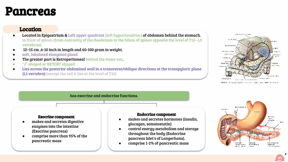
● Located in Epigastrium & Left upper quadrant (left hypochondriac) of abdomen behind the stomach. in front of spleen (from concavity of the duodenum to the hilum of spleen opposite the level of T12– L3 vertebrae).
● 12–15 cm ,6-10 inch in length and 60-100 gram in weight. ● soft, lobulated elongated gland ● The greater part is Retroperitoneal behind the lesser sac.● “J”-shaped or RETORT shaped● Lies across the posterior abdominal wall in a transverse/oblique directions at the transpyloric plane
(L1 vertebra) (except the tail it lies at the level of T12)
Location
has exocrine and endocrine functions.
Exocrine component● makes and secretes digestive
enzymes into the intestine (Exocrine pancreas)
● comprise more than 95% of the pancreatic mass
Endocrine component● makes and secretes hormones (insulin,
glucagon, somatostatin)● control energy metabolism and storage
throughout the body (Endocrine pancreas Islet's of Langerhans).
● comprise 1-2% of pancreatic mass
Pancreas Parts
4
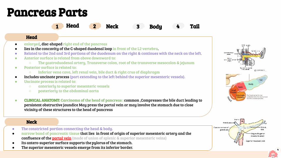
● enlarged, disc-shaped right end of the pancreas● lies in the concavity of the C-shaped duodenal loop in front of the L2 vertebra.● Related to the 2nd and 3rd portions of the duodenum on the right & continues with the neck on the left.● Anterior surface is related from above downward to:
○ The gastroduodenal artery, Transverse colon, root of the transverse mesocolon & jejunum● Posterior surface is related to:
○ Inferior vena cava, left renal vein, bile duct & right crus of diaphragm● Includes uncinate process (part extending to the left behind the superior mesenteric vessels).● Uncinate process is related to:
○ anteriorly to superior mesenteric vessels○ posteriorly to the abdominal aorta
● CLINICAL ANATOMY: Carcinoma of the head of pancreas : common ,Compresses the bile duct leading to persistent obstructive jaundice May press the portal vein or may involve the stomach due to close vicinity of these structures to the head of pancreas
● The constricted portion connecting the head & body.● narrow band of pancreatic tissue that lies in front of origin of superior mesenteric artery and the
confluence of the portal vein (made of union of splenic & superior mesenteric veins)● Its antero-superior surface supports the pylorus of the stomach.● The superior mesenteric vessels emerge from its inferior border.
Head1 2 3 4Neck Body Tail
Head
Neck
Pancreas Parts
5
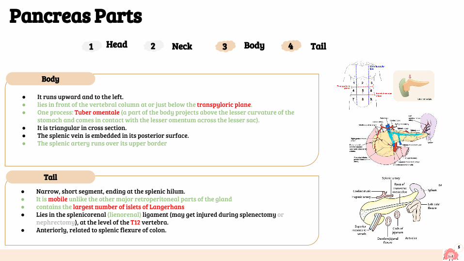
● Narrow, short segment, ending at the splenic hilum.● It is mobile unlike the other major retroperitoneal parts of the gland● contains the largest number of islets of Langerhans● Lies in the splenicorenal (lienorenal) ligament (may get injured during splenectomy or
nephrectomy), at the level of the T12 vertebra.● Anteriorly, related to splenic flexure of colon.
● It runs upward and to the left.● lies in front of the vertebral column at or just below the transpyloric plane.● One process: Tuber omentale (a part of the body projects above the lesser curvature of the
stomach and comes in contact with the lesser omentum across the lesser sac).● It is triangular in cross section.● The splenic vein is embedded in its posterior surface.● The splenic artery runs over its upper border
Head1 2 3 4Neck Body Tail
Body
Tail
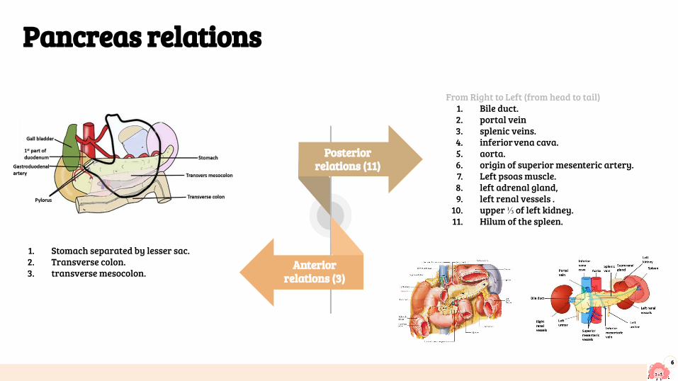
From Right to Left (from head to tail)1. Bile duct. 2. portal vein3. splenic veins.4. inferior vena cava.5. aorta.6. origin of superior mesenteric artery.7. Left psoas muscle.8. left adrenal gland, 9. left renal vessels .
10. upper ⅓ of left kidney.11. Hilum of the spleen.
6
1. Stomach separated by lesser sac.2. Transverse colon. 3. transverse mesocolon.
Pancreas relations
Anterior relations (3)
Posterior relations (11)
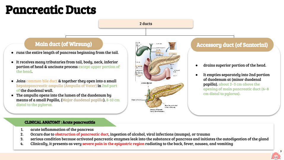
● drains superior portion of the head.
● It empties separately into 2nd portion of duodenum at (minor duodenal papilla). about 2–3 cm above the opening of main pancreatic duct (6–8 cm distal to pylorus).
Pancreatic Ducts
7
2 ducts
Accessory duct (of Santorini)
1. acute inflammation of the pancreas2. Occurs due to obstruction of pancreatic duct, ingestion of alcohol, viral infections (mumps), or trauma3. serious condition because activated pancreatic enzymes leak into the substance of pancreas and initiates the autodigestion of the gland4. Clinically, it presents as very severe pain in the epigastric region radiating to the back, fever, nausea, and vomiting
● runs the entire length of pancreas beginning from the tail.
● It receives many tributaries from tail, body, neck, inferior portion of head & uncinate process except upper portion of the head.
● Joins common bile duct & together they open into a small hepatopancreatic ampulla (Ampulla of Vater) in 2nd part of the duodenal wall.
● The ampulla opens into the lumen of the duodenum by means of a small Papilla, (Major duodenal papilla). 8-10 cm distal to the pylorus.
Main duct (of Wirsung)
CLINICAL ANATOMY : Acute pancreatitis
8
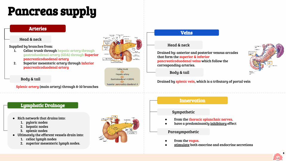
Innervation
Sympathetic
Parasympathetic
● from the thoracic splanchnic nerves.● have a predominantly inhibitory effect
● from the vagus.● stimulate both exocrine and endocrine secretions
Arteries
Head & neck
Supplied by branches from:1. Celiac trunk through hepatic artery through
gastroduodenal artery (GDA) through Superior pancreaticoduodenal artery
2. Superior mesenteric artery through Inferior pancreaticoduodenal artery
Body & tail
Splenic artery (main artery) through 8-10 branches
Veins
Head & neck
Body & tail
Drained by: anterior and posterior venous arcades that form the superior & inferior pancreaticoduodenal veins which follow the corresponding arteries.
Drained by splenic vein, which is a tributary of portal vein
Pancreas supply
Lymphatic Drainage
● Rich network that drains into:1. pyloric nodes2. hepatic nodes3. splenic nodes
● Ultimately the efferent vessels drain into: 1. celiac lymph nodes2. superior mesenteric lymph nodes.
Biliary system
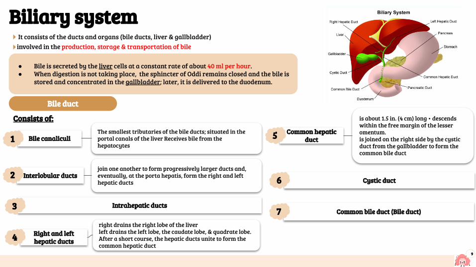
9
Consists of:
Bile canaliculi
Intrahepatic ducts
Right and left hepatic ducts
Common hepatic duct
Cystic duct
Common bile duct (Bile duct)
● Bile is secreted by the liver cells at a constant rate of about 40 ml per hour.● When digestion is not taking place, the sphincter of Oddi remains closed and the bile is
stored and concentrated in the gallbladder; later, it is delivered to the duodenum.
It consists of the ducts and organs (bile ducts, liver & gallbladder)involved in the production, storage & transportation of bile
The smallest tributaries of the bile ducts; situated in the portal canals of the liver Receives bile from the hepatocytes
right drains the right lobe of the liverleft drains the left lobe, the caudate lobe, & quadrate lobe.After a short course, the hepatic ducts unite to form the common hepatic duct
is about 1.5 in. (4 cm) long • descends within the free margin of the lesser omentum.is joined on the right side by the cystic duct from the gallbladder to form the common bile duct
1
Interlobular ductsjoin one another to form progressively larger ducts and, eventually, at the porta hepatis, form the right and left hepatic ducts
2
3
4
5
6
7
Bile duct
10
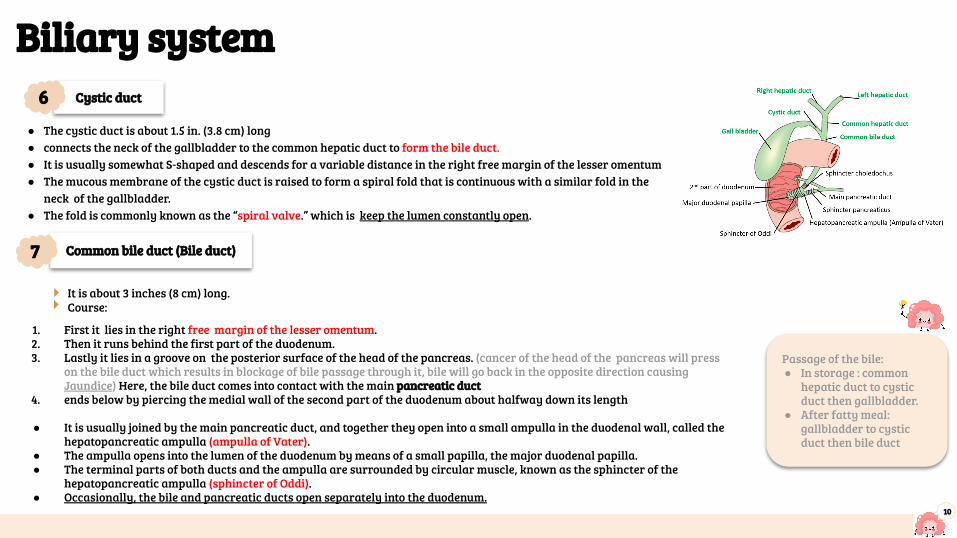
1. First it lies in the right free margin of the lesser omentum.2. Then it runs behind the first part of the duodenum.3. Lastly it lies in a groove on the posterior surface of the head of the pancreas. (cancer of the head of the pancreas will press
on the bile duct which results in blockage of bile passage through it, bile will go back in the opposite direction causing Jaundice) Here, the bile duct comes into contact with the main pancreatic duct
4. ends below by piercing the medial wall of the second part of the duodenum about halfway down its length
● It is usually joined by the main pancreatic duct, and together they open into a small ampulla in the duodenal wall, called the hepatopancreatic ampulla (ampulla of Vater).
● The ampulla opens into the lumen of the duodenum by means of a small papilla, the major duodenal papilla.● The terminal parts of both ducts and the ampulla are surrounded by circular muscle, known as the sphincter of the
hepatopancreatic ampulla (sphincter of Oddi).● Occasionally, the bile and pancreatic ducts open separately into the duodenum.
It is about 3 inches (8 cm) long.Course:
Biliary system
● The cystic duct is about 1.5 in. (3.8 cm) long ● connects the neck of the gallbladder to the common hepatic duct to form the bile duct. ● It is usually somewhat S-shaped and descends for a variable distance in the right free margin of the lesser omentum● The mucous membrane of the cystic duct is raised to form a spiral fold that is continuous with a similar fold in the
neck of the gallbladder. ● The fold is commonly known as the “spiral valve.” which is keep the lumen constantly open.
Cystic duct
Common bile duct (Bile duct)
6
7
Passage of the bile:● In storage : common
hepatic duct to cystic duct then gallbladder.
● After fatty meal: gallbladder to cystic duct then bile duct
11
Gallbladder
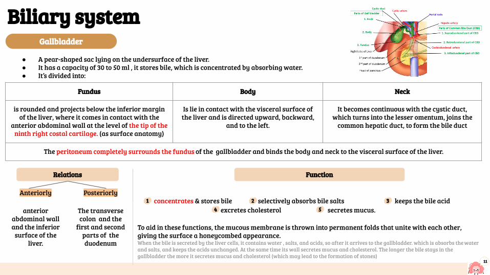
● A pear-shaped sac lying on the undersurface of the liver.● It has a capacity of 30 to 50 ml , it stores bile, which is concentrated by absorbing water.● It’s divided into:
Biliary system
Fundus Body Neck
is rounded and projects below the inferior margin of the liver, where it comes in contact with the
anterior abdominal wall at the level of the tip of the ninth right costal cartilage. (as surface anatomy)
Is lie in contact with the visceral surface of the liver and is directed upward, backward,
and to the left.
It becomes continuous with the cystic duct, which turns into the lesser omentum, joins the
common hepatic duct, to form the bile duct
The peritoneum completely surrounds the fundus of the gallbladder and binds the body and neck to the visceral surface of the liver.
Anteriorly Posteriorly concentrates & stores bile selectively absorbs bile salts keeps the bile acid excretes cholesterol secretes mucus.
To aid in these functions, the mucous membrane is thrown into permanent folds that unite with each other, giving the surface a honeycombed appearance.When the bile is secreted by the liver cells, it contains water , salts, and acids, so after it arrives to the gallbladder. which is absorbs the water and salts, and keeps the acids unchanged. At the same time its wall secretes mucus and cholesterol. The longer the bile stays in the gallbladder the more it secretes mucus and cholesterol (which may lead to the formation of stones)
anterior abdominal wall and the inferior
surface of the liver.
The transverse colon and the
first and second parts of the duodenum
Function Relations
1 2 34 5
12
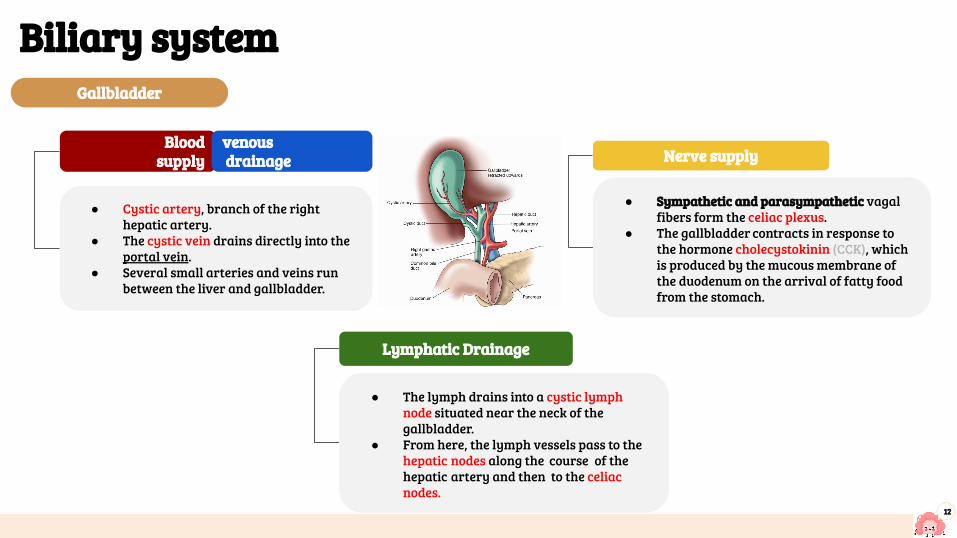
Blood supply
● Cystic artery, branch of the right hepatic artery.
● The cystic vein drains directly into the portal vein.
● Several small arteries and veins run between the liver and gallbladder.
venous drainage
● Sympathetic and parasympathetic vagal fibers form the celiac plexus.
● The gallbladder contracts in response to the hormone cholecystokinin (CCK), which is produced by the mucous membrane of the duodenum on the arrival of fatty food from the stomach.
Nerve supply
● The lymph drains into a cystic lymph node situated near the neck of the gallbladder.
● From here, the lymph vessels pass to the hepatic nodes along the course of the hepatic artery and then to the celiac nodes.
Lymphatic Drainage
Gallbladder
Biliary system
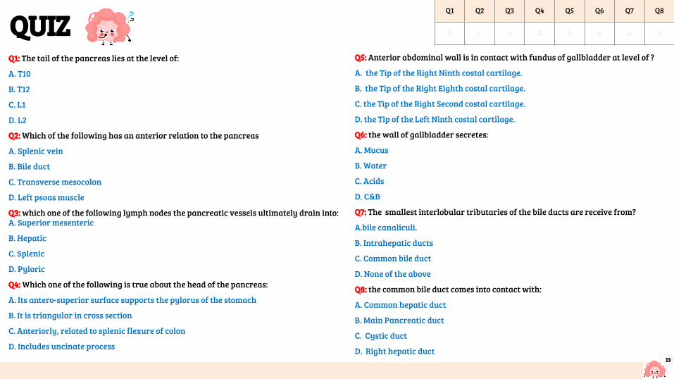
QUIZQ1: The tail of the pancreas lies at the level of:
A. T10
B. T12
C. L1
D. L2
Q2: Which of the following has an anterior relation to the pancreas
A. Splenic vein
B. Bile duct
C. Transverse mesocolon
D. Left psoas muscle
Q3: which one of the following lymph nodes the pancreatic vessels ultimately drain into:A. Superior mesenteric
B. Hepatic
C. Splenic
D. Pyloric
Q4: Which one of the following is true about the head of the pancreas:
A. Its antero-superior surface supports the pylorus of the stomach
B. It is triangular in cross section
C. Anteriorly, related to splenic flexure of colon
D. Includes uncinate process
Q5: Anterior abdominal wall is in contact with fundus of gallbladder at level of ?
A. the Tip of the Right Ninth costal cartilage.
B. the Tip of the Right Eighth costal cartilage.
C. the Tip of the Right Second costal cartilage.
D. the Tip of the Left Ninth costal cartilage.
Q6: the wall of gallbladder secretes:
A. Mucus
B. Water
C. Acids
D. C&B
Q7: The smallest interlobular tributaries of the bile ducts are receive from?
A.bile canaliculi.
B. Intrahepatic ducts
C. Common bile duct
D. None of the above
Q8: the common bile duct comes into contact with:
A. Common hepatic duct
B. Main Pancreatic duct
C. Cystic duct
D. Right hepatic duct13
Q1 Q2 Q3 Q4 Q5 Q6 Q7 Q8
B C A D A A A B

Members board
● Abdulrahman Shadid ● Ateen Almutairi
Girls team :
● Ajeed Al Rashoud● Taif Alotaibi● Noura Al Turki● Amirah Al-Zahrani● Alhanouf Al-haluli● Sara Al-Abdulkarem● Renad Al Haqbani● Nouf Al Humaidhi● Jude Al Khalifah● Nouf Al Hussaini● Danah Al Halees● Rema Al Mutawa● Maha Al Nahdi ● Razan Al zohaifi ● Ghalia Alnufaei
Team leaders
Editing file
Contact us:
Boys team:
● Mohammed Al-huqbani● Salman Alagla● Ziyad Al-jofan● Ali Aldawood● Khalid Nagshabandi● Sameh nuser● Abdullah Basamh● Alwaleed Alsaleh● Mohaned Makkawi● Abdullah Alghamdi