Panax Notoginseng Saponins Prevent Bone Loss by Promoting ...
8
Research Article Panax Notoginseng Saponins Prevent Bone Loss by Promoting Angiogenesis in an Osteoporotic Mouse Model Hao Hu , 1 Yan Chen, 1 Zhiyuan Zou, 1 Liangping Li, 1 Fuxin Wei, 2 Chun Liu, 3 Zemin Ling , 1 and Xuenong Zou 1 1 Guangdong Provincial Key Laboratory of Orthopedics and Traumatology, Department of Spinal Surgery, The First Affiliated Hospital of Sun Yat-sen University, Guangzhou 510080, China 2 Department of Orthopaedic Surgery, The Seventh Affiliated Hospital and Orthopedic Research Institute of Sun Yat-sen University, Shenzhen 518107, China 3 Precision Medicine Institute, The First Affiliated Hospital of Sun Yat-sen University, Guangzhou 510080, China Correspondence should be addressed to Zemin Ling; [email protected] and Xuenong Zou; [email protected] Hao Hu and Yan Chen contributed equally to this work. Received 22 August 2020; Revised 6 November 2020; Accepted 27 November 2020; Published 14 December 2020 Academic Editor: Shen Liu Copyright © 2020 Hao Hu et al. This is an open access article distributed under the Creative Commons Attribution License, which permits unrestricted use, distribution, and reproduction in any medium, provided the original work is properly cited. With the aging of the population and the extension of life expectancy, osteoporosis is becoming a global epidemic. Although there are several drugs used to treat osteoporosis in clinical practice, such as parathyroid hormone or bisphosphonates, they all have some serious side effects. Therefore, a safer drug is called for osteoporosis, especially for the prevention in the early stage of the disease, not only the treatment in the later stage. Panax notoginseng saponin (PNS), a traditional Chinese herb, has been used as anti- ischemic drug due to its function on improving vascular circulation. In order to verify whether Panax notoginseng saponins (PNS) could be used to prevent osteoporosis, ovariectomy (OVX) was induced in female C57BL/C6J mice, followed by orally administration with 40 mg/kg/d, 80 mg/kg/d, and 160 mg/kg/d of three different dosages of PNS for 9 weeks. Serum biochemical analysis, micro-CT, histological evaluation, and immunostaining of markers of osteogenesis and angiogenesis were performed in the sham, osteoporotic (OVX), and treatment (OVX+PNS) groups. Micro-CT and histological evaluation showed that compared to sham group, the bone mass of OVX group reduced significantly, while it was significantly restored in the moderate-dose PNS (40 mg/kg and 80 mg/kg) treatment groups. The expression of CD31 and osteocalcin (OCN) in the bone tissue of treatment group also increased, suggesting that PNS activated osteogenesis and angiogenesis, which subsequently increased the bone mass. These results confirmed the potential function of PNS on the prevention of osteoporosis. However, in the high dose of PNS (160 mg/kg) group, the antiosteoportic effect had been eliminated, which also suggested the importance of proper dose of PNS for the prevention and treatment of osteoporosis in postmenopausal women. 1. Introduction Osteoporosis is a metabolic disease characterized by decreased bone mass, increased bone fragility, and micro- architectural deterioration of bone tissue. In addition to increasing the risk of fragility fracture, osteoporosis may also increase the risk of hospitalization associated with certain complications. With the increasing aging of China’s popula- tion, the prevalence of osteoporosis has increased remarkably in the past decade (from 14.94% before 2012 to 27.96% in 2015), and the prevalence in women was significantly higher than that in men (25.41% vs. 15.33%) [1]. With aging, decreased angiogenesis in bone and bone marrow is a principal cause of osteoporosis [2]. Because estro- gen is also an important regulator for angiogenesis, its defi- ciency can lead to osteoporosis for postmenopausal women or those who have undergone tumor-related ovariectomy. Traditional Chinese medicine has been used for thousands Hindawi BioMed Research International Volume 2020, Article ID 8412468, 8 pages https://doi.org/10.1155/2020/8412468
Transcript of Panax Notoginseng Saponins Prevent Bone Loss by Promoting ...

Research ArticlePanax Notoginseng Saponins Prevent Bone Loss by PromotingAngiogenesis in an Osteoporotic Mouse Model
Hao Hu ,1 Yan Chen,1 Zhiyuan Zou,1 Liangping Li,1 FuxinWei,2 Chun Liu,3 Zemin Ling ,1
and Xuenong Zou 1
1Guangdong Provincial Key Laboratory of Orthopedics and Traumatology, Department of Spinal Surgery, The First AffiliatedHospital of Sun Yat-sen University, Guangzhou 510080, China2Department of Orthopaedic Surgery, The Seventh Affiliated Hospital and Orthopedic Research Institute of Sun Yat-sen University,Shenzhen 518107, China3Precision Medicine Institute, The First Affiliated Hospital of Sun Yat-sen University, Guangzhou 510080, China
Correspondence should be addressed to Zemin Ling; [email protected] and Xuenong Zou; [email protected]
Hao Hu and Yan Chen contributed equally to this work.
Received 22 August 2020; Revised 6 November 2020; Accepted 27 November 2020; Published 14 December 2020
Academic Editor: Shen Liu
Copyright © 2020 Hao Hu et al. This is an open access article distributed under the Creative Commons Attribution License, whichpermits unrestricted use, distribution, and reproduction in any medium, provided the original work is properly cited.
With the aging of the population and the extension of life expectancy, osteoporosis is becoming a global epidemic. Although thereare several drugs used to treat osteoporosis in clinical practice, such as parathyroid hormone or bisphosphonates, they all have someserious side effects. Therefore, a safer drug is called for osteoporosis, especially for the prevention in the early stage of the disease,not only the treatment in the later stage. Panax notoginseng saponin (PNS), a traditional Chinese herb, has been used as anti-ischemic drug due to its function on improving vascular circulation. In order to verify whether Panax notoginseng saponins(PNS) could be used to prevent osteoporosis, ovariectomy (OVX) was induced in female C57BL/C6J mice, followed by orallyadministration with 40mg/kg/d, 80mg/kg/d, and 160mg/kg/d of three different dosages of PNS for 9 weeks. Serum biochemicalanalysis, micro-CT, histological evaluation, and immunostaining of markers of osteogenesis and angiogenesis were performed inthe sham, osteoporotic (OVX), and treatment (OVX+PNS) groups. Micro-CT and histological evaluation showed thatcompared to sham group, the bone mass of OVX group reduced significantly, while it was significantly restored in themoderate-dose PNS (40mg/kg and 80mg/kg) treatment groups. The expression of CD31 and osteocalcin (OCN) in the bonetissue of treatment group also increased, suggesting that PNS activated osteogenesis and angiogenesis, which subsequentlyincreased the bone mass. These results confirmed the potential function of PNS on the prevention of osteoporosis. However, inthe high dose of PNS (160mg/kg) group, the antiosteoportic effect had been eliminated, which also suggested the importance ofproper dose of PNS for the prevention and treatment of osteoporosis in postmenopausal women.
1. Introduction
Osteoporosis is a metabolic disease characterized bydecreased bone mass, increased bone fragility, and micro-architectural deterioration of bone tissue. In addition toincreasing the risk of fragility fracture, osteoporosis may alsoincrease the risk of hospitalization associated with certaincomplications. With the increasing aging of China’s popula-tion, the prevalence of osteoporosis has increased remarkably
in the past decade (from 14.94% before 2012 to 27.96% in2015), and the prevalence in women was significantly higherthan that in men (25.41% vs. 15.33%) [1].
With aging, decreased angiogenesis in bone and bonemarrow is a principal cause of osteoporosis [2]. Because estro-gen is also an important regulator for angiogenesis, its defi-ciency can lead to osteoporosis for postmenopausal womenor those who have undergone tumor-related ovariectomy.Traditional Chinese medicine has been used for thousands
HindawiBioMed Research InternationalVolume 2020, Article ID 8412468, 8 pageshttps://doi.org/10.1155/2020/8412468

of years and still plays an important role in the prevention andthe treatment of diseases in modern China. The dried root ofPanax notoginseng, namely, Sanchi (San Qi), is a famous tra-ditional Chinese herb. The active ingredients of Panax noto-ginseng include saponins, dencichine, flavonoids, andpolysaccharides, among which the pharmaceutical effects ofthe saponins had been studied extensively [3]. The total sapo-nins of Panax notoginseng mainly include ginsenosides Ra3,Rg1, Rb1, and Rd and notoginsenoside R1. Blood concentra-tion of ginsenosides Ra3, Rb1, and Rd was significantly higherthan other compounds after an oral administration of Panaxnotoginseng extract in a rat model [4]. Panax notoginsengsaponin (PNS) is a vasoactive drug [5], which has been usedas an anti-ischemic agent to promote blood circulation in tra-ditional Chinese medicine; however, it also displayed anti-inflammatory [6, 7], antioxidant [8–11], and estrogen-like[12, 13] activities in vitro, making it a potential cure for post-menopausal osteoporosis.
Some previous studies confirmed that Panax notoginsengsaponins facilitate the osteogenic process of the skeletalprogenitor cells in vitro [14, 15], including the proliferation,differentiation, and mineralization. The possible mechanismis that PNS promotes the expression of downstreamosteogenesis-related genes by activating ERK and p38 [16]as well as TGF-β1 [17] signaling pathways. Angiogenesisplays an indispensable role in osteogenesis [18], and the mostimportant pharmacological function of PNS is its vasoactiveeffect, indicating that PNS could preserve bone mass by pro-moting the coupling of angiogenesis and osteogenesis duringosteoporosis. Although other two studies had also proved thepharmacological effect of PNS on alleviating osteoporosis inrat ovariectomy model [19, 20], the underlying mechanismswere not investigated, especially there were no studies toevaluate the function of PNS on preventing bone loss at theearly stage of menopause. In this study, we established anovariectomy-induced osteoporosis mouse model to fullyinvestigate whether early PNS treatment can prevent boneloss by targeting the vascular microarchitecture.
2. Materials and Methods
2.1. Materials. PNSs, the total saponins of Panax notogin-seng, were purchased from KPC Xuesaitong PharmaceuticalCo., Ltd (Yunnan, China). C57BL/6J mice were purchasedfrom SPF (Beijing) Biotechnology Co., Ltd (Beijing, China).Mouse NTXI(cross linked N-telopeptide of type I collagen)ELISA kit (E-EL-M3022) was purchased from Elabscience(Wuhan, China). Anti-osteocalcin antibody (ab93876),recombinant anti-CD31 antibody (ab182981), goat anti-rabbit IgG H&L (Alexa Fluor® 488) (ab150077), and goatanti-rabbit IgG H&L (HRP) (ab205718) were purchasedfrom Abcam (USA).
2.2. Animal Model and PNS Treatment. All the animal exper-iments were carried out carefully in accordance with theprinciples and guidelines of the Animal Ethics Committeeof the First Affiliated Hospital of Sun Yet-sen University. 30twelve-week-old female C57BL/6J mice were housed in indi-vidual ventilated cages under controlled conditions (temper-
ature, 20-26°C; humidity, 40-70%) for a 12-hour light-darkcycle and were allowed free access to water and food.
After one-week adaptation period, the animals were ran-domly divided into 5 equal groups (six in one cage): (1) shamoperation (sham group), (2) ovariectomy (OVX group), (3)ovariectomy+40mg/kg/d PNS (low-dose group), (4) ovariec-tomy+80mg/kg/d PNS (medium-dose group), and (5) ovari-ectomy+160mg/kg/d PNS (high-dose group). Ovariectomyor a sham operation was performed under pentobarbitalsodium (90mg/kg, i.p.) anesthesia. Two longitudinal inci-sions were made inferior to the rib cage on the dorsolateralbody wall, and then the bilateral ovaries were exteriorized,ligated, and excised. Mice in the sham surgical group hadonly a piece of fat excised. After the surgery, mice in PNS-treated groups were orally administered (oral gavage) withdifferent dosages for 9 weeks, while mice in the sham groupand the OVX group were also orally administered with waterin the same volume. After 9-week treatment, all mice wereeuthanized. Eyeball blood collection was conducted, andserum was separated by centrifugation at 1,800 rpm for 10minutes, then were aliquoted and stored at -20°C for furtheranalysis. PBS and then paraformaldehyde were perfused intothe whole body through the left ventricle. Femora were dis-sected and stored at 4% paraformaldehyde at 4°C for furtheranalysis.
2.3. Serum Biochemical Analysis. Serum samples were sent toKingmed Diagnostic (Group Co., Ltd) for conventional bio-chemical analysis, including serum alanine aminotransferase(ALT), creatinine (CREA), serum calcium (S-Ca), and serumphosphorus (S-P). Serum bone turnover markers like N-telopeptide of type I collagen (NTX) were tested by using acommercial ELISA kit (E-EL-M3022). Serum ALT andCREA were tested to verify whether PNS reveals a dose-
0 1 2 3 4 5 6 7 8 918
20
22
24
26
Weeks
Body
wei
ght (
g)
Sham
OVX
OVX+PNS (40mg/kg)
OVX+PNS (80mg/kg)
OXV+PNS (160mg/kg)
Figure 1: Plot of body weight of mice with respect to time, recordedover a period of pre- and 9 weeks postoperation. Values arepresented as means ± S:D: (N = 6).
2 BioMed Research International

dependent hepatorenal toxicity. S-Ca, S-P, and NTX weretested for bone metabolism.
2.4. Micro-CT Analysis. The microarchitectures of the distalfemurs were analyzed by a desktop Micro-CT SkyScan1276(Bruker Micro CT, Belgium). In our work, micro-CT scannerwas operated at 85 kV and 200μA. 1mm thickness alumin-ium filter was used for optimal image contrast. Images werereconstructed and processed with a spatial scanning resolu-tion of 10.0μm. Software CTAn (Bruker micro-CT, Belgium)was used to perform image analysis. Trabecular bone wasseparated from cortical bone by free-drawing region of inter-est (ROI). Volume of interest (VOI, 1mm proximal to themetaphyseal line) was chosen within 100 continuous slices.We performed bone morphologic measurements in CTAnand obtained corresponding parameters, including trabecu-lar bone volume fraction (BV/TV; %), trabecular thickness(Tb.Th; μm), trabecular number (Tb.N; μm-1), and trabecu-lar separation (Tb.Sp; μm). Then, the 3Dmodels of VOI werereconstructed with CTAn for visualization in the softwareCTVol (Bruker micro-CT, Belgium). The operator conduct-ing the micro-CT analysis was blinded to the treatmentsassociated with samples.
2.5. Histological Assessment and Immunostaining. Aftermicro-CT analysis, all femur specimens were prefixed at4% paraformaldehyde for 48 h, decalcified in 10% EDTA
(pH7.4) for 21 d at 4°C, and then embedded in paraffin.We processed 4μm thick sagittal-oriented (longitudinally)sections of bone including the metaphysis and diaphysis.All slides were stored at 4°C in case of any further analy-sis. HE staining and Safranin O-Fast Green staining wereperformed for the analysis of bone microstructure.
Immunohistochemistry and immunofluorescence stain-ing were applied to analyze osteogenesis and angiogenesisaccording to standard protocols. The sections were incubatedat 4°C overnight with primary antibodies anti-osteocalcin(ab182981, Abcam, 1 : 500) and anti-CD31 (ab93876, Abcam,1 : 500), respectively; the corresponding secondary antibod-ies were added onto the sections for 1 h. For osteocalcinimmunohistochemistry, slides were stained with DAB(ab64238, Abcam) and then counterstained with hematox-ylin (Sigma-Aldrich). For CD31 immunofluorescence,slides were counterstained with DAPI. The slide imageswere observed and captured by Eclipse Ti-SR microscope(Nikon, Japan). ImageJ was used for the following quanti-tative analysis.
2.6. Statistical Analysis. Statistical analysis was performed byusing the SPSS 22.0 software (IBM Corp., Armonk, NY,USA). All data were presented as mean ± S:D. All error barsin figures represent S.D. Group comparison was made byusing unpaired, two-tailed Student’s t-test. For all statisticalanalysis, ∗P < 0:05 was considered to be significant.
Sham
OV
X
40m
g/kg
80m
g/kg
160m
g/kg
0
2
4
6
NTX
Seru
m co
ncen
trat
ion
(mm
ol/L
) ⁎ ⁎
ns
ns
OVX+PNS
(a)
Seru
m co
ncen
trat
ion
(mm
ol/L
)
0
1
2
3
Calcium
ns
Sham
OV
X
40m
g/kg
80m
g/kg
160m
g/kg
OVX+PNS
(b)
Seru
m co
ncen
trat
ion
(mm
ol/L
)
0
1
2
3
4
Phosphorus
ns
Sham
OV
X
40m
g/kg
80m
g/kg
160m
g/kg
OVX+PNS
(c)
Seru
m co
ncen
trat
ion
(mm
ol/L
)
0
5
10
15
20
25
Creatinine⁎
Sham
OV
X
40m
g/kg
80m
g/kg
160m
g/kg
OVX+PNS
(d)
Seru
m co
ncen
trat
ion
(mm
ol/L
)
0
20
40
60
ALT⁎
Sham
OV
X
40m
g/kg
80m
g/kg
160m
g/kg
OVX+PNS
(e)
Figure 2: Serum biochemical quantitative analysis of N-telopeptide of (a) type I collagen (NTX), (b) serum calcium, (c) phosphorus, (d)creatinine, and (e) alanine aminotransferase (ALT) in the sham, OVX, and PNS-treated groups (“ns” represents no significant difference;∗P < 0:05; N = 6).
3BioMed Research International

3. Results
3.1. Body Weight. All the animals had normal activities andfeeding during the whole experimental period. We monitoredthe body weight of mice in each group before operation and 1-9 weeks postoperation (Figure 1). Generally, the body weightof mice in all groups increased until they were sacrificed.Because of the surgical trauma, the body weight of each groupdecreased significantly (P < 0:05) one week right after opera-tion. With the rehabilitation and growth of the mice, the bodyweight in each group gradually increased. There was no signif-icant difference in body weight among the OVX and PNS-treated groups at each time point, indicating that the intakeof PNS did not have a significant impact on body weight. Spe-cially, the body weight of the sham operation group was signif-icantly higher (P < 0:05) than that of the OVX group at theeighth week, which may due to less surgical trauma.
3.2. Serum Biochemical Analysis. In order to evaluate thepharmaceutic effects of PNS on bone, liver, and kidney
metabolism, the serum collected from each group were testedfor several indicators (Figure 2). NTX is a specific biochemi-cal indicator of bone resorption that is generated as a result ofosteoclast activity on bone. Compared with the sham opera-tion group, the serum NTX (Figure 2(a)) of OVX group andOVX+PNS (160mg/kg) group were significantly higher,while OVX+PNS (40 and 80mg/kg) group showed no sig-nificant difference. These results indicated that PNS (40and 80mg/kg) inhibited the activities of osteoclasts in oste-oporotic mice, but higher dose (160mg/kg) of PNS intakewould reverse the effect. As for the serum calcium andphosphorus (Figures 2(b) and 2(c)), there was no signifi-cant difference among the groups. The serum CREA andALT (Figures 2(d) and 2(e)) were tested to access the func-tion of kidney and liver, and high levels of them could reflectdrug-induced liver and kidney injury. Notably, the creatinineand ALT in OVX+PNS (160mg/kg) were significantly higherthan the OVX group, and there are no significant differencesamong other groups. High dose (160mg/kg) of PNS intakecould cause damage to kidney and liver, which may be the
(a)
Sham OVXOVX+PNS
40mg/kg 80mg/kg 160mg/kg
(b)
0
5
10
15
20
BV/TV
Perc
ent b
one v
olum
e (%
)
0
50
100
150
Tb.Th
Trab
ecul
ar th
ickn
ess (𝜇
m)
0.0020
0.0015
0.0010
0.0005
0.0000
0.0025
Tb.N
Trab
ecul
ar n
umbe
r (1/𝜇
m)
0
200
400
600
Tb.Sp
Trab
ecul
ar se
para
tion
(𝜇m
)
ns
ns.
⁎ ⁎⁎
ns.
⁎ns
⁎⁎
Sham
OV
X
40m
g/kg
80m
g/kg
160m
g/kg
OVX+PNS
Sham
OV
X
40m
g/kg
80m
g/kg
160m
g/kg
OVX+PNS
Sham
OV
X
40m
g/kg
80m
g/kg
160m
g/kg
OVX+PNS
Sham
OV
X
40m
g/kg
80m
g/kg
160m
g/kg
OVX+PNS
(c)
Figure 3: (a) Representative micro-CT 3D reconstructed images and (b) coronal images of trabecular bone in distal femur of sham, OVX, andPNS-treated groups. The red dotted line area represents the VOI for 3D reconstruction, and the quantitative analysis of thehistomorphometry (c), including percent bone volume (BV/TV), trabecular thickness (Tb.Th), trabecular number (Tb.N), and trabecularseparation (Tb.Sp), was measured (“ns” represents no significant difference; ∗P < 0:05; N = 6).
4 BioMed Research International

reason why it had the opposite pharmacological effect com-pared with the lower-dose (40 and 80mg/kg) PNS groups.
3.3. Micro-CT Analysis. Micro-CT 3D reconstructed imagesand coronal images (Figure 3) of trabecular bone in distalfemur showed distinct differences among sham operation,OVX, and PNS-treated groups. In general, compared withsham operation group, bone mass reduced significantly inOVX group, which means the osteoporotic mouse modelwas successful. When treated with 40mg/kg or 80mg/kgPNS, the bone mass restored significantly. However, if theconcentration of PNS increased to 160mg/kg, the antiosteo-porosis effect disappeared. Specifically, in the quantitativeanalysis of BV/TV and Tb.N, the sham group, 40mg/kg,and 80mg/kg PNS group showed no significant difference(P > 0:05), but OVX group showed significantly decrease(P < 0:05), and 160mg/kg PNS group showed even moredecrease. Then, in the quantitative analysis of Tb.Th andTb.Sp, the sham, OVX, 40mg/kg, and 80mg/kg PNS groupsshowed no significant difference (P > 0:05), while the160mg/kg PNS group significantly decreased (P < 0:05) inTb.Th and increased (P < 0:05) in Tb.Sp.
The above results indicated that intake of appropriatedose of PNS can increase the number of trabecular boneand bone volume and in general reveal an antiosteoporoticeffect; however, high dose of PNS intake would even worsenthe bone loss in the osteoporotic mouse model.
3.4. HE Staining and Safranin O-Fast Green Staining. Inorder to analyze the subtle changes of bone microstructureat histological and cytological levels, we performed HE stain-ing and Safranin O-Fast Green staining among each groups(Figure 4). Bones were dyed pink in HE staining and were
dyed green in Safranin O-Fast Green staining. It showed thatthe trabecular bone mass in the sham group and OVX+PNS(40 and 80mg/kg) group were evidently higher than the OVXgroup and OVX+PNS(160mg/kg) group, which was in con-sonance with the results of micro-CT morphometry.
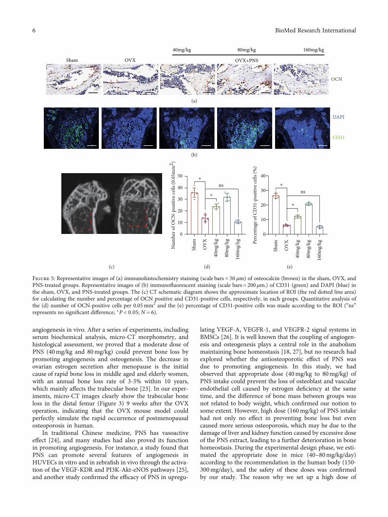
3.5. Immunostaining of Osteoblasts and Vascular EndothelialCells. Osteocalcin is one of the major noncollagenous pro-teins of the bone matrix, which is synthesized and secretedby osteoblasts and is specific for bone. In the immuno-staining of osteocalcin, OVX group showed a significantlydecrease in the number of osteoblasts when compared withthe sham operation group (Figures 5(a), 5(c), and 5(d)).Interestingly, intake of proper dosage of PNS (40mg/kg and80mg/kg) could reverse the phenotype of osteoblast decreasein the ovariectomy-induced osteoporosis model. However,excessive intake of PNS (160mg/kg) had no protection onthe decrease of osteoblast.
CD31 is the most widely used markers of endothelial dif-ferentiation, although it is not entirely specific. In this study,we used immunofluorescent staining of CD31 to representthe vascular endothelial cells. Very similar to the immunohis-tochemistry results of osteocalcin, intake of proper dosage ofPNS (40mg/kg and 80mg/kg) could reverse the decrease ofthe number of endothelial cells (Figures 5(b), 5(c), and5(e)) caused by deficiency of estrogen, while excessive intakeof PNS (160mg/kg) had no such effect.
4. Discussion
In this study, we applied ovariectomy-induced osteoporo-sis model [21, 22] to verify our hypothesis that PNS couldprevent osteoporosis by coactivating osteogenesis and
(a)
Sham OVXOVX+PNS
40mg/kg 80mg/kg 160mg/kg
(b)
Figure 4: Representative sagittal images of (a) HE staining (scale bars = 500 μm) and (b) Safranin O-Fast Green staining (scale bars = 500μm)of trabecular bone in distal femur of sham, OVX, and PNS-treated groups. Black arrowheads point out the typical trabecular bone in differentgroups.
5BioMed Research International
angiogenesis in vivo. After a series of experiments, includingserum biochemical analysis, micro-CT morphometry, andhistological assessment, we proved that a moderate dose ofPNS (40mg/kg and 80mg/kg) could prevent bone loss bypromoting angiogenesis and osteogenesis. The decrease inovarian estrogen secretion after menopause is the initialcause of rapid bone loss in middle aged and elderly women,with an annual bone loss rate of 3-5% within 10 years,which mainly affects the trabecular bone [23]. In our exper-iments, micro-CT images clearly show the trabecular boneloss in the distal femur (Figure 3) 9 weeks after the OVXoperation, indicating that the OVX mouse model couldperfectly simulate the rapid occurrence of postmenopausalosteoporosis in human.
In traditional Chinese medicine, PNS has vasoactiveeffect [24], and many studies had also proved its functionin promoting angiogenesis. For instance, a study found thatPNS can promote several features of angiogenesis inHUVECs in vitro and in zebrafish in vivo through the activa-tion of the VEGF-KDR and PI3K-Akt-eNOS pathways [25],and another study confirmed the efficacy of PNS in upregu-
lating VEGF-A, VEGFR-1, and VEGFR-2 signal systems inBMSCs [26]. It is well known that the coupling of angiogen-esis and osteogenesis plays a central role in the anabolismmaintaining bone homeostasis [18, 27], but no research hadexplored whether the antiosteoporotic effect of PNS wasdue to promoting angiogenesis. In this study, we hadobserved that appropriate dose (40mg/kg to 80mg/kg) ofPNS intake could prevent the loss of osteoblast and vascularendothelial cell caused by estrogen deficiency at the sametime, and the difference of bone mass between groups wasnot related to body weight, which confirmed our notion tosome extent. However, high dose (160mg/kg) of PNS intakehad not only no effect in preventing bone loss but evencaused more serious osteoporosis, which may be due to thedamage of liver and kidney function caused by excessive doseof the PNS extract, leading to a further deterioration in bonehomeostasis. During the experimental design phase, we esti-mated the appropriate dose in mice (40~80mg/kg/day)according to the recommendation in the human body (150-300mg/day), and the safety of these doses was confirmedby our study. The reason why we set up a high dose of
Sham OVX OVX+PNS
40mg/kg 80mg/kg 160mg/kg
OCN
(a)
DAPI
CD31
(b)
(c)
0
10
20
30
40
50N
umbe
r of O
CN-p
ositi
ve ce
lls (0
.05m
m2 )
⁎
⁎
nsSh
am
OV
X
40m
g/kg
80m
g/kg
160m
g/kg
(d)
0
10
20
30
40
Perc
enta
ge o
f CD
31-p
ositi
ve ce
lls (%
)
⁎
⁎
ns
Sham
OV
X
40m
g/kg
80m
g/kg
160m
g/kg
(e)
Figure 5: Representative images of (a) immunohistochemistry staining (scale bars = 50 μm) of osteocalcin (brown) in the sham, OVX, andPNS-treated groups. Representative images of (b) immunofluorescent staining (scale bars = 200 μm.) of CD31 (green) and DAPI (blue) inthe sham, OVX, and PNS-treated groups. The (c) CT schematic diagram shows the approximate location of ROI (the red dotted line area)for calculating the number and percentage of OCN positive and CD31-positive cells, respectively, in each groups. Quantitative analysis ofthe (d) number of OCN-positive cells per 0.05mm2 and the (e) percentage of CD31-positive cells was made according to the ROI (“ns”represents no significant difference; ∗P < 0:05; N = 6).
6 BioMed Research International

160mg/kg is because we want to observe the pharmacologicaleffects and toxicity of PNS in a high dosage, which are rarelydiscussed in other similar researches. Our study also indi-cates that when the intake of PNS was doubled from its rec-ommended amount, it showed reverse pharmacologicaleffects and hepatorenal toxicity.
Besides the angiogenesis promoting effect, PNS had beenreported to reveal estrogen-like activities in vivo [12, 13].Estrogen is an important regulator of osteoblast differentia-tion and activity, which can promote osteogenic differentia-tion of mesenchymal stem cell and prolong the life span ofosteoblasts by inhibiting apoptosis [28]. Estrogen receptors(ER) include ER α and ER β subtypes. ER α mediates mostof the effects of natural estrogen ligands, mainly expressedin cortical bone, while ER βmediates the effect of phytoestro-gens on bone, mainly expressed in trabecular bone [29, 30].PNS is one of the phytoestrogens, so we got an interestinghypothesis that PNS promotes osteogenesis and angiogenesissimultaneously or their coupling through activating ER β andits downstream signaling pathway in trabecular bone, whichwill be the next stage of our work. What is more, in the serumbiochemical analysis part, the serum NTX result may indi-cate that PNS could have effect on osteoclasts; however, itneeds more experimental evidence in the future.
Although it has been confirmed that appropriate concen-tration of PNS can prevent bone loss, the mechanism of theopposite pharmacological effect of high-dose PNS is notclear, and the specific mechanism of PNS promoting angio-genesis is not well explained. However, compared with theinjection administration route of classic antiosteoporosisdrugs, like PTH and bisphosphonate, the oral administrationcharacteristics of PNS would make it easier for patients totake and therefore improve compliance [23, 31]. And becausePNS has been used clinically for many years, it is easier toapply for clinical trials with expanded indications, makingit a potential antiosteoporotic drug in the future.
5. Conclusions
In this study, we strongly confirmed the pharmacologicaleffects of appropriate dose of PNS on promoting angiogene-sis and preventing bone loss in a mouse osteoporotic model,which may provide a new potential treatment for the preven-tion of osteoporosis in postmenopausal women.
Data Availability
All the data in this manuscript are available on requestthrough contacting the author Hao Hu directly, and the con-tact email address is [email protected].
Conflicts of Interest
The authors declare that they have no conflict of interest.
Authors’ Contributions
HH, YC, ZML, and XNZ conceived and designed the exper-iments. HH and YC performed the animal model experi-
ments and drafted the manuscript. ZML performed theμCT scan and image analysis for animals. ZYZ and LPL per-formed the histomorphometry staining and image analysis.FXW and CL helped to check and analyzed the data andrevise the manuscript. All of the authors had read andapproved the final version of this manuscript. Hao Hu andYan Chen contributed equally to this work and should beconsidered co-first authors.
Acknowledgments
This work was supported by research grants from Scienceand Technology Program of Guangzhou (201704030082and 201804020011), the National Natural Science Founda-tion of Guangdong Province-Major Fundamental ResearchFostering Program, China (2017A030308004), and Key Pro-ject of NSFC-Guangdong Joint Program (U1601220).
References
[1] P. Chen, Z. Li, and Y. Hu, “Prevalence of osteoporosis inChina: a meta-analysis and systematic review,” BMC PublicHealth, vol. 16, no. 1, p. 1039, 2016.
[2] D. A. Towler, “Angiogenesis and marrow stromal cell fates:roles in bone strength,” Osteoporosis International, vol. 14,pp. 46–53, 2003.
[3] H. Liu, J. Yang, F. du et al., “Absorption and disposition of gin-senosides after oral administration of Panax notoginsengextract to rats,” Drug Metabolism and Disposition, vol. 37,no. 12, pp. 2290–2298, 2009.
[4] T. T. X. Dong, X. M. Cui, Z. H. Song et al., “Chemical assess-ment of roots of Panax notoginseng in China: regional andseasonal variations in its active constituents,” Journal of Agri-cultural and Food Chemistry, vol. 51, no. 16, pp. 4617–4623,2003.
[5] T. B. Ng, “Pharmacological activity of sanchi ginseng (Panaxnotoginseng),” The Journal of Pharmacy and Pharmacology,vol. 58, no. 8, pp. 1007–1019, 2006.
[6] Y. G. Zhang, H. G. Zhang, G. Y. Zhang et al., “Panax notogin-seng saponins attenuate atherosclerosis in rats by regulatingthe blood lipid profile and an anti-inflammatory action,” Clin-ical and Experimental Pharmacology & Physiology, vol. 35,no. 10, pp. 1238–1244, 2008.
[7] Y. J. Jang, M. E. Kim, and S. Y. Ko, “n-Butanol extracts ofPanax notoginseng suppress LPS-induced MMP-2 expressionin periodontal ligament fibroblasts and inhibit osteoclastogen-esis by suppressing MAPK in LPS-activated RAW264.7 cells,”Archives of Oral Biology, vol. 56, no. 11, pp. 1319–1327, 2011.
[8] G. Zhao, Z. Xiang, T. Ye, Y. Yuan, and Z. Guo, “Antioxidantactivities of Salvia miltiorrhiza and Panax notoginseng,” FoodChemistry, vol. 99, no. 4, pp. 767–774, 2006.
[9] T. B. Ng, F. Liu, and H. X. Wang, “The antioxidant effects ofaqueous and organic extracts of Panax quinquefolium, Panaxnotoginseng, Codonopsis pilosula, Pseudostellaria heterophyllaand Glehnia littoralis,” Journal of Ethnopharmacology,vol. 93, no. 2-3, pp. 285–288, 2004.
[10] N. W. He, Y. Zhao, L. Guo, J. Shang, and X. B. Yang, “Antiox-idant, antiproliferative, and pro-apoptotic activities of a sapo-nin extract derived from the roots of Panax notoginseng(Burk.) F.H. Chen,” Journal of Medicinal Food, vol. 15, no. 4,pp. 350–359, 2012.
7BioMed Research International

[11] H. Qiang, C. Zhang, Z. B. Shi, H. Q. Yang, and K. Z. Wang,“Protective effects and mechanism of Panax Notoginseng sapo-nins on oxidative stress-induced damage and apoptosis of rab-bit bone marrow stromal cells,” Chinese Journal of IntegrativeMedicine, vol. 16, no. 6, pp. 525–530, 2010.
[12] R. Y. K. Chan, W. F. Chen, A. Dong, D. Guo, and M. S. Wong,“Estrogen-like activity of ginsenoside Rg1 derived from Panaxnotoginseng,” Journal of Clinical Endocrinology & Metabolism,vol. 87, no. 8, pp. 3691–3695, 2002.
[13] X. Meng, G. Sun, J. Ye, H. Xu, H. Wang, and X. Sun, “Notogin-senoside R1-mediated neuroprotection involves estrogenreceptor-dependent crosstalk between Akt and ERK1/2 path-ways: a novel mechanism of Nrf2/ARE signaling activation,”Free Radical Research, vol. 48, no. 4, pp. 445–460, 2014.
[14] Z. Ji, Y. Cheng, P. Yuan, X. Dang, X. Guo, and W. Wang,“Panax notoginseng stimulates alkaline phosphatase activity,collagen synthesis, and mineralization in osteoblasticMC3T3-E1 cells,” In Vitro Cellular & Developmental Biology.Animal, vol. 51, no. 9, pp. 950–957, 2015.
[15] X.-D. Li, B. Chang, B. Chen et al., “Panax notoginseng saponinspotentiate osteogenesis of bone marrow stromal cells by modu-lating gap junction intercellular communication activities,” Cel-lular Physiology and Biochemistry, vol. 26, no. 6, pp. 1081–1092,2010.
[16] X.-d. Li, Z.-y. Liu, B. Chang et al., “Panax notoginseng saponinspromote osteogenic differentiation of bone marrow stromalcells through the ERK and P38 MAPK signaling pathways,”Cellular Physiology and Biochemistry, vol. 28, no. 2, pp. 367–376, 2011.
[17] Y. Wang, X. Huang, Y. Tang, H. Lin, and N. Zhou, “Effects ofpanax notoginseng saponins on the osteogenic differentiationof rabbit bone mesenchymal stem cells through TGF-β1 sig-naling pathway,” BMC Complementary and Alternative Medi-cine, vol. 16, no. 1, p. 319, 2016.
[18] A. P. Kusumbe, S. K. Ramasamy, and R. H. Adams, “Couplingof angiogenesis and osteogenesis by a specific vessel subtype inbone,” Nature, vol. 507, no. 7492, pp. 323–328, 2014.
[19] J. Z. Fan, Y. Wang, Y. Meng et al., “Panax notoginsengsaponins mitigate ovariectomy-induced bone loss and inhibitmarrow adiposity in rats,” Menopause, vol. 22, no. 12,pp. 1343–1350, 2015.
[20] Y. Shen, Y. Q. Li, S. P. Li, L. Ma, L. J. Ding, and H. Ji, “Allevi-ation of ovariectomy-induced osteoporosis in rats by Panaxnotoginseng saponins,” Journal of Natural Medicines, vol. 64,no. 3, pp. 336–345, 2010.
[21] S. Negri, Y. Wang, T. Sono et al., “Human perivascular stemcells prevent bone graft resorption in osteoporotic contextsby inhibiting osteoclast formation,” STEM CELLS Transla-tional Medicine, vol. 16, no. 9, pp. 1617–1630, 2001.
[22] D. N. Kalu, “The ovariectomized rat model of postmeno-pausal bone loss,” Bone and Mineral, vol. 15, no. 3, pp. 175–191, 1991.
[23] F. Cosman, S. J. de Beur, M. S. LeBoff et al., “Erratum to: Clin-ician's guide to prevention and treatment of osteoporosis,”Osteoporosis International, vol. 26, no. 7, pp. 2045–2047, 2015.
[24] S. Chen, J. Liu, X. Liu et al., “Panax notoginseng saponinsinhibit ischemia-induced apoptosis by activating PI3K/Aktpathway in cardiomyocytes,” Journal of Ethnopharmacology,vol. 137, no. 1, pp. 263–270, 2011.
[25] S. J. Hong, J. B. Wan, Y. Zhang et al., “Angiogenic effect ofsaponin extract from Panax notoginseng on HUVECs
in vitro and zebrafish in vivo,” Phytotherapy Research,vol. 23, no. 5, pp. 677–686, 2009.
[26] H. Zheng, C. Liu, Y. Ou, Y. Zhang, and X. Fu, “Total saponinsof Panax notoginseng enhance VEGF and relative receptorssignals and promote angiogenesis derived from rat bone mar-row mesenchymal stem cells,” Journal of Ethnopharmacology,vol. 147, no. 3, pp. 595–602, 2013.
[27] M. Yang, C. J. Li, X. Sun et al., “MiR-497∼195 cluster regulatesangiogenesis during coupling with osteogenesis by maintain-ing endothelial Notch and HIF-1α activity,”Nature Communi-cations, vol. 8, no. 1, p. 16003, 2017.
[28] S. Khosla, “New insights into androgen and estrogen receptorregulation of the male skeleton,” Journal of Bone and MineralResearch, vol. 30, no. 7, pp. 1134–1137, 2015.
[29] V. Cagnetta and V. Patella, “The role of the immune system inthe physiopathology of osteoporosis,” Clinical Cases in Min-eral and Bone Metabolism, vol. 9, no. 2, pp. 85–88, 2012.
[30] M. N. Weitzmann and R. Pacifici, “Estrogen deficiency andbone loss: an inflammatory tale,” The Journal of Clinical Inves-tigation, vol. 116, no. 5, pp. 1186–1194, 2006.
[31] S. Jha, Z. Wang, N. Laucis, and T. Bhattacharyya, “Trends inmedia reports, oral bisphosphonate prescriptions, and hipfractures 1996-2012: an ecological analysis,” Journal of Boneand Mineral Research, vol. 30, no. 12, pp. 2179–2187, 2015.
8 BioMed Research International


![BY masaji+ Party Broch [r1.0.5] - Cloud Object Storage ...€¦ · Jabuticaba Kale Lemon Mangosteen Maqui Berry Monk Fruit Oat Grass Panax Notoginseng Phosphatidylcholine Pomegranate](https://static.fdocuments.net/doc/165x107/5b78f8d07f8b9a703b8ca871/by-masaji-party-broch-r105-cloud-object-storage-jabuticaba-kale-lemon.jpg)

![Review Article Sanqi Panax Notoginseng Injection for Angina ...angina Diagnosis standard Age Intervention group Control group Course (week) Outcome measures Liuetal. [ ] UAP ISFC/WHO](https://static.fdocuments.net/doc/165x107/60cf8676011a17669913bdeb/review-article-sanqi-panax-notoginseng-injection-for-angina-angina-diagnosis.jpg)
