PAMAM-Triamcinolone acetonide conjugate as a … · gene carrier for enhanced transfer activity Kun...
10
PAMAM–Triamcinolone acetonide conjugate as a nucleus-targeting gene carrier for enhanced transfer activity Kun Ma a,1 , Min-Xin Hu a, 1 , Yan Qi b , Ji-Hong Zou b , Li-Yan Qiu a , Yi Jin a, * , Xiao-Ying Ying a , Hong-Ying Sun a a Institute of Pharmaceutics, College of Pharmaceutical Sciences, Zhejiang University, Hangzhou 310058, PR China b College of Life Sciences, Heilung-kiang University, Harbin 150080, PR China article info Article history: Received 8 May 2009 Accepted 17 July 2009 Available online 4 August 2009 Keywords: Gene delivery Non-viral vector PAMAM dendrimer Triamcinolone acetonide Nuclear translocation abstract The excellent transfection efficiency and viability are essential for successful gene therapy. It suggested that when bound to its glucocorticoid receptor, glucocorticoid steroid can dilate the nuclear pore complexes and facilitated the transport of pDNA into the nucleus. In this research, the two different degrees of substitution of PAMAM–triamcinolone acetonide (PAMAM–TA) conjugates were synthesised for efficient translocation of pDNA into the nucleus. The physicochemical properties of the polyplexes were investigated by agarose gel electrophoresis, Zeta-sizer and TEM. They both could form nano-size polyplexes with pDNA. The polyplexes were very stable and showed excellent buffering capacities, facilitating endosomal escape, and no obvious difference was found between them. The TA-conjugated PAMAM-mediated transfection of luciferase and EGFP genes showed better transfer activity than native PAMAM and was comparable to the PEI 25K (polyethylenimine), and lower cytotoxicity in HEK 293 and HepG 2 cells. Even with 10% serum, their transfer activity was still high relatively. In addition, confocal microscopy examination confirmed that the enhancing mechanism for enhanced gene transfer activity of PAMAM–TA conjugate may involve the nuclear translocation of the polyplex. The low substituted degree of TA to 0.22 did not interrupt its nuclear localization potency. These findings demonstrated that the TA- grafted PAMAM dendrimer is a potential candidate as a safe and efficient gene delivery carrier for gene therapy. Ó 2009 Elsevier Ltd. All rights reserved. 1. Introduction Both the advances in molecular biology and biotechnology, along with the completion of the Human Genome Project have led gene therapy to a new level, being an alternative treatment of genetic diseases such as haemophilia, muscular dystrophy or cystic fibrosis [1,2]. As a result, the need to develop efficient, reliable and safe gene (RNA or DNA) delivery systems continues to increase with the development of applications for gene therapy. Viral vectors have been shown to be dominant gene delivery carriers due to their high gene transfer efficiencies. They have been used in the majority of gene delivery studies reported in the literature and about 70% of ongoing clinical trials [3]. However, there still exists the possibility that the viral gene carriers will insert the recombinant virus in the initial coding region of a gene. Furthermore, viruses are inherently immunogenic, leading to difficulty with repeated administrations and the high possibility of immune reactions [4]. On the other hand, Non-viral vectors, provide advantages such as improved safety, greater flexibility and more controllable manufacturing. But its low transfection efficiency compared to viral vectors hampers the clinical application. Nuclear membrane is one of the main barriers in polymer- mediated intracellular gene delivery [5]. And it was previously reported that glucocorticoid receptor dilated the nuclear pore to 60 nm and translocated into nucleus when it bound to its ligand, glucocorticoid, as a nuclear localization signal (NLS) [6]. This suggests that the transport of DNA into nucleus may very possibly be facilitated by glucocorticoid. Poly(amidoamine) (PAMAM) dendrimers were first introduced by Tomalia and co-workers in the mid-1980s [7]. At present, PAMAM dendrimer and polyethylenimine (PEI) have been tested for their potential utility and have exhibited relatively high trans- fection efficiencies in vitro while PEI showing some promising results in vivo [8]. Dendrimers are core-shell nanostructures with precise structure and low polydispersity, which are synthesized in a layer-by-layer fashion, expressed in generations, around an ethyl- enediamine or ammonia core unit. The three main properties of dendrimers are nanoscale container properties for drugs or genes, nano-scaffolding properties for producing prodrugs, and biocom- patibility for adaptability [9–11]. Interestingly, it was reported that * Corresponding author. Tel./fax: þ86 571 88208435. E-mail address: [email protected] (Y. Jin). 1 Both authors contributed equally to this work. Contents lists available at ScienceDirect Biomaterials journal homepage: www.elsevier.com/locate/biomaterials 0142-9612/$ – see front matter Ó 2009 Elsevier Ltd. All rights reserved. doi:10.1016/j.biomaterials.2009.07.036 Biomaterials 30 (2009) 6109–6118
-
Upload
vuongthien -
Category
Documents
-
view
215 -
download
0
Transcript of PAMAM-Triamcinolone acetonide conjugate as a … · gene carrier for enhanced transfer activity Kun...

lable at ScienceDirect
Biomaterials 30 (2009) 6109–6118
Contents lists avai
Biomaterials
journal homepage: www.elsevier .com/locate/biomateria ls
PAMAM–Triamcinolone acetonide conjugate as a nucleus-targetinggene carrier for enhanced transfer activity
Kun Ma a,1, Min-Xin Hu a,1, Yan Qi b, Ji-Hong Zou b, Li-Yan Qiu a, Yi Jin a,*, Xiao-Ying Ying a, Hong-Ying Sun a
a Institute of Pharmaceutics, College of Pharmaceutical Sciences, Zhejiang University, Hangzhou 310058, PR Chinab College of Life Sciences, Heilung-kiang University, Harbin 150080, PR China
a r t i c l e i n f o
Article history:Received 8 May 2009Accepted 17 July 2009Available online 4 August 2009
Keywords:Gene deliveryNon-viral vectorPAMAM dendrimerTriamcinolone acetonideNuclear translocation
* Corresponding author. Tel./fax: þ86 571 8820843E-mail address: [email protected] (Y. Jin).
1 Both authors contributed equally to this work.
0142-9612/$ – see front matter � 2009 Elsevier Ltd.doi:10.1016/j.biomaterials.2009.07.036
a b s t r a c t
The excellent transfection efficiency and viability are essential for successful gene therapy. It suggestedthat when bound to its glucocorticoid receptor, glucocorticoid steroid can dilate the nuclear porecomplexes and facilitated the transport of pDNA into the nucleus. In this research, the two differentdegrees of substitution of PAMAM–triamcinolone acetonide (PAMAM–TA) conjugates were synthesisedfor efficient translocation of pDNA into the nucleus. The physicochemical properties of the polyplexeswere investigated by agarose gel electrophoresis, Zeta-sizer and TEM. They both could form nano-sizepolyplexes with pDNA. The polyplexes were very stable and showed excellent buffering capacities,facilitating endosomal escape, and no obvious difference was found between them. The TA-conjugatedPAMAM-mediated transfection of luciferase and EGFP genes showed better transfer activity than nativePAMAM and was comparable to the PEI 25K (polyethylenimine), and lower cytotoxicity in HEK 293 andHepG 2 cells. Even with 10% serum, their transfer activity was still high relatively. In addition, confocalmicroscopy examination confirmed that the enhancing mechanism for enhanced gene transfer activity ofPAMAM–TA conjugate may involve the nuclear translocation of the polyplex. The low substituted degreeof TA to 0.22 did not interrupt its nuclear localization potency. These findings demonstrated that the TA-grafted PAMAM dendrimer is a potential candidate as a safe and efficient gene delivery carrier for genetherapy.
� 2009 Elsevier Ltd. All rights reserved.
1. Introduction
Both the advances in molecular biology and biotechnology,along with the completion of the Human Genome Project have ledgene therapy to a new level, being an alternative treatment ofgenetic diseases such as haemophilia, muscular dystrophy or cysticfibrosis [1,2]. As a result, the need to develop efficient, reliable andsafe gene (RNA or DNA) delivery systems continues to increase withthe development of applications for gene therapy. Viral vectorshave been shown to be dominant gene delivery carriers due to theirhigh gene transfer efficiencies. They have been used in the majorityof gene delivery studies reported in the literature and about 70% ofongoing clinical trials [3]. However, there still exists the possibilitythat the viral gene carriers will insert the recombinant virus in theinitial coding region of a gene. Furthermore, viruses are inherentlyimmunogenic, leading to difficulty with repeated administrationsand the high possibility of immune reactions [4]. On the other hand,Non-viral vectors, provide advantages such as improved safety,
5.
All rights reserved.
greater flexibility and more controllable manufacturing. But its lowtransfection efficiency compared to viral vectors hampers theclinical application.
Nuclear membrane is one of the main barriers in polymer-mediated intracellular gene delivery [5]. And it was previouslyreported that glucocorticoid receptor dilated the nuclear pore to60 nm and translocated into nucleus when it bound to its ligand,glucocorticoid, as a nuclear localization signal (NLS) [6]. Thissuggests that the transport of DNA into nucleus may very possiblybe facilitated by glucocorticoid.
Poly(amidoamine) (PAMAM) dendrimers were first introducedby Tomalia and co-workers in the mid-1980s [7]. At present,PAMAM dendrimer and polyethylenimine (PEI) have been testedfor their potential utility and have exhibited relatively high trans-fection efficiencies in vitro while PEI showing some promisingresults in vivo [8]. Dendrimers are core-shell nanostructures withprecise structure and low polydispersity, which are synthesized ina layer-by-layer fashion, expressed in generations, around an ethyl-enediamine or ammonia core unit. The three main properties ofdendrimers are nanoscale container properties for drugs or genes,nano-scaffolding properties for producing prodrugs, and biocom-patibility for adaptability [9–11]. Interestingly, it was reported that
K. Ma et al. / Biomaterials 30 (2009) 6109–61186110
the extent of transfection was dependent on the dendritic gener-ation of the PAMAM employed, lower generation PAMAM den-drimers were ineffective for transfection [12]. The high level ofcontrol over the dendritic architecture (size, branching density,surface functionality) makes dendrimers been researched as idealgene carriers in the past decades [13–16].
In our previous research, we synthesized five kinds of gluco-corticoid-PEI (GC-PEI) conjugates and discussed the relationshipbetween structure and transfection activity [17]. Choi et al. conju-gated dexamethasone, a potent glucocorticoid, to PAMAM G4dendrimer to facilitate nuclear translocation and enhanced thetransfection efficiency [18]. Here, we combined a glucocorticoid ofhigh potency (triamcinolone acetonide, TA) with PAMAM den-drimers to form a novel non-viral gene vectors (PAMAM–TA). Andwe investigated the polyplex formation, biosafety and genedelivery efficiency, and discovered the impact of substituted degreeonto transfection effects. The intracellular localization of PAMAM–TA/pDNA polyplexes was also examined by confocal microscopy.
2. Materials and methods
2.1. Materials
PAMAM dendrimers (ethylenediamine core, G4), polyethylenimine (PEI,branched, 1800 and 25 kDa), 2-iminothiolane (Traut’s reagent), fluorescein iso-thiocyanate (FITC), 3-[4,5-dimethylthiazol-2-yl]-2,5-diphenyltetrazolium bromide(MTT), ethidium bromide were purchased from Sigma–Aldrich Chemical Co., Ltd.
Fig. 1. Synthesic scheme of
(Milwaukee, WI, USA). RPMI 1640, penicillin–streptomycin (PS,10,000 U/mL), trypsin–EDTA (TE, 0.5% trypsin, 5.3 mM EDTA tetra-sodium) were obtained from Gibco BRL(Gaithersberg, MD, USA). Fetal bovine serum (FBS) was purchased from Sijiqing Bio-logic Co., Ltd. (Hangzhou, China). Methanesulfonyl chloride was purchased from Jia-chen Chemical Co., Ltd. (Shanghai, China). Triamcinolone acetonide (TA) was kindlydonated by Zhejiang Xianju Pharmaceutical Co., Ltd. (Hangzhou, China), and its puritywas over 99%. Promega Luciferase Assay Kit containing luciferase cell culture lysesreagent and luciferase substrates was obtained from Promega (Madison, WI, USA). TheBCA Protein Assay Kit and Hoechst 33342 were purchased from Beyotime Institute ofBiotechnology (Jiangsu, China). Plasmid DNA (pEGFP-N1 and pGL-3) was kindlyprovided by Institute of Infectious Diseases, Zhejiang University (Hangzhou, China).The plasmids were propagated in Escherichia coli DH5a, isolated, and purified usingAxyprep Plasmid DNA Maxiprep Kit (Axygene Biotechnology Limited, Hangzhou,China). The purity and concentration of DNA were determined by measuring a UVabsorbance 260 and 280 nm. All other chemicals were of analytical grade.
2.2. Synthesis of PAMAM–TA
PAMAM–TA was synthesized via a two-step reaction as shown in Fig. 1. Firstly, wesubstituted its 21-hydroxyl groups with mesylates to activate triamcinolone acetonide.Briefly, methanesulfonyl chloride (41.5 mL, 0.536 mmol) was added drop wise toa solution of TA (116.5 mg, 0.268 mmol) in anhydrous pyridine (2.5 mL) at 0 �C under N2
with stirring. After reacting for 5 h at 0 �C, ice water (50 mL) was added. The precipitatewas filtered, washed with more ice water, crude triamcinolone acetonide mesylate (TA-mesylate) as a white solid powder was obtained and dried. Using recrystallization fromethanol–acetic ether to purify the crude mesylate. TLC (ethyl acetate/ligroin/meth-anol¼ 10/10/1, v/v/v) was performed at the end of the activation. The product wassolubilized in DMSO-d6 for 1H NMR analysis (500 MHz, Bruker, Germany).
Then, the conjugation reaction was followed by the procedures as previouslyreport with some modification [18,19]. Traut’s reagent and TA-mesylate (2 equiv. or
PAMAM–TA polymers.

Fig. 2. The 1H NMR spectrum of PAMAM–TA-H (A) and PAMAM–TA-L (B).
Fig. 3. Determination of the buffer capacity of PEI 25K and PAMAM–TA series by acid-base titration. Solutions containing each polymer (0.2 mg/mL) were adjusted to pH10.0 and then titrated with 0.1 M HCl from 10.0 to 3.0.
K. Ma et al. / Biomaterials 30 (2009) 6109–6118 6111
1 equiv. to PAMAM G4) in 3.0 mL anhydrous DMSO were added slowly with 1 equiv.of PAMAM in 2.0 mL anhydrous DMSO. The reaction was allowed to proceed underN2 at room temperature under continuous stirring for 4 h. To the reaction mixture,same volume of pure water was added and filtered by 0.45 mm micropore film toremove insoluble impurities. Then dialyzed against pure water using dialysismembrane (MWCO 3500) for 48 h, the dialysis medium was refreshed every 12 h. Awhite product (PAMAM–TA) was obtained after further freeze-drying. The structureof product was confirmed by using 1H NMR (500 MHz, Bruker, Germany, D2O).
2.3. The buffering capacity of the PAMAM polymers
Polymer solution was prepared in a 50 mL flask (0.2 mg/mL, 30 mL) and purewater was used as a control. After adjusting the initial pH to 10.0 with 0.1 M NaOH ifnecessary, 25 mL increments of 0.1 M HCl were titrated into the solution and the pHresponse was measured by a micro-pH electrode at the same time. The whole pHvariation was recorded from 10.0 to 3.0.
2.4. Agarose gel electrophoresis
Polyplexes were prepared at various weight ratios between each of the polymersand pGL-3 plasmid in HEPES buffered saline (HBS, 25 mM HEPES, 150 mM NaCl, pH7.4), and the mixtures were incubated for 30 min at room temperature. The sampleswere electrophoresed on a 1% (w/v) agarose gel stained with 0.25 mg/mL ethidiumbromide in TAE buffer at 90 V for 40 min, and analyzed on a UV illuminator to showthe location of the DNA.
2.5. Size and z-potential measurements
The size and z-potential values of polyplexes were determined by Malvern Zetasizer3000HAs system (Malvern Instruments Ltd., U.K.). Polyplexes were formed at a finalconcentration of 10 mg/mL pDNA at various weight ratios inwater for size measurementsand in HBS (25 mM HEPES, 150 mM NaCl, pH 7.4) for z-potential experiments, respec-tively. The size and z-potential values were presented as the average values of 3 assays.
2.6. Transmission electron microscope (TEM)
The morphology of polyplexes with an optimal weight ratio was observed usingTEM (JEM 1230, JEOL, Japan). One drop of polyplex was placed on a copper grid and
stained with 2% phosphotungstic acid solution for 30 s. The grid was allowed to dryfurther for 20 min and was then examined with the electron microscope.
2.7. In vitro transfection experiment
We next examined the ability of PAMAM–TA to transfect human embryonickidney 293 cells and human liver carcinoma HepG 2 cells using plasmid DNA thatcontain the firefly luciferase and EGFP gene. The cells were seeded at a density of1�105 cells/well in 24-well plate in RMPI 1640 medium containing 10% FBS, and

Fig. 4. Agarose gel electrophoresis retardation assay of polyplexes at various weightratios: PAMAM (A), PAMAM–TA-H (B), PAMAM–TA-L (C). The mixtures were incubatedat room temperature for 30 min and electrophoresis on 1% (W/V) agarose gel andstained with ethidium bromide.
Table 2z-potential of the pDNA complexes with PAMAM, PAMAM–TA-H and PAMAM–TA-L.Each data is the mean � SD (n¼ 3).
Weight ratio z-potential (mV)
PAMAM 2:1 �22.4� 1.83:1 �7.0� 1.25:1 17.5� 2.36:1 18.4� 1.97:1 21.4� 2.7
PAMAM-TA-H 0.5:1 �19.8� 1.41:1 �3.9� 2.42:1 14.8� 1.63:1 18.0� 1.94:1 18.5� 2.2
PAMAM-TA-L 0.5:1 �16.8� 1.41:1 1.3� 1.02:1 15.6� 1.03:1 18.9� 1.34:1 19.5� 1.7
K. Ma et al. / Biomaterials 30 (2009) 6109–61186112
grown to reach 80% confluence prior to transfection. Before transfection, themedium was exchanged with fresh medium with or without 10% FBS. The cellswere treated with polyplex solution containing 2 mg of pDNA at various weightratios for 4 h at 37 �C and the final volume was adjusted to 500 mL by medium.After exchanging with a fresh medium with 10% FBS, cells were further incubatedfor 48 h. Then the growth medium was removed, and the cells were shaken for30 min at room temperature in 200 mL of Reporter Lysis Buffer. The lysates weretransferred into tubes and centrifuged at 13,000 rpm for 5 min. Luciferase activitywas measured with a luminometer (Turner Designs Luminometer Model TD-20/20,Promega). The total protein was determined by BCA protein assay kit. The finalluciferase activity was expressed as RLU/mg protein. Inverted fluorescent micro-scope (Leica DMI 4000 B, Germany) was used to observe the EGFP expression of thepolyplexes in 293 cells.
2.8. Cytotoxicity assay
The cytotoxicity of the polymers was measured by MTT assay. Briefly, HEK293 cells and HepG 2 cells were seeded at a density of 1�104 cells/well in100 mL of growth medium in 96-well plates (Corning), and were incubated for24 h before adding the polymers. One day later, the cells were transfected withpolyplexes at various weight ratios. The PEI 25K and PEI 1800 polyplexes wereas control at weight ratios of 1.33:1 and 5.3:1 [20]. The amount of pDNA wasfixed at 0.2 mg/well. The cells were incubated for 24 h at 37 �C. Then the mediumwas replaced with 20 mL MTT (5 mg/mL) solutions and 100 mL of fresh medium
Table 1Particle size of the pDNA complexes with PAMAM, PAMAM–TA-H and PAMAM–TA-L.Each data is the mean � SD (n ¼ 3).
Weight ratio Size
PAMAM 2:1 236.3� 7.63:1 185.4� 11.74:1 110.5� 7.35:1 133.3� 6.46:1 167.0� 2.0
PAMAM-TA-H 1:1 275.3� 19.21.5:1 196.3� 17.62:1 161.3� 5.53:1 157.7� 8.54:1 167.3� 10.2
PAMAM-TA-L 1:1 248.7� 1.51.5:1 182.7� 3.12:1 187.0� 5.33:1 181.0� 7.04:1 180.3� 2.3
without serum and further incubated for 4 h. After that, the medium wasremoved and 100 mL DMSO was added. After shaking the plate for 20 min,absorbance was immediately measured at 570 nm using an ELISA plate reader(Thermo Multiskan Spectrum, USA). Cells incubated without polymer were usedas a blank control.
Fig. 5. Morphology of PAMAM polyplex (A) and PAMAM–TA-H polyplex (B) at weightratio of 5 and 3 as observed by transmission electron microscope. The particles werenegatively stained by a 2% aqueous solution phosphotungstic acid for 30 s.

Fig. 6. Transfection efficiency of PAMAM and PAMAM–TA polymers in HEK 293 cells (A) and HepG 2 cells (B). PEI 25K was used as control. The polyplexes at various weight ratioscontaining pGL-3 were added to 1�105 cells/well in 24-well plate for 4 h. The pDNA’s concentration was 2 mg/well. Cells were harvested after 48 h transfection, luciferase activitywas examined. Relative light units (RLU) were shown as means� SD of triplicates. (*) indicated statistically significant difference (P< 0.05).
K. Ma et al. / Biomaterials 30 (2009) 6109–6118 6113
2.9. Fluorescence labeling of dendrimers
The dendrimers were labeled with FITC as former report with some modification[17,21]. PAMAM or PAMAM–TA was dissolved in PBS (pH 7.4). FITC solution dissolvedin DMSO was added dropwise to unlabeled dendrimers solution. The molar ratio ofdendrimer and FITC taken was 1:20. The solution was incubated overnight at roomtemperature with continuous stirring in the dark. The labeled dendrimers solutionwas dialyzed against PBS (pH 7.4) for 48 h and then distilled water for 24 h until freeFITC could not be detected by TLC (chloroform/methanol¼ 1/1, v/v). The solutionwas filtered through a 0.22 mm filter and then lyophilized.
2.10. Intracellular localization of polyplex
HepG 2 cells were seeded at a density of 2�105 cells/well on the surface ofa cover slide in 6-well plate in 2 mL medium containing 10% FBS before trans-fection. After incubating for 24 h, the cells were treated with polyplexes solutionand further incubated for 4 h. The polyplex solutions containing 3 mg of pGL-3 wereprepared by mixing FITC-labeled PAMAM, PAMAM–TA-H, PAMAM–TA-L and pDNAat weight ratios of 5:1, 3:1 and 2:1, respectively. Then, the cell grown medium wasexchanged with a fresh medium with 10% FBS. After 24 h, the cells were fixed with4% paraformaldehyde for 30 min. To stain the cell nuclei, the cells were incubatedwith Hoechst 33342 for 15 min at room temperature after washed three times with2 mL of PBS, and then the cover slips were mounted on glass slides with a drop of0.1 M glycerine in PBS placed in between to keep the cells from drying out. The cellswere analyzed with a confocal fluorescence microscopic system (Olympus FV1000-IX81, Tokyo). A UV laser (405 nm excitation) was used to induce the bluefluorescence of Hoechst 33342 and an argon laser (473 nm) to excite the greenfluorescence of FITC.
2.11. Statistical analysis
Statistical analysis was performed using one-way analysis of variance (ANOVA).Differences between groups were considered statistically significant at P< 0.05 (*).
3. Result and discussion
3.1. Synthesis and characterization of PAMAM polymer
It was reported that GC can facilitate the transgenic expressionas a nuclear translocation signal [22–24]. Recently, differentcommonly used GCs were conjugated to low molecular weight PEIand their transfection activity was examined. The present studyinvestigated the possibility of creating a potent transfection agentby grafting TA onto the dendritic surface of the PAMAM dendrimer(generation 4), which is commercially available at a relatively lowcost and contains a reasonable number of tertiary amines that arebelieved to contribute to the endosome-buffering effect.
The 21-hydroxy group of TA is not required for pharmacologicalactivity and was therefore a reasonable choice for conjugation toa polycation. The TA was reacted with methanesulfonyl chlorideunder mild condition to generate C21-substituted TA [25]. Triam-cinolone acetonide mesylate (TA-mesylate): 1H NMR (500 MHz,DMSO-d6) d 0.81 (s, C-19, CH3),1.12 (s, C-25, CH3),1.33 (s, C-26, CH3),
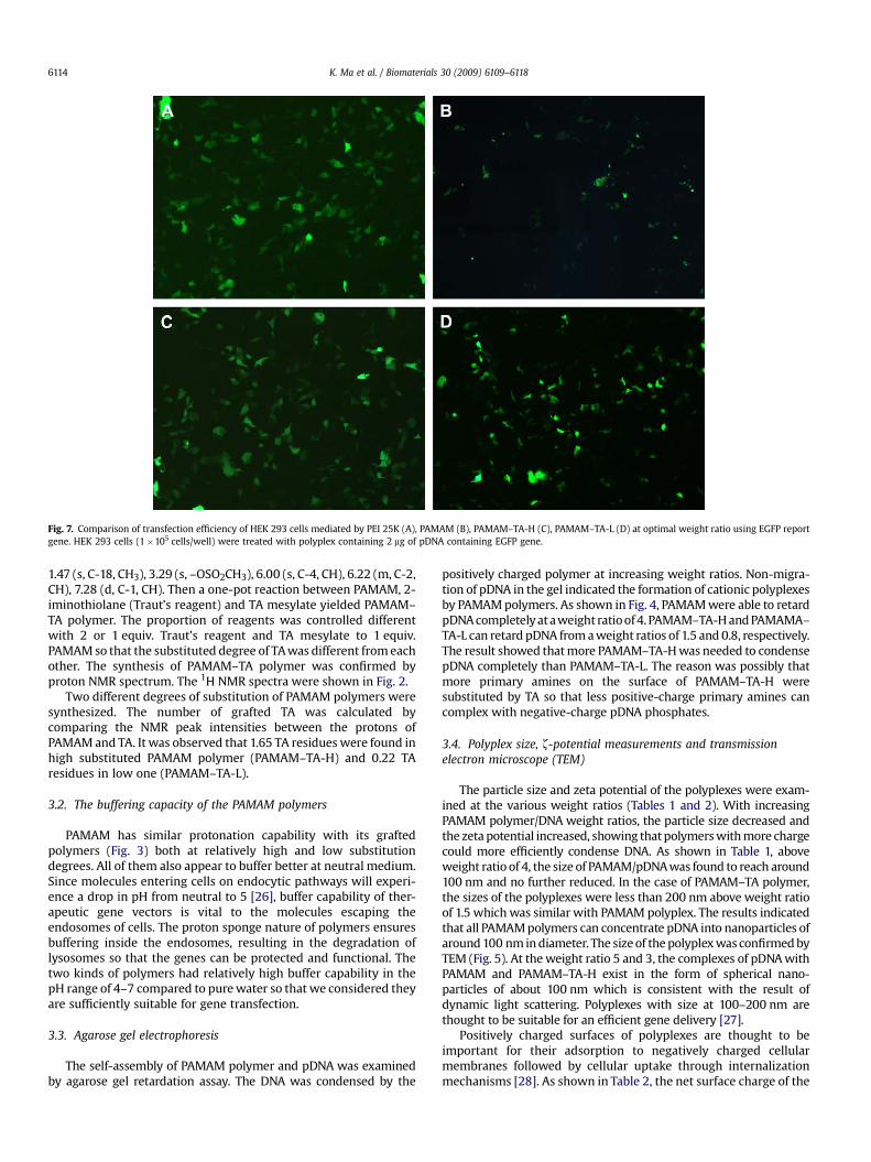
Fig. 7. Comparison of transfection efficiency of HEK 293 cells mediated by PEI 25K (A), PAMAM (B), PAMAM–TA-H (C), PAMAM–TA-L (D) at optimal weight ratio using EGFP reportgene. HEK 293 cells (1�105 cells/well) were treated with polyplex containing 2 mg of pDNA containing EGFP gene.
K. Ma et al. / Biomaterials 30 (2009) 6109–61186114
1.47 (s, C-18, CH3), 3.29 (s, –OSO2CH3), 6.00 (s, C-4, CH), 6.22 (m, C-2,CH), 7.28 (d, C-1, CH). Then a one-pot reaction between PAMAM, 2-iminothiolane (Traut’s reagent) and TA mesylate yielded PAMAM–TA polymer. The proportion of reagents was controlled differentwith 2 or 1 equiv. Traut’s reagent and TA mesylate to 1 equiv.PAMAM so that the substituted degree of TA was different from eachother. The synthesis of PAMAM–TA polymer was confirmed byproton NMR spectrum. The 1H NMR spectra were shown in Fig. 2.
Two different degrees of substitution of PAMAM polymers weresynthesized. The number of grafted TA was calculated bycomparing the NMR peak intensities between the protons ofPAMAM and TA. It was observed that 1.65 TA residues were found inhigh substituted PAMAM polymer (PAMAM–TA-H) and 0.22 TAresidues in low one (PAMAM–TA-L).
3.2. The buffering capacity of the PAMAM polymers
PAMAM has similar protonation capability with its graftedpolymers (Fig. 3) both at relatively high and low substitutiondegrees. All of them also appear to buffer better at neutral medium.Since molecules entering cells on endocytic pathways will experi-ence a drop in pH from neutral to 5 [26], buffer capability of ther-apeutic gene vectors is vital to the molecules escaping theendosomes of cells. The proton sponge nature of polymers ensuresbuffering inside the endosomes, resulting in the degradation oflysosomes so that the genes can be protected and functional. Thetwo kinds of polymers had relatively high buffer capability in thepH range of 4–7 compared to pure water so that we considered theyare sufficiently suitable for gene transfection.
3.3. Agarose gel electrophoresis
The self-assembly of PAMAM polymer and pDNA was examinedby agarose gel retardation assay. The DNA was condensed by the
positively charged polymer at increasing weight ratios. Non-migra-tion of pDNA in the gel indicated the formation of cationic polyplexesby PAMAM polymers. As shown in Fig. 4, PAMAM were able to retardpDNA completely at a weight ratio of 4. PAMAM–TA-H and PAMAMA–TA-L can retard pDNA from a weight ratios of 1.5 and 0.8, respectively.The result showed that more PAMAM–TA-H was needed to condensepDNA completely than PAMAM–TA-L. The reason was possibly thatmore primary amines on the surface of PAMAM–TA-H weresubstituted by TA so that less positive-charge primary amines cancomplex with negative-charge pDNA phosphates.
3.4. Polyplex size, z-potential measurements and transmissionelectron microscope (TEM)
The particle size and zeta potential of the polyplexes were exam-ined at the various weight ratios (Tables 1 and 2). With increasingPAMAM polymer/DNA weight ratios, the particle size decreased andthe zeta potential increased, showing that polymers with more chargecould more efficiently condense DNA. As shown in Table 1, aboveweight ratio of 4, the size of PAMAM/pDNA was found to reach around100 nm and no further reduced. In the case of PAMAM–TA polymer,the sizes of the polyplexes were less than 200 nm above weight ratioof 1.5 which was similar with PAMAM polyplex. The results indicatedthat all PAMAM polymers can concentrate pDNA into nanoparticles ofaround 100 nm in diameter. The size of the polyplex was confirmed byTEM (Fig. 5). At the weight ratio 5 and 3, the complexes of pDNA withPAMAM and PAMAM–TA-H exist in the form of spherical nano-particles of about 100 nm which is consistent with the result ofdynamic light scattering. Polyplexes with size at 100–200 nm arethought to be suitable for an efficient gene delivery [27].
Positively charged surfaces of polyplexes are thought to beimportant for their adsorption to negatively charged cellularmembranes followed by cellular uptake through internalizationmechanisms [28]. As shown in Table 2, the net surface charge of the

Fig. 8. Comparison of transfection efficiency for HEK 293 cells (A) and HepG 2 cells (B) at 2 mg/well in the presence of 10% serum among PAMAM–TA-H, PAMAM–TA-L and PAMAMat optimal weight ratio of 3, 2 and 5, respectively. Luciferase activity was shown as means� SD (n¼ 3). (*) indicated statistically significant difference (P< 0.05).
K. Ma et al. / Biomaterials 30 (2009) 6109–6118 6115
polyplex was negative at low weight ratio, but the zeta potentials ofall polyplexes were gradually increased in accordance with theincrease of charge ratios. Then, the zeta potential values of PAMAMand PAMAM–TA polyplexes were maintained constantly about20 mV over weight ratio of 5 and 2. The zeta potential of all PAMAMpolymers will give rise to similar affinity for anionic cell surfacesand in turn facilitate uptake into the cell.
3.5. In vitro transfection experiment
Polycation modified with glucocorticoid (GC) have been recentlyreported to be effective gene carriers and those polymers showeda high level of transfective activity with nuclear translocation[18,19,22–24,29–31]. The common characteristic of those systems isthought to be that the GC residues are on the surface of the polymersand their transfection activity was enhanced remarkably. And theirtransfection efficiency was correlated closely with their GC residue’sbinding affinity with glucocorticoid receptor (GR) [17]. We deducedthat if GC residue could be more potent, the transfective activitymight be more pronounced. Thus, triamcinolone acetonide (TA)which has great binding affinity with GR [32] was conjugated withPAMAM dendrimer, known as low toxicity and locating in cytoplasm[33], in order to achieve significant gene delivery potency.
The transfection activity of the newly synthesized PAMAMpolymers was investigated in HEK 293 and HepG 2 cells using pGL-3 and pEGFP-N1 reporter gene. PEI 25K polyplexes were preparedat the optimal weight ratio (1:33) demonstrating high transfection
efficiency as positive control. PAMAM polyplexes were prepared atvarious weight ratios ranging from 2 to 6, and PAMAM–TA poly-plexes were from 1 to 4.
As shown in Fig. 6, the PAMAM, PAMAM–TA-H and PAMAM–TA-Lpolyplexes exhibited the maximal transfection efficiency at theweight ratio of 6, 3 and 2, respectively. The transfection efficiency ofPAMAM–TA-H and PAMAM–TA-L showed essentially more than 4times as that of native PAMAM, which was comparable to PEI 25K atthe optimal conditions. The transfection efficiency of native PAMAM(G4) was over 5–6 times less than that of PEI 25K in both cells, whichwas similar with the former report [34]. The difference of optimalweight ratio between PAMAM–TA-H and PAMAM–TA-L might becaused by the charge density difference between them. The primaryamines on the surface of PAMAM–TA-H polymer were substitutedby TA more than that on PAMAM–TA-L, as a result, higher amountsof polymers with higher degree of substitution was needed to ach-ieve higher transgenic expression. The result corresponded with theresult of agarose gel electrophoresis. The weight ratio of the highesttransgenic expression was higher than that of polyplex formation inagarose gel electrophoresis. This may suggest that lower amounts ofpolymers were sufficient to neutralize DNA but not to transfect cellsproperly. Therefore, excess PAMAM polymer molecules would beneeded to completely enclose DNA and form a positively protectiveshield around DNA which would facilitate transfection [35].
Meanwhile, PAMAM–TA-H and PAMAM–TA-L possessed a similarhigh level of luciferase expression at respective optimal weight ratiowithout significant difference. The degree of substitution of TA from

Fig. 9. MTT assay for cytotoxicities of PAMAM–TA-H, PAMAM–TA-L and PAMAM polyplexes at various weight ratios in HEK 293 (A) and HepG 2 (B) cells. PEI 25K and PEI 1800/pDNAcomplexes were prepared at 1.33:1 and 5.3:1 of weight ratio, respectively. The amount of pDNA was fixed at 0.2 mg/well. The data points represent the mean� SD of threeexperiments.
K. Ma et al. / Biomaterials 30 (2009) 6109–61186116
0.22 to 1.65 on PAMAM backbone was not affected with their genedelivery potency. It meant that they were efficient equally as trans-fection agents, and even at low TA substitution, those PAMAMderivatives still showed relatively excellent transgenic activity.
The EGFP reporter gene expression in HepG 2 cells alsoconfirmed this result (Fig. 7). The two TA substituted PAMAM den-drimers could lead to similar EGFP expression, which were muchhigher than that of native PAMAM and comparable to PEI 25K.
Serum is generally believed to impair gene transfer activitythrough pDNA degradation, slight cellular uptake and/or dissocia-tion of pDNA from polyplexes [36]. In order to further evaluate thepotency of PAMAM–TA polymers, their transfection efficiency inthe presence of 10% serum was examined at the optimal weightratio in both cells. The results (Fig. 8) indicated that luciferaseexpression of all PAMAM polymers was decreased with serum, butthe two PAMAM–TA polymers were still higher significantly thanPAMAM in both cells. It suggests that they were more suitable forapplication in vivo to obtain satisfying transfection.
3.6. Cytotoxicity assay
In general, the ultimate success of polycations as gene deliverycarriers is characterized by maximal efficiency and minimal toxicity.The cytotoxicity of PAMAM polymer at various weight ratios was
evaluated by MTTassay in HEK 293 and HepG 2 cells. As controls, PEI25K, PEI 1800 and PAMAM were used. In Fig. 9, PEI 25K displayedserious cytotoxicity and the relative cell viability (RCV) of PEI 25Kwere around 60% at its best weight ratio. And PEI 1800 was relativelynontoxic. Meanwhile, the RCV of PAMAM was decreased with theincrease of weight ratio, showing obvious cytotoxicity at a weightratio of 8 which was similar to that of PEI 25K. In comparison withPEI 25K and PAMAM, PAMAM–TA dendrimer indicated relativelyhigh cell viability (over 80%) in both cells at all tested weight ratios.With the increase of weight ratio, all PAMAM dendrimer showedincreasing cytotoxicity, but the toxicity of PAMAM–TA was relativelylower than that of native PAMAM which meant a promising clinicalapplication. There were no significant differences of cytotoxicitybetween PAMAM–TA-H and PAMAM–TA-L. The cytotoxicity ofcationic polymers is considered to be a consequence of damagesfrom interaction with plasma membrane or other cellularcompartments. Therefore, the fact that the cytotoxicity of PAMAM ishigher than that of PAMAM–TA suggests that charge density ofPAMAM dendrimers is over its derivatives.
3.7. Intracellular localization of polyplex
After modified by TA, the transgenic activity of PAMAMdendrimer was improved drastically in both cells. The results

Fig. 10. Intra-cellular localization of FITC-labeled PAMAM (A), PAMAM–TA-H (B), PAMAM–TA-L (C) polyplexes (green) in HEK 293 cells. Hoechst 33342 was used to stain the nucleus(blue). Cells were fixed 24 h after transfection and visualized by confocal microscopy. The image 1 was the respective transmitted light images, 2 was the cellular nucleus, 3 waslabeled dendrimer and 4 was the merged images. Scale bar: 20 mm.
K. Ma et al. / Biomaterials 30 (2009) 6109–6118 6117
indicated that TA residues as nuclear localization signal (NLS)might translocate the PAMAM polymer/pDNA into nucleus sothat transgenic expression was enhanced. In order to confirm thesuggestion, PAMAM and PAMAM–TA-H labeled by FITC werecomplexed with pGL-3 at optimal weight ratios and transfectedto 293 cells for 4 h. After further 20 h incubated at 37 �C,intracellular localization of polyplex was examined by laserconfocal microscopy.
As presented in Fig. 10, there was a considerable differencebetween PAMAM and PAMAM–TA polymers. PAMAM dendrimerwas only accumulated in cytoplasm but in the nucleus which wasconsistent with the former reports [33,36]. Both PAMAM–TApolymers were found inside the nucleus region which stained inblue. The result clearly indicated that high and low degrees ofsubstitution of PAMAM–TA could translocate into the nucleusequally. The low substituted TA on PAMAM could also guide pDNAinto nuclear translocation efficiently. This efficient translocationinto the nucleus may lead to the higher transfection efficiency ofPAMAM–TA at a lower polymer concentration.
4. Conclusion
PAMAM (G4) dendrimer conjugated TA (PAMAM–TA) wassynthesized to improve its transfection efficiency and reduce
cytotoxicity. In order to examine the contribution of the substitu-tion degree of TA, low substituted PAMAM–TA was synthesized,too. They both could form nano-size polyplexes with pDNA, whichwere investigated by agarose gel electrophoresis, Zeta-sizer andTEM. And their physicochemical properties resembled each other.MTT assay showed that their cytotoxicity was lower than that ofnative PAMAM and PEI 25K significantly. The two PAMAM–TApolyplexes both represented greatly enhanced transgenic activityin comparison with native PAMAM on various cells, even with 10%serum. The investigation of the polyplex localization showed thatPAMAM polyplex was internalized into cytoplasm, but PAMAM–TA-H and PAMAM–TA-L polyplex both were found to be insidenuclei. This meant that TA conjugation to PAMAM dendrimer couldaccelerate intra-nuclear location, finally leading to satisfyingtransfection efficiency. The low substituted degree of TA to 0.22 didnot interrupt its nuclear localization potency. TA is a versatilepharmacological drug as an effective anti-inflammatory anti-rheumarthritis and anti-anaphylaxis reagent. The alteration of geneexpression may also be beneficial to these diseases gene therapy.PAMAM–TA may have synergistic effect such as enhanced trans-fection and anti-inflammatory, anti-rheumarthritis, anti-anaphy-laxis effect. In summary, PAMAM–TA with excellent transfectionefficiency and high cell viability is highly promising as a nucleargene delivery carrier.

K. Ma et al. / Biomaterials 30 (2009) 6109–61186118
Acknowledgements
This work was supported by the grants from the NationalNatural Science Foundation of China (NO. 30873175). The laserconfocal microscopy was performed by Guifeng Xiao at the insti-tution of molecular neurobiology at Zhejiang University College ofMedicine.
Appendix
Figures with essential colour discrimination. Certain figures inthis article, in particular Figs. 7 and 10, are difficult to interpret inblack and white. The full colour images can be found in the on-lineversion, at doi:10.1016/j.biomaterials.2009.07.036.
References
[1] Van Deutekom JCT, Van Ommen GJB. Advances in Duchenne musculardystrophy gene therapy. Nat Rev Genet 2003;4:774–83.
[2] Ferrari S, Geddes DM, Alton E. Barriers to and new approaches for genetherapy and gene delivery in cystic fibrosis. Adv Drug Deliv Rev2002;54:1373–93.
[3] Pack DW, Hoffman AS, Pun S, Stayton PS. Design and development of polymersfor gene delivery. Nat Rev Drug Discov 2005;4:581–93.
[4] Marshall E. Clinical research-Gene therapy a suspect in leukemia-like disease.Science 2002;298:34–5.
[5] Nishikawa M, Huang L. Nonviral vectors in the new millennium: deliverybarriers in gene transfer. Hum Gene Ther 2001;12:861–70.
[6] Shahin V, Albermann L, Schillers H, Kastrup L, Schafer C, Ludwig Y, et al.Steroids dilate nuclear pores imaged with atomic force microscopy. J CellPhysiol 2005;202:591–601.
[7] Tomalia DA, Baker H, Dewald J, Hall M, Kallos G, Martin S, et al. A new class ofpolymers-starburst-dendritic macromolecules. Polym J 1985;17:117–32.
[8] Remy JS, Abdallah B, Zanta MA, Boussif O, Behr JP, Demeneix B. Gene transferwith lipospermines and polyethylenimines. Adv Drug Deliv Rev 1998;30:85–95.
[9] Gillies ER, Frechet JM. Dendrimers and dendritic polymers in drug delivery.Drug Discov Today 2005;10:35–43.
[10] Svenson S, Tomalia DA. Dendrimers in biomedical applicationsdreflections onthe field. Adv Drug Deliv Rev 2005;57:2106–29.
[11] Tomalia DA. Birth of a new macromolecular architecture: dendrimers asquantized building blocks for nanoscale synthesic polymer chemistry. ProgPolym Sci 2005;30:294–324.
[12] Smith DK. Dendrimers and the double helixdfrom DNA binding towards genetherapy. Curr Top Med Chem 2008;8:1187–203.
[13] Kim J-B, Choi JS, Nam K, Lee M, Park J-S, Lee J-K. Enhanced transfection ofprimary cortical cultures using arginine-grafted PAMAM dendrimer, PAMAM–Arg. J Control Release 2006;114:110–7.
[14] Kim T-i, Baek J-u, Bai CZ, Park J-s. Arginine-conjugated polypropyleniminedendrimer as a non-toxic and efficient gene delivery carrier. Biomaterials2007;28:2061–7.
[15] Nama H-Y, Nama K, Hahn H-J, Kim B-H, Lim H-J, Kim H-J, et al. BiodegradablePAMAM ester for enhanced transfection efficiency with low cytotoxicity.Biomaterials 2009;30:665–73.
[16] Kim T-i, Bai CZ, Nam K, Park J-s. Comparison between arginine conjugatedPAMAM dendrimers with structural diversity for gene delivery systems.J Control Release 2009;136:132–9.
[17] Ma K, Hu MX, Qi Y, Qiu LY, Jin Y, Yu JM, et al. Structure–transfection activityrelationships with glucocorticoid-polyethylenimine conjugate nuclear genedelivery systems. Biomaterials 2009;30:3780–9.
[18] Choi JS, Ko KS, Park JS, Kim YH, Kim SW, Lee M. Dexamethasone conjugatedpoly(amidoamine) dendrimer as a gene carrier for efficient nuclear trans-location. Int J Pharm 2006;320:171–8.
[19] Gruneich JA, Price A, Zhu J, Diamond SL. Cationic corticosteroid for nonviralgene delivery. Gene Ther 2004;11:668–74.
[20] Lee M, Rentz J, Han S-O, Bull DA, Kim SW. Water-soluble lipopolymer as anefficient carrier for gene delivery to myocardium. Gene Ther 2003;10:585–93.
[21] Kolhe P, Misra E, Kannan RM, Kannan S, Lieh-Lai M. Drug complexation,in vitro release and cellular entry of dendrimers and hyperbranched polymers.Int J Pharm 2003;259:143–60.
[22] Wiseman JW, Goddard CA, Colledge WH. Steroid hormone enhancement ofgene delivery to a human airway epithelial cell line in vitro and mouseairways in vivo. Gene Ther 2001;8:1562–71.
[23] Braun S, Jenny C, Thioudellet C, Perraud F, Claudepierre M-C, Laingle–Rouault F, et al. In vitro and in vivo effects of glucocorticoids on gene transferto skeletal muscle. FEBS Lett 1999;454:277–82.
[24] Rebuffat AG, Nawrocki AR, Nielsen PE, Bernasconi AG, Bernal–Mendez E,Frey BM, et al. Gene delivery by a steroid–peptide nucleic acid conjugate.FASEB J 2002;16:1426–8.
[25] Simmons Jr SS, Pons M, Johnson DF. a-Keto mesylate: a reactive, thiol-specificfunctional group. J Org Chem 1980;45:3084–8.
[26] Maxfield FR, McGraw TE. Endocytic recycling. Nat Rev Mol Cell Biol2004;5:121–32.
[27] Mahato RI, Rolland A, Tomlinson E. Cationic lipid-based gene deliverysystems: pharmaceutical perspectives. Pharm Res 1997;14:853–9.
[28] Kim T-i, Baek J-u, Yoon JK, Choi JS, Kim K, Park J-s. Synthesis and character-ization of a novel arginine-grafted dendritic block copolymer for gene deliveryand study of its cellular uptake pathway leading to transfection. BioconjugChem 2007;18:309–17.
[29] Rebuffat A, Bernasconi A, Ceppi M, Wehrli H, Verca SB, Ibrahim M, et al.Selective enhancement of gene transfer by steroid-mediated gene delivery.Nat Biotechnol 2001;19:1155–61.
[30] Bae YM, Choi H, Lee S, Kang SH, Kim YT, Nam K, et al. Dexamethasone-conjugated low molecular weight polyethylenimine as a nucleus-targetinglipopolymer gene carrier. Bioconjug Chem 2007;18:2029–36.
[31] Gruneich JA, Diamond SL. Synthesis and structure–activity relationships ofa series of increasingly hydrophobic cationic steroid lipofection reagents.J Gene Med 2007;9:381–91.
[32] Dahlberg E, Thalen A, Brattsand R, Gustafsson J-A, Johansson U, Roempke K,et al. Correlation between chemical structure, receptor binding, and biologicalactivity of some novel, highly active, 16a, 17a-acetal-substituted glucocorti-coids. Mol Pharmacol 1983;25:70–8.
[33] Kang H, DeLong R, Fisher MH, Juliano RL. Tat-conjugated PAMAM dendrimersas delivery agents for antisense and siRNA oligonucleotides. Pharm Res2005;22:2099–106.
[34] Choia JS, Namb K, Parkb J-y, Kimc J-B, Leec J-K, Parkb J-s. Enhanced trans-fection efficiency of PAMAM dendrimer by surface modification with L-argi-nine. J Control Release 2004;99:445–56.
[35] Yamanouchi D, Wu J, Lazar AN, Kent KC, Chu C-C, Liu B. Biodegradable argi-nine-based poly(ester–amide)s as non-viral gene delivery reagents. Bioma-terials 2008;29:3269–77.
[36] Arima H, Chihara Y, Arizono M, Yamashita S, Wada K, Hirayama F, et al.Enhancement of gene transfer activity mediated by mannosylated den-drimer/a-cyclodextrin conjugate (generation 3, G3). J Control Release 2006;116:64–74.