01.09.08 Gesundheitssysteme und Krankenversicherungen By Paul Pagel.

ll • •
Volume 21, Number 2 Pagel Spring 1993
~ Observations on the Transmission and Range of Plasmacytoid leukemia of l. Chinook Salmon
Kent, M.L.1, Newbound, G.C.2, Dawe, S.C.\ Stephen, C.3 , W.D. Eaton\ G.S. Traxler\ D. Kieser\ R.F. Markham2
1Department of Fisheries and Oceans, Biological Sciences Branch, Pacific Biological Station, Nanaimo, B.C., V9R 5K6, ~Canada · ~ 2Department of Pathology and Microbiology, Atlantic Veterinary College, University of Prince Edward Island,
Charlottetown, P.E.I. CIA 4P3 -....,
3Department of Veterinary Microbiology, Western Veterinary College, Saskatoon, SK, S7N OWO ~ 4Malaspina University College, Biology and Fisheries Departments, Nanaimo, B.C. V9R 5S5
Plasmacytoid leukemia (PL), also referred to as marine anemia, has been a common disease of pen-reared chinook salmon (Oncorhny chus tshawytscha) in British Columbia since 1988 (Kent et al. 1990). Evidence is strong that the disease is caused by a new retrovirus, Salmon Leukemia Virus (SLV) (Eaton and Kent 1992). Understanding the mode of transmission and range of a disease is very important for implementing rational and effective control measures . For the past 5 years, we have
-::i- been conducting field observations and laboratory ,) experiments to elucidate the geographic range and mode LL. of transmission of the disease . The following ts a
summary of this research .
OCCURRENCE IN WILD-CAUGHT SALMON
Since 1991, we have examined 548 wild-caught salmon from southern British Columbia. One hundred and
eighteen chinook were caught by trollers in the Strait of Georgia and 430 were adult salmon that had returried to fresh water to spawn at 4 hatcheries on Vancouver Island. One ocean-caught fish had unequivocal- PL by histology, and another seven fish exhibited histological changes suggestive of mild or early PL. The kidney and spleen of two ofthese fish were combined and examined for reverse transcriptase (RT) activity as described by Eaton and Kent (1992) . This assay is used as an indication of the presence of retrovirus. This tissue sample exhibited elevated RT activity at 1.16 - 1.18 glee sucrose, which is consistent with the presence of a retrovirus·. One chinook salmon that had returned to the Robertson Creek Hatchery on Vancouver Island also had mild PL. This hatchery is an important facility of the Department of Fisheries and Ocean's Salmon Enhancement Program . In addition, this stock was a primary source of eggs for the netpen aquaculture industry in B.C.
TRANSMISSION
Laboratory Transmission. Laboratory transmission studies have shown that PL is easily transmitted by intraperitoneal injection (IP) of homogenized tissue (Newbound and Kent 1991). We have also succeeded in transmitting PL with 0.22 ~m filtrates , which indicates that the disease is caused by a virus (Kent and Dawe, 1993).

Vol. 21(2) Page2
Results from a recent study suggest that the disease can be transmitted by cohabitation in fresh water. We maintained 30 chinook smolts with 30 chinook that were exposed to PL by~ IP inj ection of affected tissue. The
\ unexposed fish were identified by pelvic fin clips. The fi sh were maintained at l5°C in a 40 L flow-through tank. Almost all of the inj~cted fish developed PL and died -with in 2 mo . . post-injection. An adaitional 17 fish were injected with tissue from PL-affected fish and placed iiL the tank after the last lish of the first exposed/ group had died. -Most of this second group died due to PL within 6 wk. Eight months after initial exposure, one of the non-injected fish developed PL. We conducted a similar cohabitation experiment in sea water. After one year we did not observe PL in the non-injected fi)ih , whereas all the injected fish developed the disease. -
Field Transmission . I n April 1990 , we transfe~ed appmximately 500 chinook smolts to a netpen farm were PL is enzootic. We maintained the fish in a separate pen next tb pens of affected tfi sh until June 1991 . We never observed gross pathological changes suggestive o:(_ Pbin the experimental fish , and histological examination of 30 fish at the end of the study revealed no changes consistent with the disease.
Vertical Transmission. Some of the hatcheries that have supplied smolts that eventually developed PL in netpens use only well water, suggesting that the disease is transmitted from the infected parent to progeny via the egg or sperm (i .e. vertically transmitted), or that the agent is at least egg-associated . Researchers at the Pacific Biological Station (PBS) purchased eyed chinook
' eggs from a freshwater hatchery that had a history of providing smolts that developed PL in netp~ns . These eggs were hatched/ and reared at PBS on dechlorinated city water. Smolts from this population were placed in an experimental netpen at PBS for about year and then returned to seawater tanks at PBS . Examination of33 of these fish revealed PL in 3 fish . In contrast, chinook from other sources maintained at PBS have never have shown PL.
OCCURRENCE IN FRESH-WATER
Our transmission studies have suggested that chinook are infected with the PL agent before they are introduced to ~
sea water. Ho\-vever, until recently, we have not observed PL in chinook reared in fresh water in B. C. Chinook are usually introduced to the netpens or released for seaward migration at 3-5 mo. after hatching. This early seawater entry has hindered the observation of the disease in freshwater hatcheries because the earliest we have
'
FHS/ AFS Newsletter /
detected PL in netpens was in fish that were about 8-moold fish . Some private hatcheries in B .C . are now rearing cninook for~ a longer time in fresh water. These fish , referred to as S-1 smolts, are introduced to netpens
-at an age of approximately 15 mo .
In March 1993 , we observed a high prevalence of PL in one stock of S-1 smolts reared at a private hatchery. The fish had mortality of j3 %over two months period prior to examination. Moribund fish had pale gills , indicative of anemia. Internal examination consistently revealed an enlarged spleen and a slightly enlarged kidney . In addition to PL , some fish exhibited g il l fungus (Saprolegnia sp .) infections, or had systemic bacterial infections by Pseudomonas sp . or Cytophaga sp . Histological examination of affected fi sh , including those without secondary bacterial or fungal infections, revealed changes consistent with PL as described by Kent et al. (199Q). Infiltration of t issues with plasm ablasts was observed in the kid ney interstiti um , spleen , liver, pancreas, and pyloric caeca. Massive proliferation of plasmablasts in non-hematopoietic organs was observed only in a few fish . For confirmatory diagnosis , kidney imprints were prepared from 11 affected fish , and \vere stained with a monoclonal antibody specific for the pr oliferating -cells fou nd in PL , as described by Newbound et al. ( 1993 ). Sam pies from all but two of the fish reacted strongly with the antibody.
CONCLUSION
Our studies have demonstrated that PL is caused by an infectious agent, probably a virus. The disease is not ea~ily (if at all) horizontally transmi.tted in sea water, whereas 'horizo11 tal transmiss ion may occur in fresh water. Baxa-Anton io el al. (1992) also observed transmission by cohabitation of a similar, if not identical , leukemic condition in chinook held in fresh water.
The occurrence of PL in fresh water supports our hypothesis that smolts carry the infection when they are introduced to netpens, usually in a subclinical form , and then develop the disease about 6 mo. to 1 year later. Several retro v iruses are known to be verticall y transmitted, and the preliminary observations at PBS suggest that this may be the case for PL. Experiments are planned to verify this observation . Plasmacy toid leukemia has occurred in wi ld fish , but the prevalence and impact on wild fish is unknown .
'

Vol. 21(2) Page 3
Acknowledgements
This study was supported in part by the Bri tish Columbia Ministry of Agriculture and Fisheries, and an NSERC Strategic Grant.
Literature Cited
Baxa-Antonio , D., J.M . Groff, and R .P. Hedrick. 1992 . Experimental horizontal transmission of Enterocytozoon salmonis to chinook salmon, Oncorhynchu > tshawytsch a J. Protozoal. 39 : 699-702 .
Eaton , W.D . and M .L . Kent . 1992 . A retrovirus in chinook salmon (Oncorhynchus tsha·,-ytscha) with plasmacytoid leukemia a!ld evidence for the etiology of the disease . Cancer Res . 52 : 6496-6500 .
Newbound, G.C., and M .L. Kent . 1991. Experimental mterspecies transmiSSIO of plasmacytc,id leukemia m salmonid fishes . Dis . Aquat. Org . 8: lY,-166 .
Newbound , G .C ., R .. F. Markham, .... U . Speare, B . Despres , B .S. Homey, F.S. Kihenge, J .A . Sheppard, G .M . Wright, and M .L . I-:ent . 1993 . Production of monoclonal antibodies specific for antigens deri vced from tissue of chinook salmon, Oncorhynchus tshawytscha, affected with plasmacytoid leukemia. Am . J. Vet. Res. (in press).
Kent, M.L. and S.C. Dawe. 1993 . Further evidence for a viral etiology in plasmacytoid leukemia of chinook salmon. Dis . Aquat. Org . (in press).
Kent, M .L., J.M. Groff, G.S. Traxler. J.G. Zinkl, and J .W. Bagshaw. 1990 . Plasmacytoid leukemia in seawater reared chinook salmon Oncorhynchus tshawytscl?a. Dis . Aquat. Org . 8: 199-209.
Isolation of IHNV From Adult Sockeye Oncorhynchus nerka In Sea Water
G.S. Traxler and J .R . Roome
Department of Fisheries and Oceans, Biological Sciences Branch, Pacific Biological Stat ion, Nanaimo, British Columbia V9R 5K6
Th r iife cycle of infectious hematopoietic necrosis (IHN) viru s in salm on id stocks is poorly understood . Th is rhabdovirus is typically isolated only fro m spawning adu lts and clinically infected j uveni le salmonids in fresh
FHS/AFS Newsletter
water. However, a few isola:ions have been made from yearling rainbow trout and 2.-year-old kok nee salmon. Amend (1975 . Journal ofWiidlife Diseasef 11:471-478) demonstrated that IHN virus can be shed from spawning adults that have survived an Ih N virus infection as fry. Vertical transmission of IHN virus from adult sockeye salmon to fry has been claimed (Mulchay, D. and R.J. Pascho 1985 . Journal of Fish Diseases 8:383-396) but subsequent experience in Oregon and Alaska has indicated that vertical transmission is the exception rather than the rule .
It has been suggested that survivors of the infection become latent carriers of IHN virus, with the virus only reappearing in tissues when the fish reach sexual maturity. However, IHN virus has not been previously isolated from salmon during their marine phase. Studies (Amos K.H. et al. 1989 . Journal of Aquatic Animal Health 1:281-283) have suggested that a life-long carrier state does not exist and that horizontal transmission of IHN virus from freshwater reservoirs accounts for the presence of the virus in spawning salmon. However, extensive sampling of freshwater organisms and sediments has failed to identify the freshwater reservoir and thus this explanation for the presence of the 'virus in mature fish must still be considered a hypothesis .
We recently isolated IHN virus from the kidney of 7/60 (11 %) adult sockeye salmon v. hile they were still in sea water in the Alberni Inlet, Vancouver Island, B.C. These samples were cc.llected in late ~eptember while the fish were still some 15 km away from their spawning river. Viral titers in tl)e kidney tissue of the positive fish were high (range 6.:3 x10³ to 9 .7x105 pfu/g) indicating that the virus was repl icating in these fish . These results suggest either that, 1) a carrier or l<J tent state exi~ ts in sockeye salmon, 2) a marine reservoi1 exists that can transmit the virus ~o soc ~eye salmon , ·1 r 3) that the virus was acquired from freshwater run-off. Of these possibilities, the first seerr s the most plausible as earlier samplings of the sockeye ~onducted in July (183 fish) and August (120 fish) h -,d proved negative for the vints and be,~ause it was ~ons . stent with experience which bas shown that fish express the virus with the approach of sexual maturity. · he lack of previo ,ts isolation of IHN virus during the marine phase of the salmonid's life cycle may be part! ~ due to the relati' ily few samples assayed (Mulcha D. et al. 1984. Ar, hives of Virology 80 :1'71 -181 ).

Vol. 21(2) Page 4 FHS/AFS Newsletter
In Vitro Cultivation of the Microsporidium Enterocytozoon salmonis
J. Wongtavatchai and R.P. Hedrick Department of Medicine, Sch<:>ol of Veterinary Medicine, University of California, Davis, CA 956 16
Prim ary cultu re s of E nterocy tozoon sa/mo n is, an intranuclear microsporidium of salmonid fish lymphocytes, have been established by using a newly developed medium. The cultures were established from parasites obtained from experi mental infections in juvenile chinook salmon (Oncorhynchus tshawytscha) . Infections were induced by intraperitoneal injection (IP) of kidney cell suspensions from naturally infected fish . At 6 - 8 wks post injection (at water temperatures of 15 - 18°C), the fish were sacrificed and peripheral blood, kidney and spleen were aseptically removed. Single cell suspensions were prepared from each tissue and the mononuclear cells were immediately isolated by Ficoll Paque density centrifugation. Mononuclear cells
-obtained from each source were adjusted to a final concentration of 5 x 10⁶ cells/ml and distributed into 25 cm2
flasks . The cultures we-re maintained at 20°C in a modified medium containing both mitogens and human recombinant interleukin 2 (Hr IL-2). The parasites within the nucleus of mononuc lear cells were - routinely examined by light microscopy following cytocentrifugation of an aliquot from the culture stained with May-Gii.i nwald Giemsa. Free spores were also noticed in the medium.
The parasites were maintained in primary cultures for up to 60 d before injecting into juvenile chinook salmon (100 g) held in 15 - 18°C running well water. The culture cell suspension was adjusted to 104 cells/ml prior to inoculation (IP) with each fish receiving 0 .5 ml of the inocula. By 8 -10 wks post injection, intranuclear forms of the parasites were detectable in peripheral blood, kidney and spleen mononuclear cells taken fro m 25 of 29 of the injected fi sh. No evidence of the micro sporidium was found in I 0 control fish . High mortality (67%) occurred between 12 - 16 wks post injection and these dying fish all showed ty pical signs associated with natural infections with E. s almon is , including anemia and leukemia (Hedrick et al . 1990). The mononuclear cells obtained from several of these fish were again placed into primary · cultures and observed for developmental •· stages of the parasite . Electron ~
microscopic examinations of 1, 14, and 30 d cultures demonstrated the presence of merogonic, sporogonic and mature spores
infected with E. sa/m onis . Continuous in vitro propagation of the parasite was achieved by the periodic addition of uninfected mononuclear cells to the i nfect~d culture . Freshly prepared cultures of normal mononuclear cells from peripheral blood (PBL) of healthy chinook salmon were mixed with parasitize cells (1 000 : l ratio) from primary cultures established for 10 - 20 d. This procedure was repeated successively for a current total of 5 passages. A dramatic increase in the percentage of mononuclear cells infected with the parasite is observed 5 - 12 d after mixing the cell populations. Cells from these cultures collected by cytocentrifugation and stained with trichrome (Weber et al . 1992) revealed numerous free spores in addition to the merogonic and sporogonic stages found in the nuclei of the mononuclear cells. The parasite was likewise propagated in rainbow trout (0. my kiss) mononuclear cells but not those from PBL of channel catfish (lcta/unJs puncta/us) or white sturgeon (A cipenser transm ontanus).
Parasites collected from the second passage were used to initiate infections in juvenile chinook salmon as previously described. Parasitized PBL was detected in 23 of 25 injected fish (92%) sampled at 10 wk post injection. A mortality of 60% was experienced by this same group of fish at 11 - 15 wk post injection.
Our studies have demonstrated the requirements for the in vitro culture of E. sa/monis in salmonid lymphocytes. This, to our knowledge, is the first microsporidian from fish to be propagated in vitro . _Our newly developed media for salmonid lymphocytes (SL-1) has also been shown to enhance the survival and growth of lymphocytes from chinook salmon with plasmacytoid leukemia (M. Kent, personal communication) which should provide the ability to further examine the viral etiology of that condition. Studies are currently being conducted to determine the
'II effects of E . sa/m o n is on ' ly mphocyte growth and
function . The cultures also provide the means to better purify t he parasite for antigenic and genomic studies . The conditions which support the in vitro of the parasite (Chilmonczyk et al. 1991 ). e
An examination of the -:ells at day 1, 2 and ~ '" . 3 in culture using the 4C10 monoclonal ~ --~ antibody specific for immunoglobulin (Ig) ' ® (',
heavy chain of rainbow trout (Thuvander ;-i,}f'•
cultivation of , salmonid ly mpho cy tes should also prov i de a better understanding of the
et al. 1990), indicated that both surface Ig \> ), •
Positive (40%) and Ig negative (600;1) r Figure 1 Elec t ron microg raph of E salmon is With in the nu clei of penphe ra l 0 we e blood mononuclea r cells after the ir maintenance In culture fo r 19 d. 3800x.
. functions of this important immune cell population.
I
Vol. 21(2) PageS FHS/AFS Newsletter
Infectious Pancreatic Necrosis Virus Prevalence in the Wells Summer Steelhead Brood stock
Steyen D. Roberts , Washington Department of Wildlife, 8411 N. General Grant Way, Spokane, WA 99208
Well s Hatchery is located on the west bank of the Columbia river in north-central Washington. The facility includes both Washington Department of Wildlife rainbow trout and steelhead hatchery and Washington Department of Fisheries chinook salmon hatchery.
Summer steelhead broodstock are trapped during their upstream spawning migration in August through October. The broodstock are held in concrete pond supplied with well water until spawning in January and February. Approximately 700 fish are trapped each year to meet the hatchery egg requ irements . Total pre-sp awnmg mortality ranges from 0.3% to 2.9%.
During spawning, all ripe females are kill spawned and kidney\spleen samples are taken . Samples are pooled in five fish lots . The corresponding eggs are also pooled, fertilized, and water hard~ned in 1 00 ppm iodophor for I hr. Eggs from each lot are incubated separately until the virology results are comp leted . Males are not sampled for virus . Occasionally samples and eggs are pooled in lots of less than five fish as numbers of ripe females dictate . Infectious pancreatic necrosis virus (IPNV) was isolated in six of the last eleven years in the female summer steelhead broodstock at Wells Hatchery.
The number of IPNV positive pools has ranged from 1.2% to 10.0% (Table 1). No pattern ofiPNV prevalence was noted. Also , the virus does not appear to spread easily to all the brood fish as is the case with infectious hematopoietic necrosis virus.
Egg lots from IPNV positive females were destroyed. A total of 930,000 eggs were destroyed in the past eleven years (Table 1) . N o infe cti ous pancreati c necrosis epizo,otics were noted in the remaining progeny. Also, pre-release samples collected in 198 7 to 1992 from smelts were negative for IPNV.
The source of the IPNV remains unknown. IPNV was also isolated in other eastern Washington summer steelhead broodstock at Leavenworth Hatchery in 1990 and Yakima Hatchery in 1991. Fish at many sites in Idaho were found to be infected with IPNV during the same period.
Table 1. IPNV prevalence in Wells stee1head broodstock 1 and the number of eggs destroyed.
Year IPNV Positive Pools\No . Sampl~d
1983 3\8+ 3.7 317,000
1984 1\83 I 1.2 25 ,000
1985 0 0 0
1986 1 0 0 0
1987 0 0 0
1988 4\65 6.2 250,000
1989 6\60 10.0 125 ,000
1990 0 0 0
1991 5\60 8.3 130,000
. 1992 0 0 0
1993 3\66 4.5 83 ,000
I 00 % or the female steelhead broodstock were sampled

Vol. 21(2) Page 6 FHS/AFS Newsletter
Infectious Hematopoietic Necrosis Virus (IHNV) Transmission Studies in Oregon. A Three Year Summary.
H. Mark Engelk ing 1, J. Kaufman 1
, W. J. Groberg, Jr2, and S. E. LaPatra3
I . Oregon Department of Fish and Wildlife, Laboratory for Fish Disease Research, Department of Microbiology, Nash Hall 220, Oregon State University, Corvallis, Oregon 97331-3 804
2. Oregon Department of Fish and Wildlife, Badgley Hall , Eastern Oregon State College, LaGrande, Oregon 97850
3 . Clear Springs Foods, Inc., P.O. Box 712, Buhl, Idaho 83316
A long term study of IHNV transmission in chinook salmon (Onco rhynchus tschawytscha) has been in progress at Elk River Fish Hatchery on the southern Oregon coast (1). Chinook salmon at this location have ex perienced epizo otic losses to IHNV since the mid-1 970s . The possible vertical transmission of IHNV in a co vert m ode (without losses , clinical signs or detection of virus) is being assessed by examining differentially marked return ing progeny from lHN virus carrying and virus negative parents . In three brood years 1985 , 1986, and 1987, 70,000 progeny from IHNV carrier parents were left ventral fi n clipped. Dl!.ring those same brood years, 50,000 progeny from IHN virus negative parents were right ventral fin clipped and released.
The results from the returning marked adults in the last th ree brood years are presented (Table 1) . In the 1990-1991 brood year, 80 fish returned ; none were IHNV carriers. In 1991-1992, 62 marked fall Chinook returned, again none were IHNV carriers . In the most recent brood year, 1992-1993, 43 adul n narked fish returned. Of these marked fish , 26 were from IHNV carrier parents and 17 from virus negative parents . No virus was detected in either gro_up of these returning marked fish. The results from these years, as well as previo-us results (2 and 3) , suggest that vertical transmission is not detectaple in fall chinook salmon reared at the Elk River Fish Hatchery. Meyers and co-workers (1990) suggest that they have observed very low levels of vertical transmission in sockeye salmon (0. nerka) in Alaska ( 4 ). Millions of fish at several locations were examined in that study. The Elk River transmission study will conclude in 1994. A summary of the fin al results of th is s tudy wi ll be submitted to the FHS Newsletter.
1. Groberg, W. J., Jr. 1987. Production trials of rearing progeny from two speciesof adult salmonids carrying IHN virus . Abstracts of the annual Oregon AFS meeting . pp28-29 .
2. LaPatra, S. E., Kaufman, J . and W. J. Groberg, Jr. 1989 . An update on IHNV transmission studies in Oregon. FHS/AFS Newsletter 17(2):5.
3 . Engelking, H. M., Kaufman, J ., Groberg , Jr., W. J., and S. E LaPatra. 1991. Vertical transmission and covert infection studies of infectious hematopoietic necrosis virus . Pro cee d ing s of the Secon d Inte rn ati o na l Symposium on VirusesofLower Vertebrates. pp 253 -259.
4. Meyers, T. R, Thomas, J. B ., Follett, J.E ., and R.R. _ Saft. Infectious hematopoetic necrosis virus ; Trends in prevalence and the risk of manageme11- t approach in alaskan sockeye sal mon cul ture . Journal of Aquatic Animal Health . 2: 85-98 .
We would like to acknowledge the excellent technical assistance of Ken Filszar, the help of Gary Susac, and the Elk River Hatchery Manager, Jerry Russum , and his crew. Funding for these studies was provided by USFWS project AFS-78 , Wallop Breaux, and State of Oregon wi ldlife and general funds .
See Table 1. next page .
\
I

Vol. 21(2) Page7 FHS/ AFS Newsletter
Table 1. IHNV Sampling Survey of all marked returning adult fall chinook salmon to Elk River Hatchery . '
PROGEN.Y ·-
NUMBER BROOD SEX NUMBER PROGENY YEAR FROM IHNV IHNV FROM IHNV IHNV
CARRIER POSITIVE FREE POSITIVE .
PAR~NTS PARENTS ..
1990-1991 F 34 0 28 0
M 7 0 l 1 0
1991-1992 F 20 0 13 0
M 14 0 15 0
1992-1993 F 18 0 14 0
M 8 0 3 0
TOTAL 101 0 84 0
The Fish Health Section thanks the following companies for their financial support:
Take Your __ dicine
~ H£W loEOCAJED RSH rofM.I.A PRIMXSo
· -""'~ -• Hlljl Pr- lnl Erugy 0~.
· --S.W,Iol --• Hlljli.JMjofJol:lolk811slol
~[)UyBalrlco. · Sic><rV L-oiA---
~tlK©, 115 · 13tl""'.s . • P.O. Bo<7oe &lj,- 83311Hl706 (20111543-6<21 ffiEX:150·1 001Wal1 FM1 ·206·~ 1·!100-!157-64<11-1·800-386·3177 Ca1lll
/ ,,
TOTAL NUMBER
RETURNING · FISH
62
18
33
29
32
ll
185
.

Vot 21(2) PageS FHS/AFS Newsletter
Detection of a Non-occluded Baculovirus in the Tropical Blue Crayfish Cherax quadricarinatus Reared in North America
J.M. Groff\ T. McDowell\ C.S. Friedman2, R.P. Hedrick 1
1Department of Medicine, School of Veterinary Medicine, University of California, Davis, California 95616 U.S.A. 2California Department of Fish and Game, Fish Disease Laboratory, Rancho Cordova, California 95670 U.S.A.
A non-occluded baculovirus was detected during a routine health examination of the tropical blue freshwater crayfish Cherax quadricarinatus reared in California. The crayfish were progeny of captive-bred adults imported from Australia. There were no external or internal signs of disease in infected crayfish but histological examination revealed eosinophilic f · to amphophilic, intranuclear inclusions within the tubular epithelial cells of the hepatopancreas . The infected cells occurred throughout the hepatopancreas but never exceeded 10% of the cells comprising the tubular epithelium . Electron microscopy revealed numerous, loosely enveloped, rod-shaped baculoviruses within affected nuclei . The enveloped virions had a length of 292 ± 15 nm wi th a diameter of 102 ± 6.8 nm . The cylindrical nucleocapsids often had squared ends and were 216 ± 13 nm in length by 47 ± 13 nm in width. There was no evidence of occlusion body formation similar to that for certain other crustacean baculoviruses. The origin of the virus is uncertain although a similar or identical agent has recently been detected in C. quadricarinatus from Australia.
-Figure 1 . Non-occluded baculovirus intranuclear inclusions within the tubular epithelial cells of the hepatopancreas. Bar = 80 f.J.
Figure 2. Electron micrograph of intranuclear baculovirus particles. Bar = 80 nm.
Vol. 21(2) Page9 FHS/AFS Newsletter
Announcements
Graduate Trai ning in Veterinary Medical Science
Mississippi State University College of Veterinary Medicine (MSU/CVM) will have 1-2 graduate assistantships leading to the M.S. and/or Ph .D. available July 1, 1993 . Emphasis will be on 1) the molecular biology of pathogens affecting fish , especially channel catfish (e.g ., channel catfish herpes virus), or 2) field investigational studies in aquaculture (i .e., diagnostic pathology and medicine, clinical pathology, and/or epidemiology of farm reared channel catfish). MSU/CVM maintains close ties to the warm-water aquaculture industry and operates two diagnostic laboratories dedicated to the farmreared channel catfish industry. Ongoing aquatic research at MSU/CVM include molecular virology and recombinant vaccine development, field trials of developing pharmaceuticals and biologics, characterization and optimization of specific and non-specific defenses against economically significant pathogens, and pathophysiology of infectious diseases (e.g., proliferative kidney disease, enteric septicemia, channel catfish anemia). Faculty support includes 17 individuals with expertise in molecular biology, immunology, pathology, microbiology/virology, parasitology, and toxicology. Successful candidate(s) must demonstrate strong written and verbal communication skills . Compensation is competitive and includes annual stipend and remission of tuition . For further information , please contact: Graduate Coordinator, Mississippi State Univers ity College of Veterinary Medicine, P.O. Drawe·r V, Mississippi State, MS, 39762.
Sullivan Memorial Membership Award
The Sullivan Memorial Award was established in 1991 by the late Carl R. Sullivan, former Executive Director, to support AFS membership for non-North-American fisheries scientists, with emphasis on Irish, Australian, English, and other candidates from English speaking countries . The award is administered by the American Fisheries Society (AFS), and includes an annual membership in AFS and a year's subscription to one of the AFS peer-reviewed journals.
To qualify, applicants must submit a one-page letter describing professional goals and current efforts toward those goals. A brief statement of hoY{ membership in the AFS might assist in the goals should also be included. Recommendation from one member of a professional fisheries organization is desirable.
Applications must be sent to the American Fisheries Society, 5410 Grosvenor Lane, Suite 110, Bethesda, Maryland 20814-2199, USA (FAX 301-897-8096) and received by July 20, 1993 , to qualify for the 1994 award. Selec7ion will be de in late August and all cand idates will be notified of results .
. t F1sh Health Management Short Course
The College of Veterinary Medicine at the University of Georgia will be presenting its annual short course on Fish Health Management August 15-18, 1993 . The course has been offered since 1978 and is open to all who are interested in maintaining fish . Past attendees have been veterinary practitioners , researchers , federal and state employees , fish retailers and wholesalers, fish fanners, individual fish breeders, manufacturers, professional aquarists , and hobbyists . Twenty-four hours of continuing education credits are available for participants .
The course is primarily directed toward the problems associated with the tropical/o~arnental freshwater and marine fish . Topics covered include: water quality control, filtration, nutrition, and the identification and control of the principal diseases . Laboratory sessions stress practical approaches for the examination of fish , identification of parasites, and methods used in the identification of bacterial infections . Special topics include sessions on reef systems, marine fish , goldfish, and Koi ponds . Ample staff are available for individual instruction. Sufficient time is scheduled for discussion of practical problems encountered by the participants . . The registration fee of $350 includes a copy of the recently

Vol. 21(2) Page 10 FHS/AFS Newsletter
published book entitled The Science of Fish Health M anagement (edited by John B, Gratzek ,and Janice R. l"1atthews) planned meals, and refre$hment breaks .
For further inform ation please contact Dr. Bruce Hollet, Director, Continuing Education , College of Veterinary Medicine, The University of Georgia, Ath.ens, Georgia 30602, Phone: 706-542-3063 , 706-642-5990.
New Journal Announcement
R eviews in Fisheries Science (edited by Robert R. Stickney, University of Washington, Seattle, Washington) provides an ii:n porrant forum for the publication of up-to-date reviews, historical articles, and original research covering the broad range of subj ect areas in fi sheries science . _These areas include management, aquaculture, taxonom y, behavior, stock identification, genetics, nutrition, and physiology. Issues concerning finfish and aquatic irivertebrates prized for their economic or recreational importance, their value as indicators of environmental health, or their natural beauty are addressed.
An important resource that will keep you apprised of the latest changes in the field, each issue of R eviews in Fisheries Science presents unique inform ation that wi ll be useful to fi sheries scientists in academia, state and federal natural resource agencies, and the private sector. -
First Issue : March ; 1993 . Subscription rate per 4 issue volu me : Individual , $79 .95 ; AFS Member, $39 .00 ; Institutional/Foreign, $195 .00 . Available from Lewis Publishers/CRC Press, Inc., 2000 Corporate Blvd., NW, Boca Raton, FL 33431 , 800/272-7737 or 407/994-055 5. ·
Call for Papers
Techniques in Fish Immunology: Fish Immunology Technical Communication - 3. Volume 3 of this very successful laboratory manual is now in the works. Papers are still being accepted on new and innovative techniques for this volume to be published in the spring of 1994. We are also looking for someone to write up chapters on the care and maintenance of common laboratory fishes for an appendix to FITC-3 .
Techniques in Fish Pathology - 1. SOS Publications is publishing a laboratory manual in the same form at as FITC's . They are looking for techniques to be contributed to this volume.
Contact: Dr. Joanne Stolen , SOS Publications, 43 DeNorm'andie Avenue, Fair Haven, NJ 07704-3303 . Phone: 908/530-3199, FAX : 908/530-5896 .
CONFERENCE ANNOUNCEMENTS
Modulators of Fish Imm une R esponses, M odels for Environmental Toxicology/Biomarkers , lmmumnostimulators, September 17-21, 1993,_Breckenridge, Colorado . This workshop is designed to bring together top researchers doing work on immunomodulation in fish . The form at will include infonn al discussion as well as demonsJrations of techniques. For registration, meeting publication, or videotapes contact : Joanne Stolen, 43 DeNormandie Avenue, Fair Haven, NJ 07704-3303 , phone: 908/530-3 199, FAX : 908/530-5896.
Western Fish Disease Workshop , June 15-16, 1993 , Port Ludlow, Washington. For more information , please contact Bob Rogers : phone: 206-902-2669, FAX: 206-902-2943 .
A quaculture Canada '93, 1Oth A nniversary of the A quaculture Association of Canada August 24-27, 1993, Charlottetown , P.E.I. Canada. For more inform ation contact: Chairman, Aquaculture Canada '93 , Host Committee, P.O. Box 2000, Charlottetown, P.E.I. Canada, CI A 7N8. Phone: 902/368-5525 , FAX: 902/368-5542 .
Vol. 21(2) Page 11 FHS/AFS -Newsletter
European Association of Fish Pathologists Sixth International Conference ''Diseases of Fish and Shellfish", September 5-10, 1993 , ·Brest, France. Second Announcement and Call for Papers . Contact: Dr. Eva-Maria Bernath, EAFP Meetings Secretary, Institute for Veterinary Medicine, Federal Health Office, Alt-Marienfelde 17-21 , W-1 000 Berlin 48, GERMANY. Phone:+49 30 7076 7958 . FAX: +49 30 7076 7961.
World Aquaculture '93. May 26-28, 1993 , Torrem9linos, Spain . Contact : European Aquaculture Society, C6upure Rechts 168, B-9000 Gent, Belgium. Phone: +32 91 237722. FAX: +32 91 237604.
1993 Western Conference, July 22-29, 1993, Sacramento, Califo rnia. This ~onference is sponsored by the Western Association of Fish and Wildlife Agencies and the Western Division American Fisheries Society. Contact: Glenn R. Phillips, Pres . WDAFS, Montana Department of Fish Wildlife and Parks, 1420 6th Ave. E., Helena, MT 59620.
Membe~hip Drive
Membership in the Fish Health Section has declined in recent years to below 1988 levels . Our high poin(for membership was in 1990 when we had 610 members in the section . The membership committee is initiating a membership drive to boost our numb.ers and all of you can have a role! For those of you who haven't yet renewed your membership, please do so ASAP. !f you are aware of colleagues who are not members (or have not renewed their previous memberships) please discuss with them the benefits of belonging to the Fish Health Section and the American Fisheries Society. Ask them to join or, better yet, offer them a membership application available in Society publi(ations from the Bethesda AFS office, or from the membership committee (Pat Chapman, Washington Departnlent of Fisheries, P.O. Box 43154, Olympia, WA 98504-3154; 206-902-2668).
This reminder is just the first of several steps the membership committee wi ll be us ing to boost membership . Look for more in the near future .
I
/

Vol. 21(2) Page 12 FHS/ AFS Newsletter
Sphaerosporasp. in Sockeye Salmon Oncorhynchus nerkaand its Association with the PKX Myxosporean
Kent, M .L., C. Kumi, D.J. Whitaker, & L. Margolis Departm ent of Fisheries and Oceans, Biological Sciences Branch, Pacific Biological Station , Nanaimo, B.C. Canada Y9R 5K6
Proliferative kidney disease (PKD) of salmonid fishes is caused b y the PK X orga n ism , w h i ch is t h e extrasporogonic stage of an unidentified 111yxosporean (Kent and Hedrick 1986). The parasite spomlates in the lumina of kidney tubules , but only incompl ete monosporous spores without well-formed valves have been observed. These forms are found for several months in fish after they have recovered from th e disease. Because fully developed spores have not been found, the identity of the parasite has not been determined .
Essentially all fishes in the genera Oncorhy nchus and Salmo are susceptible to the infection, and PKX has also been detected in pike (Esox lucius) (Seagrave et al. 1981). Tne PKX organism. elicits an unusually severe inflammatory response in rainbow trout, and Seagrave et al. (1980) suggested that this fish may be an abnormal host for the parasite. However. it is still unknown if salmonids are the natural host for PKX, or if the parasite completes its development in a non-salmonid fish .
Although the identity of PKX has not been determined, it has been suggested that the parasite may belong to the genus Sphaerospora or a related genus (Kent and Hedrick 1986; Feist 1988; Hedrick et al. 1988 ; Odening et al. 1988). Fully-formed Sphaerospora sp. spores have been observed in salmonids from waters enzootic for PKX (Fischer-Scherl et al. 1986; Hedrick et al. 1988; Walter et al. 1991 ). However, it has not been determined if these spores are a- later developmental stage of PKX. Rafferty and Mulcahy (1988) reported that polyclonal antiserum against PKX did not gross react with the Sphaerospora spores from brown trout (Salmo trutta) from Europe . Furthermore, Hedrick et al. (1992) reported that a monoclonal and the GS-1 lectin, a lectin apparently specific for PKX, did not cross react with Sphaerospora trophozoites from brown trout.
Recently we have detected fully-developed spores of a Sphaerospora sp . in the renal tubules of 28 of 90 adult sockeye salmon Oncorhynchus nerka- collected from Great Central Lake, Vancouver Island , B .C . in the summer of 1992. This watershed is enzootic for PKD (Hoskins· and Kieser 1986) , which suggests an association with the PKX organism . The Sphaerospora from sockeye salmon is monosporous (i .e. only one spore
develops within each pseudoplasmodium) . Spores are subspherical and a have prominent su tu ral ridge that bisects the spore in the perpendicular plan to the polar capsules (Figure 1). The spores contain two spherical
Figure 1. Monosporous spore of Sphaerospora sp. from the lumen of renal tubules of sockeye salmon .
polar capsules located at the anterior efid, each with a polar filament with 4-5 coils . The average measurements (f..Lm) of 20 spores if.l wet mount preparations were as follows : length, 9.1 ; thickness (perpendicular to the
. sutural plane), 10.4; width (in sutural plane), 8.6; polar capsules, 3.
This Sphaerospora sp . from sockeye is probably an undescribed species . The organism is morphologically distinct from S. truttae, the only described Sphaerospor a from salmonids (Fischer-Scherl et al. 1986; Walter et al. 1991 ). The spores of Sphaerospora sp . from sockeye are larger than those of S. truttae (which are about 7 X 9 f..Lm ), and S. truttae is disporous .
The Sphaerospora sp . from sockeye salmon may be the PKX myxosporean . The development is similar to that of the PKX myxosporean in that monosporous spores develop occurs within the lumina of renal tubules , and the pres pore stages found in the tubules of sockeye were indistinguishable from those found in the tubules of salmonids recovering from PKD (Kent and Hedrick 1986). The GS-1 lectin from Griffonia (Bandeiraea) simplicifolia is used in a histochemical stain for PKX interstitial and intraluminal fonns (Marin de Mateo et al.

Vol. 21(2) Page 13
1993). This stain may be specific for PKX, and does not cross react with S. /lUttae or Ceratomy xa shasta (Hedrick et al. 1992). Using the procedure of Marin de Mateo et al. (19 93), we found that lectin also reacts with the spores and trophozoites of the Sphaerospora sp. from sockeye salmon. Fully -formed Sphaerospora sp. spores have been observed in histological sections of renal tubules of salmonids fro m two localities in the U.S.A. with a histo ry of PKD. Hedrick et al. (1988) observed a monosporous Sphaerospora sp . in a rainbow trout (0 . my k iss) from a hatche ry in California, and similar spores were found in several sexually mature cutthroat trout (0. clarki) from an isolated lake in Montana (E. MacConnell , U .S. Fish and Wildlife Service, Bozeman, MT, pers . comm.). We have examined histological sections from these two cases, and the spores from both fishes appeared similar to those of the Sphaerospora sp. from sockey e salmon. However, because the spores from rainbow and cutthroat trout were observed only in histological sections, we could not determine with absolute certainty if they were identical with the spores described herein.
Most attempts to detect the fully developed spores of PKX have involved examining underyearling or yearling fish that had recently recovered from PKD. To our knowledge, this is the first extensive examination of adult salmonids undergoing sexual maturation from PKX enzootic waters . Further research should be conducted to determine the relationship of PKX with this S phaerospora sp . For example, we plan to examine underyearling sockeye salmon from Great Central Lake to determine if they are infected with typical PKX interstitial forms. We also plan to expose sockeye salmon to PKX in the laboratory and hold them until they are sexually mature to determine if they ultimately develop the Sphaerospora spores. In addition, we are testing the specificity of the GS-1 lectin to other myxosporeans of salmonids .
Litern.ture Cited
Clifton-Hadley, R.S., Bucke, D., and Richards, R.H. 1984. Proliferative kidney disease of salmonid fish : a review. J. Fish Dis. 7:363-377.
Clifton-Hadley, R.S., and S.W. Fiest. 1989 . Proliferative kidney disease in brown trout S alm o tlUtta: further evidence of a myxosporean etiology. Dis. Aquat. Org. 6:99-103.
Feist, S.W. 1988. The stickleback (Gasterosteus aculeatus L.) and PKD in salmonids - culprit or innocent bystander. Bull Eur. Ass. Fish. Pathol. 8: 94-96 .
Fischer-Scherl , T. , E l-Matbouli , M., and Hoffmann, R . 1986. A new Sp haerospora sp. in brown trout (S alm o tlUtta m. fario) in Germany. Bull . Eur. Ass. Fish Patho·l. 6 :16- 19.
FHS/AFS Newsletter
Hedrick, R.P. , Kent, M .L., Toth, R.J., and Morrison, J.K. 1988 . Fish infected with Sphaerospora spp . Thelohan (Myxosporea) from waters enzootic for proliferative kidney disease of salmonids. J. Protozoal. 35:13-18
Hedrick, R.P., Marin, M ., Castagnaro, M., Monge, D ., and de K inkelin, P. 1992 . Rapid lectin-based staining procedure for the detection of the myxosporean causing proliferative kidney disease in salmonid fish . Dis. Aquat. Org. 13 : 129-132 .
Hoskins, G.E., and D . Kieser. D . 1986. 'occurrence of proliferative kidney disease (PKD) in British Columbia. in: Proliferative kidney disease (PKD) in North America. Proceed . Workshop on Proliferative Kidney Disease (PKD) among salmonid fish in North America. 25-26 April 1986, University of California, Davis pp . 12-35.
Kent, M.L. , and Hedrick, R .P. 1986. Development of the PKX myxosporean in rainbow trout Salmo gairdneri. Dis. aquat. Org. 1: 169-182 .
Mari n de Mateo , M ., Adams , A ., Richards , R .H . , Castagnaro , M. and Hedrick , R .P. 1993 . Monoclonal antibody and lectrn probes recognize developmental and sporogonic stages of PKX , the causative agent of proliferative kidney disease, in Europ ean and North American salmonid fish . Dis. Aquat. Org. 15 : 23-29 .
Odening , K ., Walter, G . , and Bockhardt, I. 1988 . Koinzidentes Auftreten von PKX und Sphaerospora sp. (Myxosporidia) in Bestiinden von Salm o gairdneri (Osteichthyes). Angew. Parasitol. 29 :137-148.
Rafferty, M .D., and Mulcahy, M .F. 1988. Is P.K. 'X'. related to sphaerospora? Bull . Eur. Ass. Fish Pathol. 8:47 .
Seagrave, C.P, Bucke, D ., and Alderman, D .J . 1980 . Ultrastructure of a haplosporean-like organism: the possible causative agent of a proliferative kidney disease in rainbow trout. J. Fish Bioi. 16 : 453-459 .
Seagrave, C.P., Bucke, D ., Hudson, E.B., McGregor, D. 1981 . A survey of the prevalence and distribution of proliferative kidney disease (PKD) in England and Wales. J. Fish Dis. 4: 437-439 .
Walter, G., Odeninig, K. , Bockhardt, I . 1991. Sphaerospora tlUttae (Myxosporida) bei Salmo trutta und Thy mallus thvmallus : Erstnachweis in Thtiringen und im Harz sowie bei der Asche. Angew. Prasitol. 32 : 113-119.
Vol. 21(2) Page 14 FHS/AFS Newsletter
President's Message
Fish health management and the Fish Heal th Section itself are at a cross-road. Challenges to the suitability o f our professional certificati.m program are mounting. O.uestions about what non-DVM fish health managers are equippf d to do increase. The purpose or mission of the Fish Health Section may need definition . De fish health scientists tru; t their experimental animals humanely? Are our fish health or inspection programs adequate, too conservative or perfect? These are but a flavoring of the issues needing deliberation and action at the annual meeting in Denver. This annual meeting will be unusual in that we have some major professional issues to address and institute action. It is important that e c1 ch member provide their insight into our direction.
Recent state m ents by the American Vete rinary Medical Association (A \lMA) highlight the importan ce o f these issues. In a statement adopte d by the AVMA, it was.:; '!flgested that nonDVM FHS fish pathologists or inspe ctors were ill-prepared to address the r.eeds of fish health manage m ent in the U.S. Support amongst th e veterinary community for ~his posture was weak and extensive lobbying by DVM and non-DVM alike caused the AVM ~ to retract the sta te ment. The episode is not over. AVMA will form a committee to examine and foster the ve terinarians' role in fi sh health management. Veterinary interest must bP encouraged but not at the expense of other well qualified fish health professionals. These largely non-DVM fish health professionals have been instrumental in protecting our natural resources and fo stering commercial aquaculture. If decisions by the AVMA or FHS are not based on a thorough understanding of fish health management needs, current and future, or if either group does not know enough about the other's qualifications, then issues will become polarized and our fisheries resources will be compromised. We need to work togethe r.
Another important issue is the National Aquatic Animal Health Strategy being developed by the 1USDA, USFWS, state and commercial aquaculturists. The FHS has several members on this committee or working on subcommittee, but the Section itself has not taken a position or provided any guidance. The National Strategy is similarly examining the role of certification , veterinary and non-veterinary qualifications and various management options. Withou t our formal participation, valuable insight may be lost.
Still another issue is our Blue Book o f diagnostic procedures. The Blue Book has traditionally been a crowning accom plishment of the Se ction, specificl lly referenced in state law as the desired source for inspectio n procedures. It has tradit ionally been the re ference fo r disease status determination. Review of the m ost recent draft by our Technical Prbcedures Co mmittee has been completed. The Section must now decide how to proceed with the Blue Boo k.
These are but a few of the issues to be discussed at our annual meeting. It is very important that you attend or at least make your views known. Randy MacMillan
Vol. 21(2) Page 15 FHS/AFS Newsletter
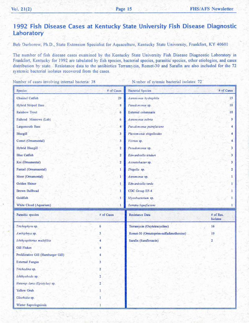
1992 Fish Disease Cases at Kentucky State University Fish Disease Diagnostic Laboratory
(
Bob Durborow, Ph.D., State Extension Specialist for Aquaculture, Kentucky State University, Frankfort, KY 40601
The- number of fish disease cases examined by the Kentucky State University Fish Disease Diagnostic Laboratory in Frankfort, Kentucky for 1992 are tabulated by fish species, bacterial species, parasitic species, other etiologies, and cases distribution by state. Resistance data to the antibiotics Terramycin, Romet-30 and Sarafin are also included for the 72 sys temic bacterial isolates recovered from the cases .
Number of cases involving internal bacteria : 38 N mber of sy temic bacterial isolates: 72
/ -#~£Cases Species #of Cases ' Baderial Species
~
Channel Catfish 29 Aeromonas hydrophila 15
Hybrid Striped Bass - 8 Pseudomonas sp. 10
Rainbow Trout I
6 Extern'al columnaris 10
Fathead Minnows (Lab) 6 A erom on as sobria 9
Largemouth Bass 4 Pse·_;domonas putrefaciens 4
Bluegill 3 Plesiomonas shige/loides 4
Comet (Ornamental) 3 Viorios sp. 4
Hybrid Bluegill 2 Pseudom onas sp. 3
Blue Catfish 2 Edwardsiel/a ictaluri 3
Koi (ornamental) 2 A cinetobacter sp. 3
Fantail (Omamaental) 1 Shigella sp. 2
Moor (Ornamental) - I Aeromonas sp. 1
Golden Shiner 1 Edwardsiel/a tarda 1 -Brown Bullhead 1 CDC Group EF -4 I
Goldfish I M ycobacterium sp. - I I (
White Cloud (Aquarium) I Serratia liquefaciens 1
Parasitic species # of Cases Resistance Data .-· # of Res. · > Isolates .. ,':·:·' : ......
' Trichophy ra sp. 6 Terramycin (Oxytetracycline)
i 16
A m biphrya sp. 5 Romet-30 (Ormetoprim-sulfadimethoxine) 19
, Ichthy opthlrius multifiliis 4 Sarafm (Sarafloxacin) 2 /
Gill Flukes 4 \
Proliferative Gill (Hamburger Gill) 4
External Fungus 3
Trichodina sp. 2
Jchthyobodo sp. 2
Heterop >laria (Epis ty lus) sp. 2
Yellow Grub I
Glochid ia sp. I
Winter Saprolegniosis I

1993 FISH HEALTH SECTION ANNUAL MEETING REGISTRATION AND LODGING INFORMATION
The 1993 annual meeting will be held at the Stouffer Concourse Hotel near Stapleton International Airport in Denver, Colorado. The meeting will begin at around 9:00a.m., Mountain Dayligh t Savings Time, on Tuesday, the 20th of July and will continue until around noon on Thursday, the 22nd.
The Stouffer is ready to accept individual room reservations from a block held in the name "Fish Health Section" at the conference rate of $77.00 per night single or double occupancy. The conference rate block will be held until June 27, 1993. Please call the Stouffer at 303/399-7500. Complimentary shuttle service from the airport is provided by the Stouffer at airport doors 4, 6, and 12, on the baggage claim level. If your arrival at Stapleton will be between 11:30 p.m. and 5:00 a.m., you should call the Stouffer to arrange pick-up. Individuals who present a federal or state tax exemption certificate at hotel check-in will not be charged room taxes. Complimentary room service coffee will be provided each morning of your stay. Don't risk missing the conference room rate --- make your reservations now!
Conference registration will be handled by Dennis (Andy) Anderson, P.O. Box 737, Ft. Morgan, CO 80701-0737 (telephone 303/867-5093). You can pre-register now till July 1st by sending Andy a check made out to "FHS Annual Meeting". Fees are as follows:
FHS Member pre-registration $50.00 Non-Member pre-registration $60.00 FHS Member registration (after July 1) $70.00 Non-Member registration (after July 1) $80.00
All conference functions, including the Wednesday evening banquet, are included in these registration prices. Additional banquet tickets may be purchased for $25.00 each. SAVE MONEY! SEND YOUR CHECK BEFORE .JULY! NON-MEMBERS, JOIN BEFORE .JULY AND PRE-REGISTER! YOU'LL REALLY SAVE!
Cut along dotted line - mail registration rorm with check
FISH HEALTH SECTION/AFS ANNUAL MEETING - DENVER, COLORADO
Name ______________________________________ __ Affiliation ---------------------------
Address -------------------------------------
# of Additional Banquet Tickets Requested
Registration Fees
Current FHS Member Yes
_ ___ @ $25.00! ea. = $ _______ _
- --- @ $ = $ ____ _
Total Amount Paid = $ _____ _
Relurn to:
DENNIS ANDERSON P.O. BOX 737
FORT MORGAN, COLORADO 80701-0737 U.S.A.
No
Vol. 21(2) Page 16 FHS/AFS Newsletter
Fish Health Section Newsletter
The Fish Health Section Newsletter is a quarterly publication of the Fish Health S~ction of the American Fisheries Society. Submissions of any length on a topic of interest to fish health specialists are encouraged with the understanding that material is not peer reviewed. Submissions should be addressed to the editiors or to a member of the publications committee.
Editors:
Ms. Leni Oman Dept of Wildlife 600 Capitol Way N. Olympia, WA~ 98501-1091 206-664-803 5 Phone 206-586-0248 FAX
Make a note! Deadline for next issue: July 30, 1993
FHS/ AFS Newsletter Fisheries Experiment Station 1465 West 200 North Logan, UT 84321-6262
/
Dr. Chris Wilson Fisheries Experiment Station 1465 W. 200 N. Logan, UT 84321-6262 801-752-1066 Phone 801-752-6977 FAX
003534 DR 931 :~ Glenn L. Hoffm~n
y
BULK RATE U.S. POSTAGE PAID NON-PROFIT ORG. LOGAN, UT PERMIT# 76