P rap ic A f Fish Dtoliths - Fisheries and Oceans Canada · VIII Acknowledgements I owe a great...
294
Î N^^W ^M Ÿ R Î V, ^N ^Î P h o t og rap h ic A tl as o f Fish Dtoliths of the Northwest Atlantic . Ocean Canadian Special Publication of Fisheries and Aquatic Sciences 133 teven E. Campana
Transcript of P rap ic A f Fish Dtoliths - Fisheries and Oceans Canada · VIII Acknowledgements I owe a great...

ÎN^^W MŸRÎV,^N Î
P h o tog rap h ic Atl as o f
Fish Dtolithsof the Northwest Atlantic . Ocean
Canadian Special Publication of Fisheries and Aquatic Sciences 133
teven E. Campana

---"-------"------"—DexàeàttnrnotGoetanVsin- tneqio%
1..‘brary
elototbee des Ke•
eIntateo des Pêctleo
et doe
Océune
Vile 9 2 VIA 9 2004
Photographic Atlas of Fish Otoliths of the
Northwest Atlantic Ocean

NRC Monograph Publishing Program
Editor: P.B. Cavers (University of Western Ontario)
Editorial Board: H. Alper, OC, FRSC (University of Ottawa); G.L. Baskerville, FRSC (University of British Colum-bia); W.G.E. Caldwell, OC, FRSC (University of Western Ontario); S. Gubins (Annual Reviews); B.K. Hall, FRSC (Dalhousie University); P. Jefferson (Agriculture and Agri-Food Canada); W.H. Lewis (Washington University); A.W. May, OC (Memorial University of Newfoundland); G.G.E. Scudder, OC, FRSC (University of British Colum-bia); B.P. Dancik, Editor-in-Chief, NRC Research Press (University of Alberta)
Inquiries: Monograph Publishing Program, NRC Research Press, National Research Council of Canada, Ottawa, Ontario Kl A 0R6, Canada. Web site: www.monographs.nrc-cnrc.gc.ca
Correct citation for this publication: Campana, S.E. 2004. Photographic Atlas of Fish Otoliths of the Northwest
Atlantic Ocean. NRC Research Press, Ottawa, Ontario. 284 pp.

Canadian Special Publication of Fisheries and Aquatic Sciences 133
Photographic Atlas
of Fish Otoliths of the
Northwest Atlantic Ocean
Steven E. Campana
Marine Fish Division
Bedford Institute of Oceanography
P.O. Box 1006, Dartmouth
Nova Scotia B2Y 4A2, Canada
ARC' CARCNRC Research Press
Ottawa 2004

© 2004 National Research Council of Canada
All rights reserved. No part of this publication may be reproduced in a retrieval system, or transmitted by any means, electronic, mechanical, photocopying, recording or otherwise, without the prior written permission of the National Research Council of Canada, Ottawa, Ontario Kl A 0R6, Canada. Printed in Canada on acid-free paper. 0
ISBN 0-660-19108-3 ISSN 0706-6481 NRC No. 46328
National Library of Canada cataloguing in publication data
Campana, Steven E., 1955– Photographic atlas of fish otoliths of the Northwest Atlantic Ocean
(Canadian Special Publication of Fisheries and Aquatic Sciences 133) Includes an abstract in French. Includes bibliographical references. ISBN 0-660-19108-3
1. Fishes — North Atlantic Ocean — Identification. 2. Fishes — North Atlantic Ocean — Age determination. 3. Otoliths. 4. Fishes – Morphology. I. National Research Council Canada. II. Title. III. Series.
QL621.5C26 2004 571.3'17 C2003-980310-4

V
Contents
Abstract/Résumé vii
Acknowledgements viii
Introduction 1
Otolith location and function 2
Otolith composition 3
Otolith morphology 3
Biological factors affecting otolith morphology 6
Effects of preservation on otolith morphology 6
Methods 7
Using the atlas 8
References 10
Photographic plates 13
Alphabetical species list 279
VÎ1
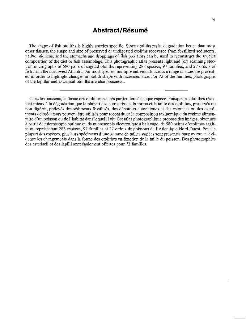
Abstract/Résumé
The shape of fish otoliths is highly species specific. Since otoliths resist degradation better than mostother tissues, the shape and size of preserved or undigested otoliths recovered from fossilized sediments,native middens, and the stomachs and droppings of fish predators can be used to reconstruct the speciescomposition of the diet or fish assemblage. This photographic atlas presents light and (or) scanning elec-tron micrographs of 580 pairs of sagittal otoliths representing 288 species, 97 families, and 27 orders offish from the northwest Atlantic. For most species, multiple individuals across a range of sizes are present-ed in order to highlight changes in otolith shape with increased size. For 72 of the families, photographsof the lapillar and asteriscal otoliths are also presented.
Chez les poissons, la forme des otolithes est très particulière à chaque espèce. Puisque les otolithes résis-tent mieux à la dégradation que la plupart des autres tissus, la forme et la taille des otolithes, préservés ounon digérés, prélevés des sédiments fossilisés, des dépotoirs autochtones et des estomacs ou des excré-ments de prédateurs peuvent être utilisés pour reconstituer la composition taxinomique du régime alimen-taire d'un poisson ou de l'habitat dans lequel il vit. Cet atlas photographique propose des images, obtenuesà partir de microscopie optique ou de microscopie électronique à balayage, de 580 paires d'otolithes sagit-taux, représentant 288 espèces, 97 familles et 27 ordres de poissons de l'Atlantique Nord-Ouest. Pour laplupart des espèces, plusieurs spécimens d'une gamme de tailles variées sont présentés pour mettre en évi-dence les changements dans la forme des otolithes en fonction de la taille du poisson. Des photographiesdes asteriscii et des lapilli sont également offertes pour 72 familles.

VIII
Acknowledgements
I owe a great deal to the people who provided me with fish or otoliths in support of this atlas. Without them, this atlas would not have been possible. I particularly appreciate the assistance of the following peo-ple: Carlos Assis (Universidade de Lisboa, Lisbon, Portugal), Tom Azarovitz (Northeast Fisheries Science Center, Woods Hole, MA), Rod Bradford (Bedford Institute of Oceanography, Dartmouth, NS), John Cas-selman (Ontario Ministry of Natural Resources, Picton, ON), Edgar Dalley (Northwest Atlantic Fisheries Centre, St. John's, NF), Ken Doe (Bedford Institute of Oceanography, Dartmouth, NS), Janet Fields (Northeast Fisheries Science Center, Woods Hole, MA), Jacques Gagné (Institut Maurice-Lamontagne, Mont Joli, PQ), Andrew Hebda (Nova Scotia Museum of Natural History, Halifax, NS), Joe Hunt (St. Andrews Biological Station, St. Andrews, NB), Brian Jessop (Bedford Institute of Oceanography, Dart-mouth, NS), Jonathan Joy (Eastern College of Applied Arts, Technology and Continuing Education, Bonavista, NF), Jeremy King (Massachusetts Division of Marine Fisheries, Pocasset, MA), Jason LeBlanc (Nova Scotia Dept. of Agriculture and Fisheries, Pictou, NS), Sylvie Levesque (Centre de recherche et de développement des produits marins, Shippagan, N.B), Tomasz Linkowski (Sea Fisheries Institute, Gdynia, Poland), John Martell (St. Andrews Biological Station, St. Andrews, NB), Allan McNeil (Nova Scotia Dept. of Agriculture and Fisheries, Pictou, NS), Roberta Miller (Institut Maurice-Lamontagne, Mont Joli, PQ), Lisa Natanson (National Marine Fisheries Service, Narragansett, RI), Vic Nordahl (Northeast Fish-eries Science Center, Woods Hole, MA), Gréa Pétursdéttir (Marine Research Institute, Reykjavik, Ice-land), Julie Porter (St. Andrews Biological Station, St. Andrews, NB), David Secor (Chesapeake Biological Laboratory, Solomons, MD), Peter Shelton (Northwest Atlantic Fisheries Centre, St. John's, NF), Greg Skomal (Massachusetts Divison of Marine Fisheries, Boston, MA), Louise Stanley (Coastal Fisheries Institute, Baton Rouge, LA), Heath Stone (St. Andrews Biological Station, St. Andrews, NB), Sarah Swan (Centre for Coastal and Marine Sciences, Dunstaffnage, Scotland), Dianne Tracey (National Institute of Water and Atmospheric Research, Wellington, New Zealand), Margaret Treble (Fisheries and Oceans Canada, Winnipeg, MB), Kim Whitman (Atlantic Veterinary College, University of Prince Edward Island, Charlottetown, PET), Charles Wilson (Coastal Fisheries Institute, Baton Rouge, LA), Steve Wis-chniowski (International Pacific Halibut Commission, Seattle, WA), and David Wyanski (Marine Resources Research Institute, Charleston, SC).
I found the professionals of the Observer Program most helpful in collecting fish from commercial ves-sels. I thank Victor Matthews, John Robidoux, John Donahue, Bill Lloyd, Dave Spallin, and Gary Tuff for their help.
My colleagues in the Marine Fish Division at the Bedford Institute of Oceanography were kind enough to collect many fish specimens for me. In particular, I thank Peter Comeau, Paul Fanning, Jim Fennell, Bill MacEachern, Mark Showell, Jim Simon, and Scott Wilson for their assistance.
Joanne Hamel, Linda Marks, Tara Caseley, Warren Joyce, and Frances MacKinnon provided excellent technical support during this project, and I greatly appreciated their assistance. I also thank Art Cosgrove, and especially Francis Kelly, for their work in preparing and formatting the illustrations and graphics. David O'Neil (National Research Council, Halifax, NS) did an excellent job preparing the SEM photos.
Last but certainly not least, I greatly appreciate the expert species identifications provided by Daphne Themelis (Dalhousie University, Halifax, NS) and Lou Van Guelpen (Huntsman Marine Science Centre, St. Andrews, NB).
1
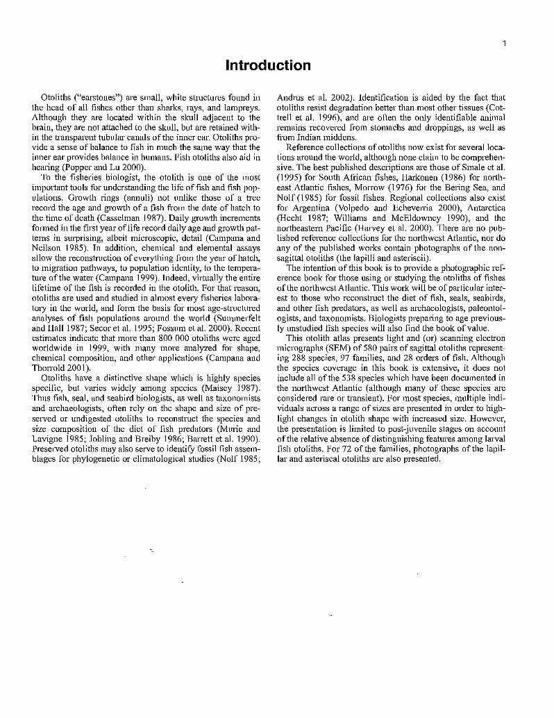
Introduction
Otoliths ("earstones") are small, white structures found inthe head of all fishes other than sharks, rays, and lampreys.Although they are located within the skull adjacent to thebrain, they are not attached to the skull, but are retained with-in the transparent tubular canals of the inner ear. Otoliths pro-vide a sense of balance to fish in much the same way that theinner ear provides balance in humans. Fish otoliths also aid inhearing (Popper and Lu 2000).
To the fisheries biologist, the otolith is one of the mostimportant tools for understanding the life of fish and fish pop-ulations. Growth rings (annuli) not unlike those of a treerecord the age and growth of a fish from the date of hatch tothe time of death (Casselman 1987). Daily growth incrementsformed in the first year of life record daily age and growth pat-terns in surprising, albeit microscopic, detail (Campana andNeilson 1985). In addition, chemical and elemental assaysallow the reconstruction of everything from the year of hatch,to migration pathways, to population identity, to the tempera-ture of the water (Campana 1999). Indeed, virtually the entirelifetime of the fish is recorded in the otolith. For that reason,otoliths are used and studied in almost every fisheries labora-tory in the world, and form the basis for most age-structuredanalyses of fish populations around the world (Summerfeltand Hall 1987; Secor et al. 1995; Fossum et al. 2000). Recentestimates indicate that more than 800 000 otoliths were agedworldwide in 1999, with many more analyzed for shape,chemical composition, and other applications (Campana andThorrold 2001).
Otoliths have a distinctive shape which is highly speciesspecific, but varies widely among species (Maisey 1987).Thus fish, seal, and seabird biologists, as well as taxonomistsand archaeologists, often rely on the shape and size of pre-served or undigested otoliths to reconstruct the species andsize composition of the diet of fish predators (Murie andLavigne 1985; Jobling and Breiby 1986; Barrett et al. 1990).Preserved otoliths may also serve to identify fossil fish assem-blages for phylogenetic or climatological studies (Nolf 1985;
Andrus et al. 2002). Identification is aided by the fact thatotoliths resist degradation better than most other tissues (Cot-trell et al. 1996), and are often the only identifiable animalremains recovered from stomachs and droppings, as well asfrom Indian middens.
Reference collections of otoliths now exist for several loca-tions around the world, although none claim to be comprehen-sive. The best published descriptions are those of Smale et al.(1995) for South African fishes, Harkonen (1986) for north-east Atlantic fishes, Morrow (1976) for the Bering Sea, andNolf (1985) for fossil fishes. Regional collections also existfor Argentina (Volpedo and Echeverria 2000), Antarctica(Hecht 1987; Williams and McEldowney 1990), and thenortheastern Pacific (Harvey et al. 2000). There are no pub-lished reference collections for the northwest Atlantic, nor doany of the published works contain photographs of the non-sagittal otoliths (the lapilli and asteriscii).
The intention of this book is to provide a photographic reference book for those using or studying the otoliths of fishesof the northwest Atlantic. This work will be of particular inter-est to those who reconstruct the diet of fish, seals, seabirds,and other fish predators, as well as archaeologists, paleontol-ogists, and taxonomists. Biologists preparing to age previous-ly unstudied fish species will also find the book of value.
This otolith atlas presents light and (or) scanning electronmicrographs (SEM) of 580 pairs of sagittal otoliths represent-ing 288 species, 97 families, and 28 orders of fish. Althoughthe species coverage in this book is extensive, it does notinclude all of the 538 species which have been documented inthe northwest Atlantic (although many of these species areconsidered rare or transient). For most species, multiple indi-viduals across a range of sizes are presented in order to high-light changes in otolith shape with increased size. However,the presentation is limited to post juvenile stages on accountof the relative absence of distinguishing features among larvalfish otoliths. For 72 of the families, photographs of the lapil-lar and asteriscal otoliths are also presented.
Medial
Otolith location and function 2
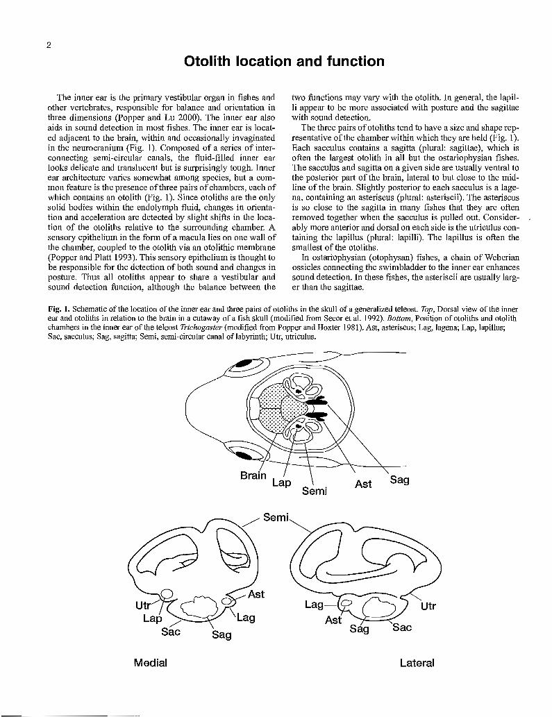
The inner ear is the primary vestibular organ in fishes and other vertebrates, responsible for balance and orientation in three dimensions (Popper and Lu 2000). The inner ear also aids in sound detection in most fishes. The inner ear is locat-ed adjacent to the brain, within and occasionally invaginated in the neurocranium (Fig. 1). Composed of a series of inter-connecting semi-circular canals, the fluid-filled inner ear looks delicate and translucent but is surprisingly tough. Inner ear architecture varies somewhat among species, but a corn-mon feature is the presence of three pairs of chambers, each of which contains an otolith (Fig. 1). Since otoliths are the only solid bodies within the endolymph fluid, changes in orienta-tion and acceleration are detected by slight shifts in the loca-tion of the otoliths relative to the surrounding chamber. A sensory epithelium in the form of a macula lies on one wall of the chamber, coupled to the otolith via an otolithic membrane (Popper and Platt 1993). This sensory epithelium is thought to be responsible for the detection of both sound and changes in posture. Thus all otoliths appear to share a vestibular and sound detection function, although the balance between the
two functions may vary with the otolith. In general, the lapil-li appear to be more associated with posture and the sagittae with sound detection.
The three pairs of otoliths tend to have a size and shape rep-resentative of the chamber within which they are held (Fig. 1). Each sacculus contains a sagitta (plural: sagittae), which is often the largest otolith in all but the ostariophysian fishes. The sacculus and sagitta on a given side are usually ventral to the posterior part of the brain, lateral to but close to the mid-line of the brain. Slightly posterior to each sacculus is a lage-na, containing an asteriscus (plural: asteriscii). The asteriscus is so close to the sagitta in many fishes that they are often removed together when the sacculus is pulled out. Consider-ably more anterior and dorsal on each side is the utriculus con-taining the lapillus (plural: lapilli). The lapillus is often the smallest of the otoliths.
In ostariophysian (otophysan) fishes, a chain of Weberian ossicles connecting the swimbladder to the inner ear enhances sound detection. In these fishes, the asteriscii are usually larg-er than the sagittae.
Fig. 1. Schematic of the location of the inner ear and three pairs of otoliths in the skull of a generalized teleost. Top, Dorsal view of the inner ear and otoliths in relation to the brain in a cutaway of a fish skull (modified from Secor et al. 1992). Bottom, Position of otoliths and otolith chambers in the inner ear of the teleost Trichogaster (modified from Popper and Hoxter 1981). Ast, asteriscus; Lag, lagena; Lap, lapillus; Sac, sacculus; Sag, sagitta; Semi, semi-circular canal of labyrinth; Utr, utriculus.
Lag- Ast
Sag
Utr
Sac
Lateral

3
Otolith composition
Otoliths are very pure compared to most biological andmineralogical structures, with the composition being dominat-ed by calcium carbonate in an organic matrix. Most otolithscontain more than 95% by weight of calcium carbonate, with3-5% in the form of an organic matrix, and less than 1% asnon-organic trace impurities. The trace element and stable iso-tope composition of the otolith has been given extensivestudy, owing to numerous applications in reconstructing theenvironmental history, migration, and population identity offishes (Campana 1999).
Calcium carbonate can crystallize as any one of three crys-tal polymorphs: calcite, aragonite, or vaterite. However, thevast majority of sagittal and lapillar otoliths are composed ofaragonite, which has a milky white appearance (Carlstrdm1963; Oliveira et al. 1996; Campana 1999). This is unlike theotoconia of mammals, which are composed of calcite.
Curiously, different polymorphs of calcium carbonateappear to be linked to the different otolith organs. While arag-onite is the norm for sagittae and lapilli, most asteriscii aremade of vaterite, thus accounting for their glassy appearance(Oliveira et al. 1996). Vaterite is also the principal polymorphin many aberrant, or "crystalline", otoliths (Mugiya 1972).Calcitic regions in otoliths are much rarer.
The implications of an otolith composition dominated bycalcium carbonate lie most clearly with otolith preservationand stability. Both calcium carbonate and otoliths are stablefor many years when stored dry. However, calcium carbonateis acid soluble, so preservation in even weakly acidic solutionswill result in dissolution of the otolith.
Otolith morphology
The three pairs of otoliths differ markedly in shape andappearance. In most adult fishes, the sagittae are the largestpair and the lapilli the smallest (Fig. 2). In contrast, asterisciiare larger than sagittae in ostariophysian fishes (a group whichincludes the minnows and catfish). Sagittal shape differs sub-stantially among species, while lapillar shape is more uniform.The shape of the asteriscii shows intermediate inter-specificvariability. Within an otolith pair, the left and right otoliths areveiy similar, but not identical, mirror images of each other.Interestingly, the left and right asteriscii can differ consider-ably more in shape than the other otolith pairs (Campana andCasselman 1993).
The orientation and major landmarks of a typical sagitta areshown in the labelled scanning electron microscope (SEM)photo of Fig. 3. The rostrum, antirostrum, and postrostrum areconsistent features of all sagittae, although their size andextension varies substantially among species. The sulcus,which represents the point of attachment of the sensory mac-ula, is also a consistent feature of sagittae. Although the finedetails of sulcus morphology are not documented here, thosedetails are defined elsewhere and can prove helpfiil in somespecies identifications (Nolf 1985; Smale et al. 1995). Anintricate morphology is also evident in the SEM images ofasteriscii, but less so in the case of the lapilli (Fig. 4). It is pos-sible that inter-specific differences in the shape of asterisciiand lapilli could be used to complement the differencesobserved in the sagittae. However, in light of the relativelysmall size of the lapilli and sagittae, SEM would probably berequired by the end user to observe all but the most gross ofmorphological differences.
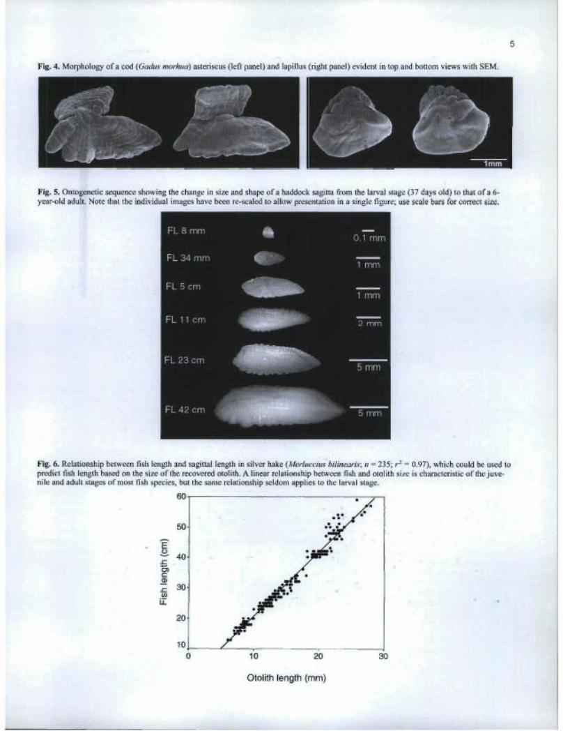
In any given species, otolith size and shape often changessubstantially with fish growth (Fig. 5). In virtually all youngfish larvae, otoliths tend to be relatively featureless: sphericalor smoothly oblate in most species, and discoid in species(such as salmonids) which hatch at a larger size. In mostspecies, the sagittae and lapilli are present at hatch, while theasteriscii first appear at an age of 2-3 weeks. At this earlystage of fish development, relative otolith sizes can be invert-ed, with lapilli being larger than sagittae (Campana 1989).Otoliths first acquire the main features of their mature shapein the juvenile stage. As the size-specific photos of this atlasdemonstrate, otolith shape can remain diagnostic but stillchange in later life as the fish (and otolith) grows. As a result,otolith size must be taken into consideration, as well as shape,when identifying a species from an otolith. In particular, theotolith shape of very large fish can differ substantially fromthose of average-sized adults.
Because of their function in maintaining the balance of thefish, otoliths tend to grow as the fish grows. Therefore, thereis almost always a strong relationship between otolith size andfish size (Hunt 1992). Given a measurement of otolith size(whether in terms of length or weight), it is possible to esti-mate the length of the fish from which the otolith was obtained(Fig. 6). These estimates provide usefiil approximations offish length, but cannot be interpreted too strictly, since thefish-otolith regression often differs among populations orgroups of fish with different growth rates (Campana 1990). Itis also important to note that the relationship between fish andotolith length is not necessarily linear, and that the relationshipfor larvae is often very different from that for adults.

Excisural notch
Posterior
Postrostrum
Antirostrum
Rostrum
Sulcus acusticus Dorsal
KM=
Distal Surface
Posterior
Postrostrum
Anterior
Antirostrum Excisural
---"" notch
Dorsal
Rostrum
4
Fig. 2. Light micrograph of the three pairs of otoliths from a 23-cm adult white perch (Morone americana). The left-hand otolith of each pair is shown on the left side.
Fig. 3. Morphology of a haddock (Melanogrammus aeglefinus) sagitta evident in proximal (top sagitta) and distal (bottom sagitta) views with SEM.
Proximal Surface Ventral
Ventral
5
Fig. 4. Morphology of a cod (Gadus morhua) asteriscus (left panel) and lapillus (right panel) evident in top and bottom views with SEM.
Fig. 5. Ontogenetic sequence showing the change in size and shape of a haddock sagitta from the larval stage (37 days old) to that of a 6-year-old adult. Note that the individual images have been re-scaled to allow presentation in a single figure; use scale bars for correct size.
Fig. 6. Relationship between fish length and sagittal length in silver hake (Merluccius bilinearis; n = 235; r` = 0.97), which could be used topredict fish length based on the size of the recovered otolith. A linear relationship between fish and otolith size is characteristic of the juve-nile and adult stages of most fish species, but the same relationship seldom applies to the larval stage.
100 10 20 30
Otolith length (mm)

Biological factors affecting otolith morphology
6
There are a broad range of biological factors which influ-ence or moderate otolith shape. These factors can operate at a range of scales, from that of general phylogeny to the individ-ual level. Few of these factors are well understood, but those that are known or suspected are mentioned here.
There are no broad phylogenetic principles which are known to guide otolith shape (Maisey 1987). Although fami-ly- or genus-level otolith characteristics are often present, it is often impossible to predict otolith shape for any given species. There may be functional relationships however. Based on numerous observations, I have noted that fast-swimming fish-es capable of rapid acceleration and turning tend to have smaller otoliths than their slower swimming counterpa rts. The tunas and swordfish are good examples of this phenomenon, whereby the sagittal otolith of a 400-kg bluefin tuna (Thunnus thynnus) is smaller than that of a 1-kg cod. Species capable of good sound production (and presumably good sound detec-tion) can also be expected to have large saggital otoliths. Members of the Sciaenidae (grunts and drums) are character-istic of this group; species such as the black drum (Pogonias cromis) have sagittae which are among the largest observed. In contrast, the families within the group Ostariophysii (such as cyprinids and catfishes), which possess a chain of Weberian
ossicles to enhance sound detection, have somewhat smaller sagittae and larger asteriscii than normal.
Within a species, otolith shape can vary with the sex, popu-lation, and growth rate, as well as the stage of ontogeny described in the previous section. The magnitude of shape dif-ferences due to ontogeny and fish size is considerably larger than that due to sex, population, and growth rate, since the effect of the latter factors may be detectable only through sta-tistical analysis (Campana and Casselman 1993; Cardinale et al. 2003).
In general, otoliths within an otolith pair are very similar but non-identical mirror images of each other. However, left versus right asymmetry is common in flatfish, a fish in which the eyes migrate to the same side of the head at around the time of metamorphosis to the settled juvenile. The presence or degree of asymmetry seems to vary among individuals, and is most evident in large individuals. In general, however, the sagitta found on the upper side of the fish (the right otolith in right-eyed flatfish) is irregularly shaped or occasionally short-er and thicker than the sagitta which faces down in the adult fish. The functional significance of this otolith asymmetry is unknown, but is presumably related to a reduced or altered function in one of the two otoliths.
Effects of preservation on otolith morphology
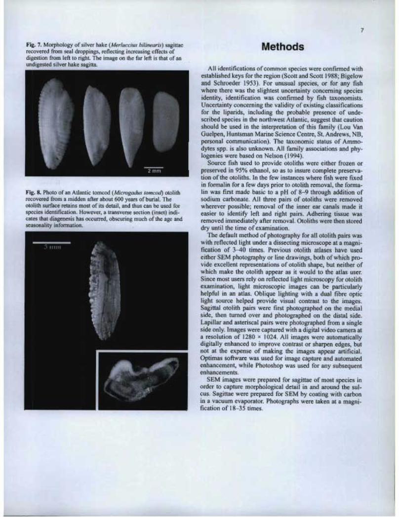
The shape and size of otoliths recovered from the stomach or feces of fish predators has long been used to reconstruct the species and size composition of the predator's diet (Murie and Lavigne 1985; Jobling and Breiby 1986; Barrett et al. 1990; Pierce et al. 1991; Bowen et al. 1993; Dolloff 1993; Burns et al. 1998). In many cases, there are few alternatives, since otoliths are often the only animal remains that are recovered, let alone identified to species. Nevertheless, there are limita-tions to this application. Several studies have fed fish of known species and size composition to seals, and then recov-ered the ingested otoliths from the stomachs or feces (Dellinger and Trillmich 1988; Cottrell et al. 1996; Tollit et al. 1997; Bowen 2000). In all cases, sources of bias have been noted, associated primarily with the relatively rapid dissolu-tion of small and (or) fragile otoliths in the acidic stomach environment. As a result, it seems likely that any dietary reconstruction could underestimate the contribution from fish species with small otoliths, or from smaller individuals. Even where complete otolith dissolution does not occur, partial dissolution can leave an otolith unrecognizable to species, or perhaps smaller than its original size. Examples of par-tial dissolution of otoliths recovered from seal droppings are shown in Fig. 7.
Preserved otoliths may also serve to identify fossil fish assemblages (Elder et al. 1996), date sedimentary strata (Gae-mers 1984), reconstruct historical populations (Hales and Reitz 1992), prepare phylogenies (Nolf 1995), reconstruct ancient climates (Ivany et al. 2000; Andrus et al. 2002), and provide indicators of seasonal occupation for ancient peoples (Van Neer et al. 1993). Such otoliths may be recovered from aquatic sediments, fossil grounds, or archaeological middens, where they have been exposed to possibly acidic conditions or chemical leaching. Such conditions have the potential to bias reconstructions of past assemblages, if otoliths have been dis-solved, or to alter climatic reconstructions if chemical leach-ing has occurred. However, other indicators can often be used to determine if otolith alteration has occurred. In the case of archaelogical applications, bias is not usually a problem, since the growth increments used to determine seasonality are either present or absent. For reasons not fully understood, otoliths in some fossil middens may sustain little damage after thousands of years of preservation, while others are rendered illegible after only a few hundred years (Fig. 8).
7
Fig. 7. Morphology of silver hake (Merluccius bilinearis) sagittaerecovered from seal droppings, reflecting increasing effects ofdigestion from left to right. The image on the far left is that of anundigested silver hake sagitta.
Fig. 8. Photo of an Atlantic tomcod (Microgadus tomcod) otolithrecovered from a midden after about 600 years of burial. Theotolith surface retains most of its detail, and thus can be used forspecies identification. However, a transverse section (inset) indi-cates that diagenesis has occurred, obscuring much of the age andseasonality information.
Methods
All identifications of common species were confirmed withestablished keys for the region (Scott and Scott 1988; Bigelowand Schroeder 1953). For unusual species, or for any fishwhere there was the slightest uncertainty concerning speciesidentity, identification was confirmed by fish taxonomists.Uncertainty concerning the validity of existing classificationsfor the liparids, including the probable presence of unde-scribed species in the northwest Atlantic, suggest that cautionshould be used in the interpretation of this family (Lou VanGuelpen, Huntsman Marine Science Centre, St. Andrews, NB,personal communication). The taxonomic status of Ammo-dytes spp. is also unknown. All family associations and phy-logenies were based on Nelson (1994).
Source fish used to provide otoliths were either frozen orpreserved in 95% ethanol, so as to insure complete preserva-tion of the otoliths. In the few instances where fish were fixedin formalin for a few days prior to otolith removal, the forma-lin was first made basic to a pH of 8-9 through addition ofsodium carbonate. All three pairs of otoliths were removedwherever possible; removal of the inner ear canals made iteasier to identify left and right pairs. Adhering tissue wasremoved immediately after removal. Otoliths were then storeddry until the time of examination.
The default method of photography for all oto]ith pairs waswith reflected light under a dissecting microscope at a magni-fication of 3-40 times. Previous otolith atlases have usedeither SEM photography or line drawings, both of which pro-vide excellent representations of otolith shape, but neither ofwhich make the otolith appear as it would to the atlas user.Since most users rely on reflected light microscopy for otolithexamination, light microscopic images can be particularlyhelpful in an atlas. Oblique lighting with a dual fibre opticlight source helped provide visual contrast to the images.Sagittal otolith pairs were first photographed on the medialside, then turned over and photographed on the distal side.Lapillar and asteriscal pairs were photographed from a singleside only. Images were captured with a digital video camera ata resolution of 1280 x 1024. All images were automaticallydigitally enhanced to improve contrast or sharpen edges, butnot at the expense of making the images appear artificial.Optimas software was used for image capture and automatedenhancement, while Photoshop was used for any subsequentenhancements.
SEM images were prepared for sagittae of most species inorder to capture morphological detail in and around the sul-cus. Sagittae were prepared for SEM by coating with carbonin a vacuum evaporator. Photographs were taken at a magni-fication of 18-35 times.
Using the atlas
8
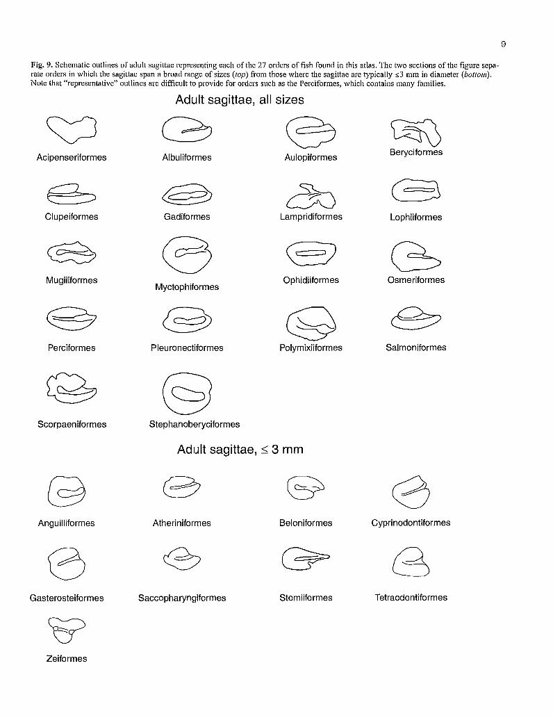
Fish systematics is an evolving science; hence species affil-iations within a family are not necessarily stable. For this rea-son, this atlas is arranged alphabetically by order, rather than phylogenetically. Within each order, families are presented alphabetically, as are species within families. A key to identi-fy unknown otoliths to the order or family level was attempt-ed but was discontinued, owing to the difficulty of providing diagnostic keys which take into account the changing shape of many otoliths with increasing size. However, outlines of "rep-resentative" sagittae for each order are provided in Fig. 9. It is important to note that there can be wide variations in otolith shape within orders and families, and that "representative" outlines are not necessarily representative. This is particularly true for the Perciformes, which comprises dozens of families.
For most species, representative otolith images occupy the entire page. Where available, an SEM image of the sagitta heads the page in order to highlight details of the sulcus. For at least one species of each family, an accompanying pair of light microscopic images to the right of the SEM image shows the lapilli and asteriscii (note, however, that left- and right-side identifications of these two otolith pairs are not necce-sarily conect). Below these images are a series of light microscopic images showing multiple pairs of sagittae from fish of different sizes, arranged from smallest to largest. Image scales often vary across the images, so the accompany-ing scale bar should be used to estimate otolith size. Each sagittal pair is shown both medial side up (sulcus side) and distal side up, with the left otoliths arranged on the left side of the image panel wherever possible. In most cases, the rostrum and antirostrum are oriented up. However, it was not always possible to identify the rostrum in some sagittae; hence cau-tion is required in interpreting the otolith orientations too strictly.
In most cases, external profile will be sufficient to identify an otolith to species. The key features include the relative size of the rostru,m, antirostrum, postrostrum, and excisuial notch. The length of the sulcus can also be diagnostic. Where mor-
phological details of the sulcus are required, it can be difficult to view these details witha light microscope. However, use of oblique lighting is often helpful in providing visual contrast. Further contrast can be provided by sprinkling powdered graphite over the medial surface of the sagitta, and then light-ly tapping the otolith on its side to remove any excess. Alter:- natively, a graphite pencil can rubbed over the sulcus region. If this is not sufficient, SEM may be required.
Clearly, intact and well-preserved otoliths will be easier to identify than will those which have been degraded or eroded. Although freshly removed otoliths provide excellent samples, otoliths which have been stored dry after removal remain in excellent condition almost indefinitely. Fish which have' remained frozen after capture also provide well-preserved otoliths, as will those which have been preserved in 95% ethanol. Note, however, that ethanol becomes increasingly acidic as concentration drops, and that otoliths will dissolve in concentrations below about 70-80%. In addition, ethanol con-centration declines soon after the fish carcass is added, owing to dilution from the water in the fish tissues. Complete ethanol replacement after 12-24 hours helps keep concentrations high. Formalin is not a recommended preservative for fish otoliths, since even buffered formalin is slightly acidic and will dis-solve otoliths. However, short-term storage in formalin is pos-sible if the formalin is first made basic with sodium carbonate to a pH of at least 8.
It is often possible to identify the characteristics of otolith dissolution or degradation. Rounded edges, particularly at the tips of the rostrum and postrostrum, often occur during diges-tion in an acidic stomach, and can signify an overall loss of material and size. Discolouration (usually brown or black) is a sign of degradation seen in both formalin-preserved material and in otoliths from archaeological sites. A chalky white appearance is a sign of exposure to mildly acidic conditions. None of these conditions should be confused with the irregu-lar glassy appearance of "crystalline" otoliths, which are uncommon but natural occurrences in most species of fishes.
9
Fig. 9. Schematic outlines of adult sagittae representing each of the 27 orders of fish found in this atlas. The two sections of the figure sepa-rate orders in which the sagittae span a broad range of sizes (top) from those where the sagittae are typically _<3 mm in diameter (bottom).Note that "representative" outlines are difficult to provide for orders such as the Perciformes, which contains many families.
Adult sagittae, all sizes
C;DAcipenseriformes
Clupeiformes
Mugiliformes
Perciformes
Scorpaeniformes
Albuliformes
Gadiformes
Myctophiformes
Pleuronectiformes
Stephanoberyciformes
Aulopiformes
Lampridiformes
Ophidiiformes
Beryciformes
aLophiiformes
Osmeriformes
Polymixiiformes Salmoniformes
Adult sagittae, <_ 3 mm
Anguilliformes Atheriniformes Cyprinodontiformes
e QJGasterosteiformes Saccopharyngiformes
Beloniformes
Stomiiformes Tetraodontiformes
Zeiformes

References
1 0
Andrus, C.F., Crowe, D.E., Sandweiss, D.H., Reitz, E.J., and Romanek, C.S. 2002. Otolith S 180 record of mid-Holocene sea surface temperatures in Peru. Science, 295: 1508-1511.
Barrett, R.T., Roy, N., Loen, J., and Montevecchi, W.A. 1990. Diets of shags Phalacrocorax aristotelis and cormorants P. carbo in Norway and possible implications for gadoid stock recruitment. Mar. Ecol. Prog. Ser. 66: 205-218.
Bigelow, H.B., and Schroeder, W.C. 1953. Fishes of the Gulf of Maine. U.S. Fish Wildl. Sen'. Fish. Bull. 74. Vol. 53.577 p.
Bowen, W.D. 2000. Reconstruction of pinniped diets: accounting for complete digestion of otoliths and cephalo-pod beaks. Can. J. Fish. Aquat. Sci. 57: 898-905.
Bowen, W.D., Lawson, J.W., and Beck, B. 1993. Seasonal and geographic variation in the species composition and size of prey consumed by grey seals (Halichoerus gripus) on the Scotian Shelf. Can. J. Fish. Aquat. Sci. 50: 1768-1778.
Bums, J.M., Trumble, S.J., Castellini, M.A., and Testa, J.W. 1998. The diet of Weddell seals in McMurdo Sound, Antarctica as determined from scat collections and stable isotope analysis. Polar Biol. 19: 272-282.
Campana, S.E. 1989. Otolith microstructure of three larval gadids in the Gulf of Maine, with inferences on early life history. Can. J. Zool. 67: 1401-1410.
Campana, S.E. 1990. How reliable are growth backcalcula-lions based on otoliths? Can. J. Fish. Aquat. Sci. 47: 2219- 2227.
Campana, S.E. 1999. Chemistry and composition of fish otoliths: pathways, mechanisms and applications. Mar. Ecol. Prog. Ser. 188: 263-297.
Campana, S.E., and Casselman, J.M. 1993. Stock discrimina-tion using otolith shape analysis. Can. J. Fish. Aquat. Sci. 50: 1062-1083.
Campana, S.E., and Neilson, J.D. 1985. Microstructure of fish otoliths. Can. J. Fish. Aquat. Sci. 42: 1014-1032.
Campana, S.E., and Thorrold, S.R. 2001. Otoliths, increments and elements: keys to a comprehensive understanding of fish populations? Can. J. Fish. Aquat. Sci. 58: 30-38.
Cardinale, M., Doering-Arjes, P., Kastowsky, M., and Mose-gaard, H. 2003. Effects of sex, stock and environment on the shape of Atlantic cod (Gadus morhua) otoliths. Can. J. Fish. Aquat. Sci. In press.
Carlstrôm, D. 1963. A crystallographic study of vertebrate otoliths. Biol. Bull. 124: 441-463.
Casselman, J.M. 1987. Determination of age and growth. In The Biology of Fish Growth. Chap. 7. Edited by A.H. Weatherley and H.S. Gill. Academic Press, New York. p. 209-242.
Cottrell, P.E., Trites, A.W., and Miller, E.H. 1996. Assessing the use of hard parts in faeces to identify harbour seal prey: results of captive feeding trials. Can. J. Zool. 74: 875-880.
Dellinger, T., and Trillmich, F. 1988. Estimating diet compo-sition from scat analysis in otariid seals (Otariidae): is it reliable? Can. J. Zool. 66: 1865-1870.
Dolloff, C.A. 1993. Predation by river otters (Lutra canaden-sis) on juvenile coho salmon (Oncorhynchus kisutch) and Dolly Varden (Salvelinus malma) in southeast Alaska. Can. J. Fish. Aquat. Sci. 50: 312-315.
Elder, K.L., Jones, GA., and Bolz, G 1996. Distribution of otoliths in surficial sediments of the U.S. Atlantic continen-tal shelf and slope and potential for reconstructing Holocene fish stocks. Paleoceanography, 11: 359-367.
Fossum, P., Kalish, J., and Moksness, E. 2000.2nd International Symposium on Fish Otolith Research and Application, Bergen, Norway, 20-25 June 1998. Fish. Res. 46: 1-371.
Gaemers, P.A.M. 1984. Fish otoliths from the Bassevelde sand (Late Tongrian) of Ruisbroek, Belgium, and the stratigra-phy of the early Oligocene of Belgium. Meded. Werkgr. Tert. Kwart. Geol. 46: 237-267.
Hales, Jr., L.S., and Reitz, E.J. 1992. Historical changes in age and growth of Atlantic croaker, Micropogonias undulatus (Perciformes: Sciaenidae). J. Archaeol. Sci. 19: 73-99.
Harkonen, T. 1986. Guide to the otoliths of the bony fishes of the northeast Atlantic. Danbiu ApS. Biological Consultants, Hellerup, Denmark. 256 p.
Harvey, J.T., Loughlin, T.R., Perez, M.A., and Oxman, D.S. 2000. Relationship between fish size and otolith length for 63 species of fishes from the eastern North Pacific Ocean. NOAA Tech. Rep. NMFS, 150: 1-36.
Hecht, T. 1987. A guide to the otoliths of southern ocean fish-es. S. Afr. T. Nav. Antarkt. 17: 1-87.
Hunt, J.J. 1992. Morphological characteristics of otoliths for selected fish in the Northwest Atlantic. J. Northw. Atl. Fish. Sci. 13: 63-75.
Ivany, L.C., Patterson, W.P., and Lohmann, K.C. 2000. Cooler win-ters as a possible cause of mass extinctions at the Eocene/Oligocene boundary. Nature (London), 407: 887-890.
Jobling, M., and Breiby, A. 1986. The use and abuse of fish otoliths in studies of feeding habits of marine piscivores. Sarsia, 71: 265-274.
Maisey, J.G. 1987. Notes on the structure and phylogeny of vertebrate otoliths. Copeia, 2: 495-499.
Morrow, J.E. 1976. Preliminary keys to otoliths of some adult fishes of the Gulf of Alaska, Bering Sea, and Beaufort Sea. NOAA Tech. Rep. NMFS Cire. 420: 1-32.
Mugiya, Y. 1972. On aberrant sagittas of teleostean fishes. Jpn. J. Ichthyol. 19: 11-14.
Murie, D.J., and Lavigne, D.M. 1985. Interpretation of otoliths in stomach content analyses of phocid seals: quan-tifying fish consumption. Can. J. Zool. 64: 1152-1157.
Nelson, J.S. 1994. Fishes of the world. John Wiley and Sons, Toronto. 600 p.
Nolf, D. 1985. Otolithi Piscium. In Handbook of Paleoichthy-logy. Vol. 10. Edited by H.-P. Schultze. Gustav Fischer Ver-lag, New York. 145 p.
Nolf, D. 1995. Studies on fossil otoliths - the state of the art. In Recent Developments in Fish Otolith Research. Edited by D.H. Secor, J.M. Dean, and S.E. Campana. University of South Carolina Press, Columbia, SC. p. 513-544.
Oliveira, A.M., Farina, M., Ludka, I.P., and Kachar, B. 1996. Vaterite, calcite and aragonite in the otoliths of three species of piranha. Naturwissenschaften, 83: 133-135.
Pierce, G.J., Boyle, P.R., and Diack, J.S.W. 1991. Identifica-tion of fish otoliths and bones in faeces and digestive tracts of seals. J. Zool. (London), 224: 320-328.

11
Popper, A.N., and Hoxter, B. 1981. The fine structure of thesacculus and lagena of a teleost fish. Hear. Res. 5: 245-263.
Popper, A.N., and Lu, Z. 2000. Structure-function relation-ships in fish otolith organs. Fish. Res. 46: 15-25.
Popper, A.N., and Platt, C. 1993. Inner ear and lateral line. InPhysiology of Fishes. Edited by D.H. Evans. CRC Press,London, UK. p. 99-136.
Scott, W.B., and Scott, M.G. 1988. Atlantic fishes of Canada.Can. Bull. Fish. Aquat. Sci. 219. 731 p.
Secor, D.H., Dean, J.M., and Laban, E.H. 1992. Otolith removaland preparation for microstructural examination. In Otolithmicrostructure examination and analysis. Canadian SpecialPublication of Fisheries and Aquatic Sciences 117. Editedby D.K. Stevenson and S.E. Campana. p. 19-57.
Secor, D.H., Dean, J.M., and Campana, S.E. (Editors). 1995.Recent developments in fish otolith research. University ofSouth Carolina Press, Columbia, SC. 735 p.
Smale, M.J., Watson, G, and Hecht, T. 1995. Otolith atlas ofsouthern African marine fishes. Ichthylological mono-graphs of the J.L.B. Smith Institute of Ichthyology. Vol. 1.p. xiv. 253 p.
Summerfelt, R.C., and Hall, G.E. (Editors). 1987. Age and growthof fish. Iowa State University Press, Ames, IA. 544 p.
Tollit, D.J., Steward, M.J., Thompson, P.M., Pierce, G.J., San-tos, M.B., and Hughes, S. 1997. Species and size differ-ences in the digestion of otoliths and beaks: implications forestimates of pinniped diet composition. Can. J. Fish. Aquat.Sci. 54: 105-119.
Van Neer, W., Augustynen, S., and Linkowski, T. 1993. Dailygrowth increments on fish otoliths as seasonality indicatorson archaeological sites: the tilapia from late palaeolithicMakhadma in Egypt. Inter. J. Osteoarch. 3: 241-248.
Volpedo, A.V., and Echeveiria, D.D. 2000. Cataloga y clavesde otolitos para las identificacion de peces del mar Argenti-no. Editorial Dunken, Buenos Aires. 88 p.
Williams, R., and McEldowney, A. 1990. A guide to the fishotoliths from waters off the Australian Antarctic Territory,Heard and Macquarie Islands. ANARE (Aust. Natl. Antarct.Res. Exped.) Res. Notes 75. Australian National AntarcticResearch Expeditions, Kingston, Tasmania. 173 p.


Photographic plates

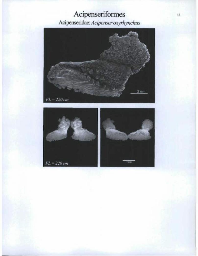
AcipenseriformesAcipenseridae: Acipenser oxyrhynchus

16 AlbuliformesHalosauridae: Halosauropsis macrochir
F'L=72cm^Foiiillli

Albuliforrnes Notacanthidae: Lipogenys gilli

AlbuliformesNotacanthidae: Notacanthus chemnitzi
FL = 87 cm
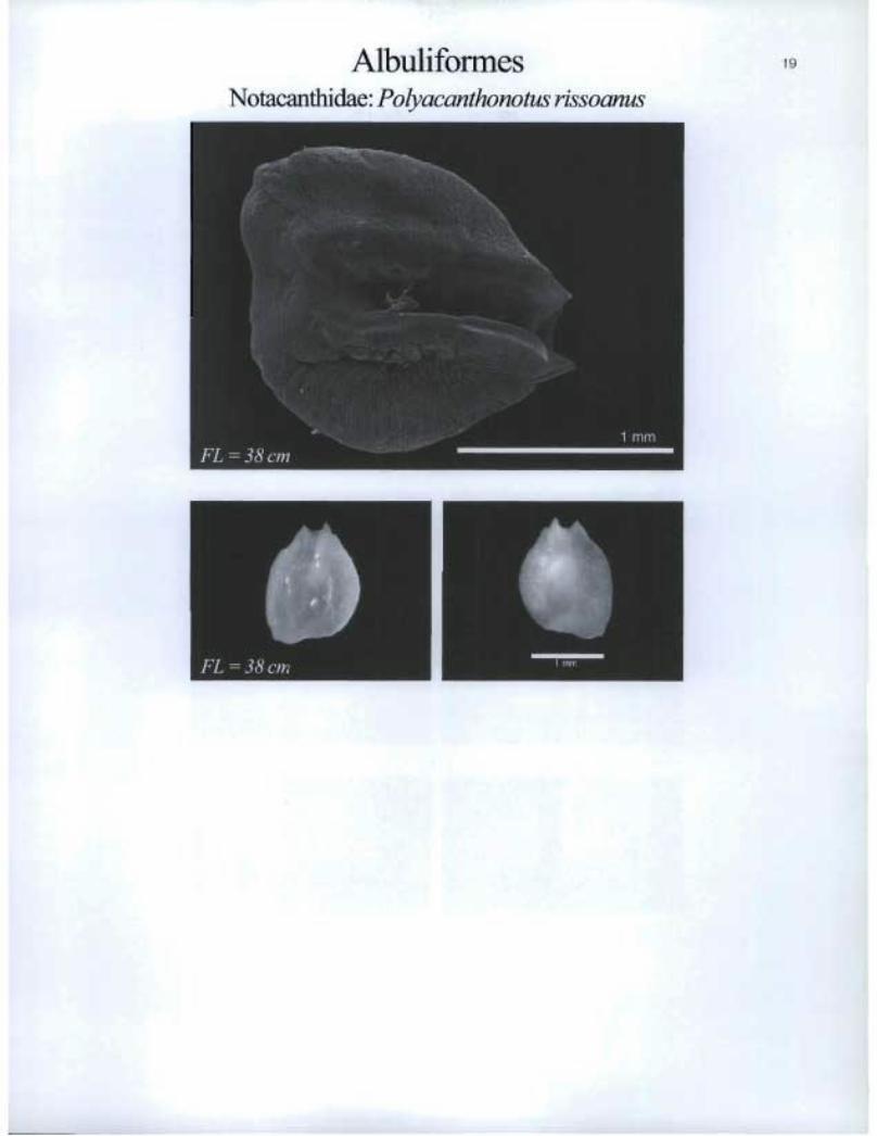
Albulifomies Notacanthidae: Polyacanthonotus rissoanus
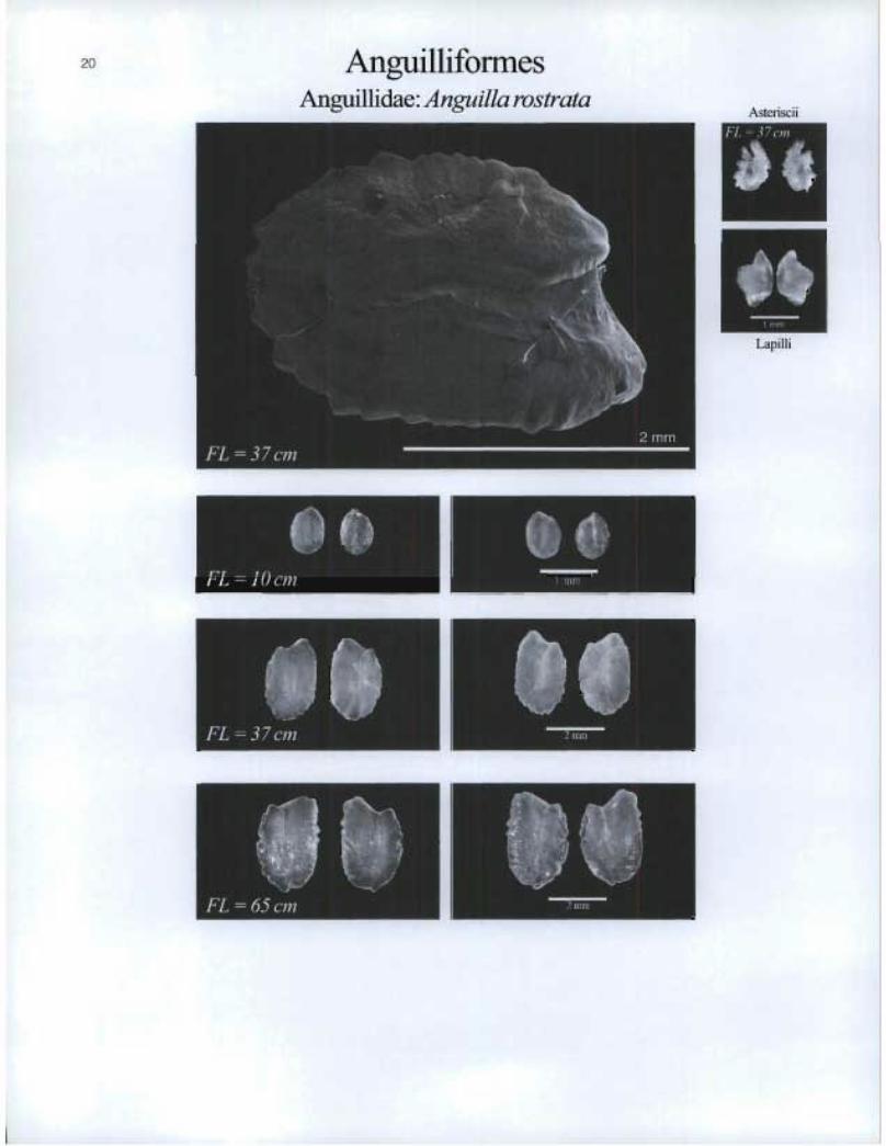
Anguilliforrnes Anguillidae: Anguilla rostrata
20
Astoiscii FL = 37 on e
Lapilli
ØS FL = 10 cm

AnguilliformesDerichthyidae: Derichthys serpentinus
FL =15 cm
4)0
FL =Z6cm
21
Asteriscii& Lapilli
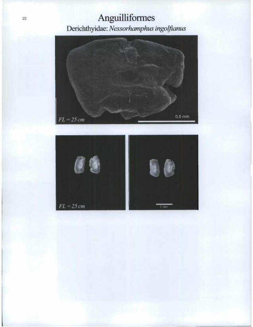
Anguilliforrnes Derichthyidae: Nessorhamphus ingolfianus

Anguilliforrnes Nemichthyidae: Nemichthys scolopaceus

24 AnguilliformesNettastomatidae: Veneficaprocera
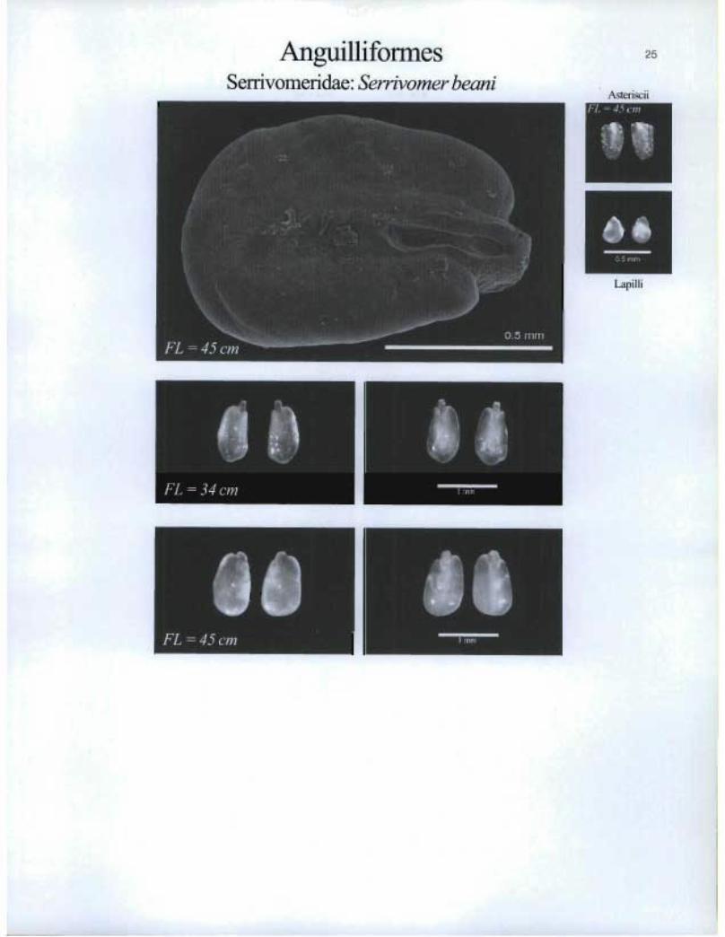
Arigu.illiformesSerrivomeridae: Serrivomer beani
25
Asteriscii!'L =4Scnr
!z®
Lapilli
FL = 45 cm n 1

• V FL = 5 7 cm
Anguilliformes Synaphobranchidae: Ilyophis brunneus

AnguilliformesSynaphobranchidae: Simenchelys parasiticus
Lapilli
sib FL = 16 cm
O. FL =43 cm
I. FL =48 cm
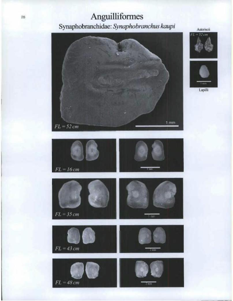
Anguilliformes Synaphobranchidae: Synapho branchus kaupi
Asteriscii
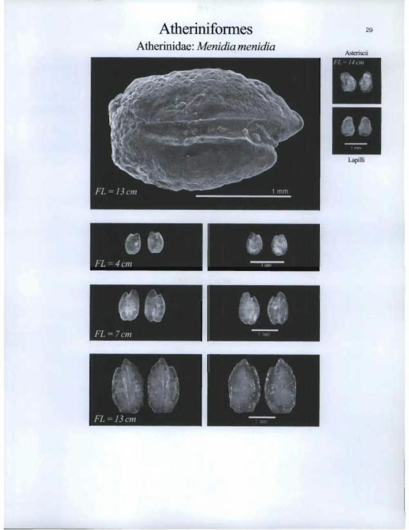
Atherinifonnes Atherinidae: Menidia menidia
29
Asteriscii
intrm
Lapilli
FL = 4 cm
s. ■1•111•■
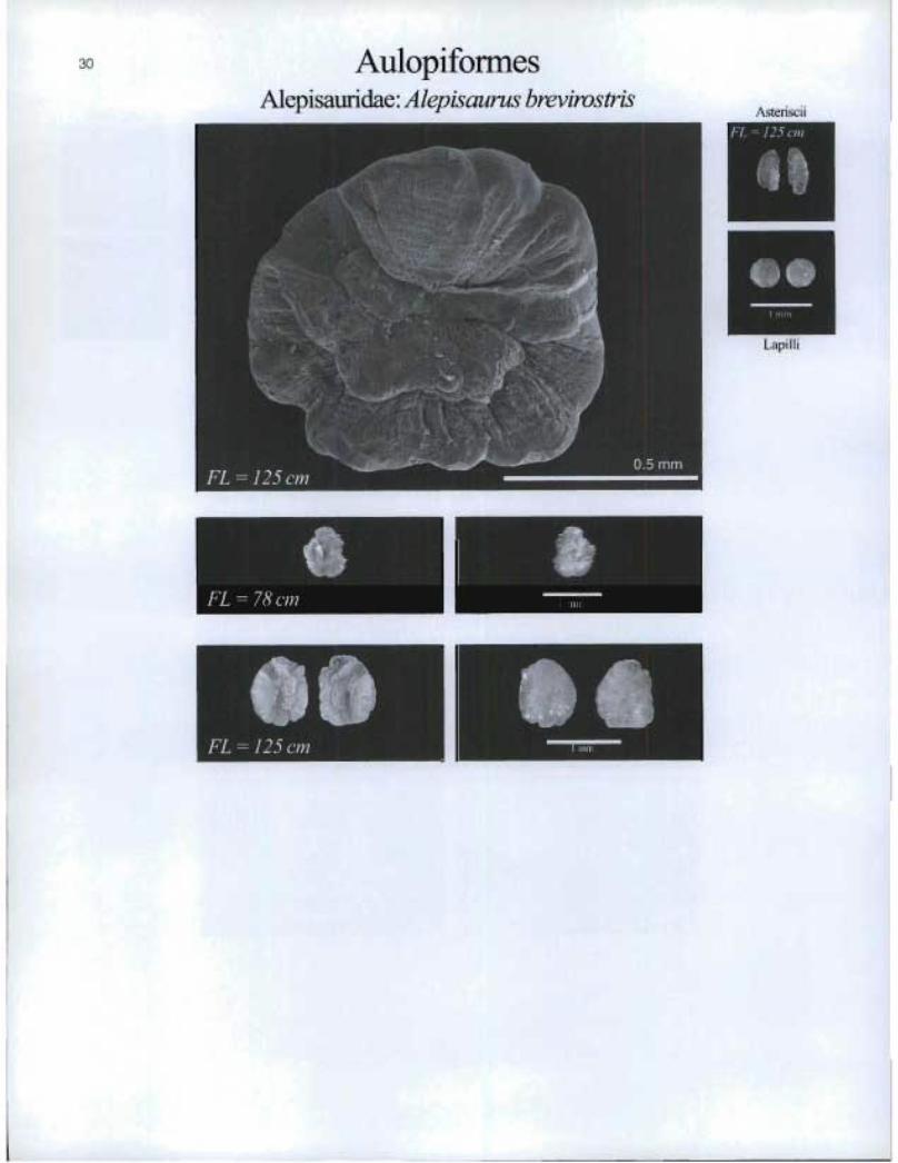
30 AulopiformesAlepisauridae: Alepisaurus brevirostris
Asteriseü
Q1
oeLapilli
FL =125 cm ®

AulopiformesAlepisauridae: Alepisaus ferox
31
Asteriscii
FL - 136 cm
111
Lapilli
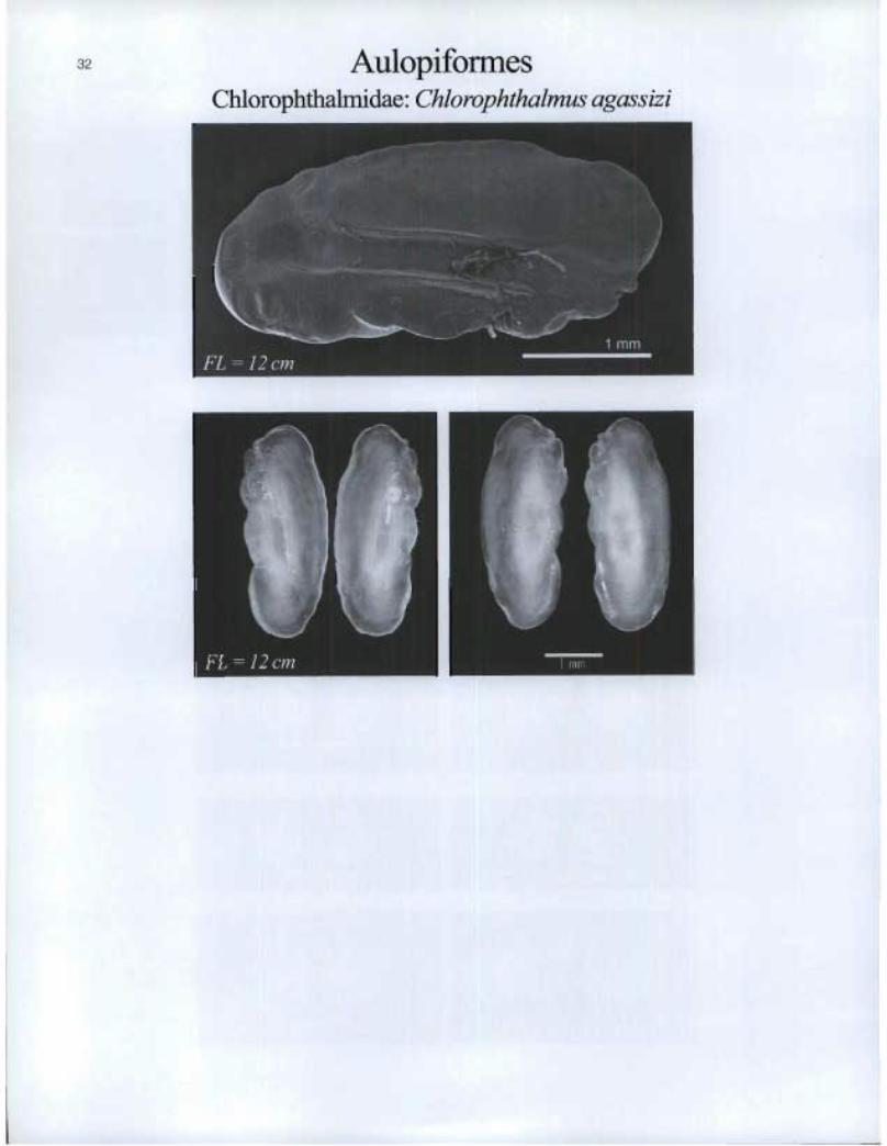
Aulopiforrnes Chlorophthalmidae: Chlorophthalmus agassizi
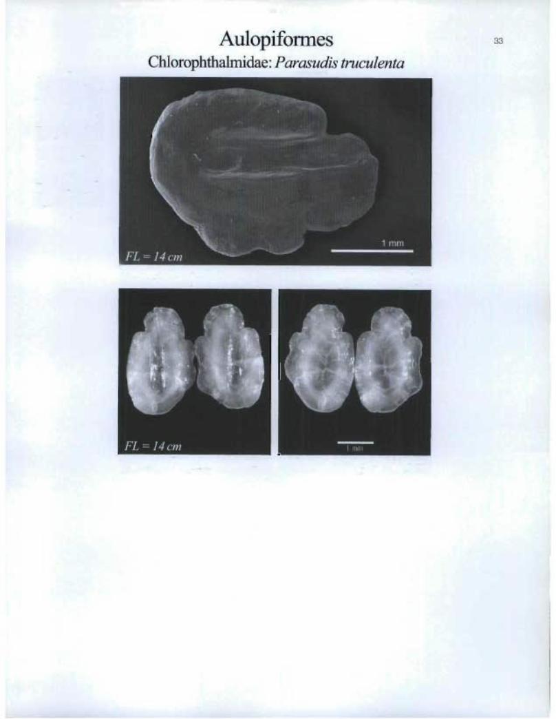
AulopiformesChlorophthalmidae: Parasudis truculenta
00 4b
Lapilli
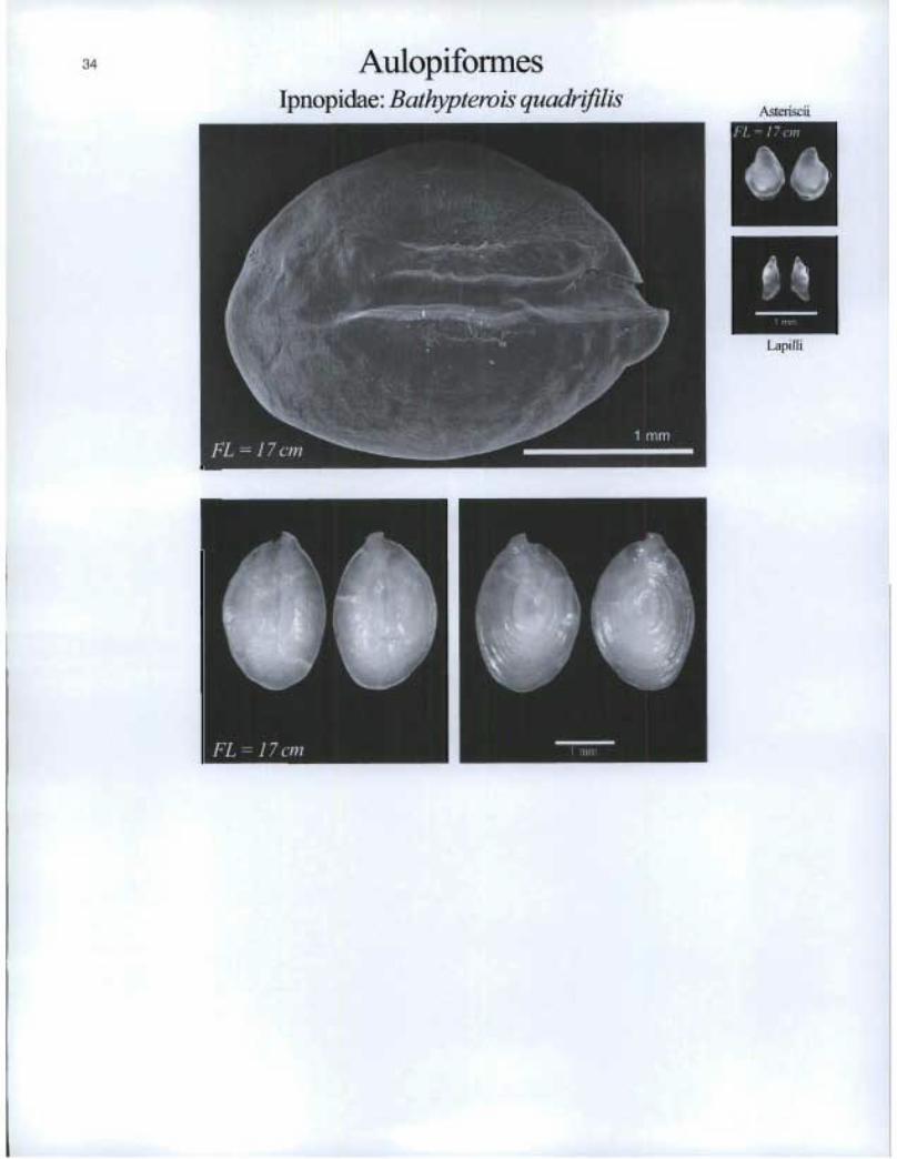
Aulopiforrnes Ipnopidae. Bathypterois quadrifilis
Asteriscii
111> FL= 17 cm
O. MTN

Aulopiformes Paralepididae: Lestidiops affinis

AulopiformesFaralepidida.e: Notolepis rissoi
FL=5cm
FL =9cm
IL®

AulopiformesParalepididae: Paralepis atlantica
37
Asteriscii
FL = !? c/??
14
Lapilli
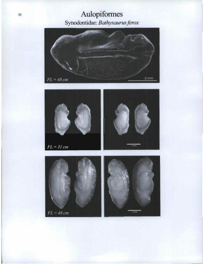
Aulopiformes Synodontidae: Bathysaurus ferox

BelonifonnesScomberesocidae: Scomberesoxsaurus
FL=17cm
FL = 25 cm
39

WIEFEIM
1
[U Lapilli
le it FL 14 cm
Beryciformes Anoplogasteridae. Anoplogaster corn uta
Asteriscii

BeryciformesTrachichthyidae: Hoplostethus atlanticus

WEI13112
se
Lapilli &Asteriscii
Of FL = 13 cm
Beryciforrnes Trachichthyidae: Hoplostethus mediterraneus
1)i FL «in
FL = 18 cm

ClupeiformesClupeidae: A losa aestivalis

VIIMM117111
Lapilli
Clupeiformes Clupeidae. Alosapseudoharengus
Asteriscii

Clupeiformes 45Clupeidae: A losa sapidissima
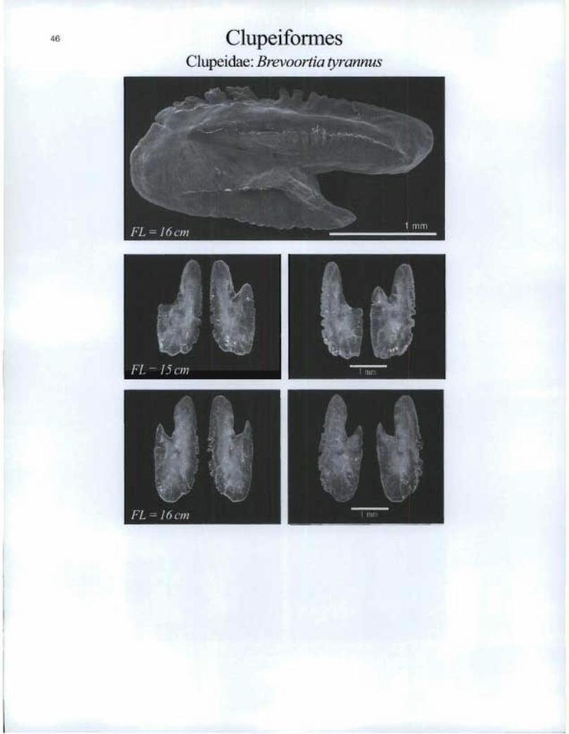
Clupeiformes Clupeidae: Brevoortia tyrannus

ClupeiformesClupeidae: Clupea harengus harengus
FL=8cm
FL =26cm
q
T,
®
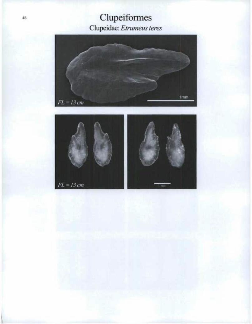
Clupeiformes Clupeidae: Etrumeus teres

FL = 8 cm
I.
Lapilli
Cyprinodontiforrnes Fundulidae: Fundulus diaphanus
49
Aste i kc t i

Cyprinodontifomies Fundulidae: Fundulus heteroclitus

FL 12 cm
Gadiformes Gadidae: Boreogadus saida
il FL = 7 cm

GadiformesGadidae: Brosme brosme
FL=Scïn
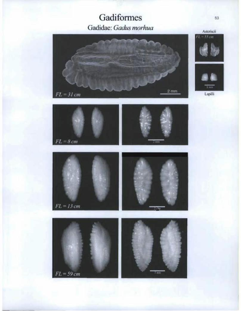
Gadiformes Gadidae: Gadus morhua
53
Asterisci i
»MIMI
U:71 PIER
Lapilli
11, 5 mn

54 GadiformesGadidae: Gadus ogac
FL=13cm
I
te FL =23cm
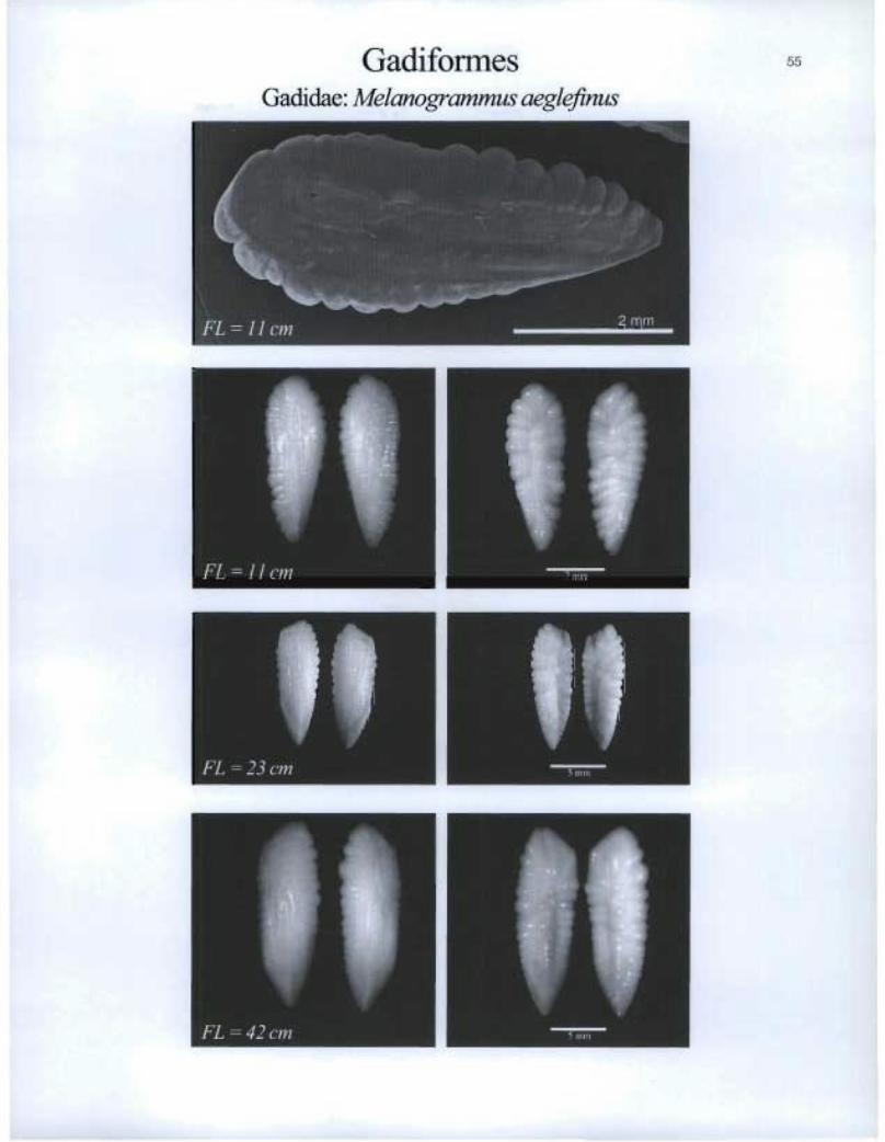
Gadiformes Gadidae. Melanogrammus aeglefinus

GadiformesGadi dae :Microgadus to m co d
Z

Gadiformes Gadidae: Micromesistius poutassou
57
Asteriscii
Lapilli
MI=
It FL = 18 cm ETU

GadifonnesGadida.e: Molva dypterygia
FL =28cm

ii FL = 44 cm
I FL = 70 cm
II RIM
1 IiIMIE
Gadiformes Gadidae: Molva molva
111 FL = 86 cm
KIM1
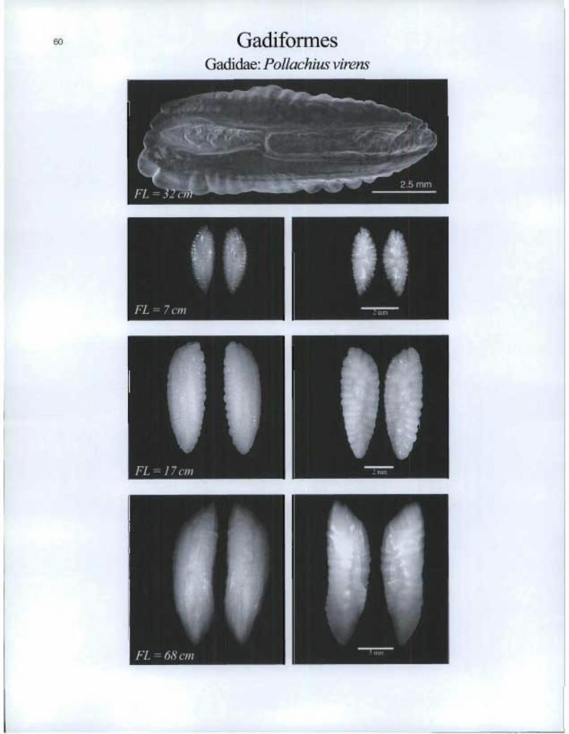
Gadiformes Gadidae: Pollachius virens
ii FL = 17 cm

GadiformesMacrouridae: Chalinura brevibarbis
11 EFL=12cm
FL=3Ocm
Macrouridae: Coryphaenoides guentheri
Z IE
61
I i„riFL = 44 cm

Gadiformes Macrouridae: Coryphaenoides rupestris
• ECM FL =25 cm

Gadiformes Macrouridae: Lionurus carapinus
63
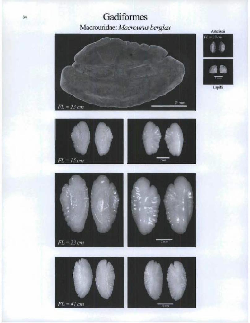
GadiformesMacrourid ae.llilacrourus berglcax
FL=.15cm
FL=23cm
zmm
Asteriscii
2Lapilli
FL = 41 cm , 5111111
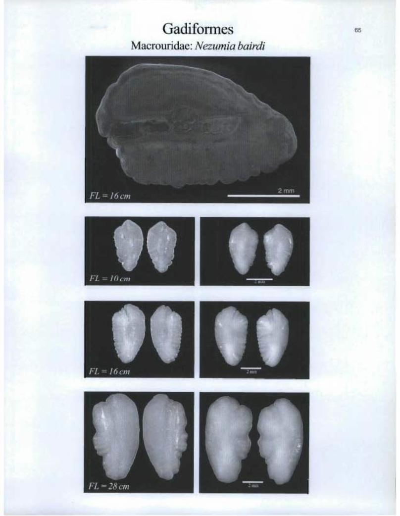
GadiformesMacrouridae: Nezumia bairdi
FL=1(1cm
FL=1tScm
I FL = 28 cm
zmn
65

I. FL =43 cm
66 Gadiformes Macrouridae: Nezumia sclerorhynchus
FL =28 cm
Macrouridae: Trachyrhynchus murrayi

Gadiformes Melanonidae: Melanonus zugmayeri
67
Asteriscii
WW1111111
Lapilii
be FL = 10 cm

68 GadiformesMerlucciidae: Merluccius albidus

Gadiformes Meducciidae: Merluccius bilinearis
69
Asteriscii
FL = 18 cm
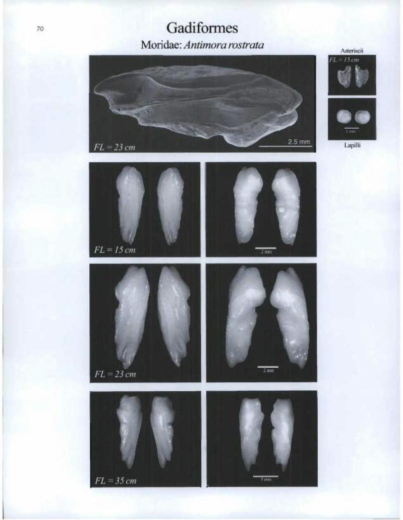
Gadiformes Moridae: Antimora rostrata
70
Asteriscii
Is gib Lapilli
FL cin

GadiformesMoridae: Brosmiculus imberbis
Side View , 2nuA Side ViewFL=15cm
71
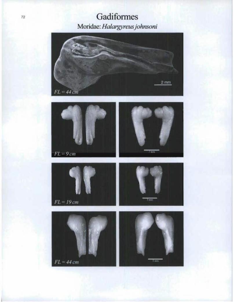
GadiformesMoridae: Halargyreus johnsoni
FL=19cm
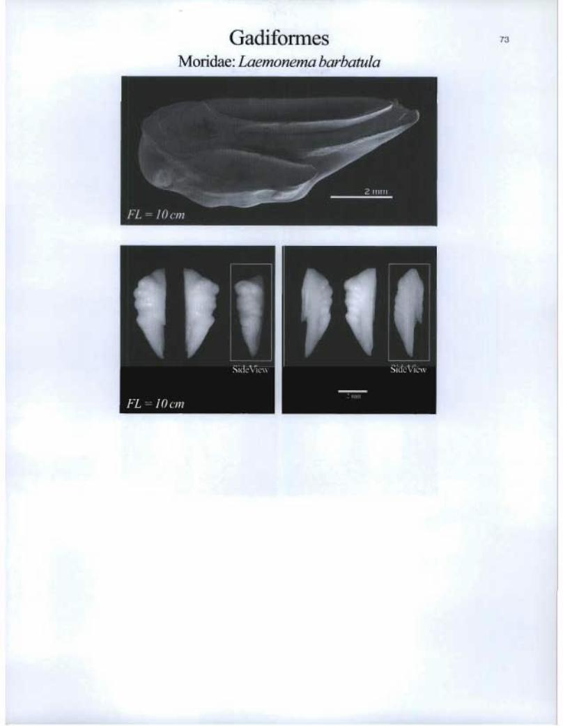
Gadiformes Moridae: Laemonema barbatula
Mr_MM
r
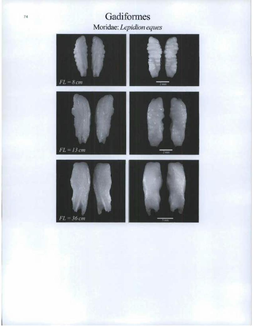
Gadi forme Moridae: Lepidion eques
FL =8cm tt
Ern

GadiformesPhycidae: Enchelyopus cimbrius

Gadiformes Phycidae: Gaidropsarus argentatus
ft FL = 15 cm
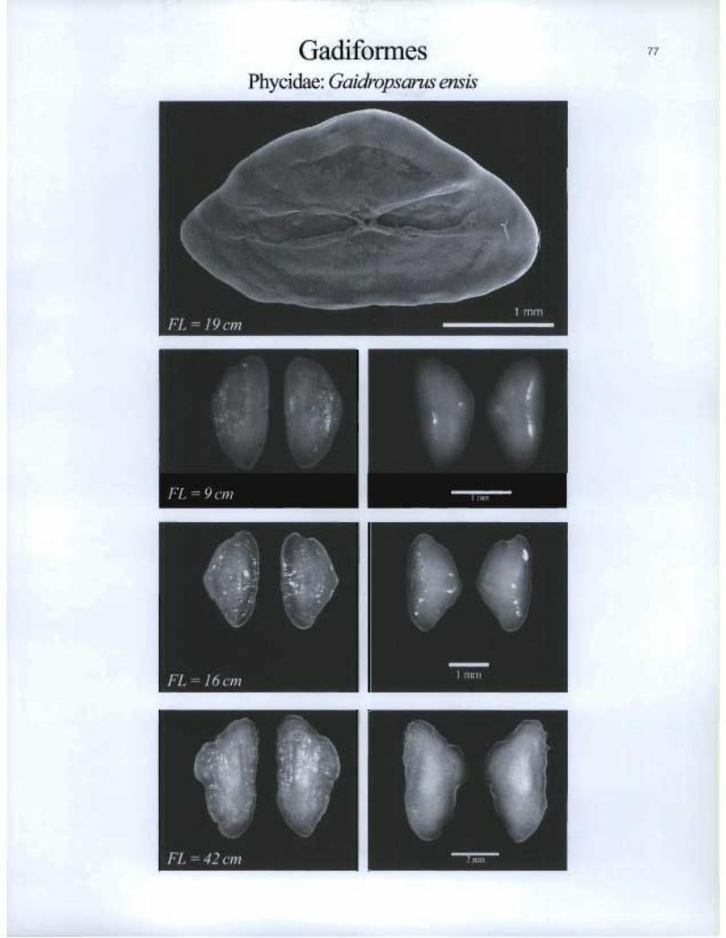
Gadiformes Phycidae: Gaidropsarus ensis

GadiformesPhycidae: Urophycis chesteri
FL=9cm
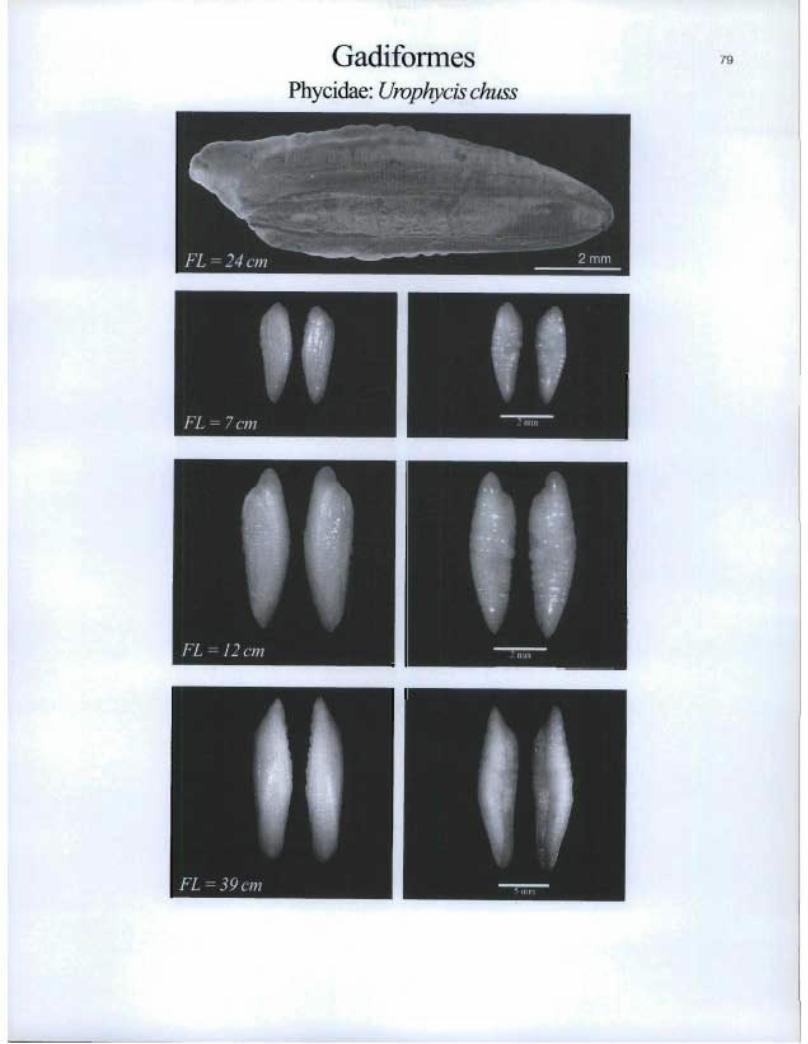
GadiformesPhycidae: Urophycis chuss
FL=12cm
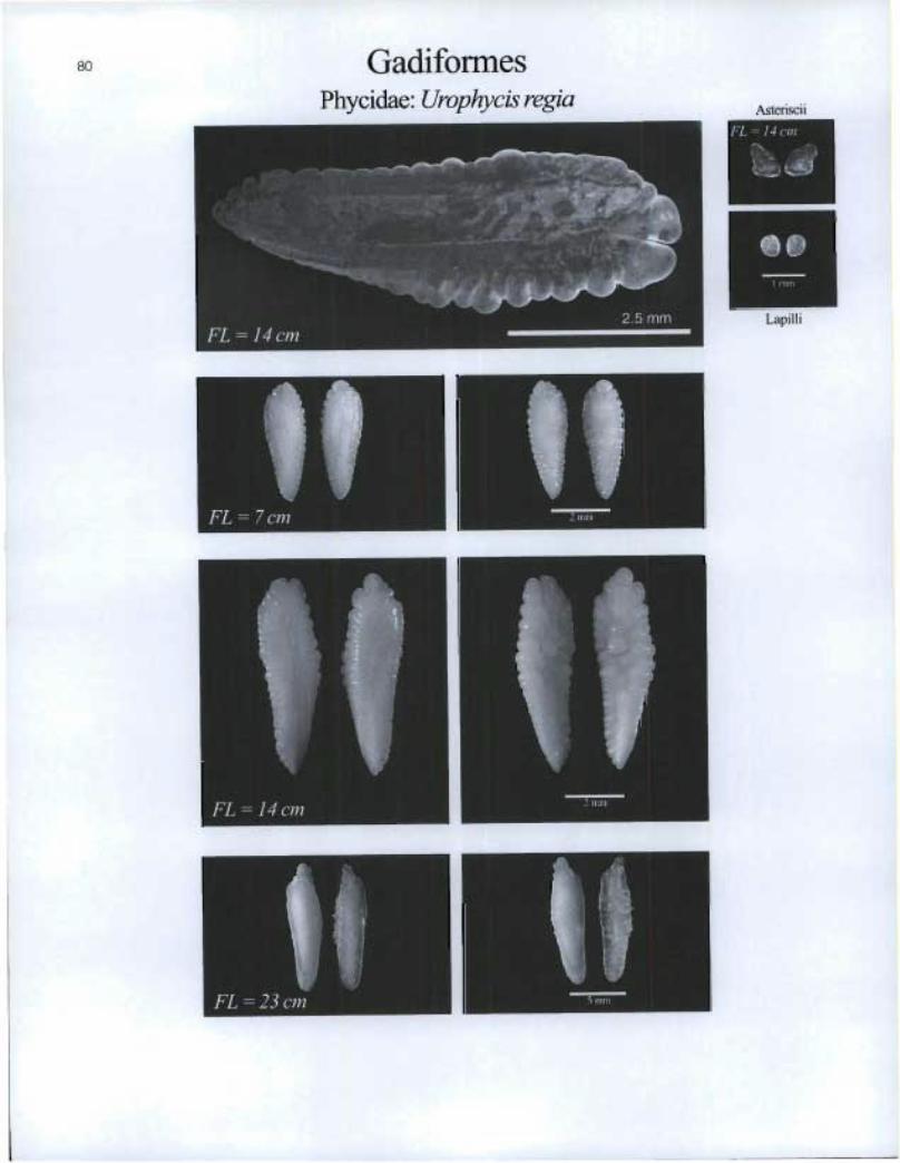
80 Gadiformes Phycidae: Urophycis regia Asteriscü
t,
FL = 7 cm
FL = 14 cm
11 ; Min
Lapilli
-, n1=111
t El=
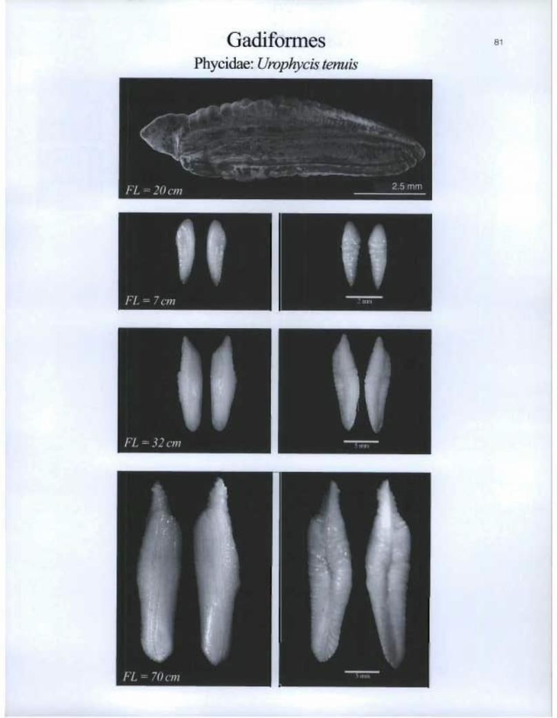
Gadiformes Phycidae: Urophycis tenuis
I 1 FL = 7 cm
ii FL = 32 cm

G.a.sterosteiformesGasterosteidae: Apeltes quadracus
Asteriscii
[r,
a]
®
Lapilli
riu IMFL=Scm 1 linfil

VIM=
0 •
Asteriscii & Lapilli
Gasterosteiformes Gasterosteidae: Gasterosteus aculeatus
83
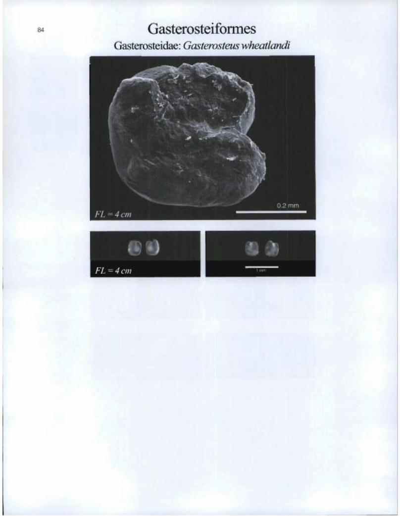
Gasterosteiformes Gasterosteidae: Gasterosteus wheatiandi

GasterosteiformesGasterosteida.e. Pungitiuspungitius
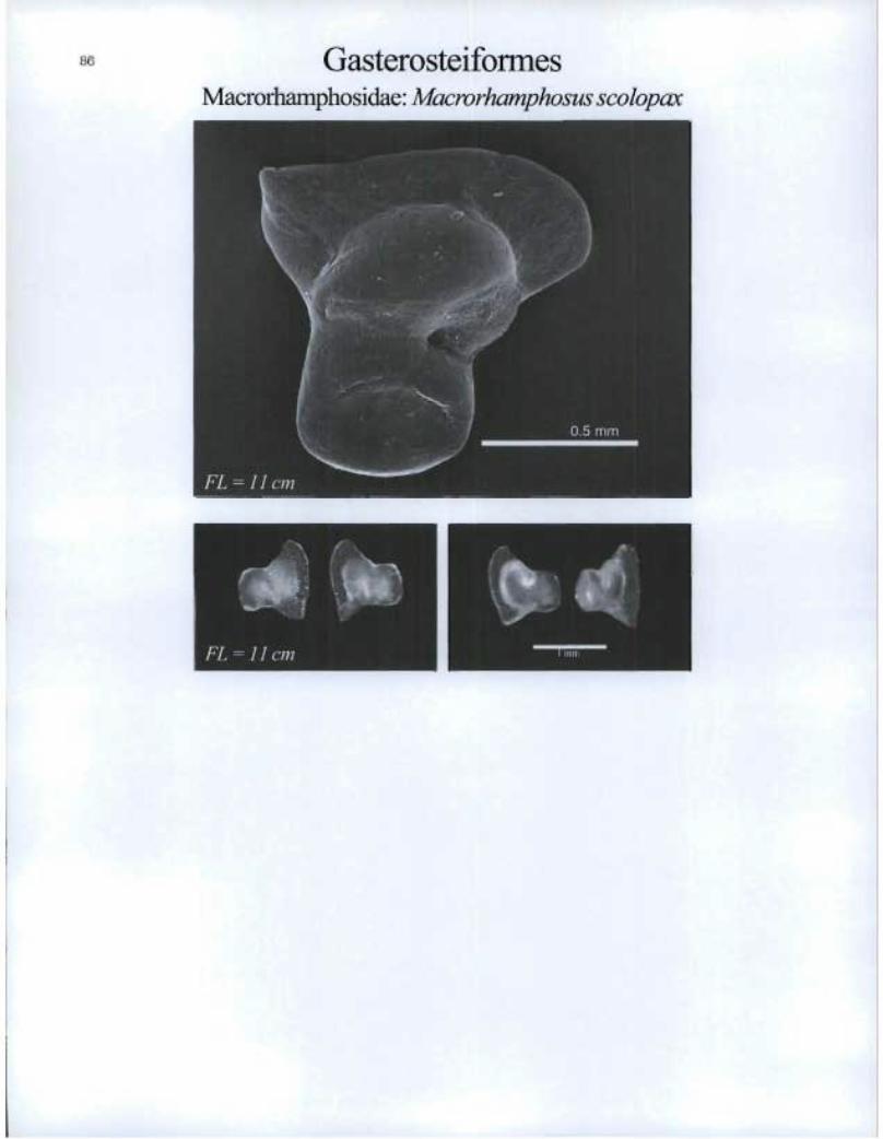
Gasterosteiformes Macrorhamphosidae: Macrorhamphosus scolopax
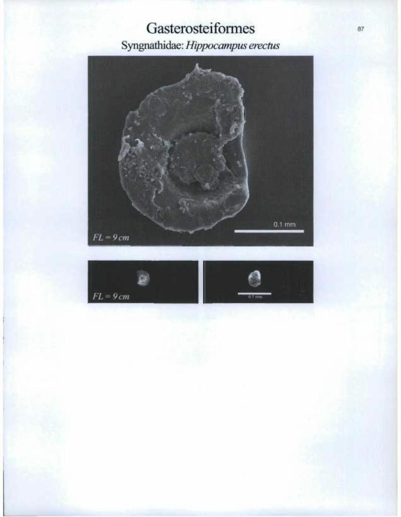
GasterosteiformesSyngnathidae: Hippocampus erectus
:,16 .1 mm
87

GasterosteiformesSyngnathidae: Syngnathusfuscus
®
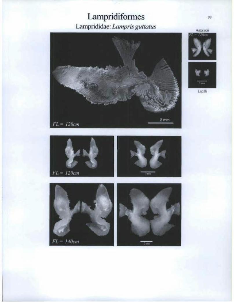
Lampridiformes Lamprididae: Lampris guttatus
89
Asteriscii
ITM1
1 iji I I i
to4 FL = 120cm

Lophiifomies Ceratiidae: Ceratias holboelli

Lophiifomies Ceratiidae: Cryptopsaras couesi

LophiiformesLophiidae: Lophius americanus 1.
mLapilli
FL=11cm
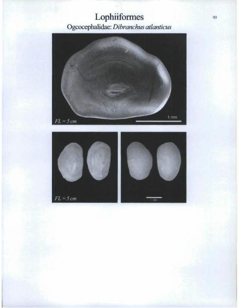
LophiiformesOgcocephalidae: Dibranchus atlanticus
FL =5cm

411 lenfiT
Lapilli
Lophiiformes Oneirodidae: Oneirodes sp.
Asteriscii

MugiliformesMugilidae: Mugil curema
FL=12cm

Myctophiformes Myctophidae: Benthosema glaciale

Myctophiformes Myctophidae: Benthosema suborbitale
Myctophidae: Bolinichthys photothorax

FL = 8 cm
Myetophiformes Myctophidae: Ceratoscopelus maderensis
Myetophidae: Ceratoscopelus warmingii

Myctophiformes 99Myctophidae: Diaphus dumerilii

lr3IyctophiformesMyctophidae: Diaphus effulgens
FL=12cm2 mm
LI I

FL =8cm
Myctophiformes Myctophidae: Diaphus metopoclampus
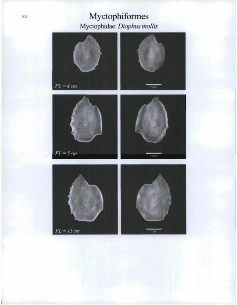
MyctophiformesMyctophidae: Diaphus mollis
FL=4cm
FL=15cm

Myetophiformes Myctophidae: Diaphus perspicillatus
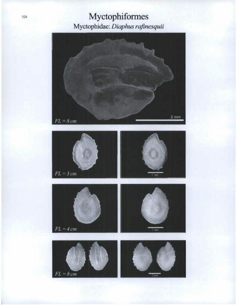
Myctophiforrnes myctophidae: Diaphus rafinesquii
411

O. é FL = 6 cm MIMI
O FL = 2 cm
Myctophiforrnes Myctophidae: Diaphus termophilus
105
Myctophidae: Diogenichthys atlanticus

MyctophiformesMyetophida.e: Electrona risso
FL=3cm
FL=5cm
FL=5cm

FL = 5 cm
MyctophiformeS Myctophidae: Gonichthys cocco
Myctophidae: Hygophum benoiti

108 MyctophiformcsMyctophidae: Hygophum hygomii
Myctophidae: Lampadena luminosa
FL=9cm
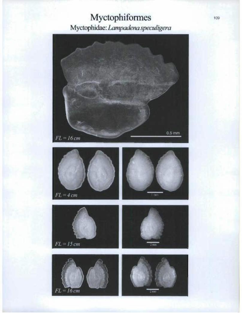
MyctophifonnesMyctophidae: Lampadena speculigera
FL =4cm
FL=1Scm n 2 nuil
emo •
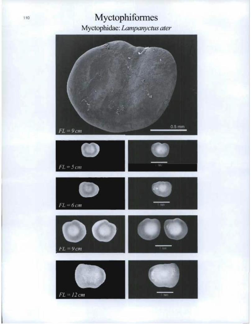
MyCtOphifOrMeS Myctophidae: Lampanyetus ater
OP FL = 9 cm
OP
FL = 1 2 cm
EMI
O FL = 8 cm
to FL = 9 cm • •
O FL = 14 cm
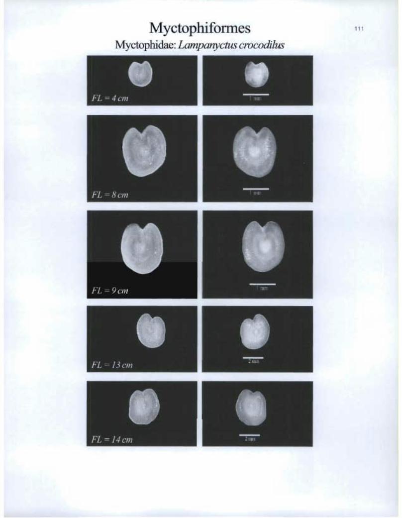
Myctophiformes Myctophidae: Lampanyctus crocodilus
FL = 13 cm

112 MyctophiformesMyctophida.e: Lampanyctusfestivus
FL=7cm
®
I^J
Myctophidae: Lampanyctus intracarius
FL=Scm
FL=7cm
FL=16cm
E®
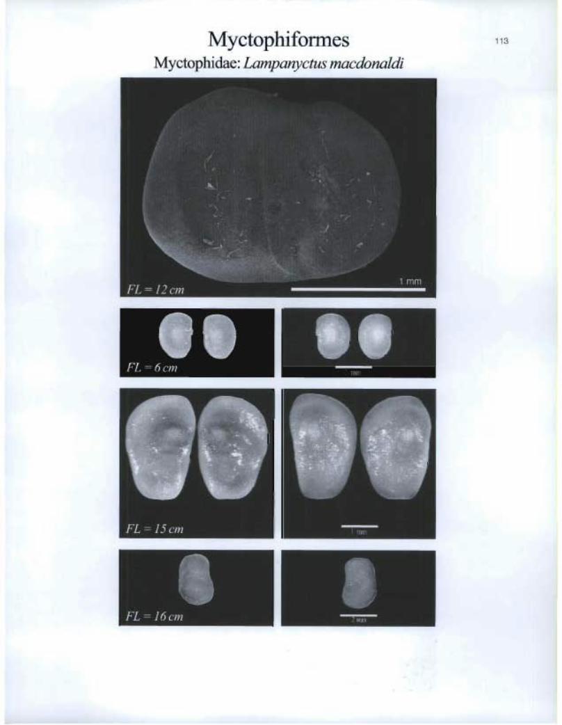
Myctophiformes Myctophidae: Lampanyctus macdonaldi
I. FL = 6 cm 111113
Effi
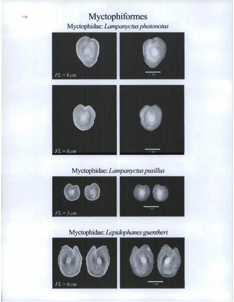
• 114 Myctophiformes Myctophidae: Lampanyctus photonotus
Myctophidae: Lampanyctus pusillus
Myctophidae: Lepidophanes guentheri
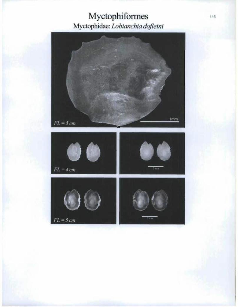
MyctophiformesMyctophidae: Lobianchia dofleini
rnFL = 4 cm 1 '111111
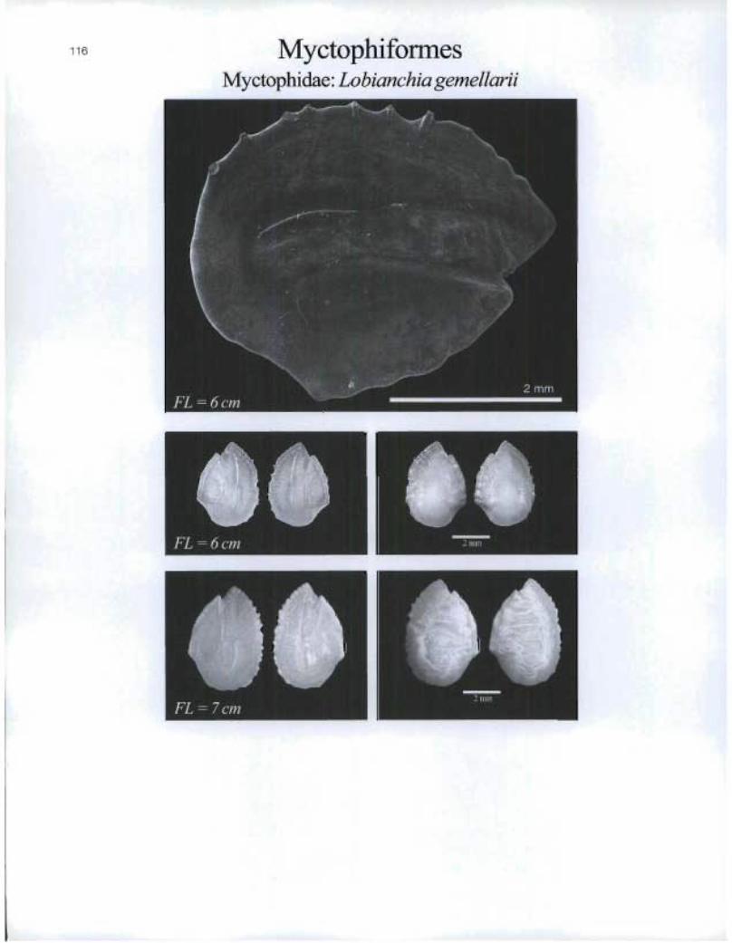
MyctophiformesMyctophidae: Lobianchia gemellarii
FL=6cm
FL=7cm

Myctophiformes Myctophidae: Myctophum asperum

Fe
Myctophiformes Myctophidae: Myctop hum punctatum
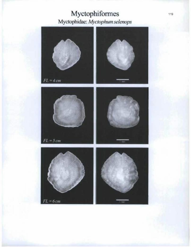
MyctophiformesMyctophidae: Myetophum selenops
FL=4cm
FL=5cm
FL=6cm
MTN
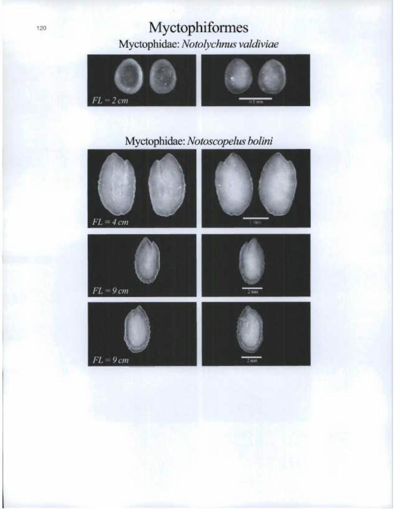
120 Myctophiformes Myctophidae: Notolychnus valdiviae
Myctophidae: Notoscopelus bohni

Myctophiforrnes Myctophidae: Notoscopelus caudispinosus
Myctophidae: Notoscopelus elongatus kroeyerii • FL = 8 cm
FL= cm
• ôt1

122 MyctophiformesMyctophida.e: Notoscopelus resplendens
11FL=9cm
Myctophidae: Protomyctophum arcticum
E
FL =4cm

e KIIIVI
I FL — 10 cm
I WM
• FL — 11 cm
Myctophiforrnes Myctophidae: Symbolophorus veranyi

124 MyctophiformesMyctophidae: Taaningichthys bathyphilus
FL =5cm
nFL=5cm ®
Myctophidae: Taaningichthys minirnus
FL=5cm M
®

Ophidiiformes Ophidiidae: Dicrolene intronigra
910 et FrIMI
FL =19cm
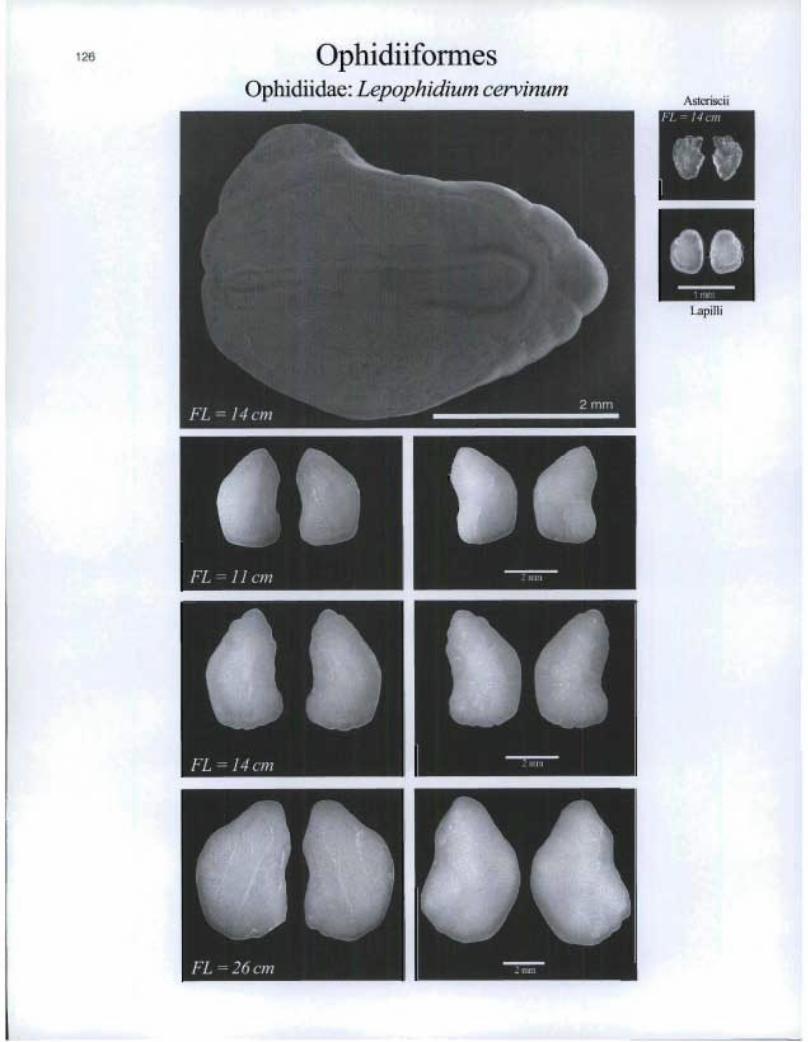
OphidiiformesOphidiidae: Lepophidium cervinum
FL=11cm
FL=14cm n ^
MMM
AsterisciiFL ° 14 on
Lapilli
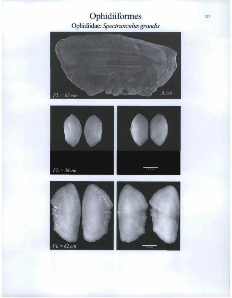
Ophidiiforrnes 127
Ophidiidae. Spectrunculus grandis
FL =62 cm
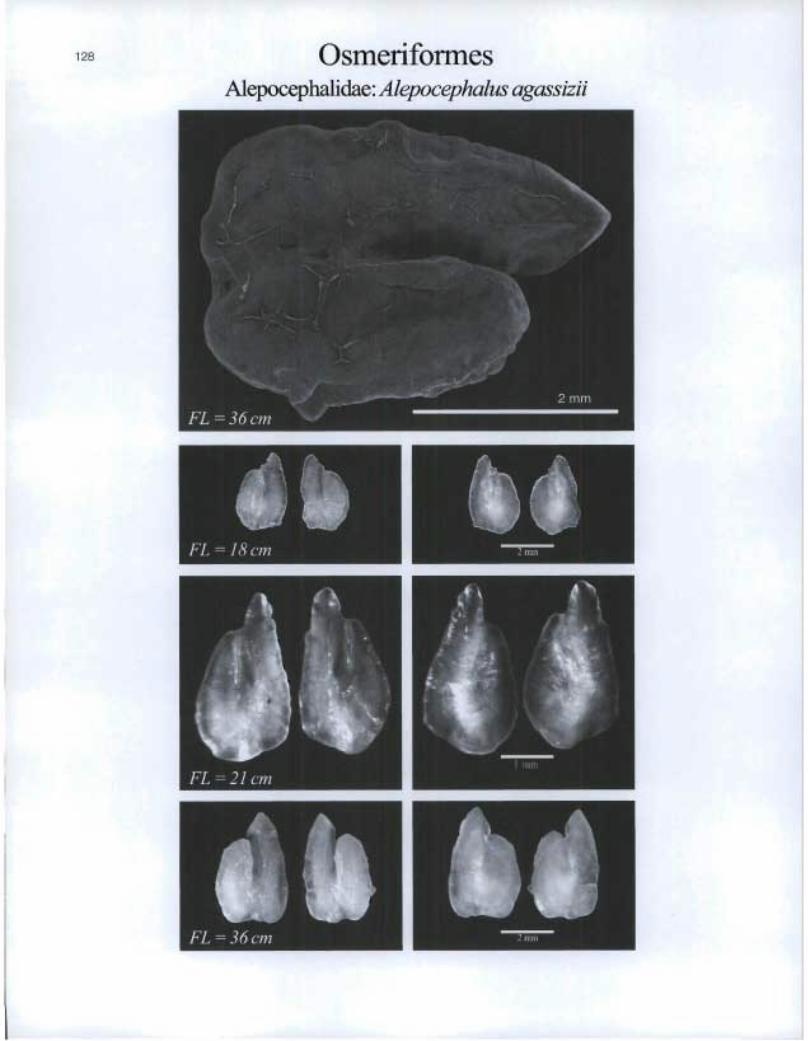
Osmeriformes Alepocephalidae: Alepocephalus agassizii
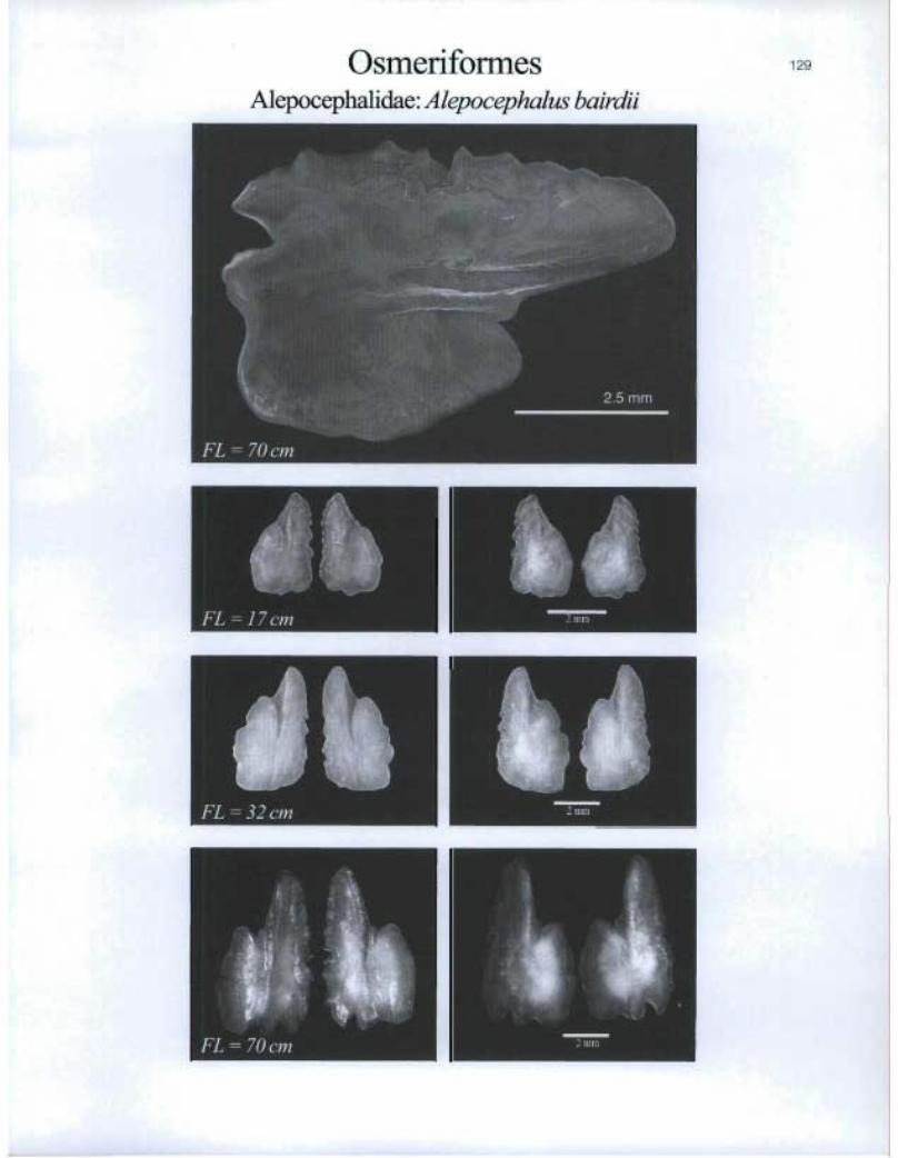
OsmeriformesAlepocephalidae: Alepocephalus bairdii
FL-17cm ®
®

11> FL = 36 cm
Osmeriformes Alepocephalidae: Baj acalifornia megalops

Osmeriformes 131
Alepocephalidae: Narcetes stomias

osmeriformes Alepocephalidae. Rouleina attrita
132
MIME
ru O.
Asteriscii & Lapilli
de FL = 25 cm
FL = 44 cm

Osmeriforrnes Alepocephalidae: Xenodermichthys copei
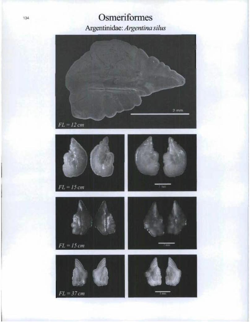
©smeriformesArgentinidae: Argentina silus

osmeriformes Bathylagidae: Bathylagus euryops
135
AsieriF;cii
11
elo 1 mm
Lapilli
FL = 10 cm

OsmeriformesOsmeridae: Mallotus villosus
AsterisciiFL = 17 cm
Lapilli
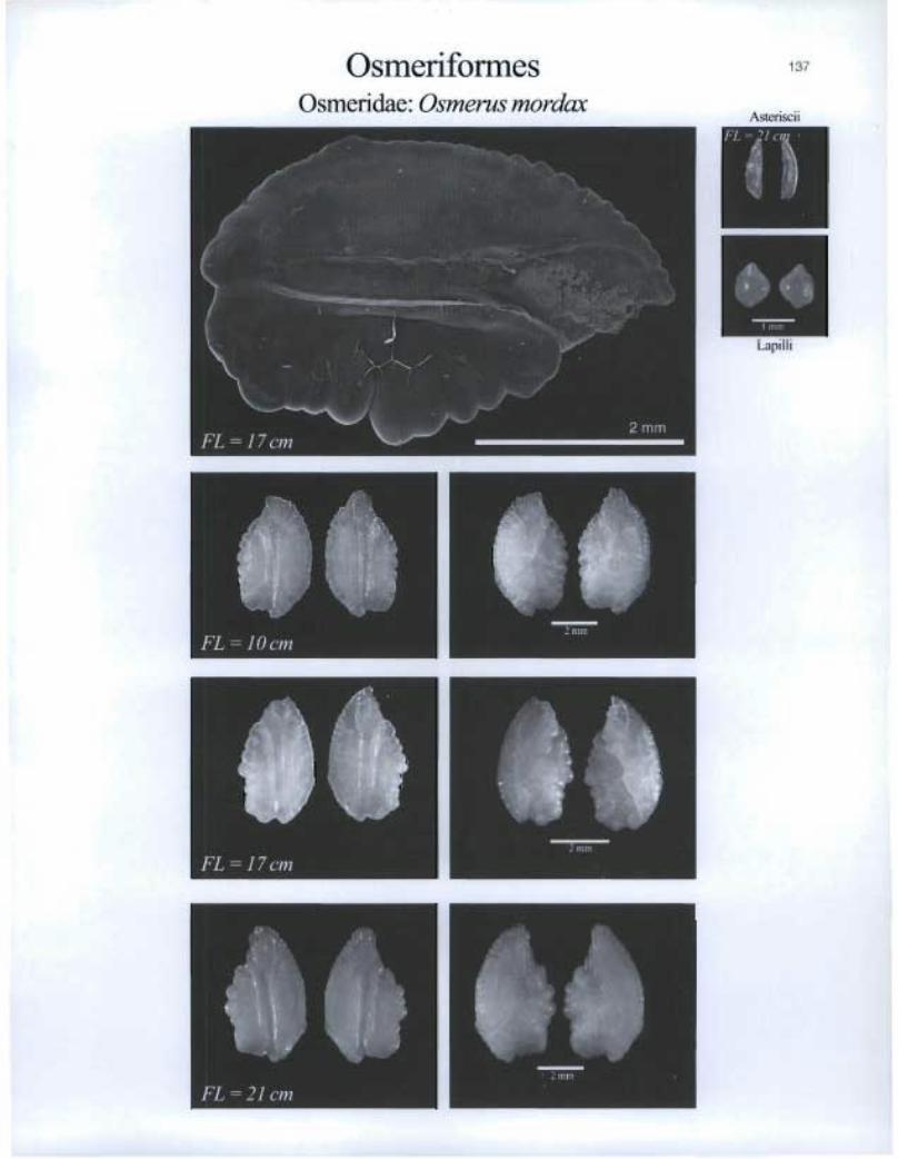
Osmeriformes Osmeridae: Osmerus mordax
137
Asteriscii
bFL — 'I cl. •
r $0.
Lapilli
FL 21 cm
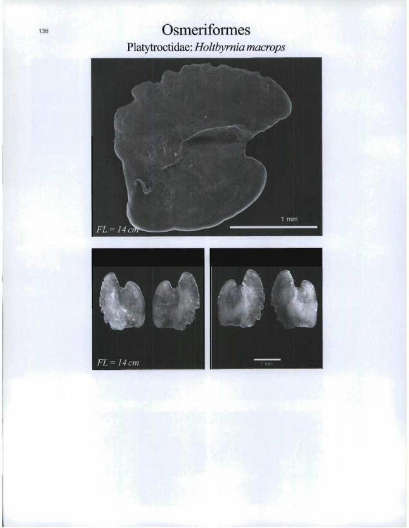
OsmeriformesPlatytroctidae: Holtbymia macrops

Perciformes Acropomatidae: Howella sherborni

140 Perciformes Acropomatidae: Polyprion americanus

PerciformesAcropomatidae: Synagrops bellus
FL = 7 cm n 2 1111tI
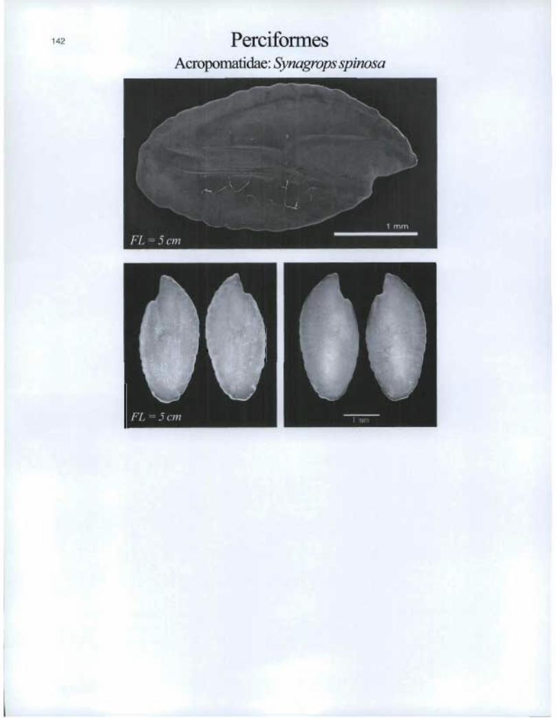
Perciformes Acropomatidae: Synagrops spinosa
0 1 IRV
FL = 6 cm
FL = 1 6 cm
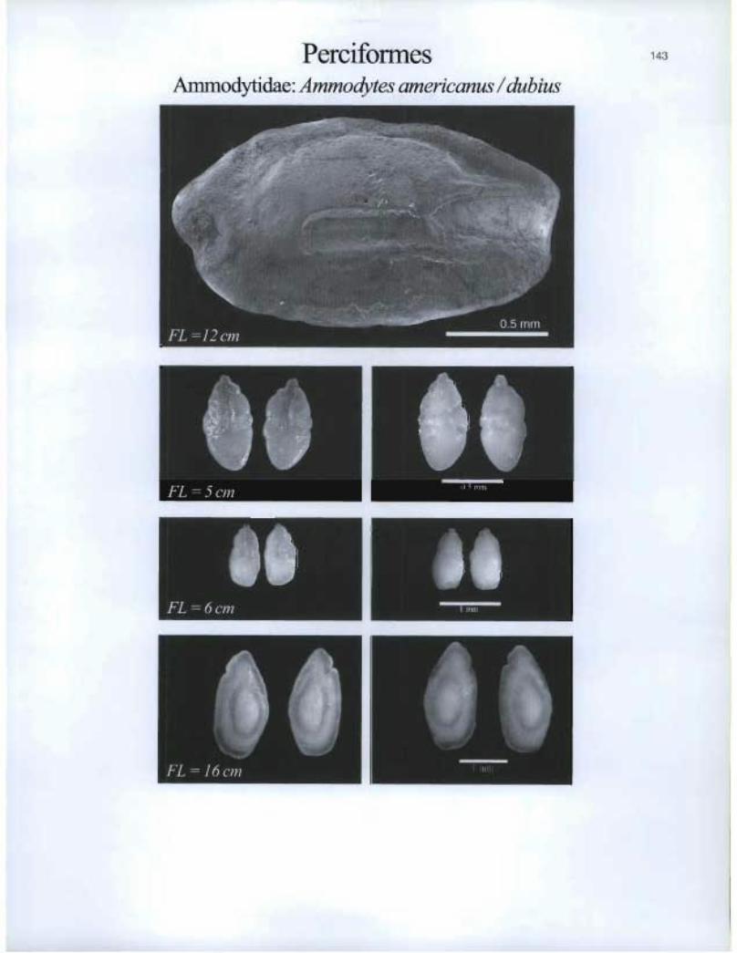
Perciformes Ammodytidae: Ammodytes americanus / dubius
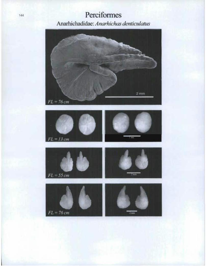
PerciformesAnarhichadidae: Anarhichas denticulatus
rilFL = 55 cm
44
N
64FL = 76 cm n 2 nvn
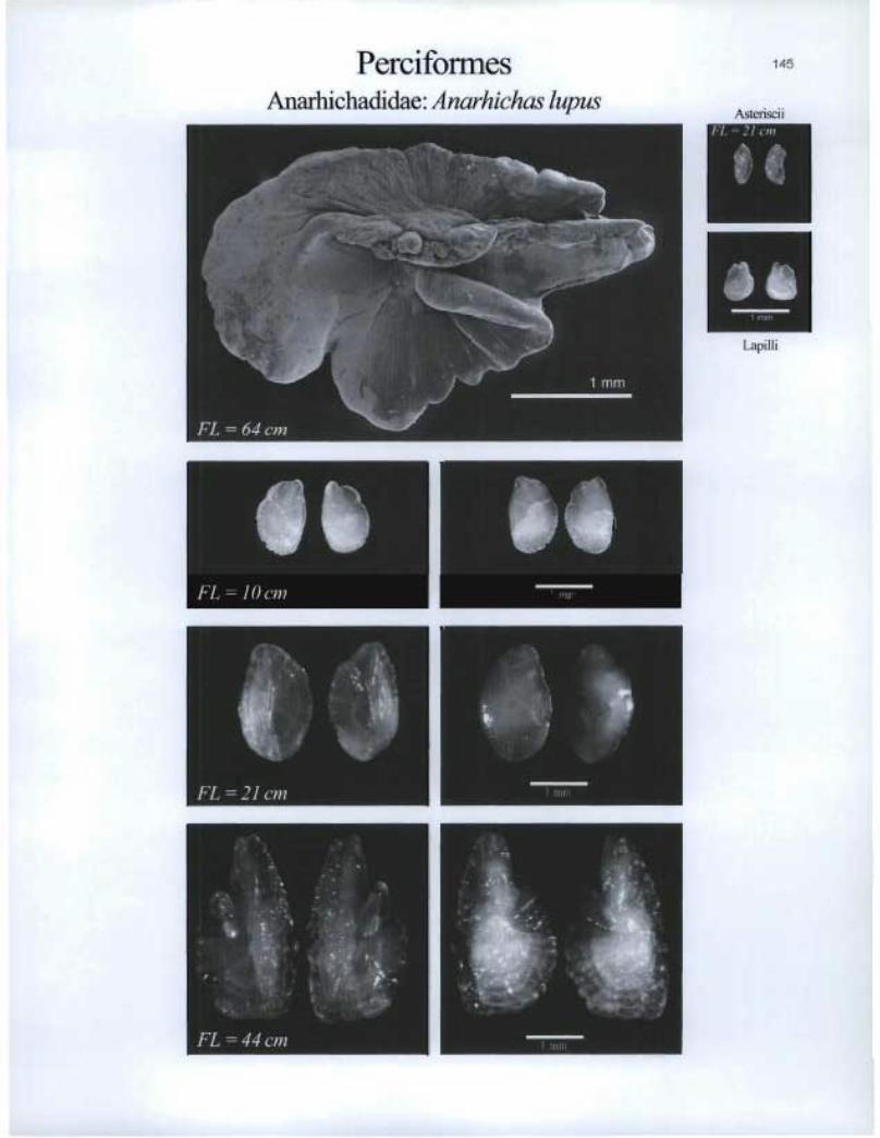
Perciformes Anarhichadidae: Anarhichas lupus
145
Asteriscii
Lapilli
FL = 10 cm

Perciformes Anarhichadidae: Anarhichas minor
FL 31 on _
6110 FL = 11 cm

PerciformesBra,midac: Brama brama
147
,4STe1'ISCI I
FL,=h¢cm
AtLapilli
Brarnidae: Taractichth^vs longïpïnnis

PerciformesCallionymidae: Callionymus agassizi
FL=1Rcm
S
Asteriscii& Lapilli

Carangidae: Caranx hippos Asteriscii
4111>41 GIZE
Perciformes Carangidae: Cararzx crysos
149
I I i
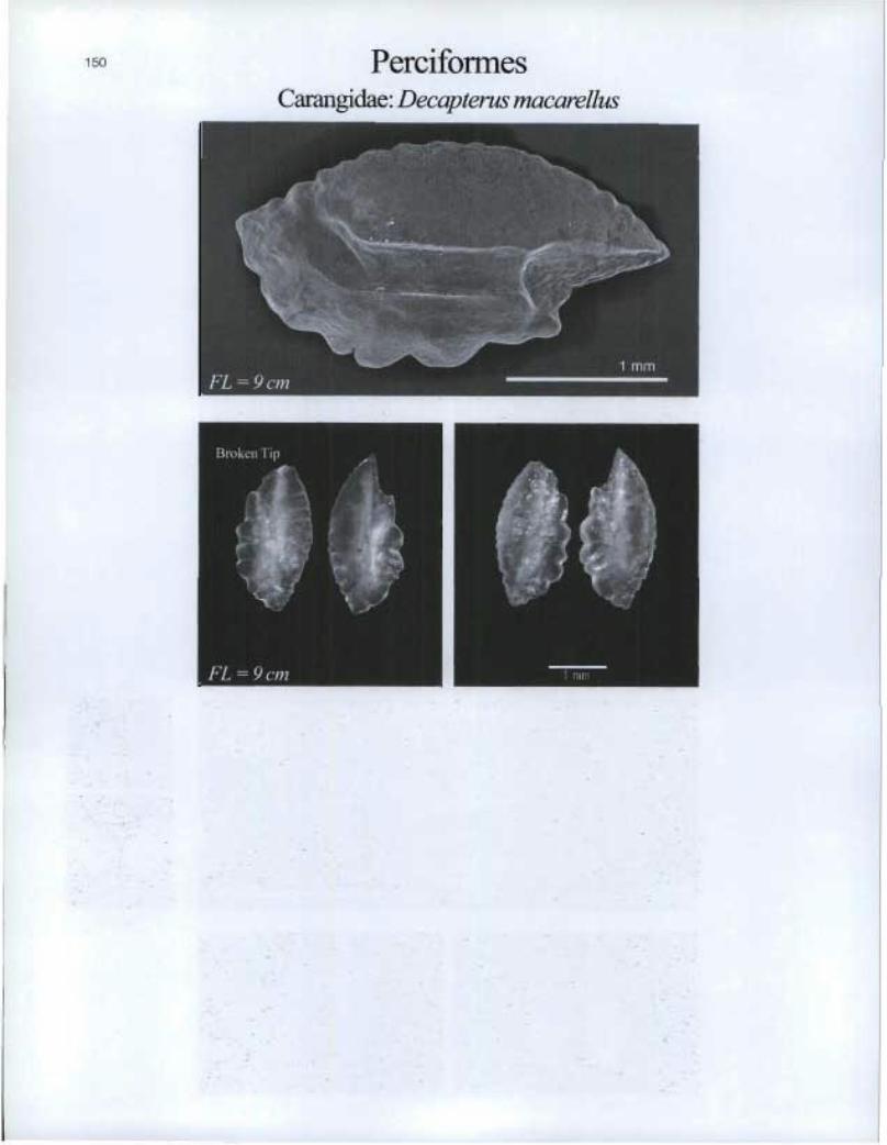
Perciformes Carangidae: Decapterus macarellus

PerciformesCarangidae: Selene setapinnis
...*,^^,^
®
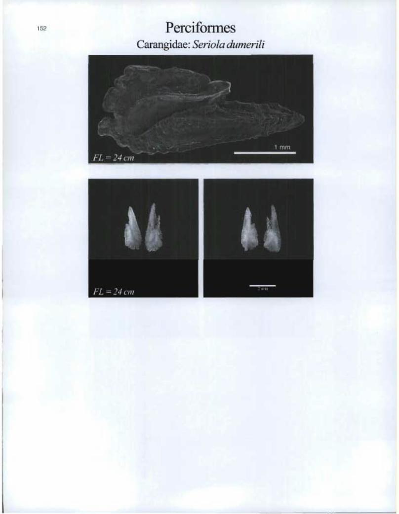
Perciformes Carangidae: Seriola dumerili

Perciformes Carangidae: Seriola zonata
153
Lapilli
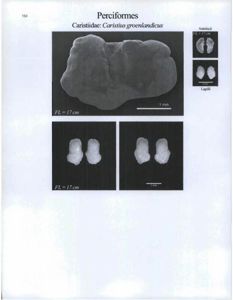
154 PerciformesCaristiidae: Caristius groenlandicus
%steriscii!'L = 17 crn
aJ
®
Lapilli
11
FL=17cm
El

Perciformes 155
Centrolophidae: Hyperoglypheperciformis
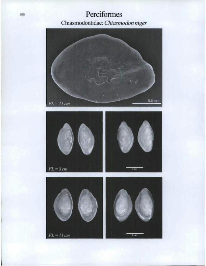
Perciformes Chiasmodontidae: Chiasmodon niger
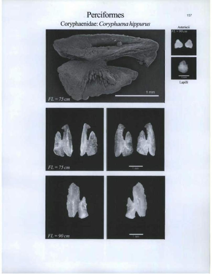
Perciformes Coryphaenidae: Coryphaena hippurus
157
Asteriscii DIIIERBBE
dba
eb apilli
tUt FL = 75 cm
t% FL = 90 cm

PerciformesCryptacanthodidae: Cryptacanthodes maculatus
FL=8cm
FL=19cm

Perciformes Echeneidae: Echeneis naucrates
159
Asteriscii
AMR
[api J Ii

Perciformes Epigonidae: Epigonus telescopus
• FL = 8 cm
É1110 KIZIM
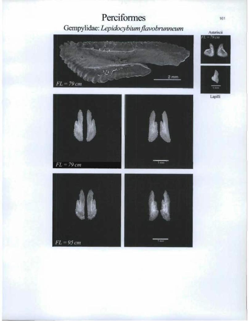
PerciformesGempylidae: Lepidocybium flavobrunneum
161
Asterisci iFL=79cm
Lapilli
FL = 79 cm
FL = 95 cm
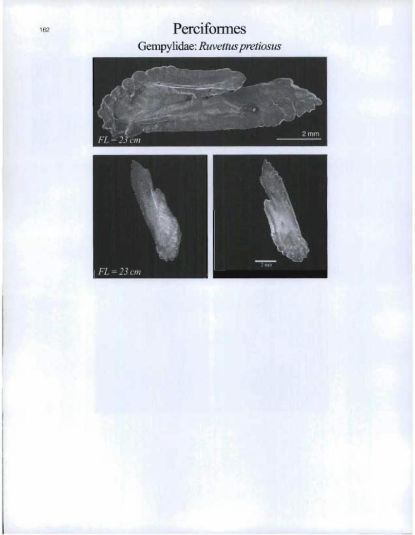
162 PerciformesGempylidae: Ruvettuspretiosus
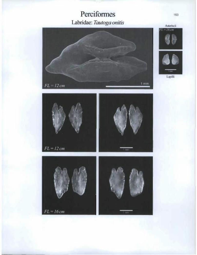
Perciformes Labridae: Tautoga onitis
163
Asteriscii
41, 'EMIT
I j)i Iii
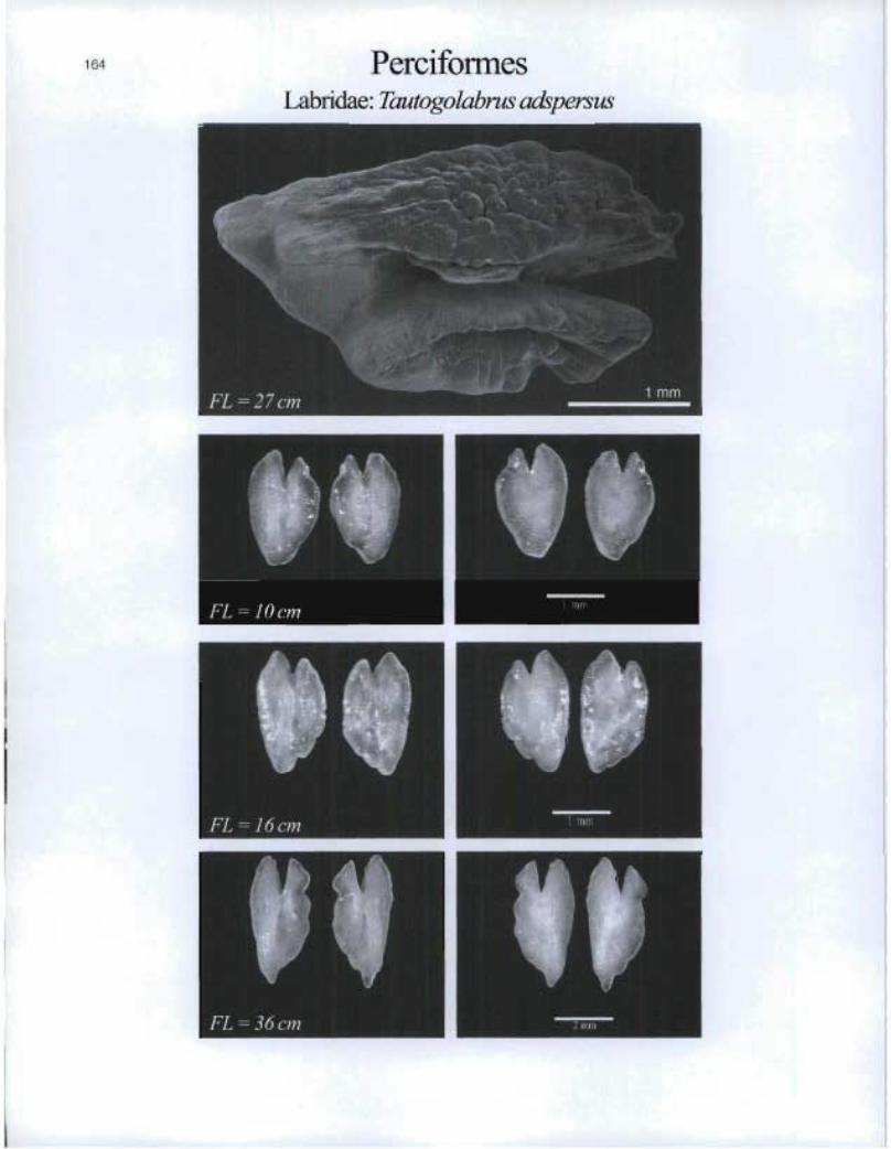
PerciformesLabridae: Tautogolabrus adspersus
FL-10cm
FL = 36 cm
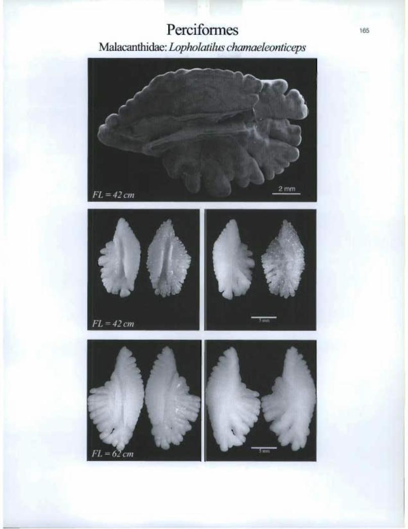
Perciformes Malacanthidae. Lopholatilus chamaeleonticeps

Perciformes Moronidae: Morone americana
166
Asteriscii
111 ERUSI
9110 FL = 19 cm
FL = 23 cm

t,
- Perciformes Moronidae: Morone saxatilis
167

PerciformesMullidae: Mullus auratus
r
FL=14cm n ü"°i,

Asteriscii & Lapilli
Perciformes Pholidae: Pholis gunnellus
169

170 PerciformesPomatomidae: Pomatomus saltatrix
FL =4Scm
®
®

Percifol mes 1 71
Sciaenidae: Pogonias cromis

172 Perciformes Scombridae: Acanthoeybium solandri
Scombridae: Katsuwonus pelamis

Perciformes Scombridae: Sarda sarda

Is
Perciformes Scombridae: Scomberj aponicus

PerciformesScombrida.e. Scomberscombrus
E
FL =1oem n 1 im„

â'
MIMI
176 Perciformes Scombridae: Scomberomorus brasiliensis
Scombridae. Scomberornorus cavalla

Perciformes 177
Scombridae: Scomberomorus maculatus

•
FL = 96 cm
, --e).-ri..4; :---, ... , 1 , %,, ,,,iii,,,.....iiii_,,,,. •
. ,(*i.,‘ ,I...
i , ,
,
‘-‘
Asteriscii MOM
Ii 44%
RICCI
Lapilli
Perciformes Scombridae: Thunnus alalunga
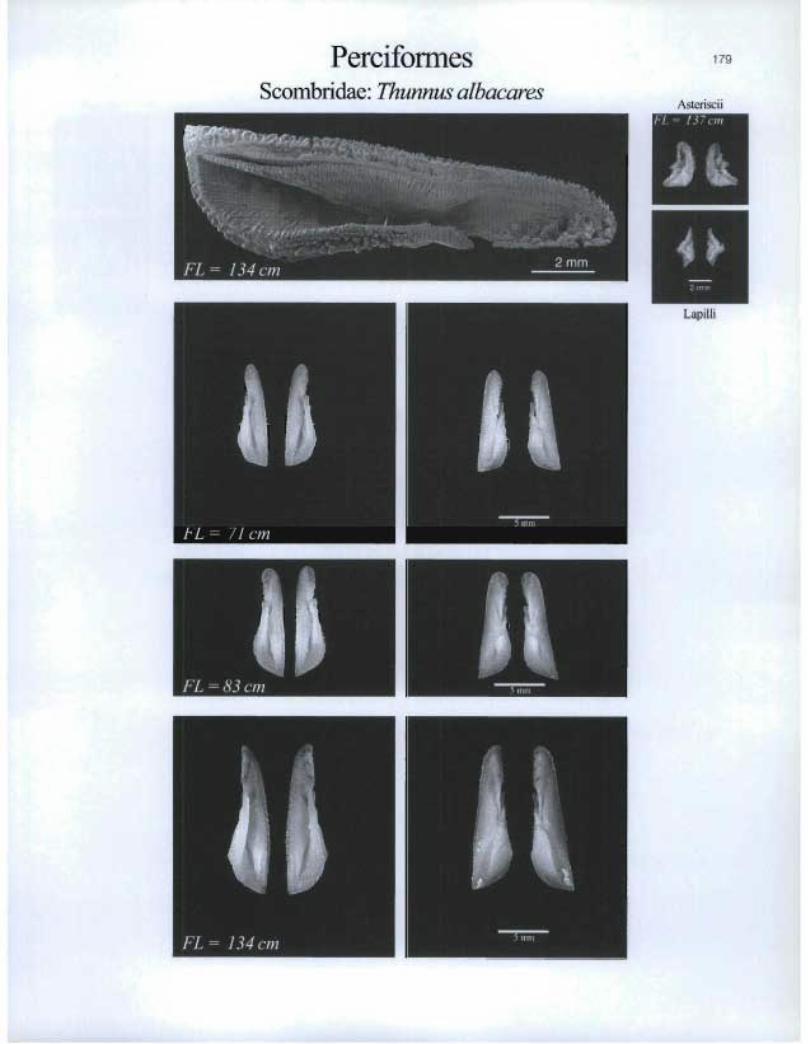
PerciformesScombridae: Thunnus albacares
FL= 71cm
FL = 83 cm ®
179
Asteriscii
m
Lapilli

180 PerciformesScombridae: Thunnus obesus
a,scen5ciiFL = 69 cm
As N
FL= 102 cm
®
Lapilli
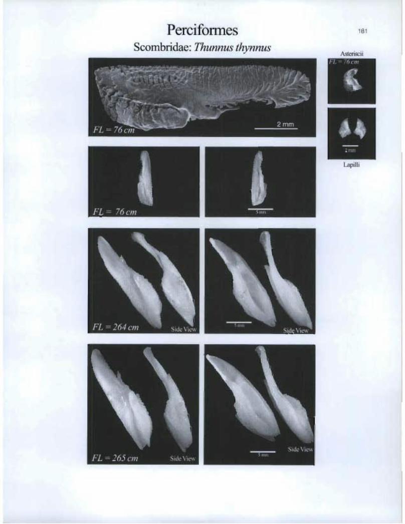
Perciformes Scombridae. Thunnus thynnus
181
Asteriscii
hm
Lapilli
. , 1111111114
FL = 264 cm Side View
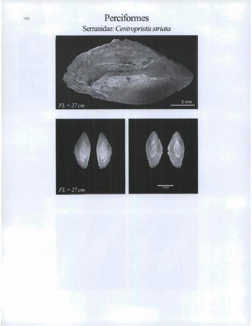
182 PerciformesSerranidae: C'entropristis striata
®
Perciformes Serranidae: Epinephelus niveatus
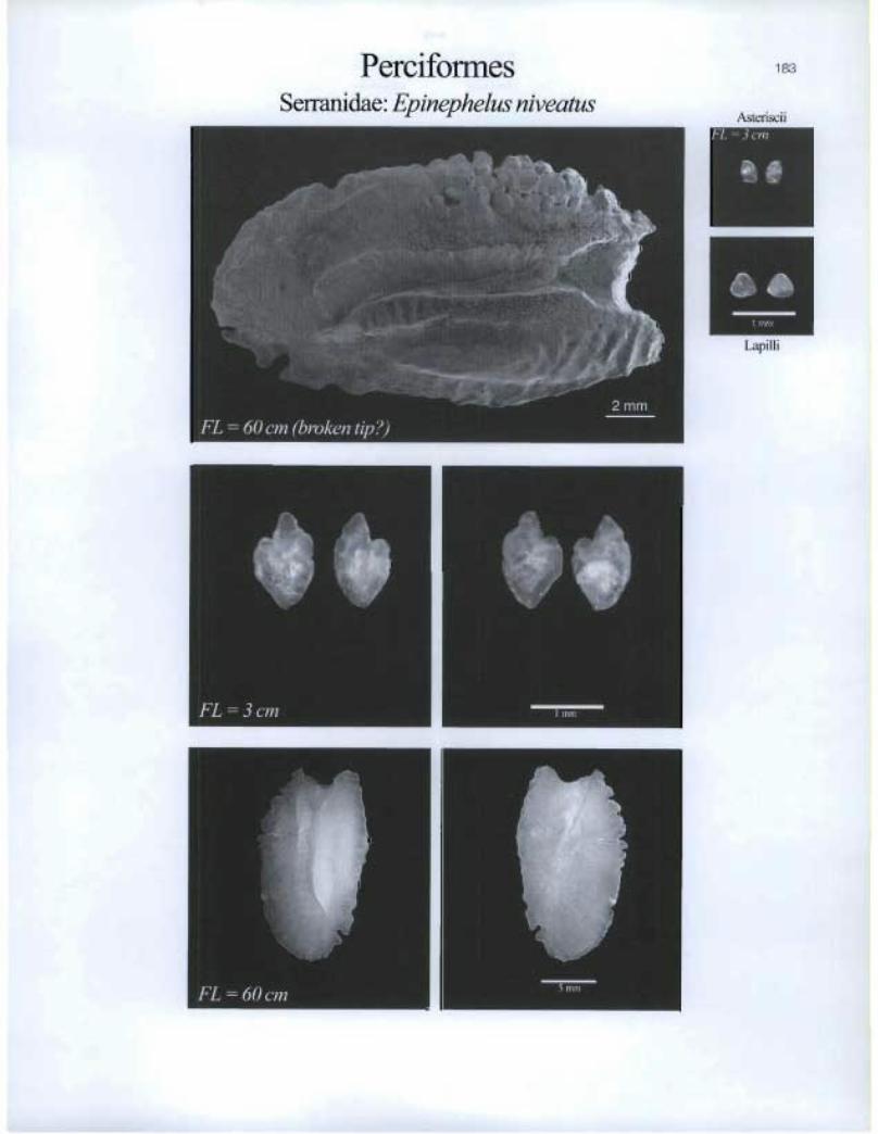
183
Asteriscii
Lapilli
• •
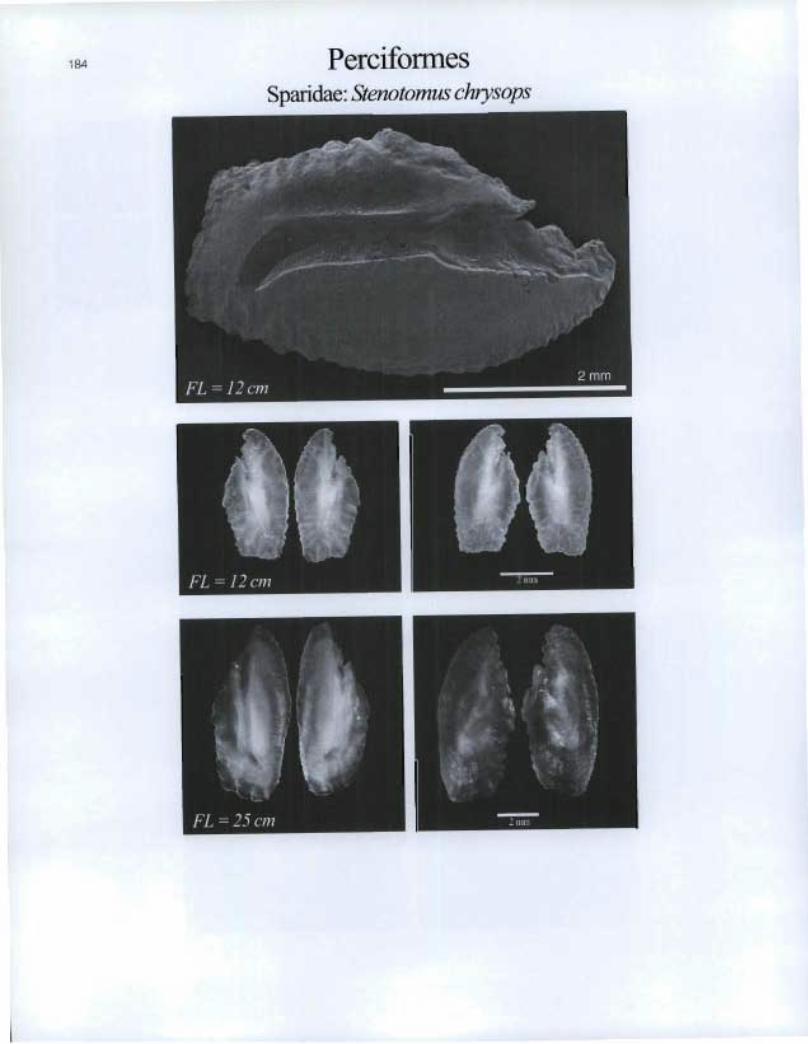
184 Perciformes Sparidae: Stenotomus chrysops
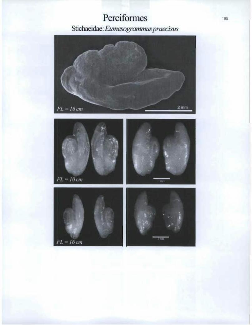
Perciformes Stichaeidae: Eumesogrammus praecisus
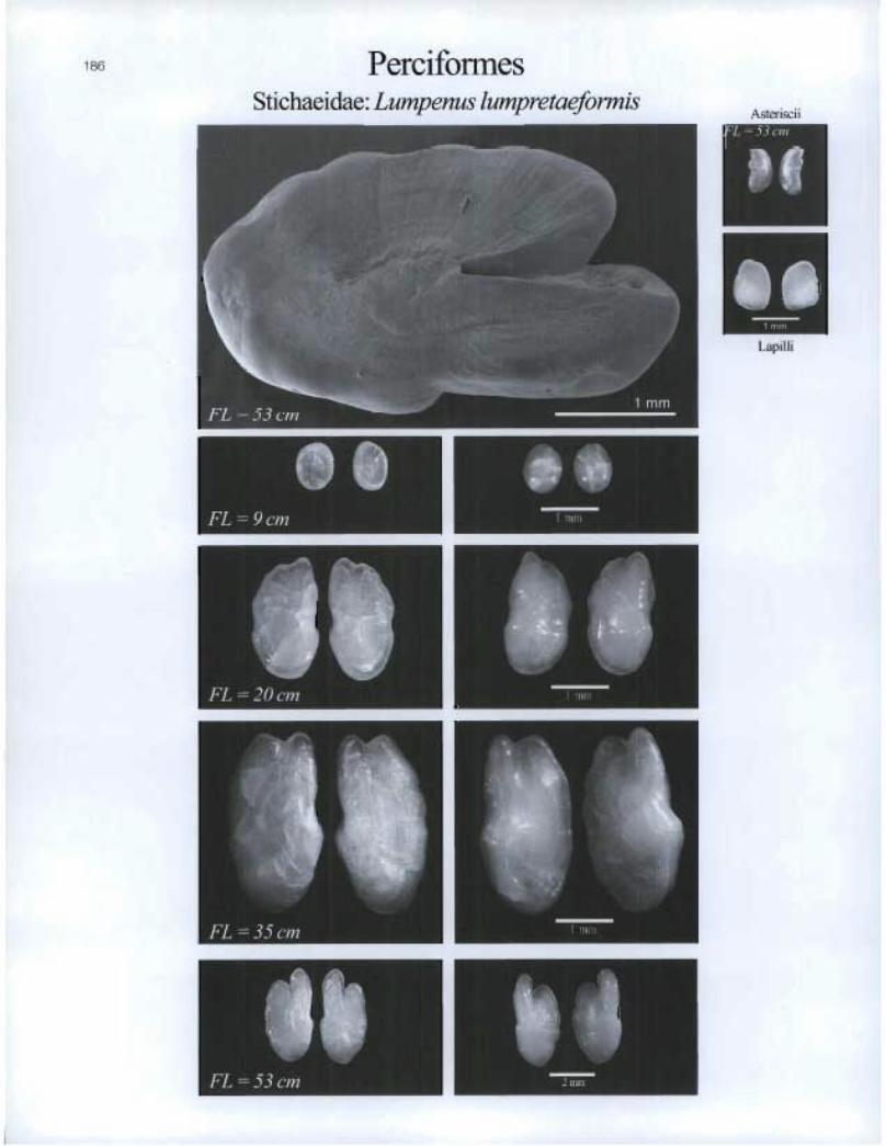
Perciformes Stichaeidae: Lumpenus lumpretaeformis
1 B6
L =53cm
I. Lapi I I i
41) • FL = 9 cm

kiiiiiiiiii
Stichaeidae: Stichaeuspunctatus
PerciformesStichaeidae: Lumpenus maculatus
" I[]
187

Perciformes Stichaeidae: Ulvaria subbifurrata
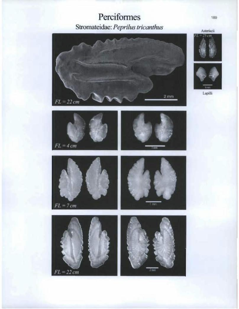
Perciformes Stromateidae: Peprilus tricanthus
189
Asteriscii
Lapilli
• o FL = 7 cm
'e

190 PerciformesTrichiuridae: Aphanopus carbo
[I]
mAsteriscii& Lapilli
FL=112cm
T®

191
FL 103 un
\,
Asteriscii & Lapilli
Perciformes Trichiuridae: Benthodesmus elongatus
tlo El=
Lapilli
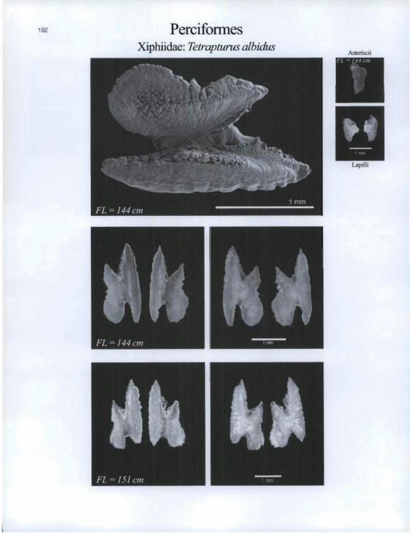
Perciformes Xiphiidae: Tetrapturus albidus
Asteriscii
Mi $0t, FL 1 5 1 cm
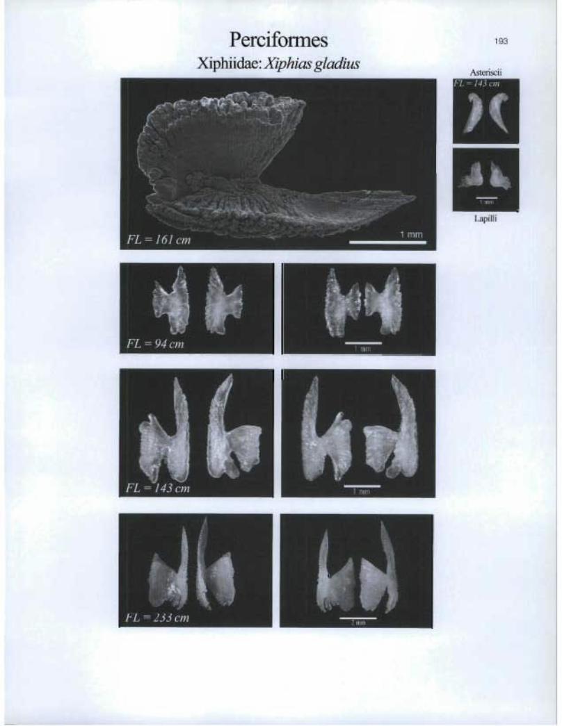
PerciformesXiphiidae: Xiphias gladius
193
asceriscii
Lapi! ] i

IMP FL 18 cm
Perciformes Zoarcidae: Gymnelus viridis

PerciformesZoarcidae: Lycenchelyspaxillus

PerciformesZoarcidae: Lycenchelys verrilli
FL=14cm

FL =34 cm 2 nun
Perciformes Zoarcidae: Lycodes esmarki
197
Asteriscii
FL = 50 cm
Lapilli
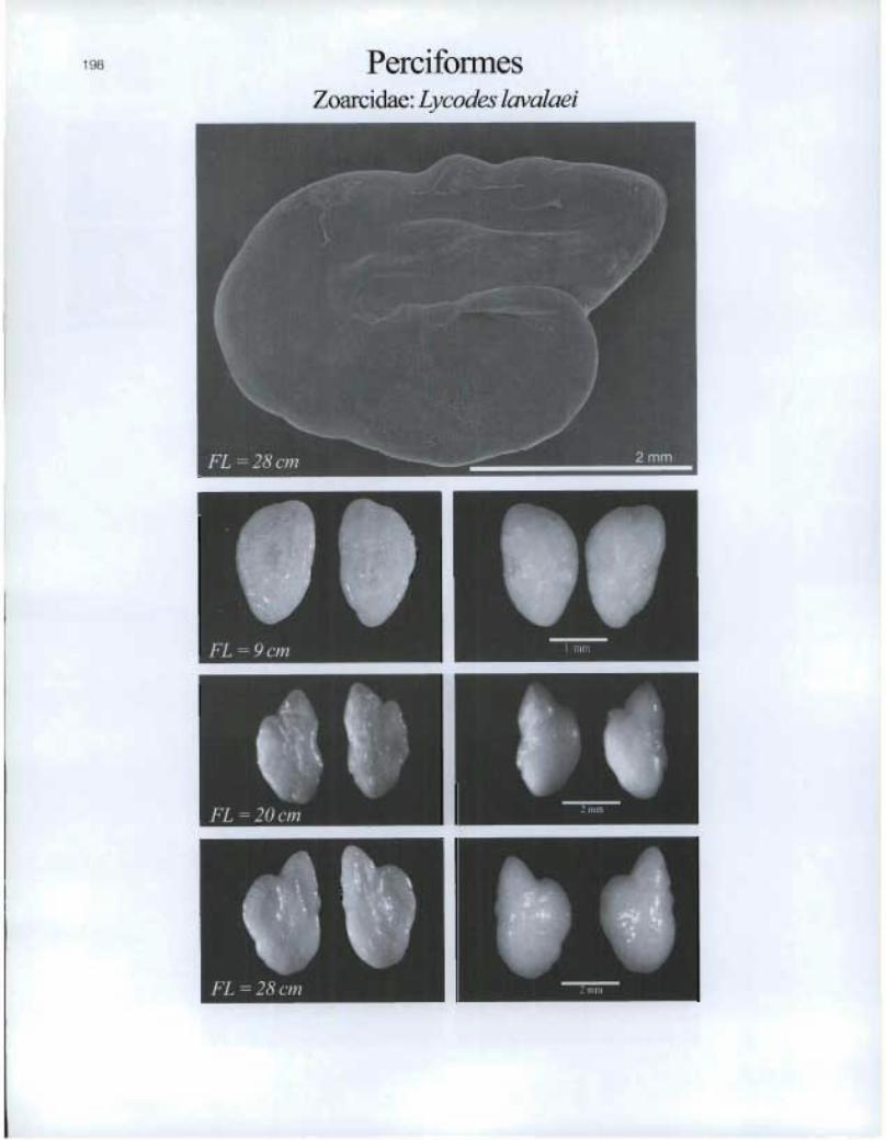
Perciformes Zoarcidae: Lyeodes lavalaei
O FL =28 cm
to

PerciformesZoarcidae: Lycodespallidus
IEFL=15cm
Zoarcida.e: Lycodes reticulatus
FL = 33 cm
199
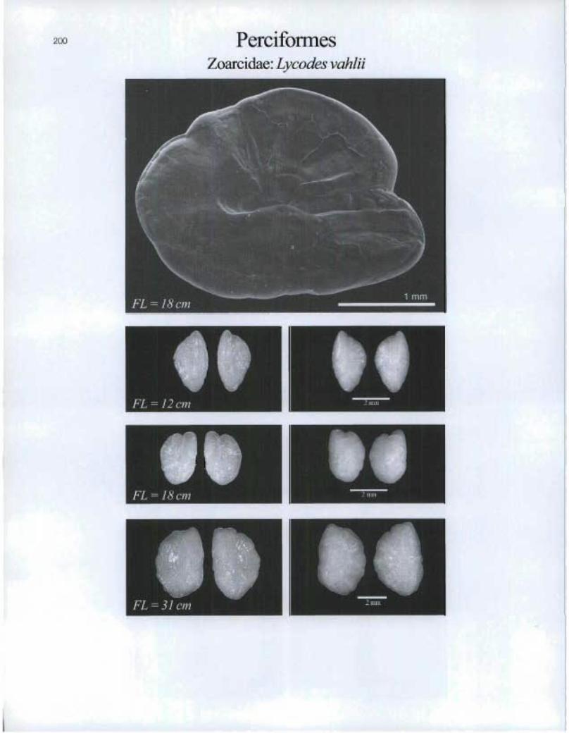
200 PerciformesZoarcidae: Lycodes vahlii
IFL =12 cm
[l]FL =18cm ®

11 FL = 7 cm
Perciformes Zoarcidae: Macrozoarces arnericanus
201
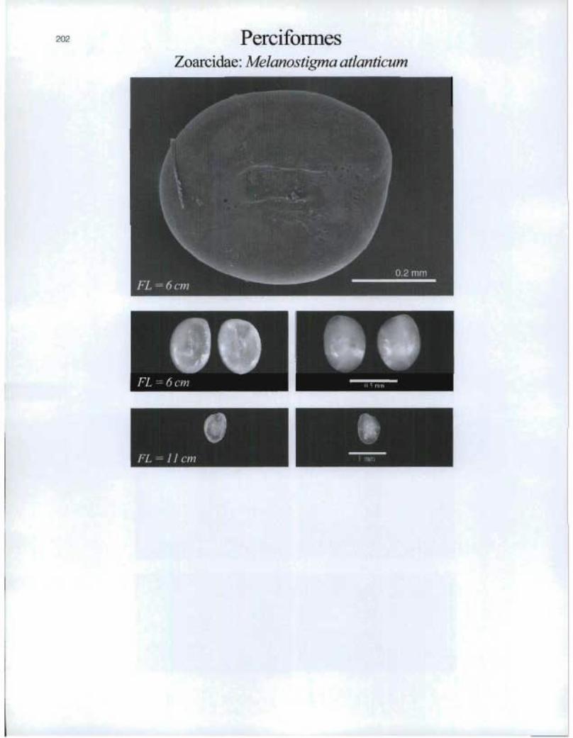
202 PerciformesZoarcidae: Melanostigma atlanticum
LEFL=6cm
I
111®
FL=11cm
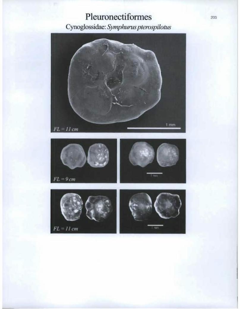
Pleuronectiforrnes Cynoglossidae: Symphurus pterospilotus
1 min
Lapilli
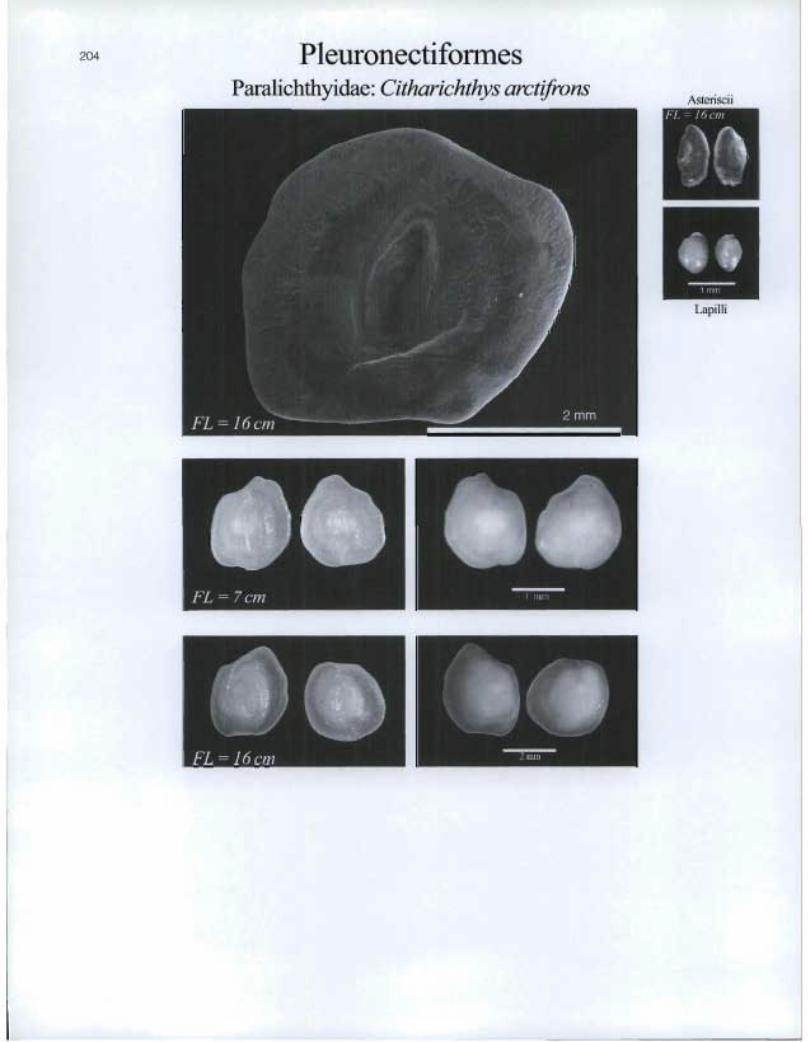
Pleuronectiforrnes Paralichthyidae: Citharichthys arctifrons
Asteriscii
O. FL = cm
Ô. LET

PleuronectiformesParalichthyidae: Etropus microstomus
FL =10 cm a ' ^^ .
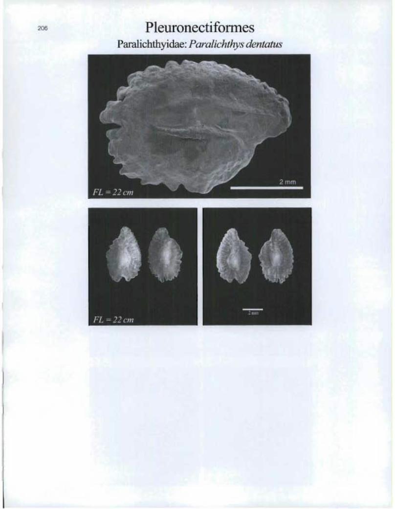
PleuronectiformesParalichthyidae: Paralichthys dentatus

Pleuronectiformes Paralichthyidae: Paralichthys oblongus

PleuronectïformesPleuronectidae: Glyptocephalus cynoglossus
FL=19cm
luFL = 49 cm
®

Pleuronectifomles Pletwonectidae: Hippoglossoides platessoides
ISS fr
Of FL = 19 cm
O FL = 43 cm
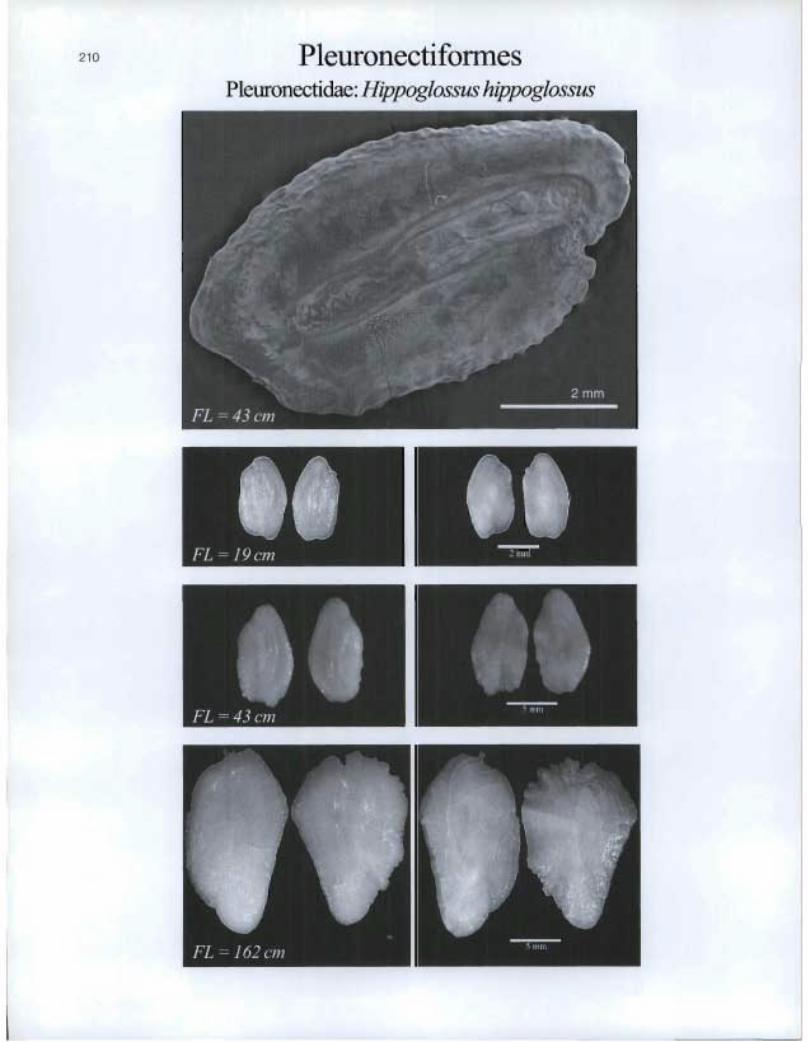
Pleuronectifonnes Pleuronectidae: Hippoglossus hippoglossus
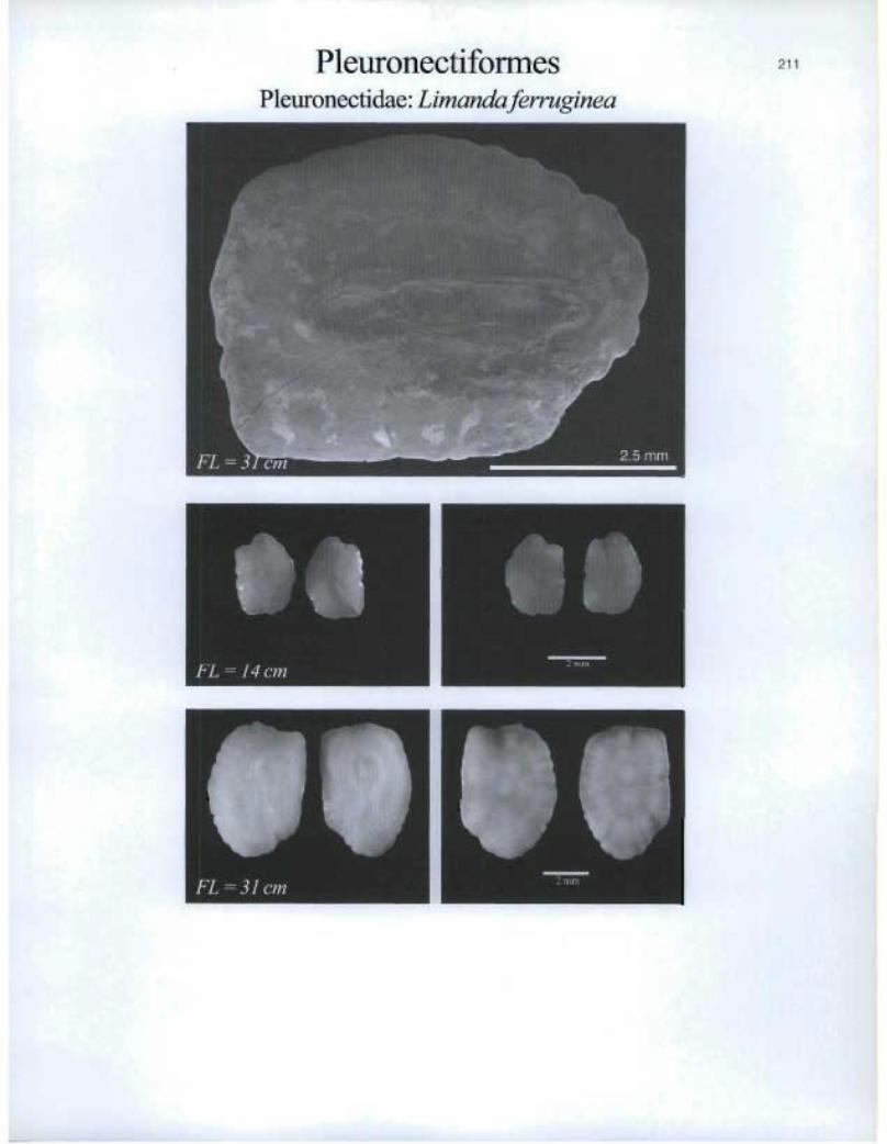
PleuronectiformesPleuronectidae: Limanda ferr,cginea
FL =14 cm
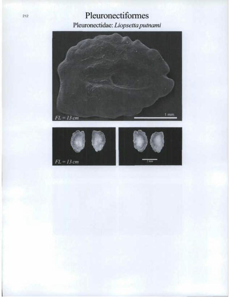
Pleuronectiformes Pleuronectidae: Liopsettaputnami
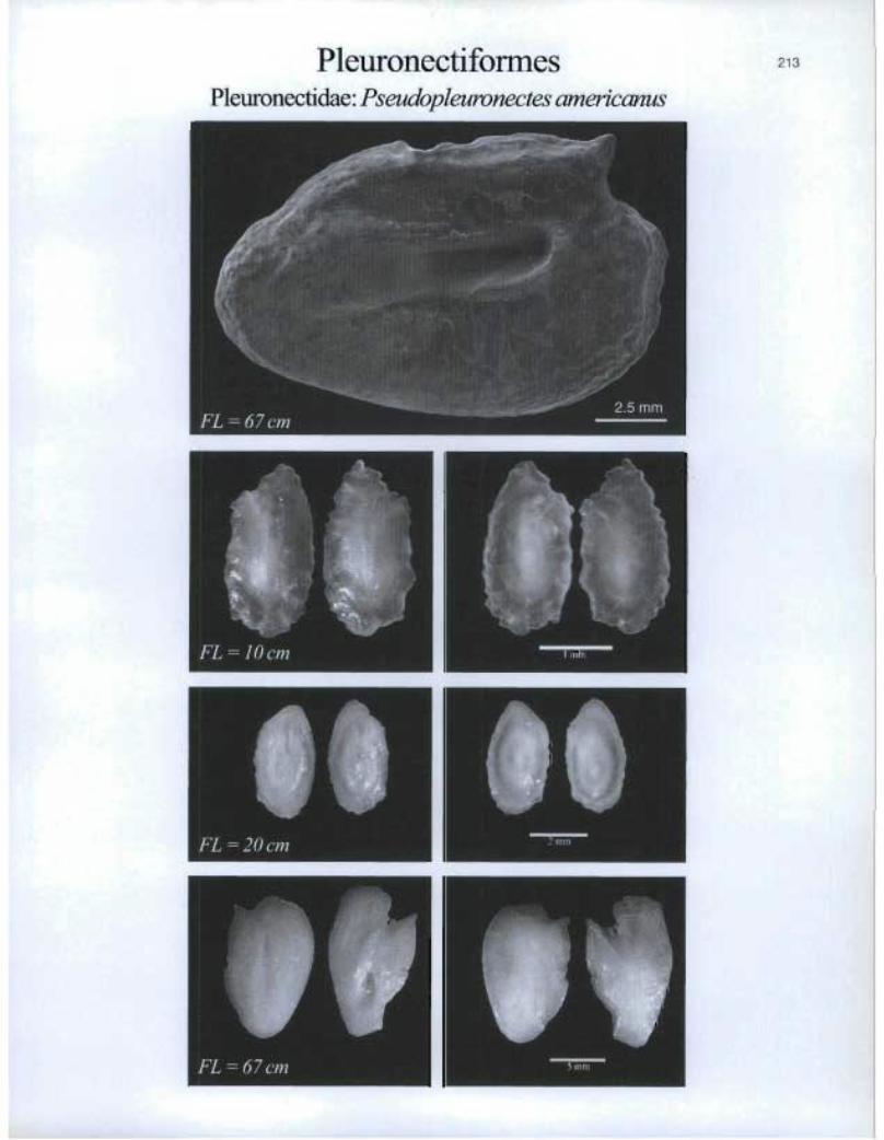
PleuronectiformesPleuronectida.e: Pseudopleuronectes americanus
IFL =20cm n '°i,i'
213

PleuronectiformesPleuronectidae: Reinhardtius hippoglossoides
FL=&cm n r-
an=

it i) FL-21 ern
O.
Pleuronectiformes Scophthalmidae: Scophthalmus aquosus
FL=lbcm
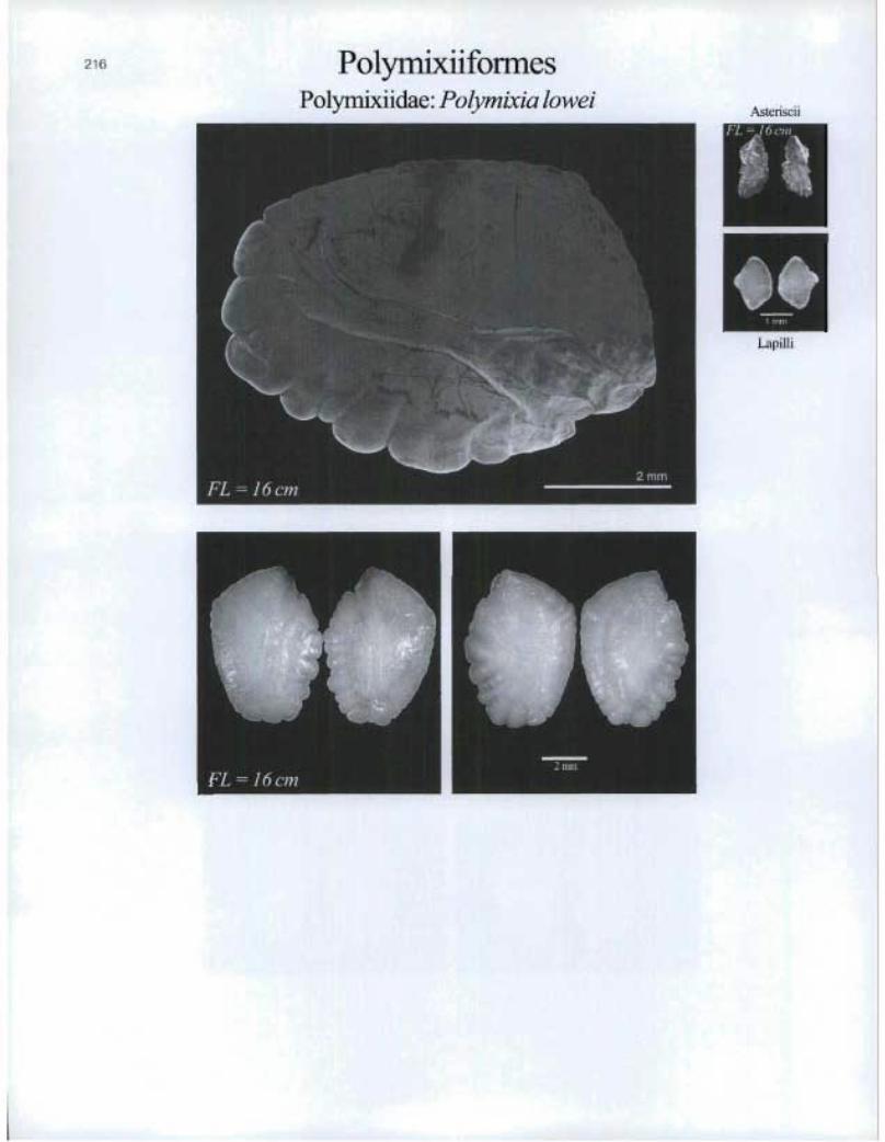
PolymixiiformesPolymixiidae: Polymixia lowei
Asteriscii
Lapilli

Saccopharyngiformes Eurypharyngidae: Eurypharyrvc pelecanoides

FL = 7 cm
6d
Salmoniformes Salmonidae: Coregonus clupeaformis
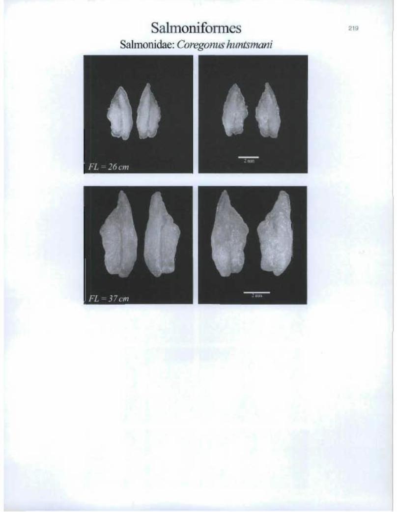
SalmoniformesSalmonid.ae: Coregonus huntsmani
®
®

SalmoniformesSalmonidae: Oncorhynchus mykiss
r1pFL=4cm
FL=13cm a 1 """

Salnrioniformes Salmonidae: Salmo salar
11110 FL = 3 cm
FL = 6 cm
it> FL = 40 cm
e. ■•■■
0 5 nun
46

SalmonifonnesSalmonidae: Salmo trutta
IEFL=5cm Imm

Salmoniformes Salmonidae: Salvelinus alpinus
223
Asterisc i i
FL = 41 ci t n.
ID II "Mr
Lapilli
FL 8 cm 46 H
EL 15 CM
46 Sot KilM1
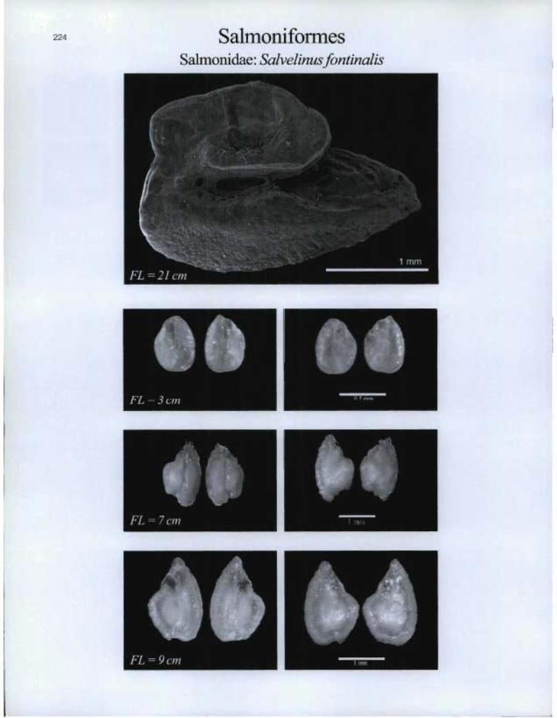
224 SalmoniformesSalmonidae: Salvelinusfontinalis
FL=3cm

Scorpaeniformes Agonidae: Agorrus decagonus
225
Asteliscii
Lapilli
FL = 17 cm
411 FL 19 cm
.4 •
"N 2=

ScorpaeniformesAgonida.e: AspidophoYocdes monopterygius
Asteriscii
rju®
Lapilli
FL=4cm
FL=11cm
s
11, FL — 8 cm
Scorpaeniformes 227
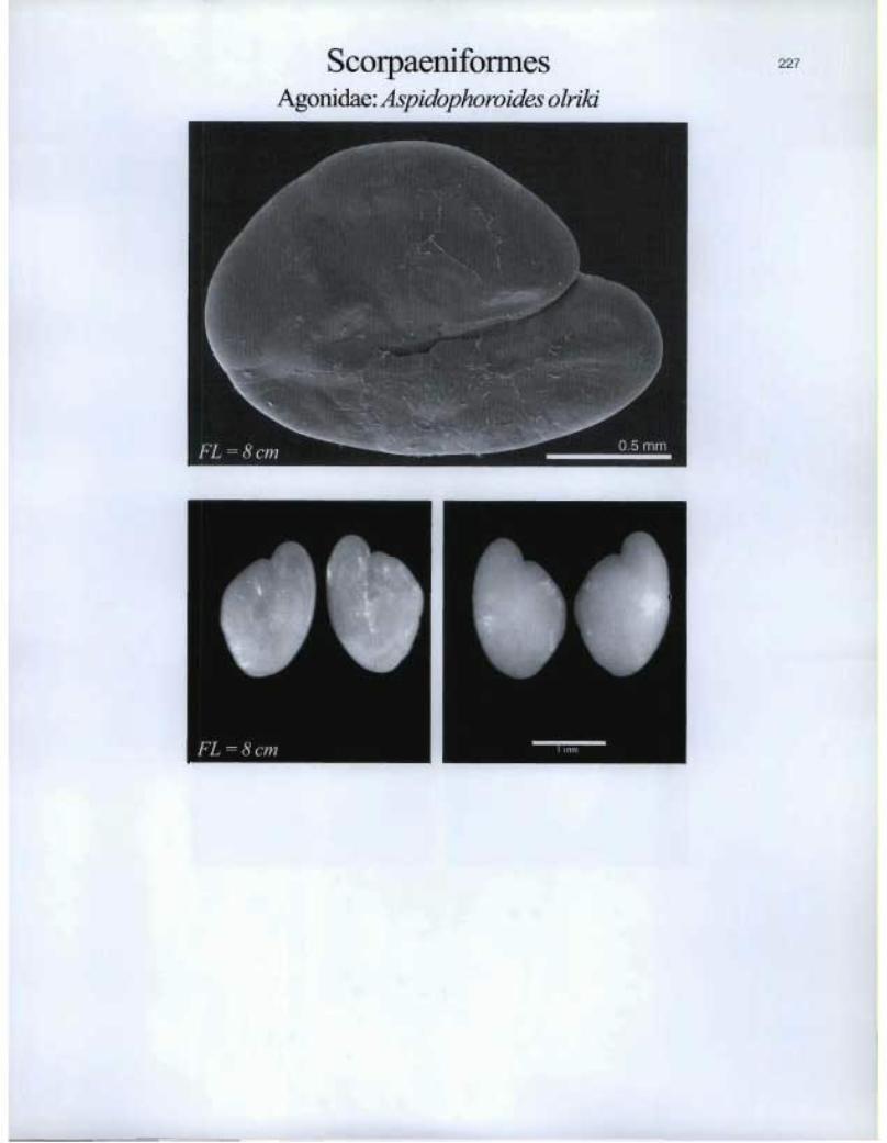
Agonidae: Aspidophoroides olriki

Scorp ►aeniformesCottidae: Artediellus atlanticus

Ô. FL .= 14 cm
Scorpaeniforrnes Cottidae: Gymnocanthus tricuspis
•11
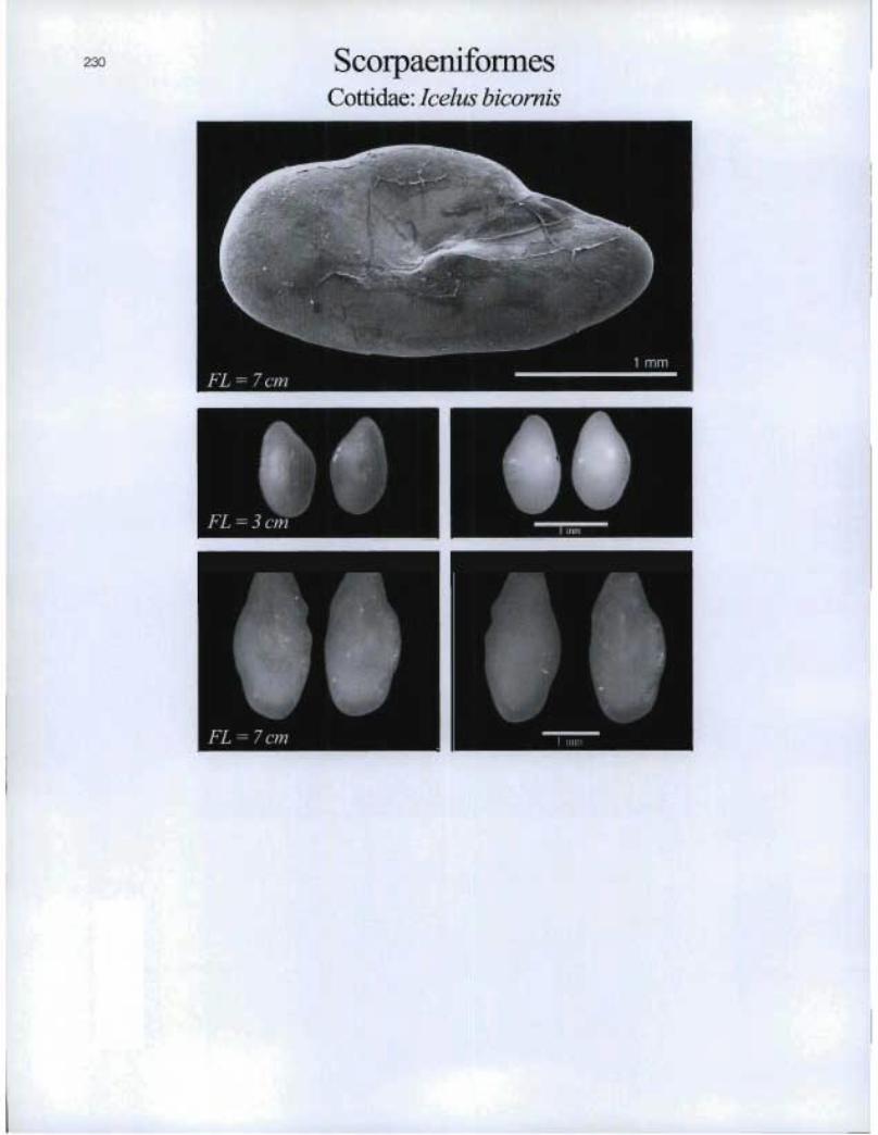
230 ScorpaeniformesCottidae: Icelus bicorytis

FL = 5 cm
01 FL = 9 cm
Scorpaeniformes Cottidae: Icelus spatula
I. 114
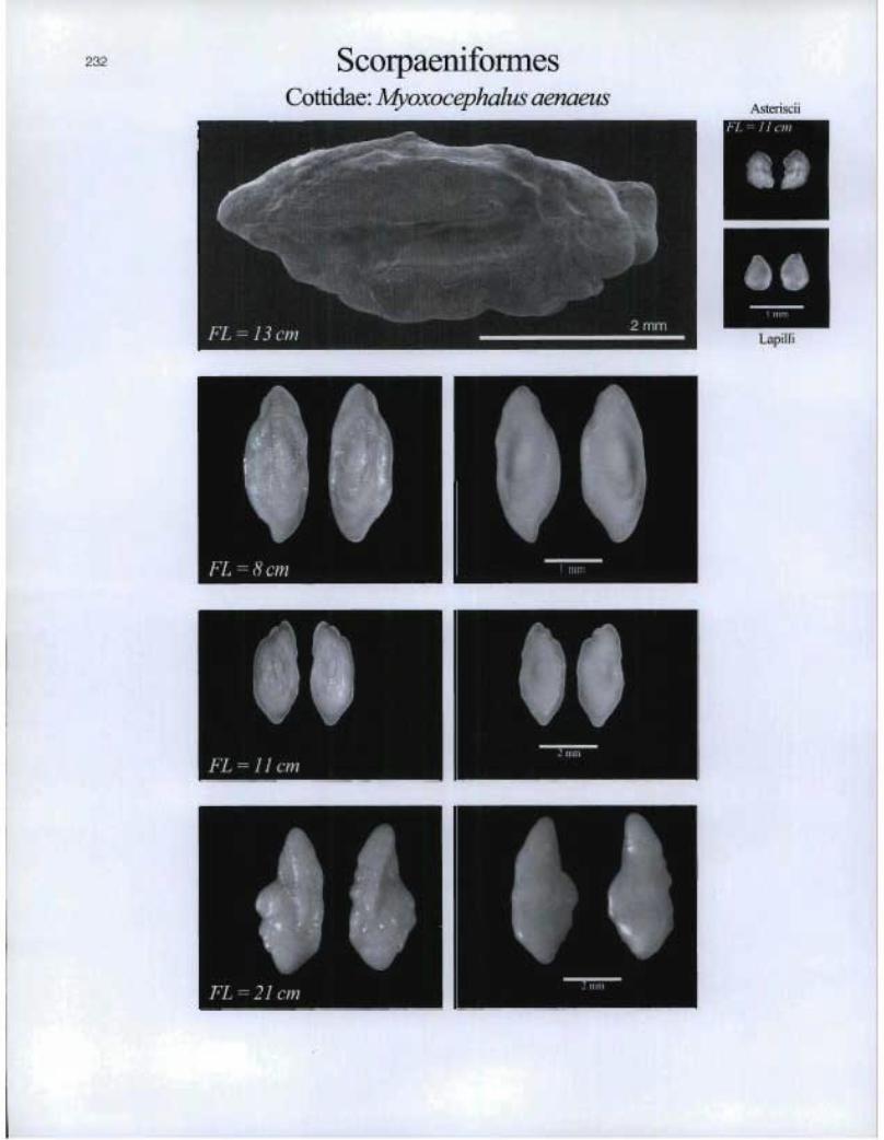
232 ScorpaeniformesCottidae: Myoxocephalus aenaeus
Asteriscii
FL=11c►n
TFL= 13 cm
FL = 8 cm
EFL=11 cm
FL = 21 cm
2 mmLapilli
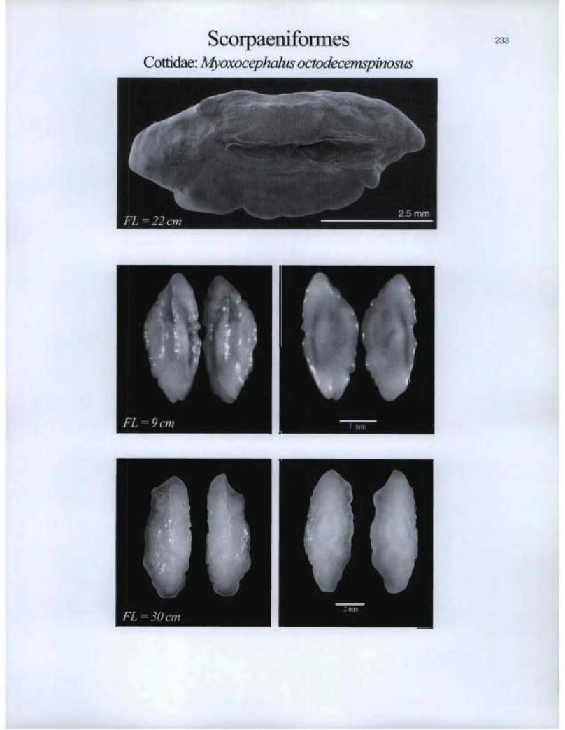
Scorpaeniforrnes Cottidae: Myoxocephalus octodecemspinosus
Is

234 ScorpaeniformesCottidae: Myoxocephalus scorpioides

Scorpaeniforrnes Cottidae: Myoxocephalus scorpius
I. FL = 3 cm
Of

236 ScorpaeniformesCottidae: Triglops murrayi
FL=I2cm
FL=icm
FL=12cm
®
®
Lapilli
& Asteriscii

Lapilli
ès FL = 14 cm
11). IA=
Scorpaeniformes Cotticlae: Triglops nybelini
237
Asteriscii
O. FL = 7 cm
46 Matti=
ft

ScorpaeniformesCottidae: Triglopspingeli

FL = 53 cm
ê.
Asteriscii & Lapilli
Il O.
1111,11
Scorpaeniformes Cyclopteridae: Cyclopterus lumpus
239
FL = 11 cm
Il FL =22 cm
111 I mm lb
FL = 53 cm
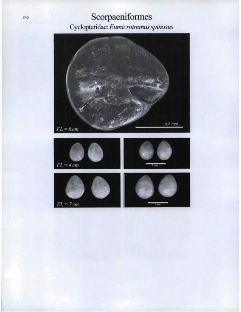
240 ScorpaeniformesCyclopteridae: Eumieroiremus spinosus
mFL=4cm ®
[lu®

scorpaeniformes Hemitripteridae: Hemitripterus americanus

ScorpaeniformesLiparidae: Careproctus reinhardti
E I
FL=9cm ® 1
FL = 5 cm
Il FL = 19 cm
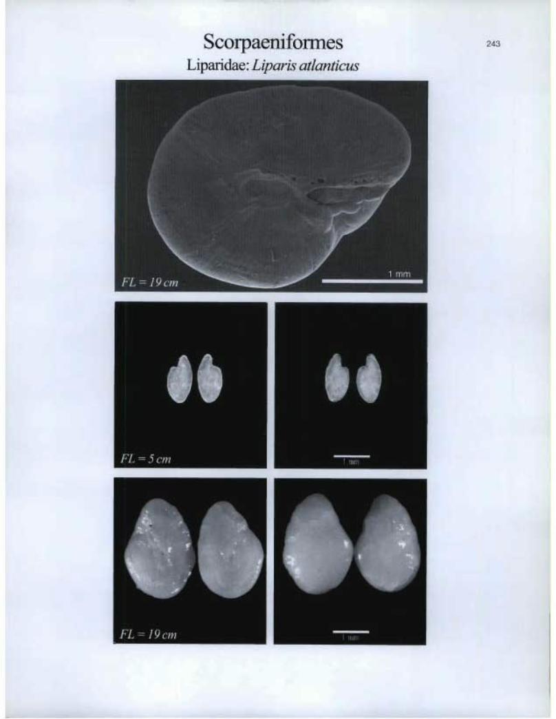
Scorpaeniformes Liparidae. Liparis atlanticus

244 ScorpaeniformesLiparidae: Liparis fabricii
IEFL=4cm
1 I®
FL=13crn
46 FL = 5 cm
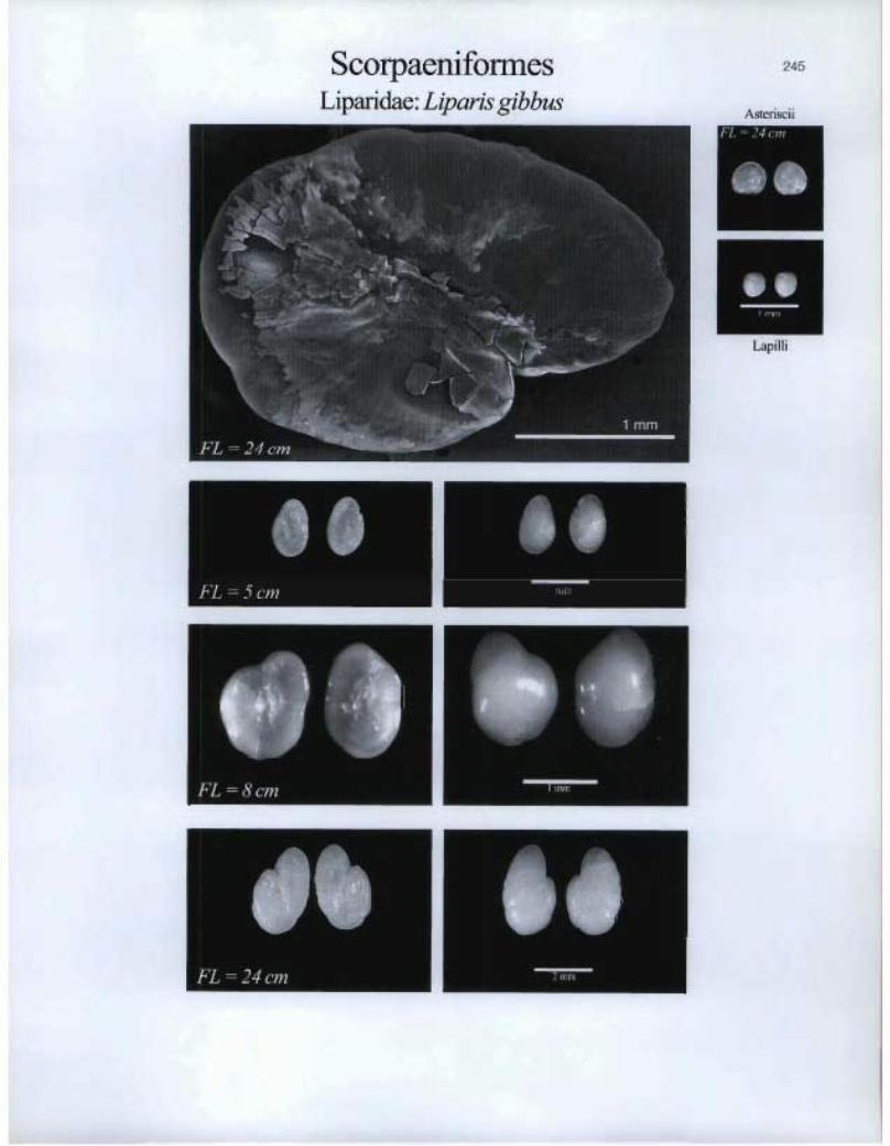
Scorpaeniformes Liparidae: Liparis gibbus
245
Asteriscii WIENNIE
O.
Lapilli
BIM
FL = 24 cm
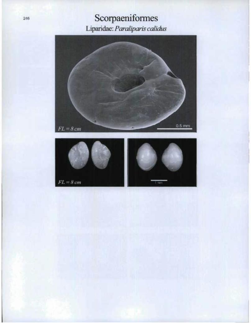
ScorpaenifonnesLipa.ridae: Paraliparis calidus
FL = 8 cm n 1 "im

s.
Scorpaeniformes Liparidae: Paraliparis copei

ScorpaeniformesPsychrolutidae: Cottunculus microps
FL=8cm
I
FL=15cm
®
L

I. 1111 FL = 8 cm Erni
Scorpaeniforrnes Psychrolutidae: Cottunculus thomsonii
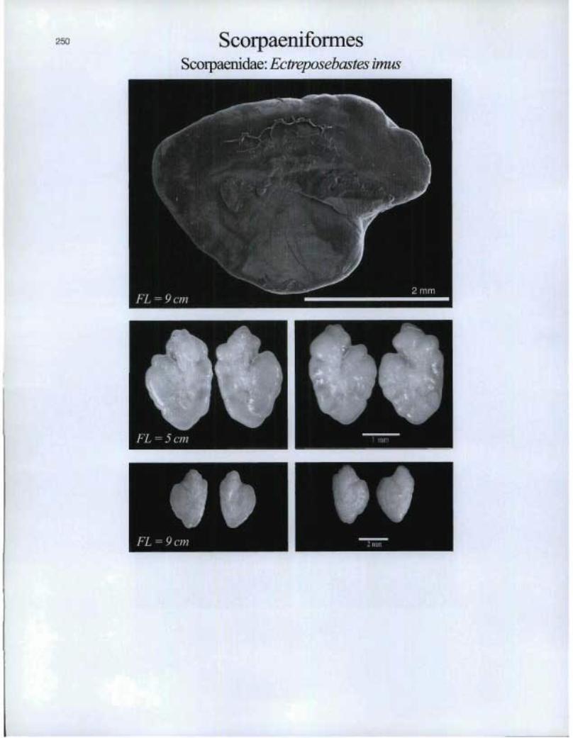
250 ScorpaeniformesScorpaenidae: Ectreposebastes imus
FL=5cm
Il
1 mm
FL=9cm
Lapi 1 1 i
Ô. FL = 7 cm
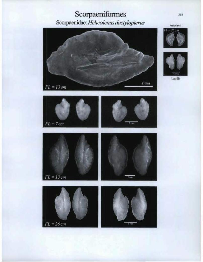
Scorpaeniforrnes Scoipaenidae: Helicolenus dactylopterus
251
Asteriscii
•• FL = 2 6 cm i=111t11
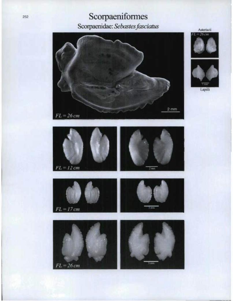
ScorpaeniformesScorpaenidae: Sebastesfasciatus
\sceriscFL=26cm
IELapilli
TFL=17cm
TFEFEC61W

Scorpaeniformes Scorpaeniclae: Sebastes marinus
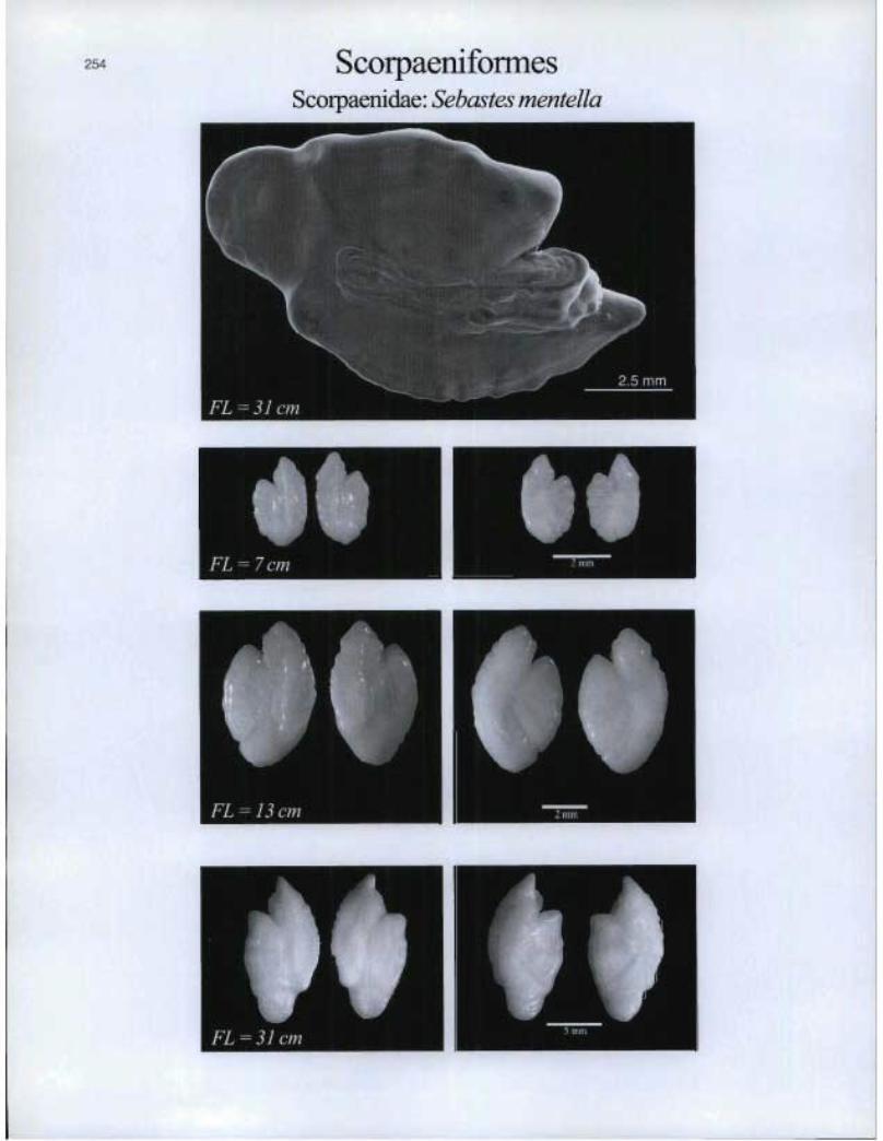
ScorpaeniformesScorpaenidae: Sebastes mentella
FL=7cm
FL=13cm
FL=31cm
IFmFmii^iW

Scorpaeniformes Triglidae: Peristedion miniatum
255
Asteriscii WEIR=
Lapilli
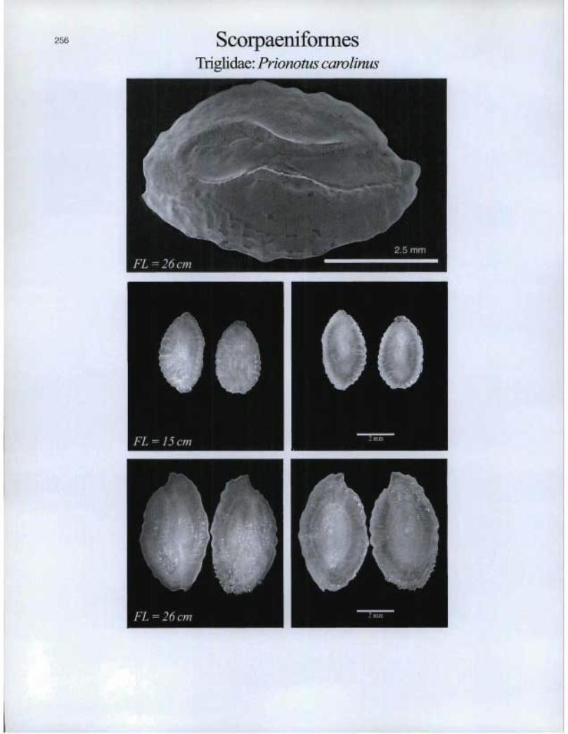
ScorpaeniformesTriglida.e: Prionotus carolinus
FL=15cm n '°l]"

RIM
Scorpaeniformes Triglidae: Prionotus evolans

Stephanob eryc i forme sMelamphaidae: Poromitra megalops
D
FL=14cm

Stephanoberyciformes Melamphaidae: Seopelogadus beanii
90 FL = 8 cm
sto 11111:1
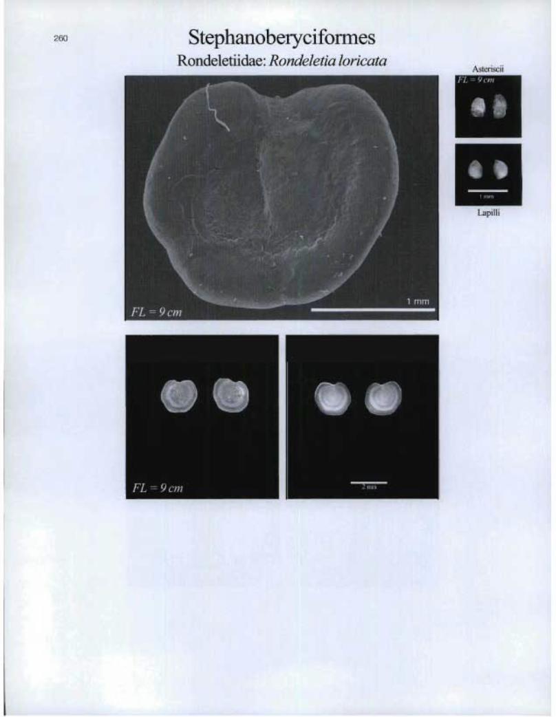
StephanoberyciformesRondeletiidae: Rondeletia loricata
7lu
Asteriscii
Lapilli
FL=9cm m 2 nun

O.
FL = 5 cm 111111
Stomiiformes Gonostomatidae: Cyclothone microdon
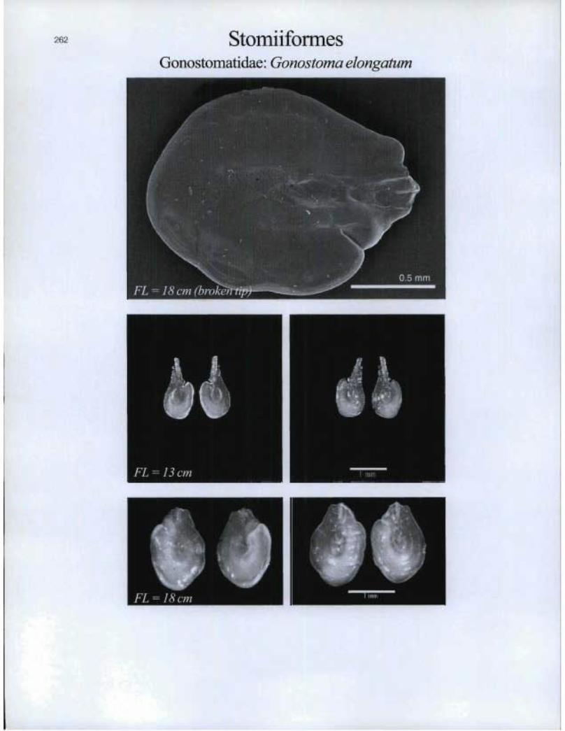
262 StomiiformesGonostomatidae: Gonostoma elongatum
FL=I3cm
FL=18cm
®

StomiiformesGonostomatidae: Vinciguerria nimbaria
FL=6cm
263
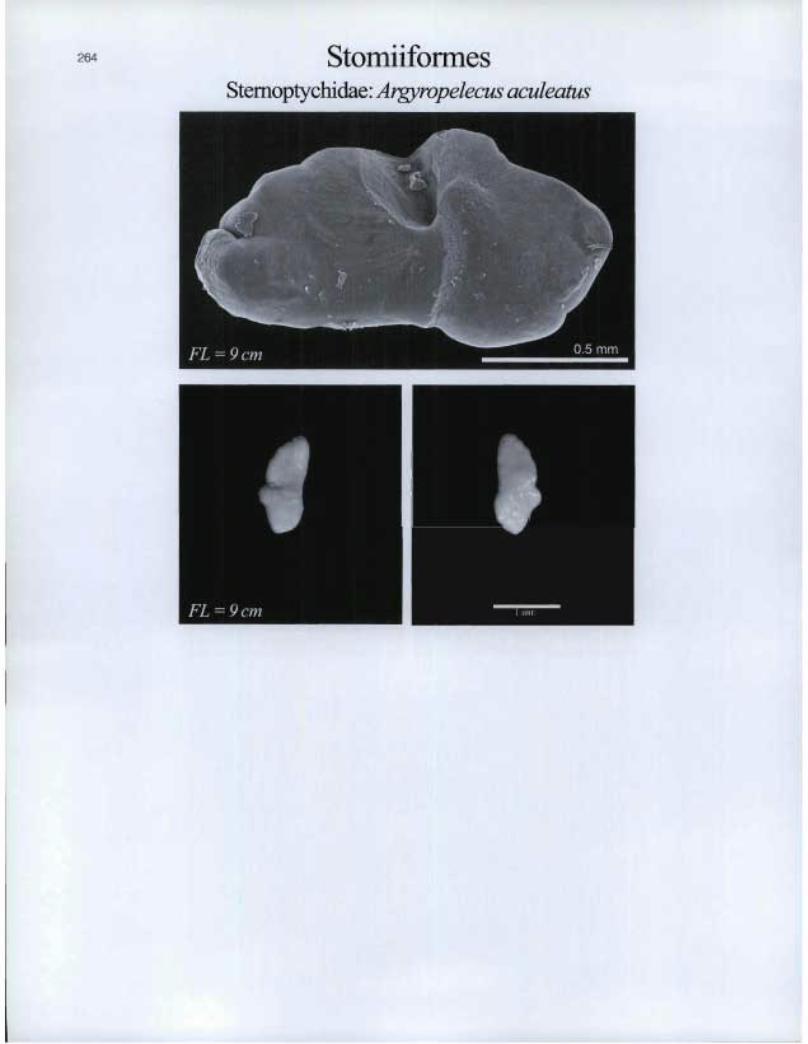
Stomliformes Stemoptychidae: Argyropelecus aculeatus

• • JIM
Lapilli
Stomiifomies Stemoptychidae: Argyropelecus gigas
265
Asteriscii
Stemoptychidae: Argyropelecus hemigymnus

266 StomiiformesStemoptychidae: Maurolicus muelleri
Asteriscii
I I
44
Lapilli
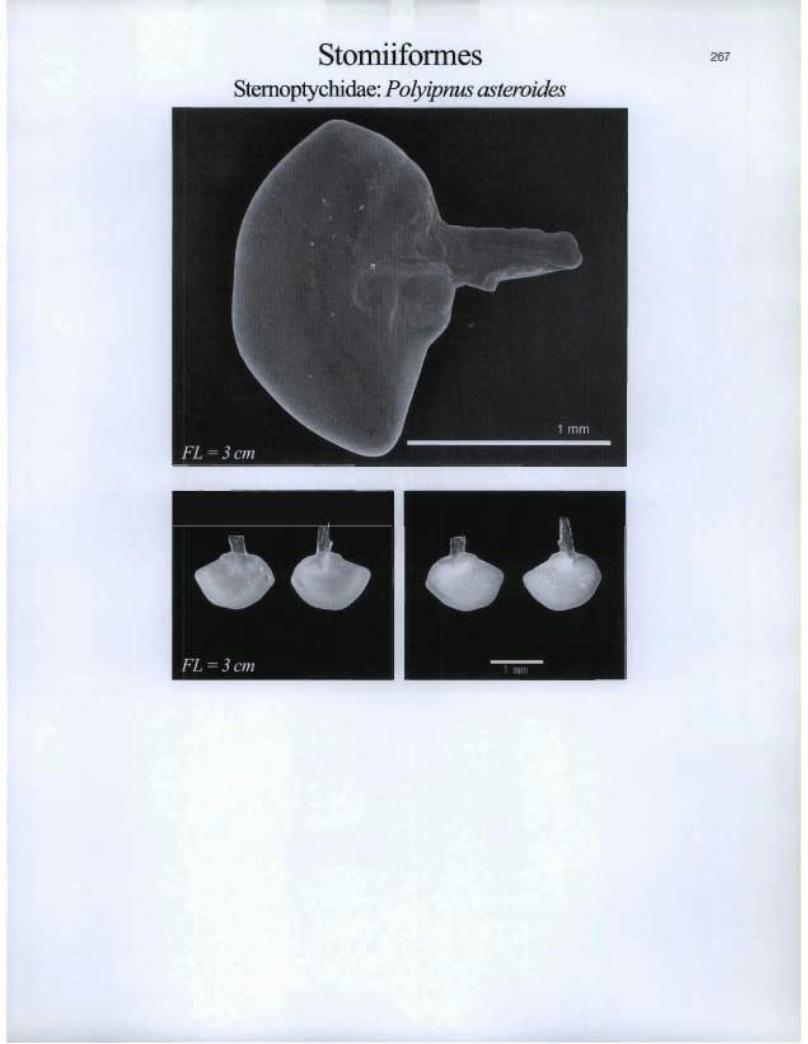
Stomiiformes Stemoptychidae: Polyipnus asteroides
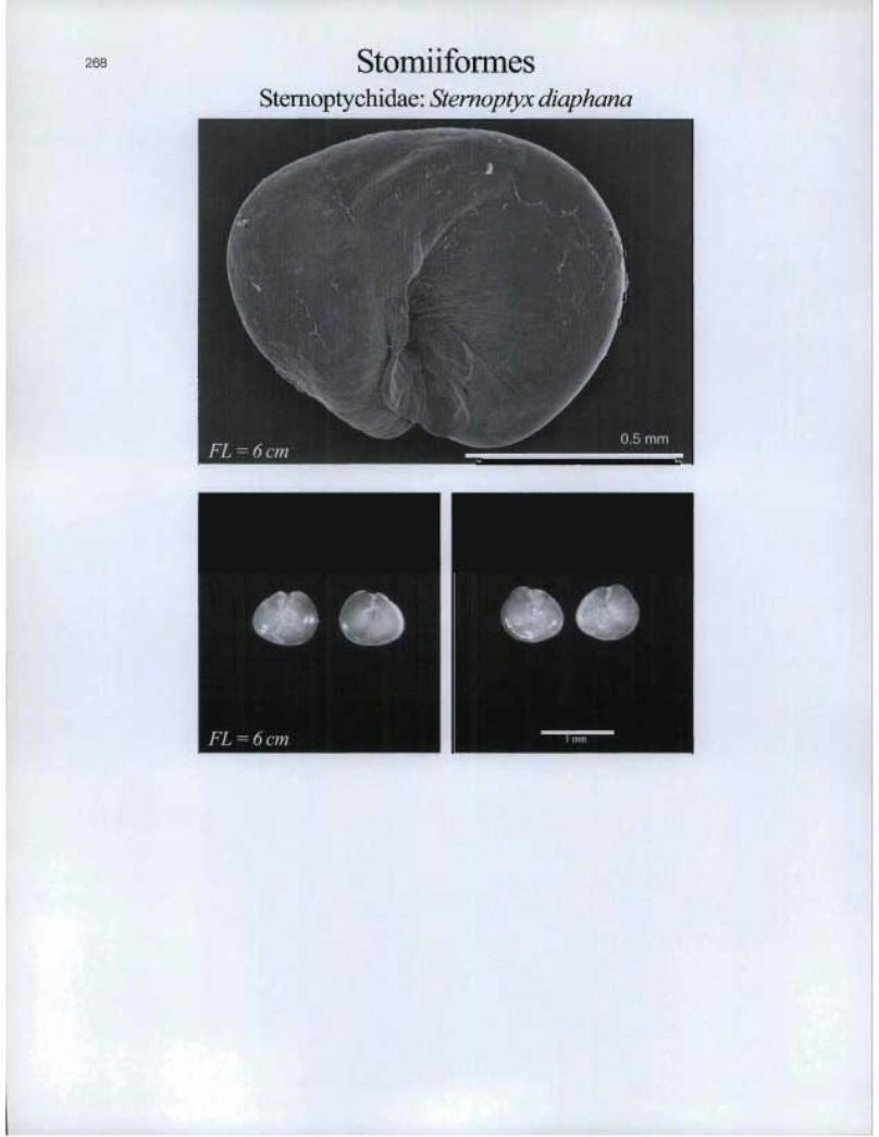
StomiiformesSternoptychidae: Stemoptyx diaphana
lromFL=6cm n -

1■1■1■ I 5 mil
s. I. FL = 11 cm
FL =17cm
Stomiiformes Stomiidae: Chauliodus sloani

270 StomiiformesStomiidae: Malacosteus niger
4111 • 1
0 11 FL = 19 cm
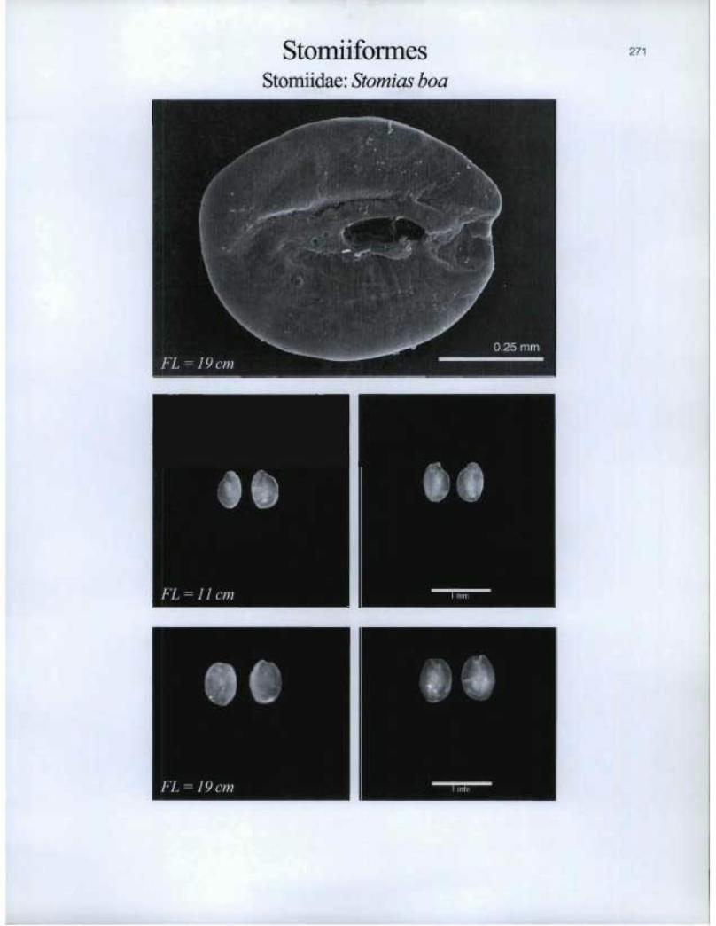
Stomiiformes Stomiidae: Stomias boa
FL = 11 cm 1.17nr

272 StomiiformesStomiidae: Trigonolampa miriceps
LIE I[]
FL=41 cm ®

it nun
r
BIM
Tetraodontiformes Balistidae: Balistes capriscus
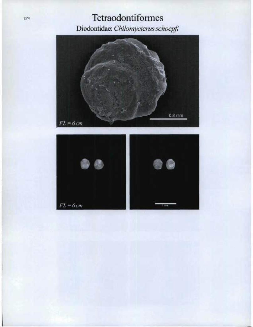
TetraodontiformesDiodontidae: Chilomycterus schoepfi
s .Z^
FL = 6 cm n --=. 17
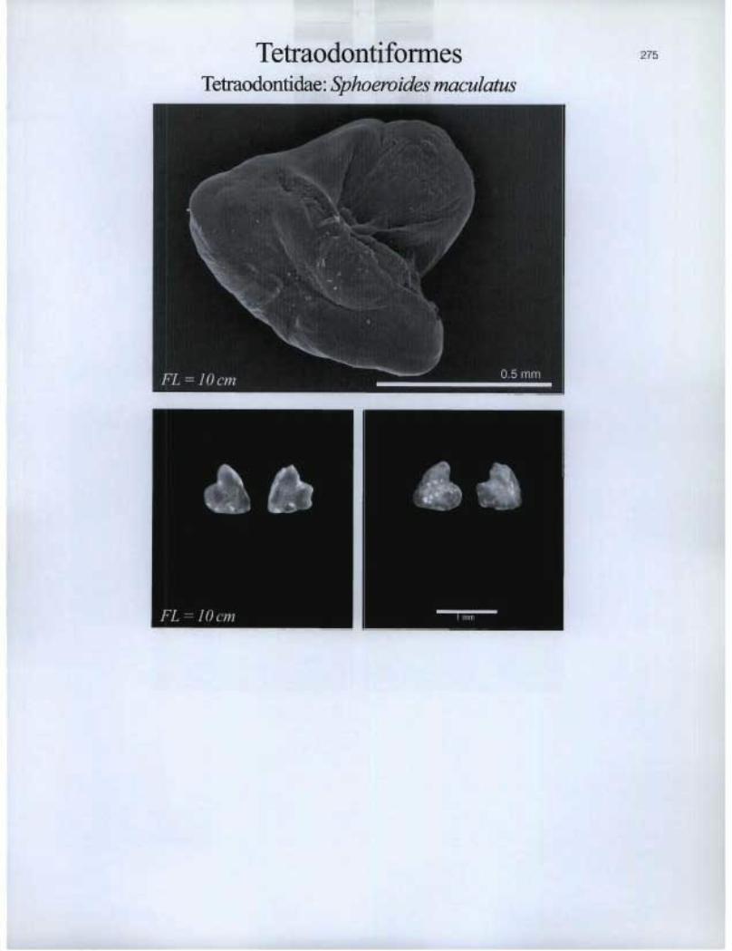
Tetraodontiformes Tetraodontidae: Sphoevides maculatus

276 ZeiformesGrammicolepididae: Daramattus americanus
^ mm

ZeiformesZeida.e: Cyttopsis roseus
277
Asteriscii
FL=9cm
2
:I1Lapilli
Li
FL=9cm n ^

IMIll
Zeiformes Zeidae: Zenopsis conchifera
«I* FL = 8 cm
40e FL = 3 3 cm
Pm mm
Alphabetical species list
Species Family Order Page number
Acanthocybium solandri Scombridae Perciformes 172 Acipenser oxyrhynchus Acipenseridae Acipenseriformes 15 Agontts decagonus Agonidae Scorpaeniformes 225 Alepisaurus brevirostris Alepisauridae Aulopiformes 30 Alepisaurus ferox Alepisauridae Aulopiformes 31 Alepocephalus agassizii Alepocephalidae Osmeriformes 128 Alepocephalus bairdii Alepocephalidae Osmeriformes 129 Alosa aestivalis Clupeidae Clupeiformes 43 Alosa pseudoharengus Clupeidae Clupeiformes 44 Alosa sapidissima Clupeidae Clupeiformes 45 Ammodytes americanus/dubius Ammodytidae Perciformes 143 Anarhichas denticulatus Anarhichadidae Perciformes 144 Anarhichas lupus Anarhichadidae Perciformes 145 Anarhichas minor Anarhichadidae Perciformes 146 Anguilla rostrata Anguillidae Anguilliformes 20 Anoplogaster cornuta Anoplogasteridae Beryciformes 40 Antimora rostrata Moridae Gadiformes 70 Apeltes quadracus Gasterosteidae Gasterosteiforrnes 82 Aphanopus carbo Trichiuridae Perciformes 190 Argentina silus Argentinidae Osmeriformes 134 Argyropelecus aculeatus Stemoptychidae Stomiiformes 264 Argyropelecus gigas Stemoptychidae Stomiiformes 265 Argyropelecus hemigymnus Stemoptychidae Stomiiformes 265 Artedielhts atlanticus Cottidae Scorpaeniformes 228 Aspidophoroides monoptetygizts Agonidae Scorpaeniformes 226 Aspidophoroides olriki Agonidae Scorpaeniformes 227 Bajacalifornia tnegalops Alepocephalidae Osmeriformes 130 Balistes capriscus Balistidae Tetraodontiformes 273 Bathylagus euryops Bathylagidae Osmeriformes 135 Bathypterois qztadrifilis Ipnopidae Aulopiformes 34 Bathysaurus ferox Synodontidae Aulopiformes 38 Benthodesmus elongatus Trichiuridae Perciformes 191 Benthosema glaciale Myctophidae Myctophiformes 96 Benthosema suborbitale Myctophidae Myctophiformes 97 Bolinichthys photothorax Myctophidae Myctophiformes 97 Boreogadus saida Gadidae Gadiformes 51 Brama brama Bramidae Perciformes 147 Brevoortia tyrannus Clupeidae Clupeiformes 46 Brosme brosme Gadidae Gadiformes 52 Brosmiculus imberbis Moridae Gadiformes 71 Callionymus agassizi Callionymidae Perciformes 148 Caranx ct:ysos Carangidae Perciformes 149 Caranx hi ppos Carangidae Perciformes 149 Careproctus reinhardti Liparidae Scorpaeniformes 242 Caristius groenlandicus Caristiidae Perciformes 154 Centropristis striata Serranidae Perciformes 182 Ceratias holboelli Ceratiidae Lophiiformes 90 Ceratoscopelus maderensis Myctophidae Myctophiformes 98 Ceratoscopehts warmingii Myctophidae Myctophiformes 98 Chalinura brevibarbis Macrouridae Gadiformes 61 Chauliodus sloani Stomiidae Stomiiformes 269
279
280
Species
Chiasmodon nigerChilomycterus schoepfiChlorophthalmus agassiziCitharichthys arctifronsClupea harengus harengusCoregonus clupeaformisCoregonus huntsmaniCoiyphaena hippurusCoryphaenoides guentheriCoiyphaenoides rupestrisCottunculus micropsCottunculus thomsoniiCryptacanthodes maculatusCryptopsaras couesiCyclopterus lumpusCyclothone microdonCyttopsis roseusDaramattus americanusDecapterus macarellusDerichthys serpentinusDiaphus dumeriliiDiaphus effulgensDiaphus metopoclampusDiaphus mollisDiaphus perspicillatus
Diaphus rafinesquii
Diaphus termophilus
Dibranchus atlanticus
Dicrolene intronigra
Diogenichthys atlanticus
Echeneis naucrates
Ectreposebastes imus
Electrona risso
Enchelyopus cimbrius
Epigonus telescopus
Epinephehrs niveatus
Etropus microstomus
Etrumeus teres
Eumesogrammus praecisus
Eumicrotremus spinosus
Eurypharynx pelecanoides
Fundulus diaphanus
Fundulus heteroclitus
Gadus morhua
Gadus ogacGaidropsarus argentatusGaidropsarus ensisGasterosteus aculeatusGasterosteus wheatlandiGlyptocephalus cynoglossusGonichthys coccoGonostoma elongatumGymnelus viridis
Family
ChiasmodontidaeDiodontidaeChlorophthalmidaeParalichthyidaeClupeidaeSalmonidaeSalmonidaeCoryphaenidaeMacrouridaeMacrouridaePsychrolutidaePsychrolutidaeCryptacanthodidaeCeratiidaeCyclopteridaeGonostomatidaeZeidaeGrammicolepididaeCarangidaeDerichthyidaeMyctophidaeMyctophidaeMyctophidaeMyctophidaeMyctophidaeMyctophidaeMyctophidaeOgcocephalidaeOphidiidaeMyctophidaeEcheneidaeScorpaenidaeMyctophidaePhycidaeEpigonidaeSerranidaeParalichthyidaeClupeidaeStichaeidaeCyclopteridaeEurypharyngidaeFundulidaeFundulidaeGadidaeGadidaePhycidaePhycidaeGasterosteidaeGasterosteidaePleuronectidaeMyctophidaeGonostomatidaeZoarcidae
Order Page number
Perciformes 156Tetraodontiformes 274Aulopiformes 32Pleuronectiformes 204Clupeiformes 47Salmoniformes 218Salmoniformes 219Perciformes 157Gadiformes 61Gadiformes 62Scorpaeniformes 248Scorpaeniformes 249Perciformes 158Lophiiformes 91Scorpaeniformes 239Stomiiformes 261Zeiformes 277Zeiformes 276Perciformes 150Anguilliformes 21Myctophiformes 99Myctophiformes 100Myctophiformes 101Myctophiformes 102Myctophiformes 103Myctophiformes 104Myctophiformes 105Lophiiformes 93Ophidiiformes 125Myctophiformes 105Perciformes 159Scorpaeniformes 250Myctophiforines 106Gadiformes 75Perciformes 160Perciformes 183Pleuronectiformes 205Clupeiformes 48Perciformes 185Scorpaeniformes 240Saccopharyngiformes 217Cyprinodontiformes 49Cyprinodontiformes 50Gadiformes 53Gadiformes 54Gadiformes 76Gadiformes 77Gasterosteiformes 83Gasterosteiformes 84Pleuronectiformes 208Myctophifonnes 107Stomiiformes 262Perciformes 194

281
Species Family Order Page number
Gymnocanthus tricuspis Cottidae Scorpaeniformes 229 Halargyreus johnsoni Moridae Gadiformes 72 Halosauropsis macrochir Halosauridae Albuliformes 16 Helicolenus dactylopterus Scorpaenidae Scorpaeniformes 251 Hemitripterus americanus Hemitripteridae Scorpaeniformes 241 Hippocampus erectus Syngnathidae Gasterosteiformes 87 Hippoglossoides platessoides Pleuronectidae Pleuronectiformes 209 Hippoglossus hippoglossus Pleuronectidae Pleuronectiformes 210 Holtbyrnia macrops Platytroctidae Osmeri formes 138 Hoplostethus atlanticus Trachichthyidae Beryciformes 41 Hoplostethus mediterwaneus Trachichthyidae Beryci formes 42 Howella sherborni Acropomatidae Perciformes 139 Hygophum benoiti Myctophidae Myctophiformes 107 Hygophum hygomii Myctophidae Myctophiformes 108 Hyperoglyphe percifonnis Centrolophidae Perciformes 155 Icelus biC0171iS Cottidae Scorpaeniformes 230 Icelus spatula Cottidae Scorpaeniformes 231 Ilyophis brunneus Synaphobranchidae Anguilliformes 26 Katsuwonus pelamis Scombridae Perciformes 172 Laemonema barbatula Moridae Gadiformes 73 Lampadena luminosa Myctophidae Myctophiformes 108 Lampadena speculigera Myctophidae Myctophiformes 109 Lampanyctus ater Myctophidae Myctophiformes 110 Lampanyctus crocodilus Myctophidae Myctophiformes 111 Lampanyctus festivus Myctophidae Myctophiformes 112 Lampanyctus intracarius Myctophidae Myctophiformes 112 Lampanyctus macdonaldi Myctophidae Myctophiformes 113 Lampanyctus photonotus Myctophidae Myctophiformes 114 Lampanyctus pusillus Myctophidae Myctophiformes 114 Lampris guttatus Lamprididae Lampridiformes 89 Lepidion eques Moridae Gadiformes 74 Lepidocybium .flavobrunneum Gempylidae Perciformes 161 Lepidophanes guentheri Myctophidae Myctophiformes 114 Lepophiditun cervinum Ophidiidae Ophidiiforrnes 126 Lestidiops affinis Paralepididae Aulopiformes 35 Limanda ferruginea Pleuronectidae Pleuronectiformes 211 Lionm.us carapinus Macrouridae Gadiformes 63 Liopsetta putnami Pleuronectidae Pleuronectiformes 212 Liparis atlanticus Liparidae Scorpaeniformes 243 Liparis .fabricii Liparidae S corpaeni formes 244 Liparis gibbus Liparidae Scorpaeniformes 245 Lipogenys gilli Notacanthidae Albuliformes 17 Lobianchia dojleini Myctophidae Myctophiformes 115 Lobianchia gemellarii Myctophidae Myctophiformes 116 Lophius americanus Lophiidae Lophiiformes 92 Lopholatilus chamaeleonticeps Malacanthidae Perciformes 165 Lumpemts lumpretaefbrmis Stichaeidae Perciformes 186 Lumpenus maculants Stichaeidae Perciformes 187 Lycenchelys paxillus Zoarcidae Perciformes 195 Lycenchelys vewilli Zoarcidae Perciformes 196 Lycodes esmarki Zoarcidae Perciformes 197 Lycodes lavalaei Zoarcidae Perciformes 198 Lycodes pallidus Zoarcidae Perciformes 199

282
Species
Lycodes reticulatusLycodes vahliiMacrorhamphosus scolopaxMacrourus berglaxMacrozoarces americanusMalacosteus nigerMallotus villosusMaurolicus muelleriMelanogrammus aeglefinusMelanonus zugmayeriMelanostigma atlanticumMenidia menidiaMerluccius albidusMerluccius bilinearisMicrogadus tomcodMicromesistius poutassouMolva dypterygiaMolva molvaMorone americanaMorone saxatilisMugil curemaMullus auratusMyctophum asperumMyctophum punctatumMyctophum selenopsMyoxocephalus aenaeusMyoxocephalus octodecemspinosusMyoxocephalus scorpioidesMyoxocephalus scorpiusNarcetes stomiasNemichthys scolopaceusNessorhamphus ingolfzanusNezumia bairdiNezumia sclerorhynchusNotacanthus chemnitziNotolepis rissoiNotolychnus valdiviaeNotoscopelus boliniNotoscopelus caudispinosusNotoscopelus elongatus kroeyeriiNotoscopelus resplendensOncorhynchus mykissOneirodes sp.Osmerus mordaxParalepis atlanticaParalichthys dentatusParalichthys oblongusParaliparis calidusParaliparis copeiParasudis truculentaPeprilus tricanthusPeristedion miniatumPholis gunnellus
Family
ZoarcidaeZoarcidaeMacrorhamphosidaeMacrouridaeZoarcidaeStomiidaeOsmeridaeSternoptychidaeGadidaeMelanonidaeZoarcidaeAtherinidaeMerlucciidaeMerlucciidaeGadidaeGadidaeGadidaeGadidaeMoronidaeMoronidaeMugilidaeMullidaeMyctophidaeMyctophidaeMyctophidaeCottidaeCottidaeCottidaeCottidaeAlepocephalidaeNemichthyidaeDerichthyidaeMacrouridaeMacrouridaeNotacanthidaeParalepididaeMyctophidaeMyctophidaeMyctophidaeMyctophidaeMyctophidaeSalmonidaeOneirodidaeOsmeridaeParalepididaeParalichthyidaeParalichthyidaeLiparidaeLiparidaeChlorophthalmidaeStromateidaeTriglidaePholidae
Order Page number
Perciformes 199Perciformes 200Gasterosteiformes 86Gadiformes 64Perciformes 201Stomiiformes 270Osmeriformes 136Stomiiformes 266Gadiformes 55Gadiformes 67Perciformes 202Atheriniformes 29Gadiformes 68Gadiformes 69Gadiformes 56Gadiformes 57Gadiformes 58Gadiformes 59Perciformes 166Perciformes 167Mugiliformes 95Perciformes 168Myctophiformes 117Myctophiformes 118Myctophiformes 119Scorpaeniformes 232Scorpaeniformes 233Scorpaeniformes 234Scorpaeniformes 235Osmeriformes 131Anguilliformes 23Anguilliformes 22Gadiformes 65Gadiformes 66Albuliformes 18Aulopiformes 36Myctophiformes 120Myctophiformes 120Myctophiformes 121Myctophiformes 121Myctophiformes 122Salmoniformes 220Lophiiformes 94Osmeriformes 137Aulopiformes 37Pleuronectiformes 206Pleuronectiformes 207Scorpaeniformes 246Scorpaeniformes 247Aulopiformes 33Perciformes 189Scorpaeniformes 255Perciformes 169
283
Species
Pogonias cromisPollachius virensPolyacanthonotus rissoanusPolyipnus asteroidesPolymixia loweiPolyprion americanusPomatomus saltatrixPoromitra megalopsPrionotars carolinusPrionotus evolansProtomyctophznn arcticumPseudopleuronectes americanusPungitius pungitiusReinhardtius hippoglossoidesRondeletia loricataRouleina attrita
Ruvettus pretiosus
Salmo salar
Salmo trutta
Salvelinus alpinus
Salvelinus fontinalis
Sarda sarda
Scomber japonicus
Scoinber scombrus
Scomberesox saurus
Scomberomorus brasiliensis
Scomberomorzrs cavalla
Scomberomorus inaculatus
Scopelogadus beanii
Scophthalmus aquosus
Sebastes fasciatus
Sebastes inarinusSebastes mentellaSelene setapinnisSeriola dumeriliSeriola zonataSerrivomer beaniSimenchelys parasiticusSpectrunczdars grandisSphoeroides maculatusStenotonnrs chuysopsSternoptyx diaphanaStichaeus punctatusStomias boaSymbolophorzrs veranyiSymphurus pterospilotusSynagrops bellusSynagrops spinosaSynaphobranchus kaupiSyngnathus firscusTaaningichthys bathyphilusTaaningichthys minimusTaractichthys longipinnis
Family
SciaenidaeGadidaeNotacanthidaeSternoptychidaePolymixiidaeAcropomatidaePomatomidaeMelamphaidaeTriglidaeTriglidaeMyctophidaePleuronectidaeGasterosteidaePleuronectidaeRondeletiidaeAlepocephalidaeGempylidaeSalmonidaeSalmonidaeSalmonidaeSalmonidaeScombridaeScombridaeScombridaeScomberesocidaeScombridaeScombridaeScombridaeMelamphaidaeScophthalmidaeScorpaenidaeScorpaenidaeScorpaenidaeCarangidaeCarangidaeCarangidaeSei7•ivomeridaeSynaphobranchidaeOphidiidaeTetraodontidaeSparidaeSternoptychidaeStichaeidaeStomiidaeMyctophidaeCynoglossidaeAcropomatidaeAcropomatidaeSynaphobranchidaeSyngnathidaeMyctophidaeMyctophidaeBramidae
Order Page number
Perciformes 171Gadiformes 60Albuliformes 19Stomiiformes 267Polymixiiformes 216Perciformes 140Perciformes 170Stephanoberyciformes 258Scoipaeniformes 256Scoipaeniformes 257Myctophiformes 122Pleuronectiformes 213Gasterosteiformes 85Pleuronectiformes 214Stephanoberyciformes 260Osmeriformes 132Perciformes 162Salmoniformes 221Salmoniformes 222Salmoniformes 223Salmoniformes 224Perciformes 173Perciformes 174Perciformes 175Beloniformes 39Perciformes 176Perciformes 176Perciformes 177Stephanoberyciformes 259Pleuronectiformes 215Scoipaeniformes 252Scorpaeniformes 253Scorpaeniformes 254Perciformes 151Perciformes 152Perciformes 153Anguilliformes 25Anguillifoilnes 27Ophidiiformes 127Tetraodontiformes 275Perciformes 184Stomiiformes 268Perciformes 187Stomiiformes 271Myctophiformes 123Pleuronectiformes 203Perciformes 141Perciformes 142Anguilliformes 28Gasterosteiformes 88Myctophiformes 124Myctophiformes 124Perciformes 147

284
Species Family Order Page number
Tautoga onitis Labridae Perciformes 163 Tautogolabrus adspersus Labridae Perciformes 164 Tetrapturus albidus Xiphiidae Perciformes 192 Thunnus alalunga Scombridae Perciformes 178 Thunnus albacares Scombridae Perciformes 179 Thunnus obesus Scombridae Perciformes 180 Thunnus thynnus Scombridae Perciformes 181 Trachyrhynchus murrayi Macrouridae Gadiformes 66 Triglops murrayi Cottidae Scorpaeniformes 236 Triglops nybelini Cottidae Scorpaeniformes 237 Triglops pingeli Cottidae Scorpaeniformes 238 Trigonolampa miriceps Stomiidae Stomiiformes 272 Ulvaria subbifitrcata Stichaeidae Perciformes 188 Urophycis chesteri Phycidae Gadiformes 78 Urophycis chuss Phycidae Gadiformes 79 Urophycis regia Phycidae Gadiformes 80 Urophycis tenuis Phycidae Gadiformes 81 Venefica procera Nettastomatidae Anguilliformes 24 Vinciguerria nimbaria Gonostomatidae Stomiiformes 263 Xenodermichthys copei Alepocephalidae Osmeriformes 133 Xi phias gladius Xiphiidae Perciformes 193 Zenopsis conchifera Zeidae Zeiformes 278

QL 626 C314 no.133 Campana, S.E. Photographic atlas of fish otoliths of the Northwes... 278479 12062031 c.1
Date Due
BRODART, CO. Cat. No. 23-233-003 Printed in U.S.A.
ISBN 0-660-19108-3
780660 191089^^^^ CanadaNRC Research Press