
Oxytocin for Induction or Augmentation of Labor
51
S E R I E S 1 Optimizing Protocols in Obstetrics OXYTOCIN FOR INDUCTION
-
Upload
truongnhan -
Category
Documents
-
view
236 -
download
2
Transcript of Oxytocin for Induction or Augmentation of Labor

S E R I E S 1
Optimizing Protocols in ObstetricsOXYTOCIN FOR INDUCTION
Index: Series 1
3 Letter to ACOG District II Members 4 Executive Summary 5 Key Elements for the Use of Oxytocin
SAMPLE PROTOCOLS 6 Med: Oxytocin for Induction or Augmentation of Labor - Policy Community General Hospital 10 Medication: Oxytocin Protocol (Intrapartum Use Of) Nathan Littauer Hospital 14 Standard of Care for the Woman for Induction / Augmentation of Labor University of Rochester Medical Center
ACOG PUBLICATIONS 18 Patient Safety Checklist -Scheduling Induction of Labor 20 Patient Safety Checklist - Inpatient Induction of Labor 22 ACOG Practice Bulletin #107 - Induction of Labor 34 ACOG Practice Bulletin #116 - Management of Intrapartum FHR Tracings
OTHER RESOURCES 43 AJOG Article - Implementation of a conservative checklist-based protocol for oxytocin administration: material and newborn outcomes 48 IHI Improvement Map
PAGE
2
DECEMBER 2011
Optimizing Protocols in ObstetricsS E R I E S 1
DECEMBER 2011
Oxytocin for Induction
3
Dear ACOG District II Member:This past January, the American Congress of Obstetricians and Gynecologists (ACOG), District II asked for you to submit your hospital’s protocol for use of oxytocin for labor augmentation. First and foremost, we thank you for your submission; since then, ACOG has received over 90 hospital proto-cols. In addition to oxytocin use, the Committee has collected protocols on managing massive obstetri-cal hemorrhage, hypertensive crisis and shoulder dystocia. The Committee has since divided into four subgroups and while still in the review phase with some of the protocols, it is now in a position to begin offering effective tools and resources on completed and reviewed protocols.
We are pleased to update you on our Patient Safety and Quality Improvement (PSQI) Committee’s progress thus far and hope your hospital will partner to improve safety as it relates to the use of oxy-tocin for labor augmentation. Rather than create a sample protocol, the PSQI Committee’s goal was to identify existing protocols that hospitals could adapt to meet their specific needs and resources. Additionally, the committee identified a number of key elements that all oxytocin protocols should include and/or consider. By providing educational materials such as key elements, assessment tools and fundamental criteria, together we can help improve quality to further reduce adverse maternal and neonatal outcomes.
Although many of the protocols received were well written and met the needs of their institution, three protocols were particularly meritorious. The Committee has chosen and enclosed the oxytocin protocols from Community General Hospital, University of Rochester School of Medicine and Nathan Littauer Hospital. In addition to the three selected protocols, enclosed you will find: • Oxytocin Executive Summary • Key Elements for the Use of Oxytocin • Induction of Labor Scheduling Form & Checklist • ACOG Practice Bulletin # 107 • HCA Checklist
The PSQI Committee strongly encourages you to utilize the enclosed materials and work with your medical team to review your existing oxytocin policies and procedures, and modify them if necessary to fit your hospital environment.
The District II PSQI Committee will continue developing assessment tools for each of the topic areas mentioned above. If you have any questions regarding the enclosed materials, please contact Donna Montalto, ACOG District II Executive Director, at 518-436-3461 or at [email protected].
Sincerely,
Richard L. Berkowitz, MD, FACOG Peter Bernstein, MD, FACOGCo-Chair, PSQI Committee Co-Chair, PSQI Committee ACOG District II ACOG District II
RB/PB/kg
Enclosures
Optimizing Protocols in ObstetricsS E R I E S 1
DECEMBER 2011
S E R I E S 1
4 5
Executive Summary
Twenty three hospitals throughout New York State responded to a request from ACOG District II to submit their protocols regarding the use of oxytocin for induction and augmentation of labor. Although many were well written and met the needs of their institution, three protocols were par-ticularly meritorious. We are submitting these documents as examples that any hospital may want to consider for their own use.
The Committee used several criteria to select those protocols that were considered “best practice” in the use of oxytocin. Two key criteria were: 1) the protocols should reflect the current understanding of the safe use of oxytocin for induction and augmentation of labor, and must be consistent with ACOG Practice Bulletin 107 (August 2009); and 2) the protocols should utilize recognized electronic fetal monitoring (EFM) nomenclature and describe appropriate interventions for the management of abnormal fetal heart rate tracings, as described in ACOG Practice Bulletin 116 (November 2010).
The committee also recommends that each hospital consider the use of a checklist when administer-ing oxytocin. Checklists provide prerequisites at the point of patient care to safely initiate oxytocin, and help to identify situations that require its discontinuation. We identified several examples of checklists currently in use that could be incorporated into an institution’s oxytocin protocol.
The committee did not identify a single protocol that meets the needs of all hospitals in New York State. Each hospital must take into account the resources available within its own institution and community to design a protocol that will assist them in the safe use of oxytocin. We encourage each institution to review its existing oxytocin policy and protocols, and modify them if necessary to provide safe patient care.
ACOG District II Patient Safety and Quality Improvement Committee
Sample Oxytocin Protocols
Community General Hospital 4900 Broad Road, Syracuse, New York 13215 (315) 492- 5011
Nathan Littauer Hospital Gloversville, New York (518) 725-8621
University of Rochester Medical Center Rochester, New York (585) 275-9306
Sample Oxytocin Checklists
Hospital Corporation of America (HCA) Institute for Healthcare Improvement (IHI)
November 8, 2011
Oxytocin for Induction
Community GeneralHospital
Checklists
5
HospitalProtocols
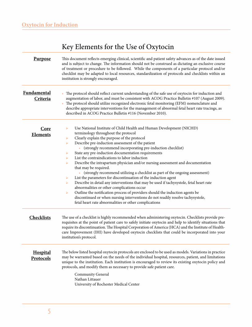
Key Elements for the Use of OxytocinThis document reflects emerging clinical, scientific and patient safety advances as of the date issued and is subject to change. The information should not be construed as dictating an exclusive course of treatment or procedure to be followed. While the components of a particular protocol and/or checklist may be adapted to local resources, standardization of protocols and checklists within an institution is strongly encouraged.
• The protocol should reflect current understanding of the safe use of oxytocin for induction and augmentation of labor, and must be consistent with ACOG Practice Bulletin #107 (August 2009).• The protocol should utilize recognized electronic fetal monitoring (EFM) nomenclature and describe appropriate interventions for the management of abnormal fetal heart rate tracings, as described in ACOG Practice Bulletin #116 (November 2010).
➢ Use National Institute of Child Health and Human Development (NICHD) terminology throughout the protocol ➢ Clearly explain the purpose of the protocol ➢ Describe pre-induction assessment of the patient • (strongly recommend incorporating pre-induction checklist) ➢ State any pre-induction documentation requirements ➢ List the contraindications to labor induction ➢ Describe the intrapartum physician and/or nursing assessment and documentation that may be required. • (strongly recommend utilizing a checklist as part of the ongoing assessment) ➢ List the parameters for discontinuation of the induction agent ➢ Describe in detail any interventions that may be used if tachysystole, fetal heart rate abnormalities or other complications occur ➢ Outline the notification process of providers should the induction agents be discontinued or when nursing interventions do not readily resolve tachysystole, fetal heart rate abnormalities or other complications
The use of a checklist is highly recommended when administering oxytocin. Checklists provide pre-requisites at the point of patient care to safely initiate oxytocin and help to identify situations that require its discontinuation. The Hospital Corporation of America (HCA) and the Institute of Health-care Improvement (IHI) have developed oxytocin checklists that could be incorporated into your institution’s protocol.
The below listed hospital oxytocin protocols are enclosed to be used as models. Variations in practice may be warranted based on the needs of the individual hospital, resources, patient, and limitations unique to the institution. Each institution is encouraged to review its existing oxytocin policy and protocols, and modify them as necessary to provide safe patient care.
Community General Nathan Littauer University of Rochester Medical Center
Oxytocin for Induction
Purpose
FundamentalCriteria
CoreElements

6
Med: Oxytocin for Induction or Augmentation of Labor - Policy
The Jim & DedeWalsh Family Birth
Center
Community GeneralHospital
DECEMBER 2011
Optimizing Protocols in ObstetricsS E R I E S 1

7
DECEMBER 2011
S E R I E S 1
Oxytocin for Induction

8
NURSING INTERVENTIONS
Oxytocin for Induction
9
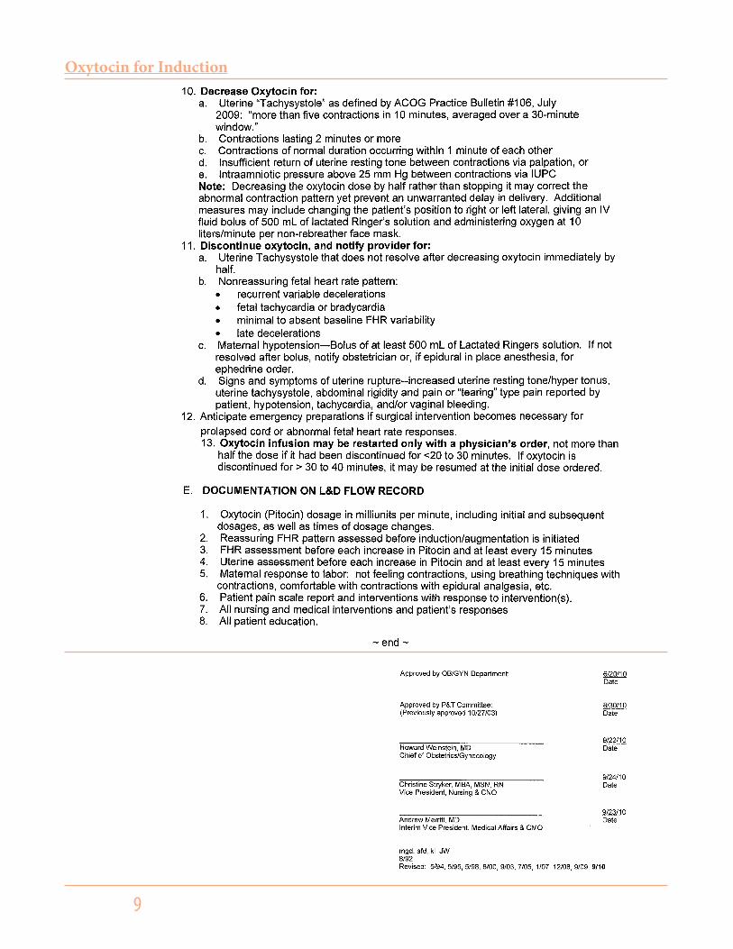
Oxytocin for Induction
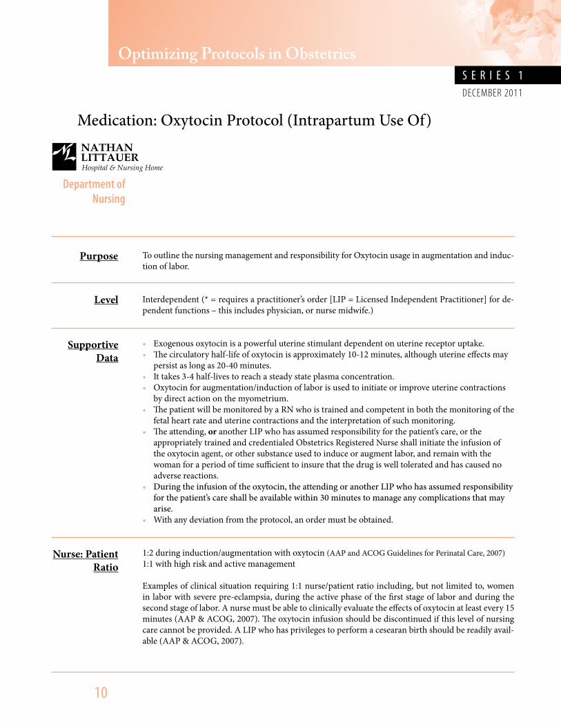
Medication: Oxytocin Protocol (Intrapartum Use Of)
Department ofNursing
To outline the nursing management and responsibility for Oxytocin usage in augmentation and induc-tion of labor.
Interdependent (* = requires a practitioner’s order [LIP = Licensed Independent Practitioner] for de-pendent functions – this includes physician, or nurse midwife.)
• Exogenous oxytocin is a powerful uterine stimulant dependent on uterine receptor uptake. • The circulatory half-life of oxytocin is approximately 10-12 minutes, although uterine effects may persist as long as 20-40 minutes. • It takes 3-4 half-lives to reach a steady state plasma concentration. • Oxytocin for augmentation/induction of labor is used to initiate or improve uterine contractions by direct action on the myometrium. • The patient will be monitored by a RN who is trained and competent in both the monitoring of the fetal heart rate and uterine contractions and the interpretation of such monitoring. • The attending, or another LIP who has assumed responsibility for the patient’s care, or the appropriately trained and credentialed Obstetrics Registered Nurse shall initiate the infusion of the oxytocin agent, or other substance used to induce or augment labor, and remain with the woman for a period of time sufficient to insure that the drug is well tolerated and has caused no adverse reactions.• During the infusion of the oxytocin, the attending or another LIP who has assumed responsibility for the patient’s care shall be available within 30 minutes to manage any complications that may arise.• With any deviation from the protocol, an order must be obtained.
1:2 during induction/augmentation with oxytocin (AAP and ACOG Guidelines for Perinatal Care, 2007)1:1 with high risk and active management
Examples of clinical situation requiring 1:1 nurse/patient ratio including, but not limited to, women in labor with severe pre-eclampsia, during the active phase of the first stage of labor and during the second stage of labor. A nurse must be able to clinically evaluate the effects of oxytocin at least every 15 minutes (AAP & ACOG, 2007). The oxytocin infusion should be discontinued if this level of nursing care cannot be provided. A LIP who has privileges to perform a cesearan birth should be readily avail-able (AAP & ACOG, 2007).
Purpose
Level
SupportiveData
Nurse: PatientRatio
10
DECEMBER 2011
Optimizing Protocols in ObstetricsS E R I E S 1

11
DECEMBER 2011
S E R I E S 1 Two non-emergent inductions may be scheduled per day. The LIP will call the charge nurse to schedule induction (stating indication). The charge nurse will record on desk calendar. In the case of more than two requests for inductions, the Chief of OB/Gyn and Nurse Manager (or her designee) will make final decision based on the nature and intensity of the indication and staffing resources.
*1) Verify that: a. the H&P (performed within 72 hours prior to the beginning of oxytocin) is completed and on medical record; b. a signed informed consent for procedure is completed and on the medical record; c. signed orders are completed and on the medical record; d. fetal position/presentation (via ultrasound) if necessary and cervical exam are documented. 2) Obtain vital signs (BP, P, R, T).3) Assess presence of vaginal bleeding.4) Assess cervical status.5) Obtain a 20-30 minute electronic fetal monitoring strip prior to the initiation of oxytocin. Notify LIP if non-reassuring.6) Explain nursing actions involved during this procedure and the expected effects of oxytocin on labor to the patient. Document education given.7) Position the patient to avoid vena cava compression.
*8) Start primary IV line of 1000ml lactated ringers using #18 gauge cathlon (#20 gauge cathlon if necessary) per IV protocol. Run at 150ml per hour.*9) Obtain pre-mixed Oxytocin 30 units in 500 ml of lactated ringers and label.10) Insert secondary IV line containing oxytocin medication into the most proximal port of the primary IV line (to avoid administering bolus of oxytocin remaining in tubing if primary infusion is run in rapidly.11) Both primary and secondary IV lines must be on IV controller. Do not begin oxytocin regimen if uterine contractions are every two minutes or more frequent.12) Begin oxytocin administration at 2.0 milliunits per minutes (2.0ml per hour).13) Increase oxytocin by 2.0 milliunits per minute (2.0ml per hour) every 30 minutes.14) Titrate oxytocin to maintain contractions of moderate to strong intensity by palpation. For contractions lasting 60-90 seconds and every 2-3 minutes, consider discontinuing the oxytocin.15) Total infusion rate should not exceed 150ml per hour. Decrease main IV rate as oxytocin rate is in- creased to maintain total rate of 150ml per hour.16) Do not exceed 20 milliunits per minute (20ml per hour) without LIP order.17) It may be necessary to use a lower dose to avoid uterine hyperstimulation.18) After spontaneous rupture of membranes (SROM), oxytocin dose may have to be significantly decreased during first stage of labor and every 10 minutes during the second stage of labor.
19) Assess and record BP, T, P, R at least every 4 hours and more frequently when risk factors identified. Maternal temperature every 2 hours if membranes ruptured.20) Assess and document uterine contractions before every dosage increase. If dosage is maintained at the same rate, document uterine activity every 30 minutes. Consider discontinuing oxytocin once labor pattern is established.21) Evaluate and record the FHR every 15 minutes during the active phase of the first stage of labor, and evaluate every 5 minutes during the second stage of labor.22) Initiate Fetal Monitoring Protocol. Fetal monitoring and uterine contractions can be achieved by intermittent auscultation and palpation or continuous fetal monitoring.23) Assess and record I&O every shift. Encourage patient to urinate every 2-3 hours during labor .24) Observe patient for signs/symptoms of water intoxication (such as headache, nausea, vomiting).25) Assess the woman’s emotional status and coping behaviors during labor. Record these assessments, your interventions, and the patient’s response to your interventions every hour .26) In reference to all other labor care and documentation, see Standards of Care for Patients With Routine/Uncomplicated Labor.
SchedulingNon-Emergent
Inductions
InitialAssessment
Initiationand
Titration ofIV Oxytocin
OngoingAssessment
Oxytocin for Induction
12
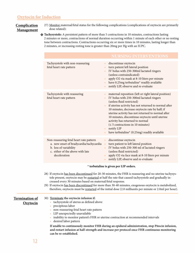
27) Monitor maternal/fetal status for the following complications (complications of oxytocin are primarily dose related):
ComplicationManagement
Tachysystole with non-reassuring fetal heart rate pattern
Tachysystole with reassuring fetal heart rate pattern
Non-reassuring fetal heart rate pattern a. new onset of bradycardia/tachycardia b. less of variability c. either of the above with late deceleration
Tachysystole: A persistent pattern of more than 5 contractions in 10 minutes, contractions lasting 2 minutes or more, contractions of normal duration occurring within 1 minute of each other or no resting tone between contractions. Contractions occurring six or more times in 10 minutes, lasting longer than 2 minutes, or increasing resting tone is greater than 20mg per Hg with an IUPC.
• discontinue oxytocin • turn patient left lateral position • IV bolus with 250-300ml lactated ringers (unless contraindicated) • apply O2 via mask at 8-10 liters per minute • have 0.25mg terbutaline* readily available • notify LIP, observe and re-evaluate
• maternal reposition (left or right lateral position) • IV bolus with 250-300ml lactated ringers (unless fluid restricted)• if uterine activity has not returned to normal after 10 minutes, decrease oxytocin rate by half; if uterine activity has not returned to normal after 10 minutes, discontinue oxytocin until uterine activity has returned to normal (≤ 5 contractions in 10 minutes)• notify LIP • have terbutaline* (0.25mg) readily available
• discontinue oxytocin • turn patient to left lateral position • IV bolus with 250-300 ml of lactated ringers (unless fluid restricted) • apply O2 via face mask at 8-10 liters per minute • notify LIP, observe and re-evaluate
v
NURSING INTERVENTIONS
* terbutaline is given per LIP orders.
28) If oxytocin has been discontinued for 20-30 minutes, the FHR is reassuring and no uterine tachysys- tole present, oxytocin may be restarted at half the rate that caused tachysystole and gradually in- creased every 30 minutes based on maternal/fetal response.29) If oxytocin has been discontinued for more than 30-40 minutes, exogenous oxytocin is metabolized, therefore, oxytocin must be restarted of the initial dose (2.0 milliunits per minute or 2.0ml per hour).
Termination ofOxytocin
30) Terminate the oxytocin infusion if: • tachysystole of uterus as defined above • precipitous labor • non-reassuring fetal heart rate pattern • LIP unexpectedly unavailable • inability to monitor patient’s FHR or uterine contraction at recommended intervals • desired labor pattern If unable to continuously monitor FHR during an epidural administration, stop Pitocin infusion, and restart infusion at half-strength and increase per protocol once FHR continuous monitoring can be re-established.
Oxytocin for Induction
13
Documentation
OxytocinTitration
Table
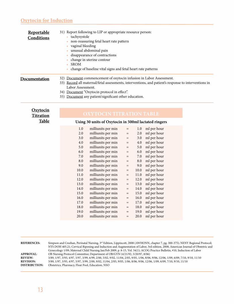
ReportableConditions
31) Report following to LIP or appropriate resource person: • tachysystole • non-reassuring fetal heart rate pattern • vaginal bleeding • unusual abdominal pain • disappearance of contractions • change in uterine contour • SROM • change of baseline vital signs and fetal heart rate patterns
32) Document commencement of oxytocin infusion in Labor Assessment.33) Record all maternal/fetal assessments, interventions, and patient’s response to interventions in Labor Assessment.34) Document “Oxytocin protocol in effect”. 35) Document any patient/significant other education.
OXYTOCIN TITRATION TABLEUsing 30 units of Oxytocin in 500ml lactated ringers
1.0 milliunits per min = 1.0 ml per hour 2.0 milliunits per min = 2.0 ml per hour 3.0 milliunits per min = 3.0 ml per hour 4.0 milliunits per min = 4.0 ml per hour 5.0 milliunits per min = 5.0 ml per hour 6.0 milliunits per min = 6.0 ml per hour 7.0 milliunits per min = 7.0 ml per hour 8.0 milliunits per min = 8.0 ml per hour 9.0 milliunits per min = 9.0 ml per hour 10.0 milliunits per min = 10.0 ml per hour 11.0 milliunits per min = 11.0 ml per hour 12.0 milliunits per min = 12.0 ml per hour 13.0 milliunits per min = 13.0 ml per hour 14.0 milliunits per min = 14.0 ml per hour 15.0 milliunits per min = 15.0 ml per hour 16.0 milliunits per min = 16.0 ml per hour 17.0 milliunits per min = 17.0 ml per hour 18.0 milliunits per min = 18.0 ml per hour 19.0 milliunits per min = 19.0 ml per hour 20.0 milliunits per min = 20.0 ml per hour
REFERENCES: Simpson and Creehan, Perinatal Nursing, 3rd Edition, Lippincott, 2008 (AWHONN, chapter 7, pg. 300-375); NENY Regional Protocol; NYS DOH 405.21; Cervical Ripening and Induction and Augmentation of Labor, 3rd edition, 2008; American Journal of Obstetric and Gynecology 1/09; Maternal Child Nursing Jan/Feb 2009, p. 8-15, Vol. 34(1); ACOG Practice Bulletin, #10, Induction of Labor APPROVAL: OB Nursing Protocol Committee; Department of OB/GYN (4/21/92, 5/20/97, 8/06)REVIEW: 3/89, 1/97, 3/93, 4/97, 5/97, 3/99, 6/99, 2/00, 5/02, 9/02, 11/04, 2/05, 9/05, 1/06, 8/06, 9/06, 12/06, 1/09, 6/09, 7/10, 9/10, 11/10 REVISION: 3/89, 1/97, 3/93, 4/97, 5/97, 3/99, 2/00, 9/02, 11/04, 2/05, 9/05, 1/06, 8/06, 9/06, 12/06, 1/09, 6/09, 7/10, 9/10, 11/10 DISTRIBUTION: Obstetrics, Pharmacy, Float Pool, Education, NSO
Oxytocin for Induction
Standard of Care for the Woman for Induction / Augmentation of Labor
14
Department ofObstetrics and Gynecology
OB/GYN Service
“Induction of labor refers to the iatrogenic stimulation of uterine contractions to accomplish delivery prior to the onset of spontaneous labor” (Wing, 2009). “Augmentation of labor is defines as the use of medical interventions when there is no progression in labor as a result of uterine dystocia or inad-equate uterine contractions” (Simpson, 2008).
Prior to induction/augmentation for any indication it is recommended that a risk benefit analysis and a discussion of the advantages and disadvantages, including the risk of cesarean birth or the possibility of a repeat induction be performed with the patient and her significant other. It is the responsibility of the nurse to ensure that the patient has been fully informed, as outlined above, and has given verbal consent.
Contraindications to induction/augmentation of labor include: · Prior classical uterine incision · Prior transmural uterine incision entering the uterine cavity · Active genital herpes infection · Placenta or vasa previa · Umbilical cord prolapse · Transverse fetal lie (ACOG, 2009)
Oxytocin is the most commonly used medication for the induction/augmentation of labor. Oxytocin is a synthetic product that is chemically and physiologically identical to the hormone oxytocin that is released by the posterior pituitary gland. Oxytocin is picked up by oxytocin receptors on the myome-trium and decidua of the uterus. It is felt that oxytocin facilitates the contraction of smooth muscle cells causing the rhythmic and coordinated contractions of labor. In this way, labor is initiated and coordinated to its ultimate outcome.IP 4.0
Misoprostol is a synthetic prostaglandin E1 analog, presently being used in the treatment and pre-vention of gastric ulcers caused by non steroidal anti inflammatory drugs. Because of its prostaglandin properties misoprostol recently has been used obstetrics for cervical ripening and as an induction agent (Simpson & Creehan, 2008). Misoprostol cannot be used in patients who have had previous uterine surgery (includes a low transverse cesarean section). It should also be noted that women with dysfunctional contraction patterns are not candidates for misoprostol. Misoprostol cannot be used augmentation of labor in women whose contractions began spontaneously or were induced by oxy-tocin. Those patients induced by artificial rupture of membranes may be given misoprostol if their contractions have not started spontaneously.
General Information
Oxytocin
Misoprostol
15
DECEMBER 2011
Optimizing Protocols in ObstetricsS E R I E S 1
15
DECEMBER 2011
S E R I E S 1 1. The patient will maintain optimal physiological and psychological functioning to include: a. Stable vital signs b. Pain rating at a level acceptable to the patient c. Absence of uterine tachysystole d. Absence of adverse effects to the oxytocin or misoprostol e. Stable emotional status 2. The patient and significant other will demonstrate appropriate knowledge of the induction/augmenta- tion procedure and will have indicated informal consent. 3. The fetus will maintain optimal physiological status as evidenced by: a. FHR 110 – 160 bpm b. Absence of indications of fetal compromise (recurrent decelerations and/or absent or minimal variability)
1. Prior to the initiation of the induction/augmentation of labor the nurse will review the record and ensure that the following are present: a. Indications for induction/augmentation of labor. b. If appropriate, documentation of fetal lung maturity c. Appropriate medical and nursing assessment of both maternal and fetal status – includes care provider’s statement indicating necessity for induction/augmentation of labor. The faxed induc- tion sheet meets criteria for the care provider’s statement. d. Appropriate orders – as per the medication, solution and dosing regimen as outlined below. e. Presence of the attending or appropriate representative (Maternal- Fetal medicine attending may cover) in the hospital. f. Notification of NICU regarding expected preterm or potentially compromised neonate. g. Completion of the Oxytocin Checklist (located in the QS system) ensuring that all areas are complete. The nurse will indicate in the appropriate box that: i. the oxytocin order is present. ii. there is documentation of informed consent. iii. the care provider privileged to do a cesarean section is present. iv. there is documentation for the indication for induction/augmentation of labor.2. Prior to the start of the induction/augmentation of labor the following will need to be in place: a. Place the patient on continuous electronic fetal monitoring – uterine and fetal. This allows for proper interpretation of the fetal tolerance to the induction/augmentation process and the absence of uterine tachysystole. For cases in which continuous electronic fetal monitoring is not possible due to inability to trace the fetus due to motion or maternal habitus, intermittent post contraction auscultation every 15 minutes during the first stage of labor and every 5 minutes during the second stage of labor is acceptable. Uterine contractions are to be assessed palpation to identify uterine activity and if uterine tachysystole is present. b. Initiation of intravenous access with a large bore intravenous catheter (preferably an 18 gauge catheter). Lactated Ringers solution should be infusing through a programmable infusion pump. c. Laboratory specimens for a type and screen, CBC with platelets and any other ordered laboratory specimens are to be obtained and sent to the appropriate laboratory setting.3. Assess and review: a. Baseline maternal vital signs b. Pain rating – provide pharmaceutical and non pharmaceutical comfort promoting interventions for relief as identified by the patient. Reassess pain rating one hour from the intervention. c. Review the fetal heart rate tracing for the presence of accelerations, moderate variability, and no recurrent decelerations. d. Report any abnormalities of maternal or fetal findings to the care provider. e. Review the orders with care provider in light of abnormal findings.4. Assess the patient/significant other’s level of understanding about the procedure and determine if in- formed consent was obtained. Verbal acknowledgement by the patient is acceptable. Information is provided to the patient/significant other addressing learning barriers that were identified.
PatientOutcomes
Interventions
Oxytocin for Induction
16
5. Prepare the appropriate equipment as appropriate for the method of labor induction/augmentation. a. Misoprostol 25 microgram tablet (prepared by Pharmacy only) i. Sterile examination gloves ii. Water soluble lubricating jelly b. Oxytocin 30 units into 500 mL normal saline. Solution labeled, as per SMH policy, set up with IV pump tubing and attached at the stopcock of the mainline IV. i. Programmable infusion pump 6. Misoprostol Induction a. Assist the care provider as needed with the insertion of the misoprostol. b. The patient is to be positioned after placement in the either the right or left lateral recumbent position to avoid supine hypotension. c. Misoprostol 25 micrograms may be repeated every 4 – 6 hours, until regular contractions or adequate intensity ensure. No doses higher than 25 micrograms may be used to stimulate labor in viable pregnancies. d. Maternal vital signs and fetal heart rate interpretation is assessed as per stage of labor. e. Repeat doses of misoprostol should be placed if adequate labor has been established. f. If adequate labor is not established after 12 to 24 hours of misoprostol administration, misoprostol may be discontinued and oxytocin administered beginning 4 hours after the last misoprostol dose.7. Oxytocin Induction/Augmentation a. Start the oxytocin infusion using a programmable infusion pump as per care provider’s orders, ensuring the dosing regimen falls within the acceptable standard of care. Always clarify orders with the care provider if orders fall outside the usual dosing regimen. Usual orders include: i. 1 or 2 mUnits/min. and increase by 1– 2 mUnits/min (specifically ordered by care- provider) every 30 minutes until every 2 – 3 minute contractions: absence of uterine tachysystole, and fetal heart rate indicated fetal tolerance of labor (maximum dose: 42 milliunits/min.). The care provider may order the incremental dosage at a frequency greater than every 30 minutes but no less than every 30 minutes. ii. With the present dosing regimen of 1mUnit/min equaling 1 mL/hr, it will never be necessary to alter the concentration of the oxytocin solutions. iii. Increase the infusion as ordered to maintain a rate that stimulates contractions every 2 – 3 minutes, lasting 45 – 90 seconds with an intensity of moderate quality or at least 50 mmHg above the resting tone with an IUPC. Blood pressure is assessed as outlined in the Standard of Care for the Intrapartum Patient. Prior to each increase the mater- nal pulse is assessed and the fetal heart rate monitor strip is reviewed. A pain rating is performed at least every 1 hour. iv. During the induction/augmentation procedure, the patient is maintained in the left or right lateral recumbent or semi recumbent position to avoid vena cava compression.8. Intake and output is maintained throughout the entire induction/augmentation process and recorded every hour. Ensure urinary output is maintained at >30 mL/hr.9. Continuously evaluate the patient for complications associated with the intrapartum use of oxytocin and misoprostol (e.g.: fetal compromise, water intoxication, cardiovascular events, pulmonary edema, tachysystole).10. If uterine tachysystole is present AND the fetal heart rate does not indicate compromise, decrease the oxytocin to the previous dose and notify the care provider. a. Uterine tachysystole is defined as: more than 5 contractions in a 10 minute period and each contraction lasting at least 45 seconds, averaged over 30 minutes.11. Discontinue the oxytocin and notify the attending and the resident for: a. Uterine tachysystole that does not respond to a decrease in oxytocin dose. b. Fetal heart rate pattern demonstrating the following: i. Fetal bradycardia ii. Recurrent late decelerations iii. Recurrent variable decelerations with absent or minimal baseline variability.
Interventions(Continued)
Oxytocin for Induction
17
c. Provide appropriate nursing interventions as related to the following complications: i. Administer oxygen via non rebreather mask, administer an intravenous fluid bolus of lactated Ringers solution, position the patient on her side, notify the care provider, and have terbutaline 0.25 mg available for administration (especially if the patient is receiving misoprostol for induction of labor) if ordered by the care provider; especially if there are indications of fetal compromise (recurrent late decelerations, minimal / absent fetal heart rate variability, prolonged decelerations, bradycardia). d. Notify the care provider if urinary output is <30 mL/hr, cardiovascular effects or pulmonary edema is noted. 12. If oxytocin is discontinued and the uterine and fetal heart rate patterns subsequently normalize, the oxytocin may be restarted at approximately one half the dose for every 10 minutes since discontinuation (e.g. half the discontinued dose if 10 minutes passed, one fourth the dose if 20 minutes have passed, one eighth the dose at 30 minutes).13. Document relevant information on the appropriate records as per SMH standards.
1. On OB electronic system: a. Nursing Admission History b. Laborrecord c. Intake and output d. IV record e. Patient teaching f. Oxytocin Checklist g. History and Physical h. Progress notes2. Faxed Induction Sheet
American College of Obstetricians and Gynecologists (2009). Induction of Labor (Practice Bulletin No. 107). Washington, DC: authorMacones, GA, Hankins, GD, Spong, CY, Hauth, J, & Moore, T. (2008). The 2008 national instititute of child health and human development workshop report on electronic fetal monitoring. Obstetrics & Gynecology 112(3): 661 666.Simpson, KR & Creehan, PA. (2008). AWHONN Perinatal Nursing (3rd Ed). Philadelphia: Lippincott Raven Co. 396 398.Simpson, KR & Poole, JH (2008). Cervical Ripening and Induction and Augmentation of Labor (3rd Ed). Washington DC: Association of Women’s Health, Obstetric, and Neonatal Nurses (AWHONN).
Written by: S. Warren, RNC CNS OB/GYN Nursing J. Weinschreider, RN, MS CNS – OB Safety
Approved by: D. Phillips RNC MS Associate Director of OB/GYN Nursing E. Pressman, MD Director of Obstetrics and Maternal Fetal Medicine
Revised: 2/00, 6/01, 10.01, 2/04, 3/05, 7/05,10/06,2/07, 6/07,12/07, 10/08, 12/09, 8/10
Documentation
References
Interventions(Continued)
Oxytocin for Induction
VOL. 118, NO. 6, DECEMBER 2011 OBSTETRICS & GYNECOLOGY 1473
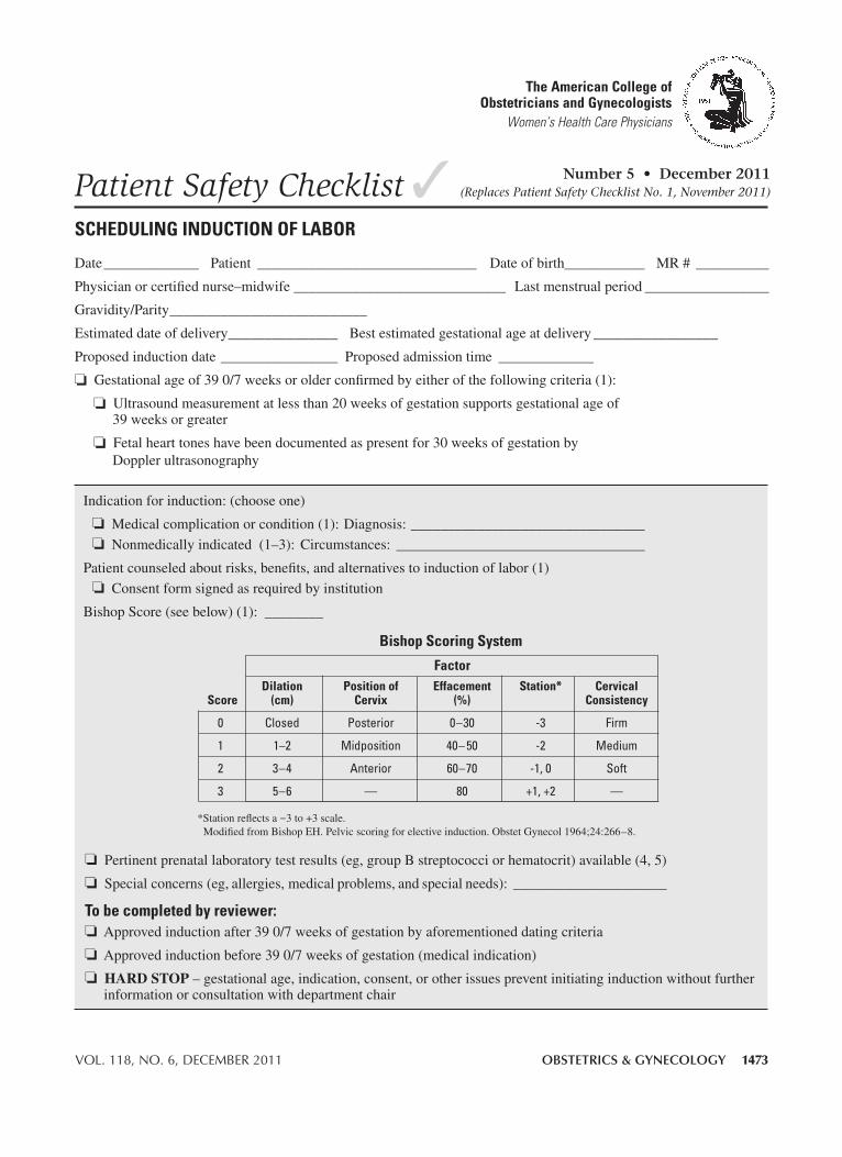
Indicationforinduction:(chooseone)
❏Medicalcomplicationorcondition(1):Diagnosis:________________________________
❏Nonmedicallyindicated(1–3):Circumstances: __________________________________
Patientcounseledaboutrisks,benefits,andalternativestoinductionoflabor(1)
❏Consentformsignedasrequiredbyinstitution
BishopScore(seebelow)(1): ________
❏Pertinentprenatallaboratorytestresults(eg,groupBstreptococciorhematocrit)available(4,5)
❏Specialconcerns(eg,allergies,medicalproblems,andspecialneeds):_____________________
To be completed by reviewer:❏Approvedinductionafter390/7weeksofgestationbyaforementioneddatingcriteria
❏Approvedinductionbefore390/7weeksofgestation(medicalindication)
❏HARD STOP–gestationalage,indication,consent,orotherissuespreventinitiatinginductionwithoutfurtherinformationorconsultationwithdepartmentchair
Date_____________ Patient______________________________ Dateofbirth___________ MR# __________
Physicianorcertifiednurse–midwife_____________________________Lastmenstrualperiod_________________
Gravidity/Parity___________________________
Estimateddateofdelivery_______________ Bestestimatedgestationalageatdelivery_________________
Proposedinductiondate________________Proposedadmissiontime _____________
❏Gestationalageof390/7weeksorolderconfirmedbyeitherofthefollowingcriteria(1):
❏Ultrasoundmeasurementatlessthan20weeksofgestationsupportsgestationalageof39weeksorgreater
❏Fetalhearttoneshavebeendocumentedaspresentfor30weeksofgestationbyDopplerultrasonography
The American College of Obstetricians and Gynecologists
Women’s Health Care Physicians
✓Patient Safety ChecklistSChedulinG induCTiOn Of lAbOr
Number 5 • December 2011(Replaces Patient Safety Checklist No. 1, November 2011)
(continued)
*Stationreflectsa−3to+3scale.ModifiedfromBishopEH.Pelvicscoringforelectiveinduction.ObstetGynecol1964;24:266–8.
bishop Scoring System
factor dilation Position of effacement Station* Cervical Score (cm) Cervix (%) Consistency
0 Closed Posterior 0–30 -3 Firm
1 1–2 Midposition 40– 50 -2 Medium
2 3–4 Anterior 60–70 -1,0 Soft
3 5–6 — 80 +1,+2 —
1474 Patient Safety Checklist Scheduling Induction of Labor OBSTETRICS & GYNECOLOGY
references 1. InductionofLabor.ACOGPracticeBulletinNo.107.AmericanCollegeofObstetriciansandGynecologists.ObstetGynecol
2009;114:386–97.
2. CaugheyAB,SundaramV,KaimalAJ,ChengYW,GiengerA,LittleSE,etal.Maternalandneonataloutcomesofelectiveinductionoflabor.EvidenceReport/TechnologyAssessmentNo.176.(PreparedbytheStanfordUniversity-UCSFEvidence-basedPracticeCenterundercontractNo.290-02-0017.)AHRQPublicationNo.09-E—5.Rockville(MD):AgencyforHealthcareResearchandQuality;2009.
3. ClarkSL,FryeDR,MeyersJA,BelfortMA,DildyGA,KoffordS,etal.Reductioninelectivedelivery<39weeksofgesta-tion:comparativeeffectivenessof3approachestochangeandtheimpactonneonatalintensivecareadmissionandstillbirth.AmJObstetGynecol2010;203:449.e1–449.e6.
4. AmericanAcademyofPediatrics,AmericanCollegeofObstetriciansandGynecologists.Antepartumcare.In:Guidelinesforperinatalcare.6thed.ElkGroveVillage(IL):AAP;Washington,DC:ACOG;2007.p.83–137.
5. AmericanAcademyofPediatrics,AmericanCollegeofObstetriciansandGynecologists.Perinatalinfections.In:Guidelinesforperinatalcare.6thed.ElkGroveVillage(IL):AAP;Washington,DC:ACOG;2007.p.303–48.
Standardization of health care processes and reduced variation has been shown to improve outcomes and quality of care. The American College of Obstetricians and Gynecologists has developed a series of patient safety checklists to help facilitate the standardization process. This checklist reflects emerg-ing clinical, scientific, and patient safety advances as of the date issued and is subject to change. The information should not be construed as dictating an exclusive course of treatment or procedure to be followed. Although the components of a particular checklist may be adapted to local resources, stan-dardization of checklists within an institution is strongly encouraged.
how to use This ChecklistThePatientSafetyChecklistonSchedulingInductionofLaborshouldbecompletedbythehealthcareproviderandsubmittedtotherespectivehospitaltoscheduleaninductionoflabor.Thehospitalshouldestablishprocedurestoreviewtheappropriatenessoftheschedulingbasedontheinformationcontainedinthechecklist.Ahardstopshouldbecallediftherearequestionsthatarisethatrequirefurtherinformationorconsultationwiththedepartmentchair.
Copyright December 2011 by theAmerican College of Obstetricians and Gynecologists, 409 12th Street, SW, PO Box 96920,Washington,DC20090-6920.Allrightsreserved.Nopartofthispublicationmaybereproduced,storedinaretrievalsystem,postedontheInternet,ortransmitted,inanyformorbyanymeans,electronic,mechanical,photocopying,recording,orotherwise,withoutpriorwrittenpermissionfromthepublisher.Requestsforauthorizationtomakephotocopiesshouldbedirectedto:CopyrightClearanceCenter,222RosewoodDrive,Danvers,MA01923,(978)750-8400.
Schedulinginductionoflabor.PatientSafetyChecklistNo.5.AmericanCollegeofObstetriciansandGynecologists.ObstetGynecol2011;118:1473–4.
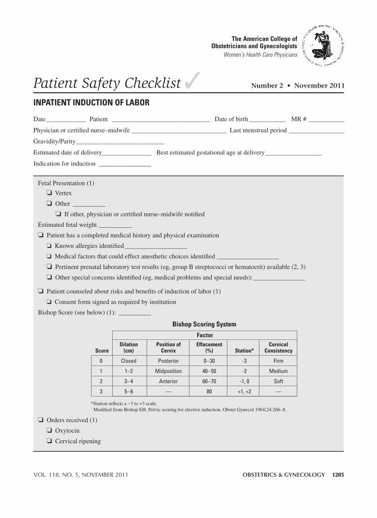
VOL. 118, NO. 5, NOVEMBER 2011 OBSTETRICS & GYNECOLOGY 1205
FetalPresentation(1)
❏Vertex
❏Other __________
❏Ifother,physicianorcertifiednurse–midwifenotified
Estimatedfetalweight__________
❏Patienthasacompletedmedicalhistoryandphysicalexamination
❏Knownallergiesidentified___________________
❏Medicalfactorsthatcouldeffectanestheticchoicesidentified___________________
❏Pertinentprenatallaboratorytestresults(eg,groupBstreptococciorhematocrit)available(2,3)
❏Otherspecialconcernsidentified(eg,medicalproblemsandspecialneeds):________________
❏Patientcounseledaboutrisksandbenefitsofinductionoflabor(1)
❏Consentformsignedasrequiredbyinstitution
BishopScore(seebelow)(1): __________
❏Ordersreceived(1)
❏Oxytocin
❏Cervicalripening
Date____________Patient ______________________________ Dateofbirth___________ MR#___________
Physicianorcertifiednurse–midwife_____________________________Lastmenstrualperiod_________________
Gravidity/Parity___________________________
Estimateddateofdelivery_______________ Bestestimatedgestationalageatdelivery_________________
Indicationforinduction________________
✓Patient Safety ChecklistInpatIent InductIon of Labor
Number 2 • November 2011
the american college of obstetricians and Gynecologists
Women’s Health Care Physicians
*Stationreflectsa−3to+3scale.ModifiedfromBishopEH.Pelvicscoringforelectiveinduction.ObstetGynecol1964;24:266–8.
bishop Scoring System
factor dilation position of effacement cervical Score (cm) cervix (%) Station* consistency
0 Closed Posterior 0–30 -3 Firm
1 1–2 Midposition 40– 50 -2 Medium
2 3–4 Anterior 60–70 -1,0 Soft
3 5–6 — 80 +1,+2 —
1206 Patient Safety Checklist Inpatient Induction of Labor OBSTETRICS & GYNECOLOGY
references 1. Inductionoflabor.ACOGPracticeBulletinNo.107.AmericanCollegeofObstetriciansandGynecologists.ObstetGynecol
2009;114:386–97.
2. AmericanAcademyofPediatrics,AmericanCollegeofObstetriciansandGynecologists.Antepartumcare.In:Guidelinesforperinatalcare.6thed.ElkGroveVillage(IL):AAP;Washington,DC:ACOG;2007.p.83–137.
3. AmericanAcademyofPediatrics,AmericanCollegeofObstetriciansandGynecologists.Perinatalinfections.In:Guidelinesforperinatalcare.6thed.ElkGroveVillage(IL):AAP;Washington,DC:ACOG;2007.p.303–48.
Standardization of health care processes and reduced variation has been shown to improve outcomes and quality of care. The American College of Obstetricians and Gynecologists has developed a series of patient safety checklists to help facilitate the standardization process. This checklist reflects emerg-ing clinical, scientific, and patient safety advances as of the date issued and is subject to change. The information should not be construed as dictating an exclusive course of treatment or procedure to be followed. Although the components of a particular checklist may be adapted to local resources, standardization of checklists within an institution is strongly encouraged.
How to use this checklistThePatientSafetyChecklistonInpatientInductionofLaborshouldbecompletedbythehealthcareprovideratthetime
ofthepatient’sadmission.
CopyrightNovember2011bytheAmericanCollegeofObstetriciansandGynecologists.40912thStreet,SW,POBox96920,Wash-ington,DC20090-6920.Allrightsreserved.Nopartofthispublicationmaybereproduced,storedinaretrievalsystem,postedontheInternet,ortransmitted,inanyformorbyanymeans,electronic,mechanical,photocopying,recording,orotherwise,withoutpriorwrittenpermissionfromthepublisher.Requestsforauthorizationtomakephotocopiesshouldbedirectedto:CopyrightClearanceCenter,222RosewoodDrive,Danvers,MA01923,(978)750-8400.
Inpatientinductionoflabor.PatientSafetyChecklistNo.2.AmericanCollegeofObstetriciansandGynecologists.ObstetGynecol2011;118:1205–6.
386 VOL. 114, NO. 2, PART 1, AUGUST 2009 OBSTETRICS & GYNECOLOGY
CLINICAL MANAGEMENT GUIDELINES FOR OBSTETRICIAN–GYNECOLOGISTS
NUMBER 107, AUGUST 2009
Replaces Practice Bulletin Number 10, November 1999; Committee Opinion Number 228, November1999; Committee Opinion Number 248, December 2000; Committee Opinion Number 283, May 2003
This Practice Bulletin was devel-oped by the ACOG Committee onPractice Bulletins—Obstetrics withthe assistance of Mildred Ramirez,MD, and Susan Ramin, MD. Theinformation is designed to aid prac-titioners in making decisions aboutappropriate obstetric and gyneco-logic care. These guidelines shouldnot be construed as dictating anexclusive course of treatment orprocedure. Variations in practicemay be warranted based on theneeds of the individual patient,resources, and limitations unique tothe institution or type of practice.
Induction of LaborMore than 22% of all gravid women undergo induction of labor in the UnitedStates, and the overall rate of induction of labor in the United States has morethan doubled since 1990 to 225 per 1,000 live births in 2006 (1). The goal ofinduction of labor is to achieve vaginal delivery by stimulating uterine con-tractions before the spontaneous onset of labor. Generally, induction of laborhas merit as a therapeutic option when the benefits of expeditious delivery out-weigh the risks of continuing the pregnancy. The benefits of labor inductionmust be weighed against the potential maternal and fetal risks associated withthis procedure (2). The purpose of this document is to review current methodsfor cervical ripening and induction of labor and to summarize the effectivenessof these approaches based on appropriately conducted outcomes-basedresearch. These practice guidelines classify the indications for and contraindi-cations to induction of labor, describe the various agents used for cervicalripening, cite methods used to induce labor, and outline the requirements for thesafe clinical use of the various methods of inducing labor.
BackgroundIn 1948, Theobald and associates described their use of the posterior pituitaryextract, oxytocin, by intravenous drip for labor induction (3). Five years later,oxytocin was the first polypeptide hormone synthesized by du Vigneaud andassociates (4). This synthetic polypeptide hormone has since been used to stim-ulate uterine contractions. Other methods used for induction of labor includemembrane stripping, amniotomy, nipple stimulation, and administration ofprostaglandin E analogues.
Cervical RipeningThe goal of cervical ripening is to facilitate the process of cervical softening,thinning, and dilating with resultant reduction in the rate of failed induction and
ACOGPRACTICE BULLETIN
THE AMERICAN COLLEGE OFOBSTETRICIANS AND
GYNECOLOGISTSWOMEN’S HEALTH CARE PHYSICIANS
VOL. 114, NO. 2, PART 1, AUGUST 2009 ACOG Practice Bulletin Induction of Labor 387
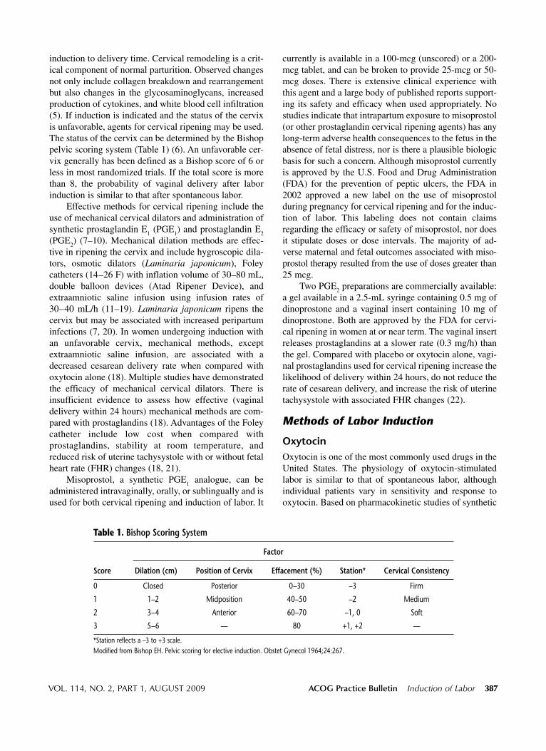
induction to delivery time. Cervical remodeling is a crit-ical component of normal parturition. Observed changesnot only include collagen breakdown and rearrangementbut also changes in the glycosaminoglycans, increasedproduction of cytokines, and white blood cell infiltration(5). If induction is indicated and the status of the cervixis unfavorable, agents for cervical ripening may be used.The status of the cervix can be determined by the Bishoppelvic scoring system (Table 1) (6). An unfavorable cer-vix generally has been defined as a Bishop score of 6 orless in most randomized trials. If the total score is morethan 8, the probability of vaginal delivery after laborinduction is similar to that after spontaneous labor.
Effective methods for cervical ripening include theuse of mechanical cervical dilators and administration ofsynthetic prostaglandin E1 (PGE1) and prostaglandin E2
(PGE2) (7–10). Mechanical dilation methods are effec-tive in ripening the cervix and include hygroscopic dila-tors, osmotic dilators (Laminaria japonicum), Foleycatheters (14–26 F) with inflation volume of 30–80 mL,double balloon devices (Atad Ripener Device), andextraamniotic saline infusion using infusion rates of30–40 mL/h (11–19). Laminaria japonicum ripens thecervix but may be associated with increased peripartuminfections (7, 20). In women undergoing induction withan unfavorable cervix, mechanical methods, exceptextraamniotic saline infusion, are associated with adecreased cesarean delivery rate when compared withoxytocin alone (18). Multiple studies have demonstratedthe efficacy of mechanical cervical dilators. There isinsufficient evidence to assess how effective (vaginaldelivery within 24 hours) mechanical methods are com-pared with prostaglandins (18). Advantages of the Foleycatheter include low cost when compared withprostaglandins, stability at room temperature, andreduced risk of uterine tachysystole with or without fetalheart rate (FHR) changes (18, 21).
Misoprostol, a synthetic PGE1 analogue, can beadministered intravaginally, orally, or sublingually and isused for both cervical ripening and induction of labor. It
currently is available in a 100-mcg (unscored) or a 200-mcg tablet, and can be broken to provide 25-mcg or 50-mcg doses. There is extensive clinical experience withthis agent and a large body of published reports support-ing its safety and efficacy when used appropriately. Nostudies indicate that intrapartum exposure to misoprostol(or other prostaglandin cervical ripening agents) has anylong-term adverse health consequences to the fetus in theabsence of fetal distress, nor is there a plausible biologicbasis for such a concern. Although misoprostol currentlyis approved by the U.S. Food and Drug Administration(FDA) for the prevention of peptic ulcers, the FDA in2002 approved a new label on the use of misoprostolduring pregnancy for cervical ripening and for the induc-tion of labor. This labeling does not contain claimsregarding the efficacy or safety of misoprostol, nor doesit stipulate doses or dose intervals. The majority of ad-verse maternal and fetal outcomes associated with miso-prostol therapy resulted from the use of doses greater than25 mcg.
Two PGE2 preparations are commercially available:a gel available in a 2.5-mL syringe containing 0.5 mg ofdinoprostone and a vaginal insert containing 10 mg ofdinoprostone. Both are approved by the FDA for cervi-cal ripening in women at or near term. The vaginal insertreleases prostaglandins at a slower rate (0.3 mg/h) thanthe gel. Compared with placebo or oxytocin alone, vagi-nal prostaglandins used for cervical ripening increase thelikelihood of delivery within 24 hours, do not reduce therate of cesarean delivery, and increase the risk of uterinetachysystole with associated FHR changes (22).
Methods of Labor Induction
OxytocinOxytocin is one of the most commonly used drugs in theUnited States. The physiology of oxytocin-stimulatedlabor is similar to that of spontaneous labor, althoughindividual patients vary in sensitivity and response tooxytocin. Based on pharmacokinetic studies of synthetic
Table 1. Bishop Scoring System
Factor
Score Dilation (cm) Position of Cervix Effacement (%) Station* Cervical Consistency
0 Closed Posterior 0–30 –3 Firm
1 1–2 Midposition 40–50 –2 Medium
2 3–4 Anterior 60–70 –1, 0 Soft
3 5–6 — 80 +1, +2 —
*Station reflects a –3 to +3 scale.Modified from Bishop EH. Pelvic scoring for elective induction. Obstet Gynecol 1964;24:267.
388 ACOG Practice Bulletin Induction of Labor OBSTETRICS & GYNECOLOGY
oxytocin, uterine response ensues after 3–5 minutes ofinfusion, and a steady level of oxytocin in plasma isachieved by 40 minutes (23). The uterine response tooxytocin depends on the duration of the pregnancy; thereis a gradual increase in response from 20 to 30 weeks ofgestation, followed by a plateau from 34 weeks of gesta-tion until term, when sensitivity increases (24). Lowerbody mass index and greater cervical dilation, parity, orgestational age are predictors of successful response tooxytocin for induction (25).
Membrane StrippingStripping or sweeping the amniotic membranes is com-monly practiced to induce labor. Significant increases inphospholipase A2 activity and prostaglandin F2α (PGF2α)levels occur from membrane stripping (26). Strippingmembranes increases the likelihood of spontaneouslabor within 48 hours and reduces the incidence of induc-tion with other methods (27). Although membrane sweep-ing has been associated with increased risk of prelaborrupture of membranes (28), other published systematicreviews, including one with 1,525 women, have not cor-roborated this finding (27). Women who undergo mem-brane stripping may experience discomfort from theprocedure as well as vaginal bleeding and irregular uter-ine contractions within the ensuing 24 hours (27). Thereare insufficient data to guide clinical practice for mem-brane stripping in women whose group B streptococcusculture is positive.
AmniotomyArtificial rupture of the membranes may be used as amethod of labor induction, especially if the condition ofthe cervix is favorable. Used alone for inducing labor,amniotomy can be associated with unpredictable andsometimes long intervals before the onset of contrac-tions. There is insufficient evidence on the efficacy andsafety of amniotomy alone for labor induction (29). In atrial of amniotomy combined with early oxytocin infusioncompared with amniotomy alone, the induction-to-deliv-ery interval was shorter with the amniotomy-plus-oxy-tocin method (30). There are insufficient data to guide thetiming of amniotomy in patients who are receiving intra-partum prophylaxis for group B streptococcal infection.
Nipple StimulationNipple stimulation or unilateral breast stimulation hasbeen used as a natural and inexpensive nonmedicalmethod for inducing labor. In a systematic review of 6trials including 719 women that compared breast stimu-lation with no intervention, a significant decrease in thenumber of women not in labor at 72 hours was noted, but
only in women with favorable cervices (31). None of thewomen had uterine tachysystole with or without FHRchanges, and there were no differences in meconium-stained amniotic fluid or cesarean delivery rates (31).Breast stimulation was associated with a decrease inpostpartum hemorrhage (31). This method has only beenstudied in low-risk pregnancies.
Labor Induction TerminologyAt a 2008 workshop sponsored by the American Collegeof Obstetricians and Gynecologists, the Eunice KennedyShriver National Institute of Child Health and HumanDevelopment, and the Society for Maternal–Fetal Med-icine on intrapartum electronic FHR monitoring, the defi-nitions for FHR pattern categorization were reviewed andupdated. The existing classification systems for FHR pat-terns were assessed and new recommendations for use inthe United States were made (32). In particular, it wasdetermined that the terms hyperstimulation and hypercon-tractility should be abandoned. It was recommended thatthe term tachysystole, with or without corresponding FHRdecelerations, be used instead.
Uterine ContractionsUterine contractions are quantified as the number of con-tractions present in a 10-minute window, averaged over30 minutes. Contraction frequency alone is a partial assess-ment of uterine activity. Other factors such as duration,intensity, and relaxation time between contractions areequally important in clinical practice. The following rep-resents terminology to describe uterine activity:
• Normal: Five contractions or less in 10 minutes,averaged over a 30-minute window
• Tachysystole: More than five contractions in 10 min-utes, averaged over a 30-minute window
Listed are characteristics of uterine contractions:
• Tachysystole should always be qualified as to thepresence or absence of associated FHR decelera-tions.
• The term tachysystole applies to both spontaneousand stimulated labor. The clinical response to tachy-systole may differ depending on whether contrac-tions are spontaneous or stimulated.
The majority of literature cited in this PracticeBulletin was published prior to the 2008 NICHD defini-tions and interpretations of FHR tracings. Consequently,it is difficult to generalize the results of the cited litera-ture, which used nonstandardized and ambiguous defini-tions for FHR patterns.
VOL. 114, NO. 2, PART 1, AUGUST 2009 ACOG Practice Bulletin Induction of Labor 389
Clinical Considerations andRecommendations
What are the indications and contraindica-tions to induction of labor?
Indications for induction of labor are not absolute butshould take into account maternal and fetal conditions,gestational age, cervical status, and other factors.Following are examples of maternal or fetal conditionsthat may be indications for induction of labor:
• Abruptio placentae
• Chorioamnionitis
• Fetal demise
• Gestational hypertension
• Preeclampsia, eclampsia
• Premature rupture of membranes
• Postterm pregnancy
• Maternal medical conditions (eg, diabetes mellitus,renal disease, chronic pulmonary disease, chronichypertension, antiphospholipid syndrome)
• Fetal compromise (eg, severe fetal growth restric-tion, isoimmunization, oligohydramnios)
Labor also may be induced for logistic reasons, forexample, risk of rapid labor, distance from hospital, orpsychosocial indications. In such circumstances, at leastone of the gestational age criteria in the box should bemet, or fetal lung maturity should be established. Amature fetal lung test result before 39 weeks of gestation,in the absence of appropriate clinical circumstances, isnot an indication for delivery.
The individual patient and clinical situation shouldbe considered in determining when induction of labor iscontraindicated. Generally, the contraindications to laborinduction are the same as those for spontaneous laborand vaginal delivery. They include, but are not limited to,the following situations:
• Vasa previa or complete placenta previa
• Transverse fetal lie
• Umbilical cord prolapse
• Previous classical cesarean delivery
• Active genital herpes infection
• Previous myomectomy entering the endometrial cavity
What criteria should be met before the cervixis ripened or labor is induced?
Assessment of gestational age and consideration of anypotential risks to the mother or fetus are of paramount
importance for appropriate evaluation and counselingbefore initiating cervical ripening or labor induction. Thepatient should be counseled regarding the indications forinduction, the agents and methods of labor stimulation,and the possible need for repeat induction or cesareandelivery. Although prospective studies are limited inevaluating the benefits of elective induction of labor, nul-liparous women undergoing induction of labor withunfavorable cervices should be counseled about a two-fold increased risk of cesarean delivery (33, 34, 35). Inaddition, labor progression differs significantly forwomen with an elective induction of labor comparedwith women who have spontaneous onset of labor (36).Allowing at least 12–18 hours of latent labor beforediagnosing a failed induction may reduce the risk ofcesarean delivery (37, 38).
Additional requirements for cervical ripening andinduction of labor include assessment of the cervix,pelvis, fetal size, and presentation. Monitoring FHR anduterine contractions is recommended as for any high-riskpatient in active labor. Although trained nursing person-nel can monitor labor induction, a physician capable of performing a cesarean delivery should be readily available.
What is the relative effectiveness of availablemethods for cervical ripening in reducing theduration of labor?
A systematic review found that in patients with an unfa-vorable cervix, Foley catheter placement before oxytocininduction significantly reduced the duration of labor(21). This review also concluded that catheter placementresulted in a reduced risk of cesarean delivery. When theFoley catheter was compared with PGE2 gel, the majorityof the studies have found no difference in duration ofinduction to delivery or cesarean delivery rate. The useof prostaglandins is associated with an increased risk oftachysystole with or without FHR changes when com-pared with the Foley catheter (21). The use of differentsize Foley catheters, insufflation volumes, as well as dif-
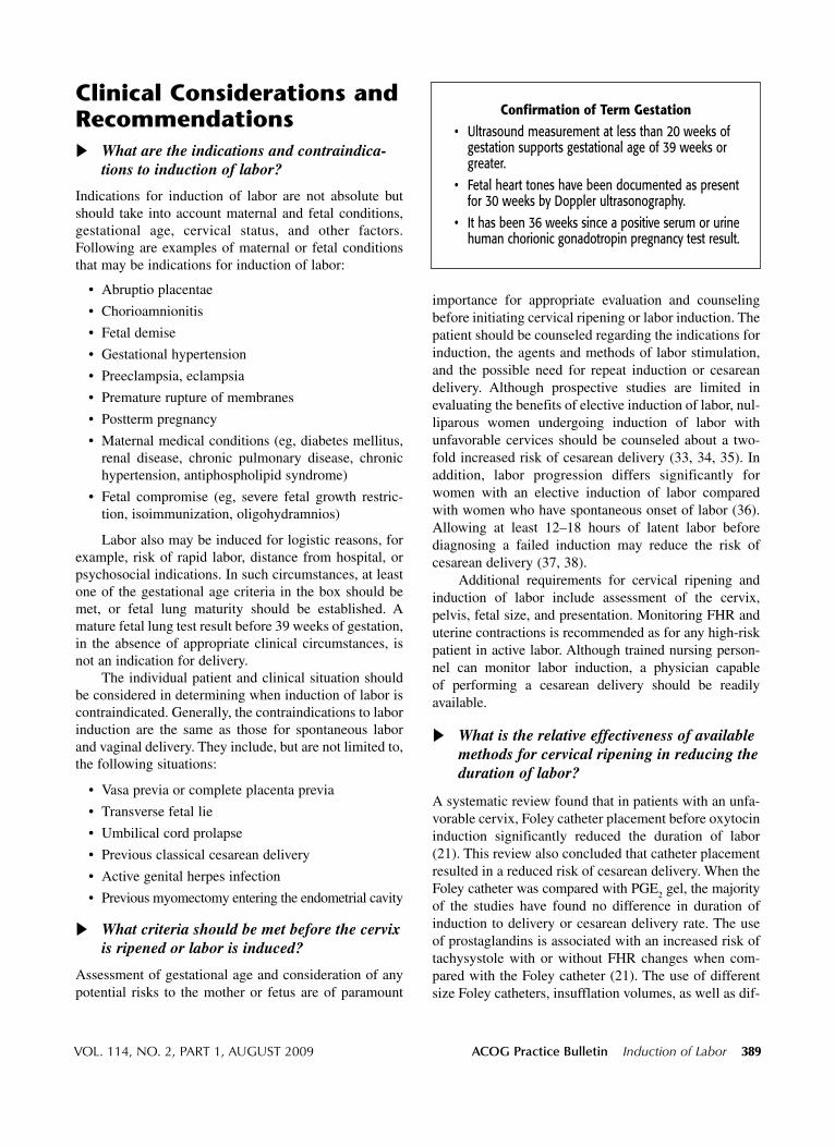
Confirmation of Term Gestation
• Ultrasound measurement at less than 20 weeks ofgestation supports gestational age of 39 weeks orgreater.
• Fetal heart tones have been documented as presentfor 30 weeks by Doppler ultrasonography.
• It has been 36 weeks since a positive serum or urinehuman chorionic gonadotropin pregnancy test result.
390 ACOG Practice Bulletin Induction of Labor OBSTETRICS & GYNECOLOGY
ferent misoprostol protocols, yields inconsistent resultsto determine induction to delivery times, cesarean deliv-ery rate, and risk of meconium passage (18, 21). Theaddition of oxytocin along with the use of the Foleycatheter does not appear to shorten the time of deliveryin a randomized controlled trial (39).
Studies examining extraamniotic saline infusedthrough the Foley catheter compared with use of theFoley catheter with concurrent oxytocin administrationreport conflicting results on the time from induction todelivery (19, 40, 41). Differences in methodology couldexplain the opposing findings. The Foley catheter is areasonable and effective alternative for cervical ripeningand inducing labor.
Intracervical or intravaginal PGE2 (dinoprostone)commonly is used and is superior to placebo or no therapyin promoting cervical ripening (42). Several prospectiverandomized clinical trials and two meta-analyses havedemonstrated that PGE1 (misoprostol) is an effectivemethod for cervical ripening (43–48). Misoprostol admin-istered intravaginally has been reported to be either supe-rior to or as efficacious as dinoprostone gel (48–51).Vaginal misoprostol has been associated with less use ofepidural analgesia, more vaginal deliveries within 24hours, and more uterine tachysystole with or without FHRchanges compared with dinoprostone and oxytocin (48).In contrast, misoprostol compared with oxytocin for cer-vical ripening resulted in longer intervals to active laborand delivery in a randomized controlled trial (52). It is dif-ficult, however, to compare the results of studies on miso-prostol because of differences in endpoints, includingBishop score, duration of labor, total oxytocin use, suc-cessful induction, and cesarean delivery rate. Pharma-cologic methods for cervical ripening do not decrease thelikelihood of cesarean delivery.
How should prostaglandins be administered?
One quarter of an unscored 100-mcg tablet (ie, approxi-mately 25 mcg) of misoprostol should be considered asthe initial dose for cervical ripening and labor induction.The frequency of administration should not be more thanevery 3–6 hours. In addition, oxytocin should not beadministered less than 4 hours after the last misoprostoldose. Misoprostol in higher doses (50 mcg every 6hours) may be appropriate in some situations, althoughhigher doses are associated with an increased risk ofcomplications, including uterine tachysystole with FHRdecelerations.
If there is inadequate cervical change with minimaluterine activity after one dose of intracervical dinopros-tone, a second dose may be given 6–12 hours later. Themanufacturers recommend a maximum cumulative dose
of 1.5 mg of dinoprostone (three doses or 7.5 mL of gel)within a 24-hour period. A minimum safe time intervalbetween prostaglandin administration and initiation ofoxytocin has not been determined. According to themanufacturers’ guidelines, after use of 1.5 mg of dino-prostone in the cervix or 2.5 mg in the vagina, oxytocininduction should be delayed for 6–12 hours because theeffect of prostaglandins may be heightened with oxy-tocin. After use of dinoprostone in sustained-releaseform, delaying oxytocin induction for 30–60 minutesafter removal is sufficient. Limited data are available onthe use of buccal or sublingual misoprostol for cervicalripening or induction of labor, and these methods are notrecommended for clinical use until further studies sup-port their safety (53).
What are the potential complications witheach method of cervical ripening, and howare they managed?
Tachysystole with or without FHR changes is more com-mon with vaginal misoprostol compared with vaginalprostaglandin E2, intracervical prostaglandin E2, and oxy-tocin (48). Tachysystole (defined in some studies as greaterthan 5 uterine contractions in 10 minutes in consecutive10-minute intervals) and tachysystole with associatedFHR decelerations are increased with a 50-mcg or greaterdose of misoprostol (43, 47, 48, 54). There seems to bea trend toward lower rates of uterine tachysystole withFHR changes with lower dosages of misoprostol (25mcg every 6 hours versus every 3 hours) (48).
The use of misoprostol in women with prior cesare-an delivery or major uterine surgery has been associatedwith an increase in uterine rupture and, therefore, shouldbe avoided in the third trimester (55, 56). An increase inmeconium-stained amniotic fluid also has been reportedwith misoprostol use (47, 48). Although misoprostol appearsto be safe and effective in inducing labor in women withunfavorable cervices, further studies are needed to deter-mine the optimal route, dosage, timing interval, and phar-macokinetics of misoprostol. Moreover, data are needed onthe management of complications related to misoprostoluse and when it should be discontinued. If uterine tachy-systole and a Category III FHR tracing (defined as either a sinusoidal pattern or an absent baseline FHR variabilityand any of the following: recurrent late decelerations, recur-rent variable decelerations, or bradycardia) occurs withmisoprostol use and there is no response to routine cor-rective measures (maternal repositioning and supplemen-tal oxygen administration), cesarean delivery should beconsidered (32). Subcutaneous terbutaline also can be usedin an attempt to correct the Category III FHR tracing oruterine tachysystole.
VOL. 114, NO. 2, PART 1, AUGUST 2009 ACOG Practice Bulletin Induction of Labor 391
The intracervical PGE2 gel (0.5 mg) has a 1% rate ofuterine tachysystole with associated FHR changes whilethe intravaginal PGE2 gel (2–5 mg) or vaginal insert isassociated with a 5% rate (42, 57, 58). Uterine tachysys-tole typically begins within 1 hour after the gel or insertis placed but may occur up to 9 1/2 hours after the vagi-nal insert has been placed (57–59).
Removing the PGE2 vaginal insert usually will helpreverse the effect of uterine tachysystole. Irrigation of thecervix and vagina is not beneficial. Maternal side effectsfrom the use of low-dose PGE2 (fever, vomiting, and diar-rhea) are quite uncommon (60). Prophylactic antiemetics,antipyretics, and antidiarrheal agents usually are not needed. The manufacturers recommend that caution beexercised when using PGE2 in patients with glaucoma,severe hepatic or renal dysfunction, or asthma. However,PGE2 is a bronchodilator, and there are no reports of bron-choconstriction or significant blood pressure changes afterthe administration of the low-dose gel.
Increased maternal and neonatal infections have beenreported in connection with the use of Laminaria japon-icum and hygroscopic dilators when compared with thePGE2 analogues (7, 13, 20). The Foley catheter can causesignificant vaginal bleeding in women with a low-lyingplacenta (21). Other reported complications include rup-ture of membranes, febrile morbidity, and displacement ofthe presenting part (61).
What are the recommended guidelines forfetal surveillance after prostaglandin use?
The prostaglandin preparations should be administeredwhere uterine activity and the FHR can be monitoredcontinuously for an initial observation period. Furthermonitoring can be governed by individual indications forinduction and fetal status.
The patient should remain recumbent for at least 30minutes. The FHR and uterine activity should be moni-tored continuously for a period of 30 minutes to 2 hoursafter administration of the PGE2 gel (62). Uterine con-tractions usually are evident in the first hour and exhibitpeak activity in the first 4 hours (62, 63). The FHR mon-itoring should be continued if regular uterine contrac-tions persist; maternal vital signs also should berecorded.
Are cervical ripening methods appropriate inan outpatient setting?
Limited information is available on the safety of outpa-tient management of induction of labor. In a randomized,double-blind, controlled trial comparing 2 mg of intrav-aginal PGE2 gel with placebo for 5 consecutive days as
an outpatient procedure, it was noted that PGE2 gel waseffective and safe for initiation of labor in women at termwith a Bishop score of 6 or less (64). No significant dif-ferences in adverse outcomes were noted in another ran-domized trial of 300 women at term comparing the useof controlled-release PGE2 in an outpatient versus inpa-tient setting (65). Larger controlled studies are needed toestablish an effective and safe dose and vehicle for PGE2
before use on an outpatient basis can be recommended.However, outpatient use may be appropriate in carefullyselected patients. Mechanical methods may be particu-larly appropriate in the outpatient setting. A randomizedtrial comparing the Foley catheter in an outpatient versusinpatient setting for preinduction cervical ripeningdemonstrated similar efficacy and safety with a reduc-tion of hospital stay of 9.6 hours (66).
What are the potential complications of various methods of induction?
The side effects of oxytocin use are principally doserelated; uterine tachysystole and Category II or III FHRtracings are the most common side effects. Uterine tachy-systole may result in abruptio placentae or uterine rupture.Uterine rupture secondary to oxytocin use is rare even inparous women (67). Water intoxication can occur withhigh concentrations of oxytocin infused with large quanti-ties of hypotonic solutions, but is rare in doses used forlabor induction.
Misoprostol appears to be safe and beneficial forinducing labor in a woman with an unfavorable cervix.Although the exact incidence of uterine tachysystolewith or without FHR changes is unknown and the crite-ria used to define this complication are not always clearin the various reports, there are reports of uterinetachysystole with or without FHR changes occurringmore frequently in women given misoprostol comparedwith women given PGE2 (43, 45, 48, 68). There does notappear to be a significant increase in adverse fetal out-comes from tachysystole without associated FHR decel-erations (68, 69). The occurrence of complications doesappear to be dose-dependent (10, 48). Clinical trials haveshown that at an equivalent dosage, the vaginal routeproduces greater clinical efficacy than the oral route(53). Oral misoprostol administration is associated withfewer abnormal FHR patterns and episodes of uterinetachy-systole with associated FHR changes when com-pared with vaginal administration (70, 71).
The potential risks associated with amniotomyinclude prolapse of the umbilical cord, chorioamnionitis,significant umbilical cord compression, and rupture ofvasa previa. The physician should palpate for an umbili-cal cord and avoid dislodging the fetal head. The FHR
392 ACOG Practice Bulletin Induction of Labor OBSTETRICS & GYNECOLOGY
should be assessed before and immediately after amni-otomy. Amniotomy for induction of labor may be con-traindicated in women known to have HIV infectionbecause duration of ruptured membranes has been iden-tified as an independent risk factor for vertical transmis-sion of HIV infection (29).
Stripping the amniotic membranes is associated withbleeding from undiagnosed placenta previa or low-lyingplacenta, and accidental amniotomy. Bilateral breast stim-ulation has been associated with uterine tachysystole withassociated FHR decelerations. In a systematic review,breast stimulation was associated with an increased trendin perinatal death (31). Until safety issues are studied fur-ther, this practice is not recommended in an unmonitoredsetting.
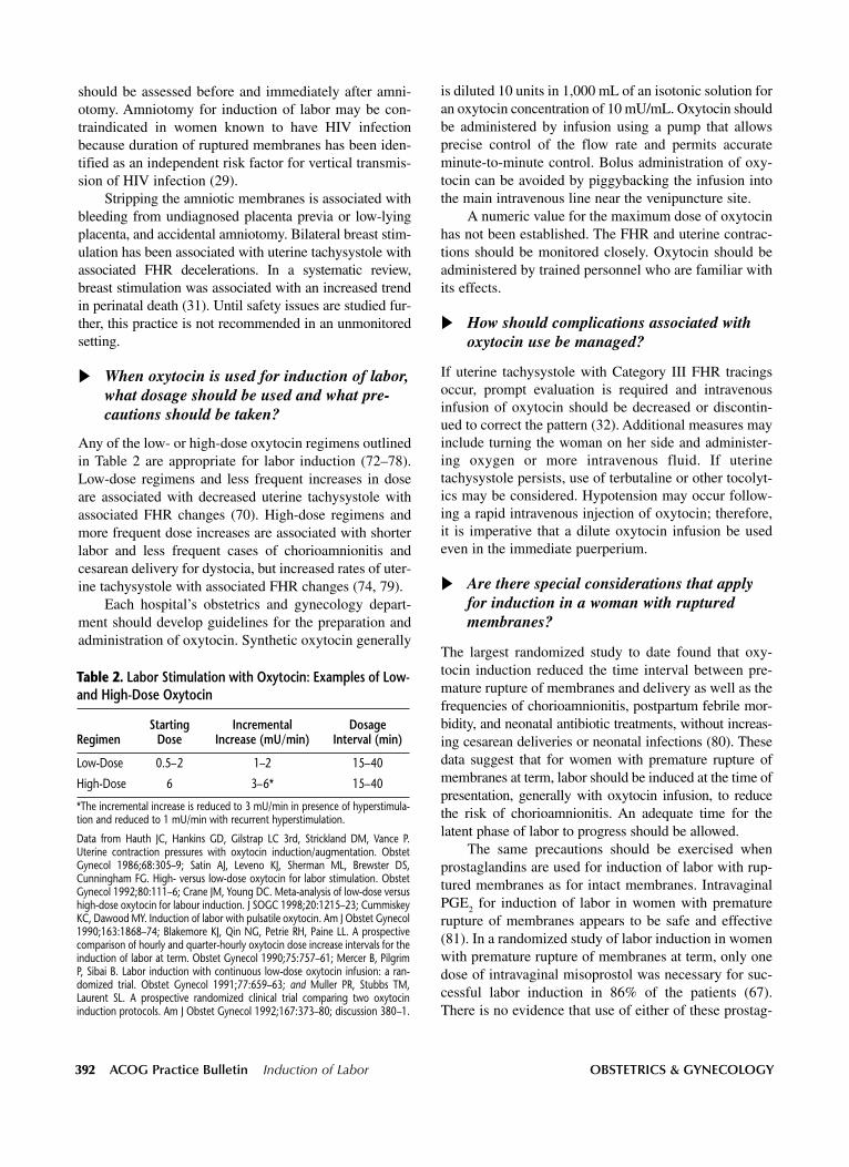
When oxytocin is used for induction of labor,what dosage should be used and what pre-cautions should be taken?
Any of the low- or high-dose oxytocin regimens outlinedin Table 2 are appropriate for labor induction (72–78).Low-dose regimens and less frequent increases in doseare associated with decreased uterine tachysystole withassociated FHR changes (70). High-dose regimens andmore frequent dose increases are associated with shorterlabor and less frequent cases of chorioamnionitis andcesarean delivery for dystocia, but increased rates of uter-ine tachysystole with associated FHR changes (74, 79).
Each hospital’s obstetrics and gynecology depart-ment should develop guidelines for the preparation andadministration of oxytocin. Synthetic oxytocin generally
is diluted 10 units in 1,000 mL of an isotonic solution foran oxytocin concentration of 10 mU/mL. Oxytocin shouldbe administered by infusion using a pump that allowsprecise control of the flow rate and permits accurateminute-to-minute control. Bolus administration of oxy-tocin can be avoided by piggybacking the infusion intothe main intravenous line near the venipuncture site.
A numeric value for the maximum dose of oxytocinhas not been established. The FHR and uterine contrac-tions should be monitored closely. Oxytocin should beadministered by trained personnel who are familiar withits effects.
How should complications associated withoxytocin use be managed?
If uterine tachysystole with Category III FHR tracingsoccur, prompt evaluation is required and intravenousinfusion of oxytocin should be decreased or discontin-ued to correct the pattern (32). Additional measures mayinclude turning the woman on her side and administer-ing oxygen or more intravenous fluid. If uterinetachysystole persists, use of terbutaline or other tocolyt-ics may be considered. Hypotension may occur follow-ing a rapid intravenous injection of oxytocin; therefore,it is imperative that a dilute oxytocin infusion be usedeven in the immediate puerperium.
Are there special considerations that applyfor induction in a woman with rupturedmembranes?
The largest randomized study to date found that oxy-tocin induction reduced the time interval between pre-mature rupture of membranes and delivery as well as thefrequencies of chorioamnionitis, postpartum febrile mor-bidity, and neonatal antibiotic treatments, without increas-ing cesarean deliveries or neonatal infections (80). Thesedata suggest that for women with premature rupture of membranes at term, labor should be induced at the time ofpresentation, generally with oxytocin infusion, to reducethe risk of chorioamnionitis. An adequate time for thelatent phase of labor to progress should be allowed.
The same precautions should be exercised whenprostaglandins are used for induction of labor with rup-tured membranes as for intact membranes. IntravaginalPGE2 for induction of labor in women with prematurerupture of membranes appears to be safe and effective(81). In a randomized study of labor induction in womenwith premature rupture of membranes at term, only onedose of intravaginal misoprostol was necessary for suc-cessful labor induction in 86% of the patients (67).There is no evidence that use of either of these prostag-
Table 2. Labor Stimulation with Oxytocin: Examples of Low-and High-Dose Oxytocin
Starting Incremental Dosage Regimen Dose Increase (mU/min) Interval (min)
Low-Dose 0.5–2 1–2 15–40
High-Dose 6 3–6* 15–40
*The incremental increase is reduced to 3 mU/min in presence of hyperstimula-tion and reduced to 1 mU/min with recurrent hyperstimulation.
Data from Hauth JC, Hankins GD, Gilstrap LC 3rd, Strickland DM, Vance P.Uterine contraction pressures with oxytocin induction/augmentation. ObstetGynecol 1986;68:305–9; Satin AJ, Leveno KJ, Sherman ML, Brewster DS,Cunningham FG. High- versus low-dose oxytocin for labor stimulation. ObstetGynecol 1992;80:111–6; Crane JM, Young DC. Meta-analysis of low-dose versushigh-dose oxytocin for labour induction. J SOGC 1998;20:1215–23; CummiskeyKC, Dawood MY. Induction of labor with pulsatile oxytocin. Am J Obstet Gynecol1990;163:1868–74; Blakemore KJ, Qin NG, Petrie RH, Paine LL. A prospectivecomparison of hourly and quarter-hourly oxytocin dose increase intervals for theinduction of labor at term. Obstet Gynecol 1990;75:757–61; Mercer B, PilgrimP, Sibai B. Labor induction with continuous low-dose oxytocin infusion: a ran-domized trial. Obstet Gynecol 1991;77:659–63; and Muller PR, Stubbs TM,Laurent SL. A prospective randomized clinical trial comparing two oxytocininduction protocols. Am J Obstet Gynecol 1992;167:373–80; discussion 380–1.
VOL. 114, NO. 2, PART 1, AUGUST 2009 ACOG Practice Bulletin Induction of Labor 393
landins increases the risk of infection in women withruptured membranes (67, 81). There is insufficient evi-dence to guide the physician on use of mechanical dila-tors in women with ruptured membranes.
A meta-analysis that included 6,814 women with pre-mature rupture of membranes at term compared inductionof labor with prostaglandins or oxytocin to expectantmanagement (82). A significant reduction in the risk ofwomen developing chorioamnionitis or endometritis and areduced number of neonates requiring admission to theneonatal intensive care unit was noted in the women whounderwent induction of labor compared with expectantmanagement (82).
What methods can be used for induction oflabor with intrauterine fetal demise in thelate second or third trimester?
The method and timing of delivery after a fetal deathdepends on the gestational age at which the death occur-red, on the maternal history of a previous uterine scar, andmaternal preference. Although most patients will desireprompt delivery, the timing of delivery is not critical;coagulopathies are associated with prolonged fetal reten-tion and are uncommon. In the second trimester, dilationand evacuation can be offered if an experienced healthcare provider is available, although patients should becounseled that dilation and evacuation may limit efficacyof autopsy for the detection of macroscopic fetal abnor-malities.
Labor induction is appropriate at later gestationalages, if second-trimester dilation and evacuation is un-available, or based on patient preference. Much of thedata for management of fetal demise has been extrapo-lated from randomized trials of management of secondtrimester pregnancy termination. Available evidence fromrandomized trials supports the use of vaginal misopros-tol as a medical treatment to terminate nonviable preg-nancies before 24 weeks of gestation (83). Based onlimited data, the use of misoprostol between 24 to 28weeks of gestation also appears to be safe and effective(84, 85). Before 28 weeks of gestation, vaginal miso-prostol appears to be the most efficient method of laborinduction, regardless of cervical Bishop score (84, 86),although high-dose oxytocin infusion also is an accept-able choice (87, 88). Typical dosages for misoprostol useare 200–400 mcg vaginally every 4–12 hours. After 28weeks of gestation, induction of labor should be managedaccording to usual obstetric protocols. Cesarean deliveryfor fetal demise should be reserved for unusual circum-stances because it is associated with potential maternalmorbidity without any fetal benefit.
Several studies have evaluated the use of misopros-tol at a dosage of 400 mcg every 6 hours in women witha stillbirth up to 28 weeks of gestation and a prior uter-ine scar (85, 89). There does not appear to be an increasein complications in those women. Further research isrequired to assess effectiveness and safety, optimal routeof administration, and dose.
In patients after 28 weeks of gestation, cervical ripen-ing with a transcervical Foley catheter has been associatedwith uterine rupture rates comparable to spontaneouslabor (90) and this may be a helpful adjunct in patientswith an unfavorable cervical assessment. Therefore, inpatients with a prior low transverse cesarean delivery, trialof labor remains a favorable option. There are limited datato guide clinical practice in a patient with a prior classicalcesarean delivery, and the delivery plan should be individ-ualized.
Summary ofRecommendations andConclusionsThe following recommendations and conclusionsare based on good and consistent scientific evi-dence (Level A):
Prostaglandin E analogues are effective for cervicalripening and inducing labor.
Low- or high-dose oxytocin regimens are appropri-ate for women in whom induction of labor is indi-cated (Table 2).
Before 28 weeks of gestation, vaginal misoprostolappears to be the most efficient method of laborinduction regardless of Bishop score, although high-dose oxytocin infusion also is an acceptable choice.
Approximately 25 mcg of misoprostol should beconsidered as the initial dose for cervical ripeningand labor induction. The frequency of administra-tion should not be more than every 3–6 hours.
Intravaginal PGE2 for induction of labor in womenwith premature rupture of membranes appears to besafe and effective.
The use of misoprostol in women with prior cesareandelivery or major uterine surgery has been associatedwith an increase in uterine rupture and, therefore,should be avoided in the third trimester.
The Foley catheter is a reasonable and effectivealternative for cervical ripening and inducing labor.
394 ACOG Practice Bulletin Induction of Labor OBSTETRICS & GYNECOLOGY
The following recommendation is based on evi-dence that may be limited or inconsistent (Level B)
Misoprostol (50 mcg every 6 hours) to induce labormay be appropriate in some situations, althoughhigher doses are associated with an increased risk ofcomplications, including uterine tachysystole withFHR decelerations.
Proposed PerformanceMeasurePercentage of patients in whom gestational age is estab-lished by clinical criteria when labor is being induced forlogistic or psychosocial indications
References1. Martin JA, Hamilton BE, Sutton PD, Ventura SJ,
Menacker F, Kirmeyer S, et al. Births: final data for 2006.Natl Vital Stat Rep 2009;57:1–102. (Level II-3)
2. Agency for Healthcare Research and Quality. Maternaland neonatal outcomes of elective induction of labor.AHRQ Evidence Report/Technology Assessment No.176. Rockville (MD): AHRQ; 2009. (Systematic review)
3. Theobald GW, Graham A, Campbell J, Gange PD,Driscoll WJ. Use of post-pituitary extract in obstetrics. Br Med J 1948;2:123–7. (Level III)
4. du Vigneaud V, Ressler C, Swan JM, Roberts CW,Katsoyannis PG, Gordon S. The synthesis of an octapep-tide amide with the hormonal activity of oxytocin. J Am Chem Soc 1953;75:4879–80. (Level III)
5. Smith R. Parturition. N Engl J Med 2007;356:271–83.(Level III)
6. Bishop EH. Pelvic scoring for elective induction. ObstetGynecol 1964;24:266–8. (Level III)
7. Krammer J, Williams MC, Sawai SK, O’Brien WF. Pre-induction cervical ripening: a randomized comparison oftwo methods. Obstet Gynecol 1995;85:614–8. (Level I)
8. Fletcher HM, Mitchell S, Simeon D, Frederick J, Brown D.Intravaginal misoprostol as a cervical ripening agent. Br JObstet Gynaecol 1993;100:641–4. (Level I)
9. Porto M. The unfavorable cervix: methods of cervicalpriming. Clin Obstet Gynecol 1989;32:262–8. (Level III)
10. Wing DA, Jones MM, Rahall A, Goodwin TM, Paul RH.A comparison of misoprostol and prostaglandin E2 gel forpreinduction cervical ripening and labor induction. Am JObstet Gynecol 1995;172:1804–10. (Level I)
11. Atad J, Hallak M, Ben-David Y, Auslender R, Abramovici H.Ripening and dilatation of the unfavourable cervix forinduction of labour by a double balloon device: experi-ence with 250 cases. Br J Obstet Gynaecol 1997;104:29–32. (Level III)
12. Blumenthal PD, Ramanauskas R. Randomized trial ofDilapan and Laminaria as cervical ripening agents beforeinduction of labor. Obstet Gynecol 1990;75:365–8. (Level I)
13. Chua S, Arulkumaran S, Vanaja K, Ratnam SS. Preinduc-tion cervical ripening: prostaglandin E2 gel vs hygroscopicmechanical dilator. J Obstet Gynaecol Res 1997;23:171–7. (Level I)
14. Gilson GJ, Russell DJ, Izquierdo LA, Qualls CR, CuretLB. A prospective randomized evaluation of a hygroscopiccervical dilator, Dilapan, in the preinduction ripening ofpatients undergoing induction of labor. Am J ObstetGynecol 1996;175:145–9. (Level I)
15. Lin A, Kupferminc M, Dooley SL. A randomized trial ofextra-amniotic saline infusion versus laminaria for cervi-cal ripening. Obstet Gynecol 1995;86:545–9. (Level I)
16. Lyndrup J, Nickelsen C, Weber T, Molnitz E, Guldbaek E.Induction of labour by balloon catheter with extra-amni-otic saline infusion (BCEAS): a randomised comparisonwith PGE2 vaginal pessaries. Eur J Obstet GynecolReprod Biol 1994;53:189–97. (Level I)
17. Mullin PM, House M, Paul RH, Wing DA. A comparisonof vaginally administered misoprostol with extra-amnioticsaline solution infusion for cervical ripening and laborinduction. Am J Obstet Gynecol 2002;187:847–52. (Level I)
18. Boulvain M, Kelly A, Lohse C, Stan C, Irion O.Mechanical methods for induction of labour. CochraneDatabase of Systematic Reviews 2001, Issue 4. Art. No.: CD001233. DOI: 10.1002/14651858.CD001233.(Level III)
19. Guinn DA, Davies JK, Jones RO, Sullivan L, Wolf D.Labor induction in women with an unfavorable Bishopscore: randomize controlled trial of intrauterine Foleycatheter with concurrent oxytocin infusion versus Foleycatheter with extra-amniotic saline infusion with concur-rent oxytocin infusion. Am J Obstet Gynecol 2004;191:225–9. (Level I)
20. Kazzi GM, Bottoms SF, Rosen MG. Efficacy and safetyof Laminaria digitata for preinduction ripening of thecervix. Obstet Gynecol 1982;60:440–3. (Level II-2)
21. Gelber S, Sciscione A. Mechanical methods of cervicalripening and labor induction. Clin Obstet Gynecol 2006;49:642–57. (Level III)
22. Kelly AJ, Kavanagh J, Thomas J. Vaginal prostaglandin(PGE2 and PGF2a) for induction of labour at term.Cochrane Database of Systematic Reviews 2003, Issue 4.Art. No.: CD003101. DOI: 10.1002/14651858.CD003101.(Level III)
23. Seitchik J, Amico J, Robinson AG, Castillo M. Oxytocinaugmentation of dysfunctional labor. IV. Oxytocin phar-macokinetics. Am J Obstet Gynecol 1984;150:225–8.(Level III)
24. Caldeyro-Barcia R, Poseiro JJ. Physiology of the uterinecontraction. Clin Obstet Gynecol 1960;3:386–408. (LevelIII)
25. Satin AJ, Leveno KJ, Sherman ML, McIntire DD. Factorsaffecting the dose response to oxytocin for labor stimula-tion. Am J Obstet Gynecol 1992;166:1260–1. (Level II-3)
VOL. 114, NO. 2, PART 1, AUGUST 2009 ACOG Practice Bulletin Induction of Labor 395
26. McColgin SW, Bennett WA, Roach H, Cowan BD,Martin JN Jr, Morrison JC. Parturitional factors associatedwith membrane stripping. Am J Obstet Gynecol 1993;169:71–7. (Level I)
27. Boulvain M, Stan C, Irion O. Membrane sweeping forinduction of labour. Cochrane Database of SystematicReviews 2005, Issue 1. Art. No.: CD000451. DOI: 10.1002/14651858.CD000451.pub2. (Level III)
28. Hill MJ, McWilliams GD, Garcia-Sur D, Chen B, Munroe M,Hoeldtke NJ. The effect of membrane sweeping on prela-bor rupture of membranes: a randomized controlled trial.Obstet Gynecol 2008;111:1313–9. (Level I)
29. Bricker L, Luckas M. Amniotomy alone for induction oflabour. Cochrane Database of Systematic Reviews 2000,Issue 4. Art. No.: CD002862. DOI: 10.1002/14651858.CD002862. (Level III)
30. Moldin PG, Sundell G. Induction of labour: a randomisedclinical trial of amniotomy versus amniotomy with oxy-tocin infusion. Br J Obstet Gynaecol 1996;103:306–12.(Level I)
31. Kavanagh J, Kelly AJ, Thomas J. Breast stimulation forcervical ripening and induction of labour. CochraneDatabase of Systematic Reviews 2005, Issue 3. Art. No.:CD003392. DOI: 10.1002/14651858.CD003392.pub2.(Level III)
32. Macones GA, Hankins GDV, Spong CY, Hauth J, Moore T.The 2008 National Institute of Child Health and HumanDevelopment Workshop Report on Electronic FetalMonitoring. Obstet Gynecol 2008;11:661–6. (Level III)
33. Moore LE, Rayburn WF. Elective induction of labor. ClinObstet Gynecol 2006;49:698–704. (Level III)
34. Luthy DA, Malmgren JA, Zingheim RW. Cesarean deliv-ery after elective induction in nulliparous women: thephysician effect. Am J Obstet Gynecol 2004;191:1511–5.(Level II-2)
35. Vrouenraets FP, Roumen FJ, Dehing CJ, van den Akker ES,Aarts MJ, Scheve EJ. Bishop score and risk of cesareandelivery after induction of labor in nulliparous women.Obstet Gynecol 2005;105:690–7. (Level II-2)
36. Vahratian A, Zhang J, Troendle JF, Sciscione AC,Hoffman MK. Labor progression and risk of cesareandelivery in electively induced nulliparas. Obstet Gynecol2005;105:698–704. (Level II-2)
37. Rouse DJ, Owen J, Hauth JC. Criteria for failed laborinduction: prospective evaluation of a standardized proto-col. Obstet Gynecol 2000;96:671–7. (Level II-3)
38. Simon CE, Grobman WA. When has an induction failed?Obstet Gynecol 2005;105:705–9. (Level II-2)
39. Pettker CM, Pocock SB, Smok DP, Lee SM, Devine PC.Transcervical Foley catheter with and without oxytocinfor cervical ripening: a randomized controlled trial.Obstet Gynecol 2008;111:1320–6. (Level I)
40. Karjane NW, Brock EL, Walsh SW. Induction of laborusing a Foley balloon, with and without extra-amniotic salineinfusion. Obstet Gynecol 2006;107:234–9. (Level II-1)
41. Lin MG, Reid KJ, Treaster MR, Nuthalapaty FS, Ramsey PS,Lu GC. Transcervical Foley catheter with and without
extraamniotic saline infusion for labor induction: a ran-domized controlled trial. Obstet Gynecol 2007;110:558–65. (Level I)
42. Rayburn WF. Prostaglandin E2 gel for cervical ripeningand induction of labor: a critical analysis. Am J ObstetGynecol 1989;160:529–34. (Level III)
43. Buser D, Mora G, Arias F. A randomized comparisonbetween misoprostol and dinoprostone for cervical ripen-ing and labor induction in patients with unfavorable cer-vices. Obstet Gynecol 1997;89:581–5. (Level I)
44. Sanchez-Ramos L, Kaunitz AM, Wears RL, Delke I,Gaudier FL. Misoprostol for cervical ripening and laborinduction: a meta-analysis. Obstet Gynecol 1997;89:633–42. (Level III)
45. Sanchez-Ramos L, Peterson DE, Delke I, Gaudier FL,Kaunitz AM. Labor induction with prostaglandin E1misoprostol compared with dinoprostone vaginal insert: arandomized trial. Obstet Gynecol 1998;91:401–5. (Level I)
46. Srisomboon J, Piyamongkol W, Aiewsakul P. Comparisonof intracervical and intravaginal misoprostol for cervicalripening and labour induction in patients with anunfavourable cervix. J Med Assoc Thai 1997;80:189–94.(Level I)
47. Wing DA, Rahall A, Jones MM, Goodwin TM, Paul RH.Misoprostol: an effective agent for cervical ripening andlabor induction. Am J Obstet Gynecol 1995;172:1811–6.(Level I)
48. Hofmeyr GJ, Gulmezoglu AM. Vaginal misoprostol forcervical ripening and induction of labour. CochraneDatabase of Systematic Reviews 2003, Issue 1. Art. No.:CD000941. DOI: 10.1002/14651858.CD000941. (Level III)
49. Gregson S, Waterstone M, Norman I, Murrells T. A ran-domised controlled trial comparing low dose vaginalmisoprostol and dinoprostone vaginal gel for inducinglabour at term. BJOG 2005;112:438–44. (Level I)
50. Garry D, Figueroa R, Kalish RB, Catalano CJ, Maulik D.Randomized controlled trial of vaginal misoprostol versusdinoprostone vaginal insert for labor induction. J MaternFetal Neonatal Med 2003;13:254–9. (Level I)
51. Pandis GK, Papageorghiou AT, Ramanathan VG,Thompson MO, Nicolaides KH. Preinduction sonographicmeasurement of cervical length in the prediction of suc-cessful induction of labor. Ultrasound Obstet Gynecol2001;18:623–8. (Level II-3)
52. Fonseca L, Wood HC, Lucas MJ, Ramin SM, Phatak D,Gilstrap LC 3rd, et al. Randomized trial of preinductioncervical ripening: misoprostol vs oxytocin. Am J ObstetGynecol 2008;199(3):305.e1–5. (Level I)
53. Muzonzini G, Hofmeyr GJ. Buccal or sublingual miso-prostol for cervical ripening and induction of labour.Cochrane Database of Systematic Reviews 2004, Issue 4.Art. No.: CD004221. DOI: 10.1002/14651858.CD004221.pub2. (Level III)
54. Magtibay PM, Ramin KD, Harris DY, Ramsey PS,Ogburn PL Jr. Misoprostol as a labor induction agent. JMatern Fetal Med 1998;7:15–8. (Level I)
55. Wing DA, Lovett K, Paul RH. Disruption of prior uterineincision following misoprostol for labor induction in
396 ACOG Practice Bulletin Induction of Labor OBSTETRICS & GYNECOLOGY
70. Toppozada MK, Anwar MY, Hassan HA, el-Gazaerly WS.Oral or vaginal misoprostol for induction of labor. Int J Gynaecol Obstet 1997;56:135–9. (Level I)
71. Weeks A, Alfirevic Z. Oral misoprostol administration forlabor induction. Clin Obstet Gynecol 2006;49:658–71.(Level III)
72. Hauth JC, Hankins GD, Gilstrap LC 3rd, Strickland DM,Vance P. Uterine contraction pressures with oxytocininduction/augmentation. Obstet Gynecol 1986;68:305–9.(Level II-2)
73. Satin AJ, Leveno KJ, Sherman ML, Brewster DS,Cunningham FG. High- versus low-dose oxytocin forlabor stimulation. Obstet Gynecol 1992;80:111–6. (Level II-1)
74. Crane JM, Young DC. Meta-analysis of low-dose versushigh-dose oxytocin for labour induction. J SOGC 1998;20:1215–23. (Level III)
75. Cummiskey KC, Dawood MY. Induction of labor withpulsatile oxytocin. Am J Obstet Gynecol 1990;163:1868–74. (Level I)
76. Blakemore KJ, Qin NG, Petrie RH, Paine LL. A prospec-tive comparison of hourly and quarter-hourly oxytocindose increase intervals for the induction of labor at term.Obstet Gynecol 1990;75:757–61. (Level I)
77. Mercer B, Pilgrim P, Sibai B. Labor induction with con-tinuous low-dose oxytocin infusion: a randomized trial.Obstet Gynecol 1991;77:659–63. (Level I)
78. Muller PR, Stubbs TM, Laurent SL. A prospective ran-domized clinical trial comparing two oxytocin inductionprotocols. Am J Obstet Gynecol 1992;167:373–80; dis-cussion 380–1. (Level I)
79. Hannah ME, Ohlsson A, Farine D, Hewson SA, Hodnett ED,Myhr TL, et al. Induction of labor compared with expec-tant management for prelabor rupture of the membranes atterm. TERMPROM Study Group. N Engl J Med 1996;334:1005–10. (Level I)
80. Merrill DC, Zlatnik FJ. Randomized, double-maskedcomparison of oxytocin dosage in induction and augmen-tation of labor. Obstet Gynecol 1999;94:455–63. (Level I)
81. Ray DA, Garite TJ. Prostaglandin E2 for induction oflabor in patients with premature rupture of membranes atterm. Am J Obstet Gynecol 1992;166:836–43. (Level I)
82. Dare MR, Middleton P, Crowther CA, Flenady VJ,Varatharaju B. Planned early birth versus expectant man-agement (waiting) for prelabour rupture of membranesat term (37 weeks or more). Cochrane Database ofSystematic Reviews 2006, Issue 1. Art. No.: CD005302.DOI: 10.1002/14651858.CD005302.pub2. (Level III)
83. Neilson JP, Hickey M, Vazquez J. Medical treatment forearly fetal death (less than 24 weeks). Cochrane Databaseof Systematic Reviews 2006, Issue 3. Art. No.: CD002253.DOI: 10.1002/14651858.CD002253.pub3. (Level I)
84. Dickinson JE, Evans SF. The optimization of intravaginalmisoprostol dosing schedules in second-trimester preg-nancy termination [published erratum appears in Am JObstet Gynecol 2005;193:597]. Am J Obstet Gynecol2002;186:470–4. (Level I)
women with previous cesarean delivery. Obstet Gynecol1998;91:828–30. (Level III)
56. Induction of labor for VBAC. ACOG Committee OpinionNo. 342. American College of Obstetricians and Gynecol-ogists. Obstet Gynecol 2006;108:465–7. (Level III)
57. Rayburn WF, Wapner RJ, Barss VA, Spitzberg E, Molina RD,Mandsager N, et al. An intravaginal controlled-releaseprostaglandin E2 pessary for cervical ripening and initia-tion of labor at term. Obstet Gynecol 1992;79:374–9.(Level I)
58. Witter FR, Rocco LE, Johnson TR. A randomized trial ofprostaglandin E2 in a controlled-release vaginal pessaryfor cervical ripening at term. Am J Obstet Gynecol 1992;166:830–4. (Level I)
59. Witter FR, Mercer BM. Improved intravaginal controlled-release prostaglandin E2 insert for cervical ripening atterm. The Prostaglandin E2 insert Study Group. J MaternFetal Med 1996;5:64–9. (Level I)
60. Brindley BA, Sokol RJ. Induction and augmentation oflabor: basis and methods for current practice. ObstetGynecol Surv 1988;43:730–43. (Level III)
61. Sherman DJ, Frenkel E, Tovbin J, Arieli S, Caspi E,Bukovsky I. Ripening of the unfavorable cervix withextraamniotic catheter balloon: clinical experience andreview. Obstet Gynecol Surv 1996;51:621–7. (Level III)
62. Bernstein P. Prostaglandin E2 gel for cervical ripeningand labour induction: a multicentre placebo-controlledtrial. CMAJ 1991;145:1249–54. (Level I)
63. Miller AM, Rayburn WF, Smith CV. Patterns of uterineactivity after intravaginal prostaglandin E2 during prein-duction cervical ripening. Am J Obstet Gynecol 1991;165:1006–9. (Level II-1)
64. O’Brien JM, Mercer BM, Cleary NT, Sibai BM. Efficacyof outpatient induction with low-dose intravaginalprostaglandin E2: a randomized, double-blind, placebo-controlled trial. Am J Obstet Gynecol 1995;173:1855–9.(Level I)
65. Biem SR, Turnell RW, Olatunbosun O, Tauh M, Biem HJ.A randomized controlled trial of outpatient versus inpa-tient labour induction with vaginal controlled-releaseprostaglandin-E2: effectiveness and satisfaction. J ObstetGynaecol Can 2003;25:23–31. (Level I)
66. Sciscione AC, Muench M, Pollock M, Jenkins TM,Tildon-Burton J, Colmorgen GH. Transcervical Foleycatheter for preinduction cervical ripening in an outpatientversus inpatient setting. Obstet Gynecol 2001;98:751–6.(Level I)
67. Flannelly GM, Turner MJ, Rassmussen MJ, Stronge JM.Rupture of the uterus in Dublin; an update. J ObstetGynaecol 1993;13:440–3. (Level II-3)
68. Sanchez-Ramos L, Chen AH, Kaunitz AM, Gaudier FL,Delke I. Labor induction with intravaginal misoprostol interm premature rupture of membranes: a randomizedstudy. Obstet Gynecol 1997;89:909–12. (Level I)
69. Wing DA, Ortiz-Omphroy G, Paul RH. A comparison ofintermittent vaginal administration of misoprostol withcontinuous dinoprostone for cervical ripening and laborinduction. Am J Obstet Gynecol 1997;177:612–8. (Level I)
VOL. 114, NO. 2, PART 1, AUGUST 2009 ACOG Practice Bulletin Induction of Labor 397
85. Dickinson JE. Misoprostol for second-trimester pregnancytermination in women with a prior cesarean delivery.Obstet Gynecol 2005;105:352–6. (Level II-2)
86. Tang OS, Lau WN, Chan CC, Ho PC. A prospective ran-domised comparison of sublingual and vaginal misopros-tol in second trimester termination of pregnancy. BJOG2004;111:1001–5. (Level I)
87. Toaff R, Ayalon D, Gogol G. Clinical use of high concen-tration oxytocin drip. Obstet Gynecol 1971;37:112–20.(Level II-3)
88. Winkler CL, Gray SE, Hauth JC, Owen J, Tucker JM.Mid-second-trimester labor induction: concentrated oxy-tocin compared with prostaglandin E2 vaginal supposito-ries. Obstet Gynecol 1991;77:297–300. (Level II-3)
89. Daskalakis GJ, Mesogitis SA, Papantoniou NE,Moulopoulos GG, Papapanagiotou AA, Antsaklis AJ.Misoprostol for second trimester pregnancy terminationin women with prior caesarean section. BJOG 2005;112:97–9. (Level II-2)
90. Bujold E, Blackwell SC, Gauthier RJ. Cervical ripeningwith transcervical Foley catheter and the risk of uterinerupture. Obstet Gynecol 2004;103:18–23. (Level II-2)
The MEDLINE database, the Cochrane Library, andACOG’s own internal resources and documents were usedto conduct a literature search to locate relevant articles pub-lished between January 1985 and January 2009. The searchwas restricted to articles published in the English language.Priority was given to articles reporting results of original re-search, although review articles and commentaries alsowere consulted. Abstracts of research presented at symposiaand scientific conferences were not considered adequate forinclusion in this document. Guidelines published by organi-zations or institutions such as the National Institutes ofHealth and the American College of Obstetricians and Gynecologists were reviewed, and additional studies werelocated by reviewing bibliographies of identified articles.When reliable research was not available, expert opinionsfrom obstetrician–gynecologists were used.
Studies were reviewed and evaluated for quality accordingto the method outlined by the U.S. Preventive ServicesTask Force:
I Evidence obtained from at least one properlydesigned randomized controlled trial.
II-1 Evidence obtained from well-designed controlledtrials without randomization.
II-2 Evidence obtained from well-designed cohort orcase–control analytic studies, preferably from morethan one center or research group.
II-3 Evidence obtained from multiple time series with orwithout the intervention. Dramatic results in uncon-trolled experiments also could be regarded as thistype of evidence.
III Opinions of respected authorities, based on clinicalexperience, descriptive studies, or reports of expertcommittees.
Based on the highest level of evidence found in the data,recommendations are provided and graded according to thefollowing categories:
Level A—Recommendations are based on good and con-sistent scientific evidence.
Level B—Recommendations are based on limited or incon-sistent scientific evidence.
Level C—Recommendations are based primarily on con-sensus and expert opinion.
Copyright © August 2009 by the American College of Obstetri-cians and Gynecologists. All rights reserved. No part of thispublication may be reproduced, stored in a retrieval system,posted on the Internet, or transmitted, in any form or by anymeans, electronic, mechanical, photocopying, recording, orotherwise, without prior written permission from the publisher.
Requests for authorization to make photocopies should bedirected to Copyright Clearance Center, 222 Rosewood Drive,Danvers, MA 01923, (978) 750-8400.
The American College of Obstetricians and Gynecologists409 12th Street, SW, PO Box 96920, Washington, DC 20090-6920
Induction of labor. ACOG Practice Bulletin No. 107. American Collegeof Obstetricians and Gynecologists. Obstet Gynecol 2009;114:386–97.
1232 VOL. 116, NO. 5, NOVEMBER 2010 OBSTETRICS & GYNECOLOGY
P RAC T I C EBUL L E T I N
the american college of obstetricians and gynecologistswomen’s health care physicians
BackgroundIn 2008, a workshop sponsored by the American College of Obstetricians and Gynecologists, the Eunice Kennedy Shriver National Institute of Child Health and Human Development, and the Society for Maternal–Fetal Med-icine focused on updating EFM nomenclature, recom-mending an interpretative system, and setting research priorities (1). Nomenclature for baseline FHR and FHR variability, accelerations, and decelerations were reaf-firmed (Table 1). New terminology was recommended for the description and quantification of uterine contrac-tions. Normal uterine activity was defined as five or fewer contractions in 10 minutes, averaged over a 30-minute window. Tachysystole was defined as more than five contractions in 10 minutes, averaged over 30 minutes and should be categorized by the presence or absence of FHR decelerations. Tachysystole can be applied to spontaneous or induced labor. The terms hyperstimulation and hyper-contractility were abandoned.
A three-tiered system for intrapartum EFM interpre-tation also was recommended (Box 1), with the nomen- clature and interpretation described elsewhere (1). This second Practice Bulletin on intrapartum FHR tracings reviews the management of heart rate patterns based on the three-tiered classification system (Figure 1).
Clinical Considerations and Recommendations
How is a Category I EFM tracing managed?
Category I FHR tracings are normal (Box 1). These trac- ings are not associated with fetal acidemia (2–6). Cate- gory I FHR patterns may be managed in a routine manner with either continuous or intermittent monitoring. Trac-ings should be periodically evaluated and documented during active labor by a health care provider (eg, this may include physician, nurse, or midwife) based on clinical
Management of Intrapartum Fetal Heart Rate Tracings Intrapartum electronic fetal monitoring (EFM) is used for most women who give birth in the United States. As such, clinicians are faced daily with the management of fetal heart rate (FHR) tracings. The purpose of this document is to provide obstetric care providers with a framework for evaluation and management of intrapartum EFM patterns based on the new three-tiered categorization.
Number 116, November 2010
clinical management guidelines for obstetrician–gynecologists
Committee on Practice Bulletins—Obstetrics. This Practice Bulletin was developed by the Committee on Practice Bulletins—Obstetrics with the assis-tance of George Macones, MD, and Sean Blackwell, MD, in collaboration with Thomas Moore, MD, Catherine Spong, MD, John Hauth, MD, Gary Hankins, MD, and representatives from the Association of Women’s Health, Obstetric and Neonatal Nurses–Audrey Lyndon RN, PhD, Kathleen R. Simpson, PhD RN, and Anne Santa-Donato, RNC, MSN, and the American College of Nurse–Midwives––Tekoa King, CNM, MPH. The information is designed to aid practitioners in making decisions about appropriate obstetric and gynecologic care. These guidelines should not be construed as dictating an exclusive course of treatment or procedure. Variations in practice may be warranted based on the needs of the individual patient, resources, and limitations unique to the institution or type of practice.
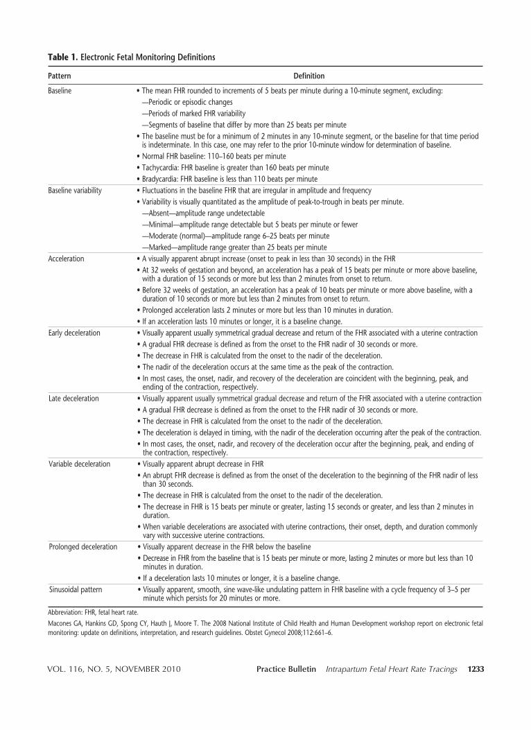
VOL. 116, NO. 5, NOVEMBER 2010 Practice Bulletin Intrapartum Fetal Heart Rate Tracings 1233
Table 1. Electronic Fetal Monitoring Definitions
Pattern Definition
Baseline •ThemeanFHRroundedtoincrementsof5beatsperminuteduringa10-minutesegment,excluding: —Periodicorepisodicchanges —PeriodsofmarkedFHRvariability —Segmentsofbaselinethatdifferbymorethan25beatsperminute •Thebaselinemustbeforaminimumof2minutesinany10-minutesegment,orthebaselineforthattimeperiod isindeterminate.Inthiscase,onemayrefertotheprior10-minutewindowfordeterminationofbaseline. •NormalFHRbaseline:110–160beatsperminute •Tachycardia:FHRbaselineisgreaterthan160beatsperminute •Bradycardia:FHRbaselineislessthan110beatsperminuteBaselinevariability •FluctuationsinthebaselineFHRthatareirregularinamplitudeandfrequency •Variabilityisvisuallyquantitatedastheamplitudeofpeak-to-troughinbeatsperminute. —Absent—amplituderangeundetectable —Minimal—amplituderangedetectablebut5beatsperminuteorfewer —Moderate(normal)—amplituderange6–25beatsperminute —Marked—amplituderangegreaterthan25beatsperminuteAcceleration •Avisuallyapparentabruptincrease(onsettopeakinlessthan30seconds)intheFHR •At32weeksofgestationandbeyond,anaccelerationhasapeakof15beatsperminuteormoreabovebaseline, withadurationof15secondsormorebutlessthan2minutesfromonsettoreturn. •Before32weeksofgestation,anaccelerationhasapeakof10beatsperminuteormoreabovebaseline,witha durationof10secondsormorebutlessthan2minutesfromonsettoreturn. •Prolongedaccelerationlasts2minutesormorebutlessthan10minutesinduration. •Ifanaccelerationlasts10minutesorlonger,itisabaselinechange.Earlydeceleration •VisuallyapparentusuallysymmetricalgradualdecreaseandreturnoftheFHRassociatedwithauterinecontraction •AgradualFHRdecreaseisdefinedasfromtheonsettotheFHRnadirof30secondsormore. •ThedecreaseinFHRiscalculatedfromtheonsettothenadirofthedeceleration. •Thenadirofthedecelerationoccursatthesametimeasthepeakofthecontraction. •Inmostcases,theonset,nadir,andrecoveryofthedecelerationarecoincidentwiththebeginning,peak,and endingofthecontraction,respectively.Latedeceleration •VisuallyapparentusuallysymmetricalgradualdecreaseandreturnoftheFHRassociatedwithauterinecontraction •AgradualFHRdecreaseisdefinedasfromtheonsettotheFHRnadirof30secondsormore. •ThedecreaseinFHRiscalculatedfromtheonsettothenadirofthedeceleration. •Thedecelerationisdelayedintiming,withthenadirofthedecelerationoccurringafterthepeakofthecontraction. •Inmostcases,theonset,nadir,andrecoveryofthedecelerationoccurafterthebeginning,peak,andendingof thecontraction,respectively.Variabledeceleration •VisuallyapparentabruptdecreaseinFHR •AnabruptFHRdecreaseisdefinedasfromtheonsetofthedecelerationtothebeginningoftheFHRnadirofless than30seconds. •ThedecreaseinFHRiscalculatedfromtheonsettothenadirofthedeceleration. •ThedecreaseinFHRis15beatsperminuteorgreater,lasting15secondsorgreater,andlessthan2minutesin duration. •Whenvariabledecelerationsareassociatedwithuterinecontractions,theironset,depth,anddurationcommonly varywithsuccessiveuterinecontractions.Prolongeddeceleration •VisuallyapparentdecreaseintheFHRbelowthebaseline •DecreaseinFHRfromthebaselinethatis15beatsperminuteormore,lasting2minutesormorebutlessthan10 minutesinduration. •Ifadecelerationlasts10minutesorlonger,itisabaselinechange.Sinusoidalpattern •Visuallyapparent,smooth,sinewave-likeundulatingpatterninFHRbaselinewithacyclefrequencyof3–5per minutewhichpersistsfor20minutesormore.
Abbreviation:FHR,fetalheartrate.
MaconesGA,HankinsGD,SpongCY,HauthJ,MooreT.The2008NationalInstituteofChildHealthandHumanDevelopmentworkshopreportonelectronicfetalmonitoring:updateondefinitions,interpretation,andresearchguidelines.ObstetGynecol2008;112:661–6.
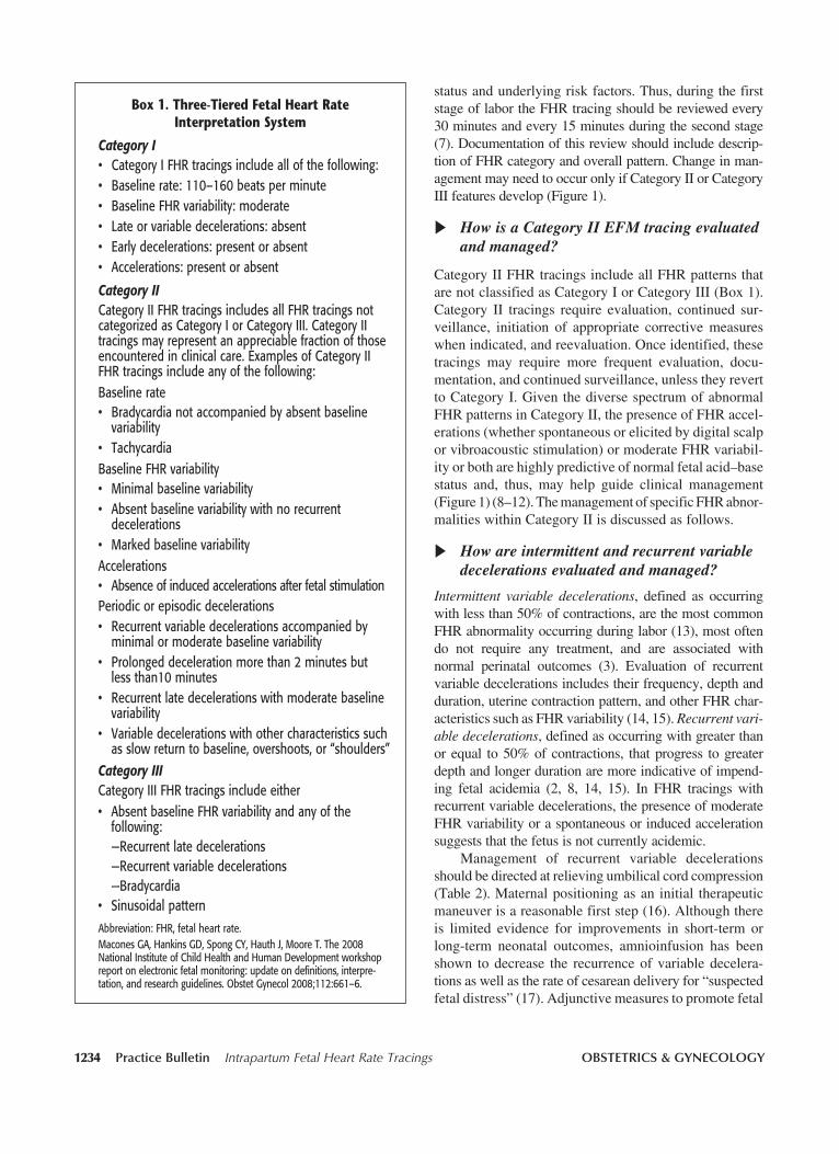
1234 Practice Bulletin Intrapartum Fetal Heart Rate Tracings OBSTETRICS & GYNECOLOGY
status and underlying risk factors. Thus, during the first stage of labor the FHR tracing should be reviewed every 30 minutes and every 15 minutes during the second stage (7). Documentation of this review should include descrip-tion of FHR category and overall pattern. Change in man-agement may need to occur only if Category II or Category III features develop (Figure 1).
How is a Category II EFM tracing evaluated and managed?
Category II FHR tracings include all FHR patterns that are not classified as Category I or Category III (Box 1). Category II tracings require evaluation, continued sur-veillance, initiation of appropriate corrective measures when indicated, and reevaluation. Once identified, these tracings may require more frequent evaluation, docu-mentation, and continued surveillance, unless they revert to Category I. Given the diverse spectrum of abnormal FHR patterns in Category II, the presence of FHR accel- erations (whether spontaneous or elicited by digital scalp or vibroacoustic stimulation) or moderate FHR variabil- ity or both are highly predictive of normal fetal acid–base status and, thus, may help guide clinical management (Figure 1) (8–12). The management of specific FHR abnor- malities within Category II is discussed as follows.
How are intermittent and recurrent variable decelerations evaluated and managed?
Intermittent variable decelerations, defined as occurring with less than 50% of contractions, are the most common FHR abnormality occurring during labor (13), most often do not require any treatment, and are associated with normal perinatal outcomes (3). Evaluation of recurrent variable decelerations includes their frequency, depth and duration, uterine contraction pattern, and other FHR char-acteristics such as FHR variability (14, 15). Recurrent vari-able decelerations, defined as occurring with greater than or equal to 50% of contractions, that progress to greater depth and longer duration are more indicative of impend-ing fetal acidemia (2, 8, 14, 15). In FHR tracings with recurrent variable decelerations, the presence of moderate FHR variability or a spontaneous or induced acceleration suggests that the fetus is not currently acidemic.
Management of recurrent variable decelerations should be directed at relieving umbilical cord compression (Table 2). Maternal positioning as an initial therapeutic maneuver is a reasonable first step (16). Although there is limited evidence for improvements in short-term or long-term neonatal outcomes, amnioinfusion has been shown to decrease the recurrence of variable decelera-tions as well as the rate of cesarean delivery for “suspected fetal distress” (17). Adjunctive measures to promote fetal
Box 1. Three-Tiered Fetal Heart Rate Interpretation System
Category I• CategoryIFHRtracingsincludeallofthefollowing:• Baselinerate:110–160beatsperminute• BaselineFHRvariability:moderate• Lateorvariabledecelerations:absent• Earlydecelerations:presentorabsent• Accelerations:presentorabsent
Category IICategoryIIFHRtracingsincludesallFHRtracingsnotcategorizedasCategoryIorCategoryIII.CategoryIItracingsmayrepresentanappreciablefractionofthoseencounteredinclinicalcare.ExamplesofCategoryIIFHRtracingsincludeanyofthefollowing:Baselinerate• Bradycardianotaccompaniedbyabsentbaseline
variability• TachycardiaBaselineFHRvariability• Minimalbaselinevariability• Absentbaselinevariabilitywithnorecurrent
decelerations• MarkedbaselinevariabilityAccelerations• AbsenceofinducedaccelerationsafterfetalstimulationPeriodicorepisodicdecelerations• Recurrentvariabledecelerationsaccompaniedby
minimalormoderatebaselinevariability• Prolongeddecelerationmorethan2minutesbut
lessthan10minutes• Recurrentlatedecelerationswithmoderatebaseline
variability• Variabledecelerationswithothercharacteristicssuch
asslowreturntobaseline,overshoots,or“shoulders”Category IIICategoryIIIFHRtracingsincludeeither• AbsentbaselineFHRvariabilityandanyofthe
following: —Recurrentlatedecelerations —Recurrentvariabledecelerations —Bradycardia• SinusoidalpatternAbbreviation:FHR,fetalheartrate.MaconesGA,HankinsGD,SpongCY,HauthJ,MooreT.The2008NationalInstituteofChildHealthandHumanDevelopmentworkshopreportonelectronicfetalmonitoring:updateondefinitions,interpre-tation,andresearchguidelines.ObstetGynecol2008;112:661–6.
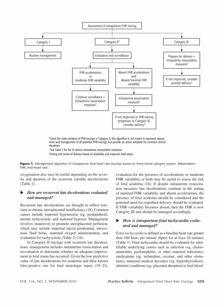
VOL. 116, NO. 5, NOVEMBER 2010 Practice Bulletin Intrapartum Fetal Heart Rate Tracings 1235
oxygenation also may be useful depending on the sever-ity and duration of the recurrent variable decelerations (Table 2).
How are recurrent late decelerations evaluated and managed?
Recurrent late decelerations are thought to reflect tran- sient or chronic uteroplacental insufficiency (18). Common causes include maternal hypotension (eg, postepidural), uterine tachysystole, and maternal hypoxia. Management involves maneuvers to promote uteroplacental perfusion, which may include maternal lateral positioning, intrave-nous fluid bolus, maternal oxygen administration, and evaluation for tachysystole (Table 2) (16).
In Category II tracings with recurrent late decelera-tions, management includes intrauterine resuscitation and reevaluation to determine whether an adequate improve-ment in fetal status has occurred. Given the low predictive value of late decelerations for acidemia and their known false-positive rate for fetal neurologic injury (19–23),
evaluation for the presence of accelerations or moderate FHR variability or both may be useful to assess the risk of fetal acidemia (24). If despite intrauterine resuscita-tion measures late decelerations continue in the setting of minimal FHR variability and absent accelerations, the presence of fetal acidemia should be considered and the potential need for expedited delivery should be evaluated. If FHR variability becomes absent, then the FHR is now Category III and should be managed accordingly.
How is intrapartum fetal tachycardia evalu-ated and managed?
Fetal tachycardia is defined as a baseline heart rate greaterthan 160 beats per minute (bpm) for at least 10 minutes (Table 1). Fetal tachycardia should be evaluated for iden-tifiable underlying causes such as infection (eg, chorio- amnionitis, pyelonephritis, or other maternal infections), medications (eg, terbutaline, cocaine, and other stimu-lants), maternal medical disorders (eg, hyperthyroidism), obstetric conditions (eg, placental abruption or fetal bleed-
*GiventhewidevariationofFHRtracingsinCategoryII,thisalgorithmisnotmeanttorepresentassess-mentandmanagementofallpotentialFHRtracings,butprovideanactiontemplateforcommonclinicalsituations.†SeeTable2forlistofvariousintrauterineresuscitativemeasures‡Timingandmodeofdeliverybasedonfeasibilityandmaternal–fetalstatus
AssessmentofintrapartumFHRtracing
CategoryI CategoryII* CategoryIII
Routinemanagement Evaluationandsurveillance Preparefordelivery+Intrauterineresuscitative
measures†
Ifnotimproved,considerpromptdelivery‡
FHRaccelerations or
moderateFHRvariability
AbsentFHRaccelerationsand
Absent/minimalFHRvariability
Continuesurveillance+Intrauterineresuscitative
measures†
Intrauterineresuscitativemeasures†
IfnotimprovedorFHRtracingprogressestoCategoryIII,
considerdelivery‡
Figure 1.Managementalgorithmofintrapartumfetalheartratetracingsbasedonthree-tieredcategorysystem.Abbreviation:FHR,fetalheartrate.
1236 Practice Bulletin Intrapartum Fetal Heart Rate Tracings OBSTETRICS & GYNECOLOGY
also may occur in fetuses with congenital heart abnor-malities or myocardial conduction defects, such as those associated with maternal collagen vascular disease. Most often the onset of bradycardia associated with congenital heart block occurs in the second trimester; it is extremely unlikely that new onset intrapartum bradycardia would be due to this condition.
Treatment for Category II tracing with bradycardia or prolonged decelerations is directed at the underlying cause (Table 2). Fetal heart rate variability during baseline periods should be evaluated in order to better assess the risk of fetal acidemia (25). If bradycardia with minimal or absent variability or prolonged decelerations or both do not resolve, then prompt delivery is recommended.
How is minimal FHR variability evaluated and managed?
As with other characteristics of the FHR tracing, base-line variability often changes with fetal sleep or wake state and over the course of labor, and it may transition from moderate to minimal and back again. Evaluation of minimal FHR variability should include evaluation of potential causes such as maternal medications (eg, opioid, magnesium sulfate), fetal sleep cycle, or fetal acidemia (26–28). For minimal variability thought to be due to recent maternal opiod administration, FHR varia-bility often improves and returns to moderate variability within 1–2 hours. A fetal sleep cycle generally lasts 20 minutes but can persist up to 60 minutes, and moderate variability should return when the fetal sleep cycle is
ing), and fetal tachyarrhythmias (often associated with FHR greater than 200 bpm). In isolation, tachycardia is poorly predictive for fetal hypoxemia or acidemia, unless accompanied by minimal or absent FHR variability or recurrent decelerations or both.
Treatment for a Category II tracing with tachycardia should be directed at the underlying cause. In addition, other characteristics of the FHR tracing need to be evalu-ated in concert with the tachycardia, especially baseline variability. For example, a FHR tracing with tachycar-dia, minimal variability, and no accelerations cannot reliably exclude fetal acidemia.
How are intrapartum bradycardia and pro-longed decelerations evaluated and managed?
Fetal bradycardia is defined as a baseline heart rate less than 110 bpm for at least 10 minutes (Table 1). Prolonged decelerations are defined as FHR decreases ofat least 15 bpm below baseline that last at least 2 minutes but less than 10 minutes. Clinical intervention often is indicated before the distinction can be made between a prolonged deceleration and fetal bradycardia; thus, the immediate management of the two is similar.
Prolonged decelerations or fetal bradycardia should be evaluated for identifiable causes such as maternal hypotension (eg, postepidural), umbilical cord prolapse or occlusion, rapid fetal descent, tachysystole, placental abruption, or uterine rupture. Bradycardia due to these conditions often occurs in labor and usually is preceded by an initially normal FHR baseline. Rarely, bradycardia
Table 2. Various Intrauterine Resuscitative Measures for Category II or Category III Tracings or Both
Goal Associated Fetal Heart Rate Abnormality* Potential Intervention (s)†
Promotefetaloxygenationandimprove Recurrentlatedecelerations Initiatelateralpositioning(eitherleftorright)uteroplacentalbloodflow Prolongeddecelerationsorbradycardia Administermaternaloxygenadministration Minimalorabsentfetalheartratevariability Administerintravenousfluidbolus Reduceuterinecontractionfrequency
Reduceuterineactivity TachysystolewithCategoryIIorIIItracing Discontinueoxytocinorcervicalripeningagents Administertocolyticmedication(eg,terbutaline)
Alleviateumbilicalcordcompression Recurrentvariabledecelerations Initiatematernalrepositioning Prolongeddecelerationsorbradycardia Initiateamnioinfusion Ifprolapsedumbilicalcordisnoted,elevatethe presentingfetalpartwhilepreparationsare underwayforoperativedelivery
*Evaluationfortheunderlyingsuspectedcause(s)isalsoanimportantstepinmanagementofabnormalFHRtracings.†Dependingonthesuspectedunderlyingcause(s)ofFHRabnormality,combiningmultipleinterventionssimultaneouslymaybeappropriateandpotentiallymoreeffec-tivethandoingindividuallyorserially(SimpsonKR,JamesDC.Efficacyofintrauterineresuscitationtechniquesinimprovingfetaloxygenstatusduringlabor.ObstetGynecol2005;105:1362–8).DatafromYoungBK,KatzM,KleinSA,SilvermanF.FetalbloodandtissuepHwithmoderatebradycardia.AmJObstetGynecol1979;135:45–7;ChauhanSP,RoachH,NaefRW2nd,MagannEF,MorrisonJC,MartinJNJr.Cesareansectionforsuspectedfetaldistress.Doesthedecision-incisiontimemakeadifference?JReprodMed1997;42:347–52; SchaubergerCW,ChauhanSP. Emergency cesarean section and the30-minute rule: definitions.Am J Perinatol 2009;26:221–6; andSchifrinBS,Hamilton-RubinsteinT,ShieldsJR.Fetalheartratepatternsandthetimingoffetalinjury.JPerinatol1994;14:174–81.
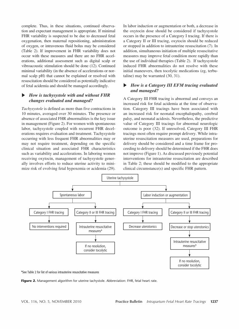
VOL. 116, NO. 5, NOVEMBER 2010 Practice Bulletin Intrapartum Fetal Heart Rate Tracings 1237
complete. Thus, in these situations, continued observa-tion and expectant management is appropriate. If minimal FHR variability is suspected to be due to decreased fetal oxygenation, then maternal repositioning, administration of oxygen, or intravenous fluid bolus may be considered (Table 2). If improvement in FHR variability does not occur with these measures and there are no FHR accel-erations, additional assessment such as digital scalp or vibroacoustic stimulation should be done (12). Continued minimal variability (in the absence of accelerations or nor-mal scalp pH) that cannot be explained or resolved with resuscitation should be considered as potentially indicative of fetal acidemia and should be managed accordingly.
How is tachysystole with and without FHR changes evaluated and managed?
Tachysystole is defined as more than five contractions in 10 minutes, averaged over 30 minutes. The presence or absence of associated FHR abnormalities is the key issue in management (Figure 2). For women with spontaneous labor, tachysystole coupled with recurrent FHR decel-erations requires evaluation and treatment. Tachysystole occurring with less frequent FHR abnormalities may or may not require treatment, depending on the specific clinical situation and associated FHR characteristics such as variability and accelerations. In laboring women receiving oxytocin, management of tachysystole gener-ally involves efforts to reduce uterine activity to mini-mize risk of evolving fetal hypoxemia or acidemia (29).
In labor induction or augmentation or both, a decrease in the oxytocin dose should be considered if tachysystole occurs in the presence of a Category I tracing. If there is a Category II or III tracing, oxytocin should be reduced or stopped in addition to intrauterine resuscitation (7). In addition, simultaneous initiation of multiple resuscitative measures may improve fetal condition more rapidly than the use of individual therapies (Table 2). If tachysystole induced FHR abnormalities do not resolve with these initial maneuvers, then tocolytic medications (eg, terbu-taline) may be warranted (30, 31).
How is a Category III EFM tracing evaluated and managed?
A Category III FHR tracing is abnormal and conveys an increased risk for fetal acidemia at the time of observa-tion. Category III tracings have been associated with an increased risk for neonatal encephalopathy, cerebral palsy, and neonatal acidosis. Nevertheless, the predictive value of Category III tracings for abnormal neurologic outcome is poor (32). If unresolved, Category III FHR tracings most often require prompt delivery. While intra-uterine resuscitation measures are used, preparations for delivery should be considered and a time frame for pro-ceeding to delivery should be determined if the FHR does not improve (Figure 1). As discussed previously potential interventions for intrauterine resuscitation are described in Table 2; these should be modified to the appropriate clinical circumstance(s) and specific FHR pattern.
Figure 2.Managementalgorithmforuterinetachystole.Abbreviation:FHR,fetalheartrate.
Uterinetachysystole
Spontaneouslabor Laborinductionoraugmentation
CategoryIFHRtracing CategoryIIorIIIFHRtracing
Intrauterineresuscitativemeasures*
Nointerventionsrequired Intrauterineresuscitativemeasures*
Ifnoresolution, considertocolytic
CategoryIFHRtracing CategoryIIorIIIFHRtracing
Decreaseuterotonics Decreaseorstoputerotonics
Ifnoresolution, considertocolytic
*SeeTable2forlistofvariousintrauterineresuscitativemeasures
1238 Practice Bulletin Intrapartum Fetal Heart Rate Tracings OBSTETRICS & GYNECOLOGY
If a Category III tracing continues and intra-uterine resuscitative measures are unsuccess-ful, over what time interval should delivery be accomplished?
The acceptable time frame to accomplish delivery in the setting of a Category III FHR tracing has not been established. Historically, a 30-minute rule from decision-to-incision time for emergent cesarean delivery in the setting of abnormal FHR pattern has existed (7); how-ever, the scientific evidence to support this threshold is lacking. In a study of 2,808 women who had cesarean deliveries for emergent indications, investigators found that more than 30% of the cesarean deliveries began more than 30 minutes after the decision to operate, yet adverse neonatal outcomes were not increased among those infants delivered after more than 30 minutes (33). Multiple other studies affirm the lack of associa-tion of increased adverse outcomes with this 30-minute decision-to-incision time frame (34–38). It also should be recognized that in some cases immediate delivery in a woman with an unknown duration of a Category III tracing may not improve outcome if the fetus has already experienced hypoxic ischemic injury (39, 40).
Nevertheless, when a decision for operative delivery in the setting of a Category III EFM tracing is made, it should be accomplished as expeditiously as feasible. The decision-to-incision interval and mode of delivery should be based on the timing that best incorporates maternal and fetal risks and benefits. For instance many of these clinical scenarios will include high-risk condi-tions or pregnancy complications (eg, morbid obesity, eclampsia, cardiopulmonary compromise, hemorrhage), which may require maternal stabilization or additional surgical preparation before performance of emergent cesarean delivery. These factors also may vary based on the institution and local circumstances. Preparation for impending delivery of a woman with a Category III trac-ing often requires assessment of several logistical issues depending on the setting and clinical circumstances (see Box 2).
Summary of Conclusions and RecommendationsThe following recommendations and conclusions are based on good and consistent scientific evi-dence (Level A):
Category I FHR tracings may be managed in a rou-tine manner because they are not associated with fetal acidemia.
A Category III FHR tracing is abnormal and conveys an increased risk of fetal acidemia at the time of observation.
Amnioinfusion has been shown to decrease the recurrence of variable decelerations as well as the rate of cesarean delivery for abnormal FHR patterns.
The following recommendations and conclusions are based on evidence that may be limited or inconsistent (Level B):
Intravenous fluid bolus, lateral positioning and oxy-gen administration, when used together, may improve fetal oxygenation during labor.
Regardless of whether labor is spontaneous or stimu-lated, tachysystole accompanied by Category II or Category III FHR tracing requires evaluation and initiation of appropriate treatment.
Category II tracings require evaluation, continued surveillance, initiation of appropriate corrective measures when indicated, and reevaluation. The presence of FHR accelerations (whether spontane-ous or elicited) or moderate FHR variability or both are highly predictive of normal fetal acid–base status and, thus, may help guide clinical management.
The following conclusion is based primarily on consensus and expert opinion (Level C):
The optimal time frame to effect delivery in the set-ting of a Category III FHR tracing has not been established.
Box 2. Potential Logistical Considerations in Preparation for Operative Delivery in
Setting of Category III Tracing
• Obtaininformedconsent(verbalorwrittenasfea-sible)
• Assemblesurgicalteam(surgeon,scrubtechnician,andanesthesiapersonnel)
• Assesspatienttransittimeandlocationforoperativedelivery
• Ensureintravenousaccess• Reviewstatusoflaboratorytests(eg,completeblood
typeandscreen)andassessneedforavailabilityofbloodproducts
• Assessneedforpreoperativeplacementofindwell-ingfoleycatheter
• Assemblepersonnelforneonatalresuscitation
VOL. 116, NO. 5, NOVEMBER 2010 Practice Bulletin Intrapartum Fetal Heart Rate Tracings 1239
References 1. Intrapartum fetal heart rate monitoring: nomenclature,
interpretation, and general management principles. ACOG Practice Bulletin No. 106. American College of Obstet-ricians and Gynecologists. Obstet Gynecol 2009:114:192–202. (Level III)
2. Macones GA, Hankins GD, Spong CY, Hauth J, Moore T. The 2008 National Institute of Child Health and Human Development workshop report on electronic fetal moni-toring: update on definitions, interpretation, and research guidelines. Obstet Gynecol 2008;112:661–6. (Level III)
3. Berkus MD, Langer O, Samueloff A, Xenakis EM, Field NT. Electronic fetal monitoring: what’s reassuring? Acta Obstet Gynecol Scand 1999;78:15–21. (Level II-3)
4. Krebs HB, Petres RE, Dunn LJ, Smith PJ. Intrapartum fetal heart rate monitoring. VI. Prognostic significance of accelerations. Am J Obstet Gynecol 1982;142:297–305. (Level II-3)
5. Tejani N, Mann LI, Bhakthavathsalan A, Weiss RR. Correlation of fetal heart rate-uterine contraction patterns with fetal scalp blood pH. Obstet Gynecol 1975;46: 392–6. (Level III)
6. Dellinger EH, Boehm FH, Crane MM. Electronic fetal heart rate monitoring: early neonatal outcomes associated with normal rate, fetal stress, and fetal distress. Am J Obstet Gynecol 2000;182:214–20. (Level III)
7. American Academy of Pediatrics, American College of Obstetricians and Gynecologists. Fetal heart rate monitor-ing. In: Guidelines for perinatal care. 6th ed. Elk Grove Village (IL): AAP; Washington, DC: ACOG; 2007. p. 146–7. (Level III)
8. Parer JT, King T, Flanders S, Fox M, Kilpatrick SJ. Fetal acidemia and electronic fetal heart rate patterns: is there evidence of an association? J Matern Fetal Neonatal Med 2006;19:289–94. (Level III)
9. Lin CC, Vassallo B, Mittendorf R. Is intrapartum vibro-acoustic stimulation an effective predictor of fetal acido-sis? J Perinat Med 2001;29:506–12. (Level II-3)
10. Clark SL, Gimovsky ML, Miller FC. Fetal heart rate response to scalp blood sampling. Am J Obstet Gynecol 1982;144:706–8. (Level III)
11. Clark SL, Gimovsky ML, Miller FC. The scalp stimula-tion test: a clinical alternative to fetal scalp blood sam-pling. Am J Obstet Gynecol 1984;148:274–7. (Level III)
12. Skupski DW, Rosenberg CR, Eglinton GS. Intrapartum fetal stimulation tests: a meta-analysis. Obstet Gynecol 2002;99:129–34. (Meta-analysis)
13. Garite TJ, Dildy GA, McNamara H, Nageotte MP, Boehm FH, Dellinger EH, et al. A multicenter controlled trial of fetal pulse oximetry in the intrapartum management of nonre-assuring fetal heart rate patterns. Am J Obstet Gynecol 2000;183:1049–58. (Level I)
14. Kubli FW, Hon EH, Khazin AF, Takemura H. Observations on heart rate and pH in the human fetus during labor. Am J Obstet Gynecol 1969;104:1190–206. (Level III)
15. Krebs HB, Petres RE, Dunn LJ. Intrapartum fetal heart rate monitoring. VIII. Atypical variable decelerations. Am J Obstet Gynecol 1983;145:297–305. (Level II-3)
16. Simpson KR. Intrauterine resuscitation during labor: review of current methods and supportive evidence. J Midwifery Womens Health 2007;52:229–37. (Level III)
17. Hofmeyr GJ. Amnioinfusion for potential or suspected umbilical cord compression in labour. Cochrane Database of Systematic Reviews 1998, Issue 1. Art. No.: CD000013. DOI: 10.1002/14651858.CD000013. (Meta-analysis)
18. Harris JL, Krueger TR, Parer JT. Mechanisms of late decelerations of the fetal heart rate during hypoxia. Am J Obstet Gynecol 1982;144:491–6. (animal)
19. Nelson KB, Dambrosia JM, Ting TY, Grether JK. Uncertain value of electronic fetal monitoring in pre- dicting cerebral palsy. N Engl J Med 1996;334:613–8. (Level II-2)
20. Larma JD, Silva AM, Holcroft CJ, Thompson RE, Donohue PK, Graham EM. Intrapartum electronic fetal heart rate monitoring and the identification of metabolic acidosis and hypoxic-ischemic encephalopathy. Am J Obstet Gynecol 2007;197:301.e1–301.e8. (Level II-2)
21. Parer JT, King T. Fetal heart rate monitoring: is it salvage-able? Am J Obstet Gynecol 2000;182:982–7. (Level III)
22. Spencer JA, Badawi N, Burton P, Keogh J, Pemberton P, Stanley F. The intrapartum CTG prior to neonatal enceph-alopathy at term: a case-control study. Br J Obstet Gynaecol 1997;104:25–8. (Level II-2)
23. Williams KP, Galerneau F. Comparison of intrapartum fetal heart rate tracings in patients with neonatal seizures vs. no seizures: what are the differences? J Perinat Med 2004;32:422–5. (Level II-2)
24. Paul RH, Suidan AK, Yeh S, Schifrin BS, Hon EH. Clinical fetal monitoring. VII. The evaluation and sig-nificance of intrapartum baseline FHR variability. Am J Obstet Gynecol 1975;123:206–10. (Level III)
25. Young BK, Katz M, Klein SA, Silverman F. Fetal blood and tissue pH with moderate bradycardia. Am J Obstet Gynecol 1979;135:45–7. (Level III)
26. Giannina G, Guzman ER, Lai YL, Lake MF, Cernadas M, Vintzileos AM. Comparison of the effects of meperidine and nalbuphine on intrapartum fetal heart rate tracings. Obstet Gynecol 1995;86:441–5. (Level I)
27. Kopecky EA, Ryan ML, Barrett JF, Seaward PG, Ryan G, Koren G, et al. Fetal response to maternally adminis-tered morphine. Am J Obstet Gynecol 2000;183:424–30. (Level III)
28. Hallak M, Martinez-Poyer J, Kruger ML, Hassan S, Blackwell SC, Sorokin Y. The effect of magnesium sul-fate on fetal heart rate parameters: A randomized, placebo-controlled trial. Am J Obstet Gynecol 1999;181:1122–7. (Level I)
29. Simpson KR, James DC. Effects of oxytocin-induced uterine hyperstimulation during labor on fetal oxygen status and fetal heart rate patterns. Am J Obstet Gynecol 2008;199:34.e1–34.e5. (Level II-3)
1240 Practice Bulletin Intrapartum Fetal Heart Rate Tracings OBSTETRICS & GYNECOLOGY
30. Pullen KM, Riley ET, Waller SA, Taylor L, Caughey AB, Druzin ML, et al. Randomized comparison of intrave-nous terbutaline vs nitroglycerin for acute intrapartum fetal resuscitation. Am J Obstet Gynecol 2007;197:414.e1–414.e6. (Level I)
31. Kulier R, Hofmeyr GJ. Tocolytics for suspected intra-partum fetal distress. Cochrane Database of Systematic Reviews 1998, Issue 2. Art. No.: CD000035. DOI: 10.1002/ 14651858.CD000035. (Meta-analysis)
32. American Academy of Pediatrics, American College of Obstetricians and Gynecologists. Neonatal encepha-lopathy and cerebral palsy: defining the pathogenesis and pathophysiology. Elk Grove Village (IL): AAP; Washington, DC: ACOG; 2003. (Level III)
33. Bloom SL, Leveno KJ, Spong CY, Gilbert S, Hauth JC, Landon MB, et al. Decision-to-incision times and mater-nal and infant outcomes. National Institute of Child Health and Human Development Maternal-Fetal Medicine Units Network. Obstet Gynecol 2006;108:6–11. (Level II-2)
34. MacKenzie IZ, Cooke I. What is a reasonable time from decision-to-delivery by caesarean section? Evidence from 415 deliveries. BJOG 2002;109:498–504. (Level II-3)
35. Holcroft CJ, Graham EM, Aina-Mumuney A, Rai KK, Henderson JL, Penning DH. Cord gas analysis, decision-to-delivery interval, and the 30-minute rule for emergency cesareans. J Perinatol 2005;25:229–35. (Level II-2)
36. Hillemanns P, Strauss A, Hasbargen U, Schulze A, Genzel- Boroviczeny O, Weninger E, et al. Crash emergency cesar- ean section: decision-to-delivery interval under 30 min and its effect on Apgar and umbilical artery pH. Arch Gyne- col Obstet 2005;273:161–5. (Level II-2)
37. Chauhan SP, Roach H, Naef RW 2nd, Magann EF, Morrison JC, Martin JN Jr. Cesarean section for suspected fetal distress. Does the decision-incision time make a dif-ference? J Reprod Med 1997;42:347–52. (Level II-2)
38. Schauberger CW, Chauhan SP. Emergency cesarean sec-tion and the 30-minute rule: definitions. Am J Perinatol 2009;26:221–6. (Level III)
39. Schifrin BS, Hamilton-Rubinstein T, Shields JR. Fetal heart rate patterns and the timing of fetal injury. J Perinatol 1994;14:174–81. (Level III)
40. Phelan JP, Ahn MO. Perinatal observations in forty-eight neurologically impaired term infants. Am J Obstet Gynecol 1994;171:424–31. (Level III)
41. Althabe O Jr, Schwarcz RL, Pose SV, Escarcena L, Caldeyro-Barcia R. Effects on fetal heart rate and fetal pO2 of oxygen administration to the mother. Am J Obstet Gynecol 1967;98:858–70. (Level III)
42. Carbonne B, Benachi A, Leveque ML, Cabrol D, Papiernik E. Maternal position during labor: effects on fetal oxygen saturation measured by pulse oximetry. Obstet Gynecol 1996;88:797–800. (Level III)
43. Abitbol MM. Supine position in labor and associated fetal heart rate changes. Obstet Gynecol 1985;65:481–6. (Level III)
44. Simpson KR, James DC. Efficacy of intrauterine resusci-tation techniques in improving fetal oxygen status during labor. Obstet Gynecol 2005;105:1362–8. (Level I)
The MEDLINE database, the Cochrane Library, and the American College of Obstetricians and Gynecologists’ own internal resources and documents were used to con-duct a lit er a ture search to lo cate rel e vant ar ti cles pub lished be tween January 1985–December 2009. The search was re strict ed to ar ti cles pub lished in the English lan guage. Pri or i ty was given to articles re port ing results of orig i nal re search, although re view ar ti cles and com men tar ies also were consulted. Ab stracts of re search pre sent ed at sym po-sia and sci en tif ic con fer enc es were not con sid ered adequate for in clu sion in this doc u ment. Guide lines pub lished by or ga ni za tions or in sti tu tions such as the Na tion al In sti tutes of Health and the Amer i can Col lege of Ob ste tri cians and Gy ne col o gists were re viewed, and ad di tion al studies were located by re view ing bib liographies of identified articles. When re li able research was not available, expert opinions from ob ste tri cian–gynecologists were used.
Studies were reviewed and evaluated for qual i ty ac cord ing to the method outlined by the U.S. Pre ven tive Services Task Force:
I Evidence obtained from at least one prop er ly de signed randomized controlled trial.
II-1 Evidence obtained from well-designed con trolled tri als without randomization.
II-2 Evidence obtained from well-designed co hort or case–control analytic studies, pref er a bly from more than one center or research group.
II-3 Evidence obtained from multiple time series with or with out the intervention. Dra mat ic re sults in un con-trolled ex per i ments also could be regarded as this type of ev i dence.
III Opinions of respected authorities, based on clin i cal ex pe ri ence, descriptive stud ies, or re ports of ex pert committees.
Based on the highest level of evidence found in the data, recommendations are provided and grad ed ac cord ing to the following categories:
Level A—Recommendations are based on good and con-sis tent sci en tif ic evidence.
Level B—Recommendations are based on limited or in con-sis tent scientific evidence.
Level C—Recommendations are based primarily on con-sen sus and expert opinion.
Copyright November 2010 by the American College of Ob ste t- ri cians and Gynecologists. All rights reserved. No part of this publication may be reproduced, stored in a re triev al sys tem, posted on the Internet, or transmitted, in any form or by any means, elec tron ic, me chan i cal, photocopying, recording, or oth er wise, without prior written permission from the publisher.
Requests for authorization to make photocopies should be directed to Copyright Clearance Center, 222 Rosewood Drive, Danvers, MA 01923, (978) 750-8400.
The American College of Obstetricians and Gynecologists 409 12th Street, SW, PO Box 96920, Washington, DC 20090-6920
Management of intrapartum fetal heart rate tracings. Practice Bulletin No. 116. American College of Obstetricians and Gynecologists. Obstet Gynecol 2010;116:1232–40.
O
IoSJ
Ocio
Stot
Ra�i�E
Cn
Osfat
FNLMUT
R2
RcMCAP8
0©d
Research www.AJOG.org
4
BSTETRICS
mplementation of a conservative checklist-based protocol forxytocin administration: maternal and newborn outcomes
teven Clark, MD; Michael Belfort, MD, PhD; George Saade, MD; Gary Hankins, MD; Darla Miller, RN; Donna Frye, RN;anet Meyers, RN
BJECTIVE: The purpose of this study was to examine the effects of aonservative and specific checklist-based protocol for oxytocin admin-stration on maternal and newborn outcome. The protocol was basedn maternal and fetal response to oxytocin rather than infusion rate.
TUDY DESIGN: This was a retrospective chart review and data extrac-ion of the last 100 patients receiving oxytocin before implementationf the protocol and the first 100 patients receiving oxytocin after pro-ocol implementation.
ESULTS: The 2 groups were demographically similar. For the pre-nd postprotocol groups, the mean time of infusion to delivery was 8.5
5.3 hours versus 8.2 � 4.5 hours (NS), the maximum oxytocinnfusion rate was 13.8 � 6.3 mU/min versus 11.4 � 6.1 mU/min (P
.003) and the cesarean delivery rate was 15% versus 13% (NS).
level facility.See related editorial, page 445
80.e1 American Journal of Obstetrics & Gynecology NOVEMBER 2007
roup, but these differences did not individually reach statistical sig-ificance. However, newborns with any index of adverse outcome wereignificantly fewer in the post protocol group (31 vs 18, P � .049).ystem wide implementation of this program was associated with aecline in the rate of primary cesarean delivery from 23.6% in 2005 to1.0% in 2006.
ONCLUSION: Implementation of a specific and conservative check-ist-based protocol for oxytocin infusion based on maternal and fetalesponse results in a significant reduction in maximum infusion ratesf oxytocin without lengthening labor or increasing operative interven-
ion. Cesarean delivery rate declined system-wide following implemen-ation of this protocol. Newborn outcome also appears to be improved.
very index of newborn outcome was improved in the post-protocol
ite this article as: Clark S, Belfort M, Saade G, et al. Implementation of a conservative checklist-based protocol for oxytocin administration: maternal andewborn outcomes. Am J Obstet Gynecol 2007;197:480.e1-480.e5.
xytocin is one of the most com-monly administered drugs in ob-
tetrics. Although this agent, when care-ully administered, is generally safe,dverse perinatal outcomes related to fe-al hypoxia may occur in the presence of
uterine hyperstimulation.1 Due to a lackof outcomes based data demonstratingthe clear superiority of any specific reg-imen of oxytocin administration, cur-rent guidelines in this regard are non-specific.2-4 While no single regimen ofoxytocin administration has beendemonstrated superior in terms ofclinical outcomes, one of the most fun-damental principles of quality im-provement is that, in general, greaterpractice variation is associated withpoorer outcomes than more uniformpractice patterns.5,6 In recent decades,the airline industry has established anenviable record of safety, due, in largepart, to the extensive use of a uniform,checklist-based approach to the man-agement of certain high risk situa-tions.7,8 We examined the effects ofimplementation of a conservative uni-form checklist-based system of oxyto-cin administration in a large, tertiary
MATERIALS AND METHODS
The Hospital Corporation of America isthe nation’s largest single health care de-livery organization, with 125 obstetricfacilities in 20 states. In 2004, the Perina-tal Safety Division assisted with the es-tablishment of a system wide uniform,checklist-based protocol for oxytocinadministration by work groups com-posed of representative practicing physi-cians, nurses, and pharmacists from theentire organization, as well as consult-ants from other institutions in areasserved by our hospitals. With respect tomandated response to both fetal heartrate abnormalities and uterine contrac-tion patterns, the protocols were pur-posefully far more conservative thanwould be required by current standard ofcare (Tables 2 and 3).4 These protocolswere then piloted in select facilities tofurther refine the safety and practicalityof the checklists. The resultant checklistbased protocols were then presented foradoption by individual departments of
rom the Hospital Corporation of America,ashville, TN, and St Mark’s Hospital, Saltake City, UT (Drs Clark and Belfort, Msiller, Ms Frye, and Ms Meyers), and theniversity of Texas, Galveston, Galveston,X (Drs Saade and Hankins).
eceived Mar. 20, 2007; revised Jul. 18,007; accepted Aug. 7, 2007.
eprints not available from authors. Addressorrespondence to: Steven L. Clark, MD,edical Director, Women and Newbornslinical Program, Hospital Corporation ofmerica, St Mark’s Hospital, Women’savilion 1140 E 3900 S, Salt Lake City, UT4124; [email protected]
002-9378/$32.002007 Mosby, Inc. All rights reserved.
oi: 10.1016/j.ajog.2007.08.026
gnsSd2
Clrott
Key words: cesarean delivery, medication safety, oxytocin
o
bstetrics and gynecology in each of our
fdtaaIatsifwcefi
nCuldomoirm1t1
tcwfnciuttgoa0ansasa
RAgtttDsdcmosattrd
stAstee(ip
ddi
www.AJOG.org Obstetrics Research
acilities. These protocols are designed asefault models of oxytocin administra-ion, to be automatically implementednd uniformly followed in the absence ofspecific physician order to the contrary.
ndividual variations from protocol arellowed as long as the physician prospec-ively documents her/his rationale foruch an alternative approach in the med-cal record. The protocols are designedor use in singleton, vertex, term labor inomen with an unscarred uterus. Medi-
al indications for induction did not ex-mpt the patient from protocol use. Uni-orm protocols for oxytocin mixing and
TABLE 1Oxytocin in-use checklistDemographic/clinical factor
Maternal age...................................................................................................................
Parity...................................................................................................................
Gestational age (wk)...................................................................................................................
Oxytocin onset to delivery (h)...................................................................................................................
Total time oxytocin infused (h)...................................................................................................................
Maximum oxytocin dose (mU/min)...................................................................................................................
Latent phase length (h)...................................................................................................................
Active phase length (h)...................................................................................................................
Second stage length (h)...................................................................................................................
Birthweight...................................................................................................................
Apgar 1 minute...................................................................................................................
Apgar 5 minutes...................................................................................................................
Cervical ripening agents...................................................................................................................
Cesarean delivery...................................................................................................................
Cesarean for fetal heart rateabnormalities
...................................................................................................................
Operative vaginal delivery...................................................................................................................
Cesarean for labor arrest...................................................................................................................
Newborn intensive care admission...................................................................................................................
Respiratory distress...................................................................................................................
Sepsis suspected...................................................................................................................
Sepsis confirmed...................................................................................................................
Mean days in newborn intensivecare
...................................................................................................................
Mean newborn intensive caredays for those admitted toNICU
...................................................................................................................
Infants with 1 or more newborncomplication
nfusion were utilized. i
St Mark’s Hospital is a tertiary level,onteaching referral facility in Salt Lakeity, UT, with an annual delivery vol-me of approximately 3700. The check-
ist-based protocols were adopted by theepartment of Obstetrics and Gynecol-gy at St Marks hospital and imple-ented March 1, 2005. As part of our
ngoing internal patient safety and qual-ty assurance program, we collected dataegarding clinical course of labor andaternal/newborn outcomes in the last
00 patients receiving oxytocin prior tohe adoption of the protocol and the first00 patients receiving oxytocin after the
checklist Postchecklist
�5.2 27.5�5.2..................................................................................................................
�1.2 1.3�1.2..................................................................................................................
�1.7 39.0�1.4..................................................................................................................
�4.3 8.6�4.5..................................................................................................................
�5.3 8.2�4.5..................................................................................................................
�6.3 11.4�6.1 (0.003)*..................................................................................................................
�3.3 5.5�3.2..................................................................................................................
�1.3 2.3�1.8..................................................................................................................
9�0.69 0.74�0.92..................................................................................................................
98�1.259 7.421�1.231 (0.017)*..................................................................................................................
�1.1 7.9�0.88 (0.048)*..................................................................................................................
�1.0 8.8�0.98..................................................................................................................
10..................................................................................................................
13..................................................................................................................
3
..................................................................................................................
10..................................................................................................................
9..................................................................................................................
11..................................................................................................................
6..................................................................................................................
1..................................................................................................................
2..................................................................................................................
9 0.81
..................................................................................................................
�10.4 7.4�5.7
..................................................................................................................
18 (P � .049)*
ntroduction of the protocol. Institu- c
NOVEMBER 2007 America
ional review board approval for publi-ation of this analysis was obtained. Ourorking hypothesis was that such uni-
orm, highly conservative practice wouldot significantly prolong labor or in-rease the intervention rate and wouldmprove perinatal outcomes. Basedpon the composite morbidity found in
he pre-protocol group, we calculatedhat 100 patients in each group wouldive our analysis an 80% power to dem-nstrate a 50% reduction in compositedverse outcome at an alpha error of.05. Data extraction was undertaken bysingle, experienced labor and deliveryurse from an institution in a differenttate. Univariate (Chi-square, Fisher ex-ct, Student t, or Mann-Whitney rankum test as appropriate) and multivari-te analyses were performed.
ESULTSll patients were delivered within a sin-le month both before and after the pro-ocol institution. During this period ofime, there were no variations from pro-ocol ordered by the attending physician.emographic and clinical data are pre-
ented in Table 1. The only significantifference was a small but significant in-reased birthweight in the checklistanaged group. The maximum dose of
xytocin used to achieve delivery wasignificantly lower in the checklist man-ged group. There was no difference inhe length of any stage or phase of labor,otal time of oxytocin administration, orate of operative vaginal or abdominalelivery.Following analysis of these data, these
ame protocols were implementedhroughout the Hospital Corporation ofmerica system. During the first year of
ystem wide implementation of this pro-ocol (2006), the primary cesarean deliv-ry rate in approximately 220,000 deliv-ries fell from 23.6% (1995) to 21.0%1996) in contrast to an annual increasen rate of primary cesarean of 1-4% inrevious years (Figure).A comparison of newborn outcomes
emonstrated a statistically significantifference in 1 minute Apgar scores, with
mproved 1 minute Apgar scores in the
Pre
27.2.........
1.1.........
38.8.........
8.7.........
8.5.........
13.8.........
5.8.........
2.1.........
0.6.........
6.9.........
7.6.........
8.7.........
16.........
15.........
6
.........
11.........
9.........
16.........
13.........
2.........
6.........
2.2
.........
14.3
.........
30
hecklist managed group. In addition,
n Journal of Obstetrics & Gynecology 480.e2
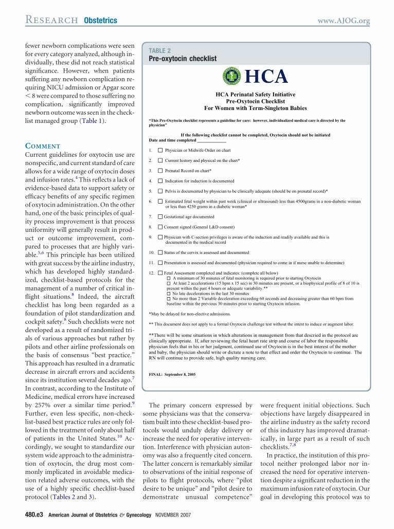
ffdssq�cnl
CCnaaeeohiuupawwimflcfcdaptTdsIMbFllocstmtup
sttitoTtpd
wotoic
tctm
Research Obstetrics www.AJOG.org
4
ewer newborn complications were seenor every category analyzed, although in-ividually, these did not reach statisticalignificance. However, when patientsuffering any newborn complication re-uiring NICU admission or Apgar score
8 were compared to those suffering noomplication, significantly improvedewborn outcome was seen in the check-
ist managed group (Table 1).
OMMENT
urrent guidelines for oxytocin use areonspecific, and current standard of carellows for a wide range of oxytocin dosesnd infusion rates.4 This reflects a lack ofvidence-based data to support safety orfficacy benefits of any specific regimenf oxytocin administration. On the otherand, one of the basic principles of qual-
ty process improvement is that processniformity will generally result in prod-ct or outcome improvement, com-ared to processes that are highly vari-ble.5,6 This principle has been utilizedith great success by the airline industry,hich has developed highly standard-
zed, checklist-based protocols for theanagement of a number of critical in-
ight situations.8 Indeed, the aircrafthecklist has long been regarded as aoundation of pilot standardization andockpit safety.8 Such checklists were noteveloped as a result of randomized tri-ls of various approaches but rather byilots and other airline professionals onhe basis of consensus “best practice.”his approach has resulted in a dramaticecrease in aircraft errors and accidentsince its institution several decades ago.7
n contrast, according to the Institute ofedicine, medical errors have increased
y 257% over a similar time period.9
urther, even less specific, non-check-ist-based best practice rules are only fol-owed in the treatment of only about halff patients in the United States.10 Ac-ordingly, we sought to standardize ourystem wide approach to the administra-ion of oxytocin, the drug most com-
only implicated in avoidable medica-ion related adverse outcomes, with these of a highly specific checklist-based
rotocol (Tables 2 and 3). d80.e3 American Journal of Obstetrics & Gynecol
The primary concern expressed byome physicians was that the conserva-ism built into these checklist-based pro-ocols would unduly delay delivery orncrease the need for operative interven-ion. Interference with physician auton-my was also a frequently cited concern.he latter concern is remarkably similar
o observations of the initial response ofilots to flight protocols, where “pilotesire to be unique” and “pilot desire to
TABLE 2Pre-oxytocin checklist
HCA PerinatalPre-Oxytoc
For Women with T
“This Pre-Oxytocin checklist represents a guideline for care: physician”
If the following checklist cannot be comDate and time completed _____________
1. Physician or Midwife Order on chart
2. Current history and physical on the chart*
3. Prenatal Record on chart*
4. Indication for induction is documented
5. Pelvis is documented by physician to be clinically
6. Estimated fetal weight within past week (clinical or less than 4250 grams in a diabetic woman*
7. Gestational age documented
8. Consent signed (General L&D consent)
9. Physician with C-section privileges is aware of the documented in the medical record
10. Status of the cervix is assessed and documented
11. Presentation is assessed and documented (physicia
12. Fetal Assessment completed and indicates: (comp A minimum of 30 minutes of fetal monitorin At least 2 accelerations (15 bpm x 15 sec) in
present within the past 4 hours or adequate variaNo late decelerations in the last 30 minutesNo more than 2 Variable deceleration exceed
baseline within the previous 30 minutes prior to
*May be delayed for non-elective admissions.
** This document does not apply to a formal Oxytocin cha
**There will be some situations in which alterations clinically appropriate. If, after reviewing the fetal hephysician feels that in his or her judgment, continuedand baby, the physician should write or dictate a noteRN will continue to provide safe, high quality nursin
FINAL: September 8, 2005
emonstrate unusual competence” g
ogy NOVEMBER 2007
ere frequent initial objections. Suchbjections have largely disappeared inhe airline industry as the safety recordf this industry has improved dramat-
cally, in large part as a result of suchhecklists.7,8
In practice, the institution of this pro-ocol neither prolonged labor nor in-reased the need for operative interven-ion despite a significant reduction in the
aximum infusion rate of oxytocin. Our
fety InitiativeChecklist-Singleton Babies
ver, individualized medical care is directed by the
ted, Oxytocin should not be initiated
quate (should be on prenatal record)*
trasound) less than 4500grams in a non-diabetic woman
uction and readily available and this is
quired to come in if nurse unable to determine)
ll below)equired prior to starting Oxytocininutes are present, or a biophysical profile of 8 of 10 is .**
0 seconds and decreasing greater than 60 bpm from ng Oxytocin infusion.
e test without the intent to induce or augment labor.
anagement from that descried in the protocol are ate strip and course of labor the responsible of Oxytocin is in the best interest of the mother hat effect and order the Oxytocin to continue. The re.
Sain
erm
howe
ple
ade
or ul
ind
n re
lete ag is r30 mbility
ing 6starti
lleng
in mart r use to tg ca
oal in developing this protocol was to
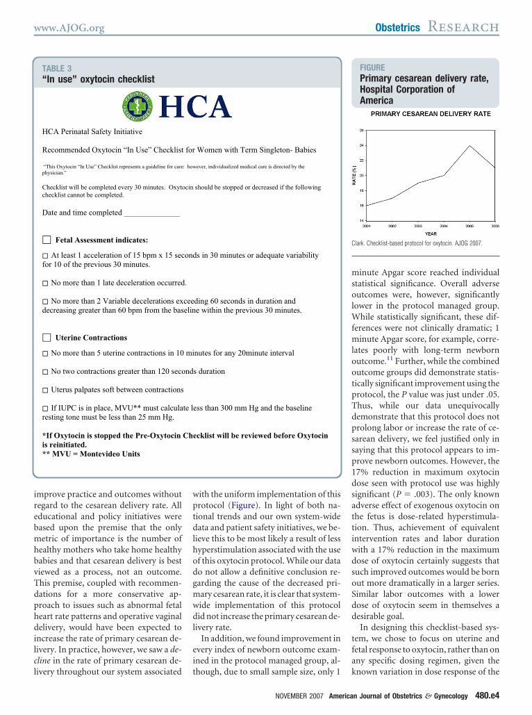
irebmhbvTdphdilcl
wptdlhodgmwdl
ei
msolWfmlootpTdpssp1dsattiwdsoSdd
tfa
C
www.AJOG.org Obstetrics Research
mprove practice and outcomes withoutegard to the cesarean delivery rate. Allducational and policy initiatives wereased upon the premise that the onlyetric of importance is the number of
ealthy mothers who take home healthyabies and that cesarean delivery is bestiewed as a process, not an outcome.his premise, coupled with recommen-ations for a more conservative ap-roach to issues such as abnormal fetaleart rate patterns and operative vaginalelivery, would have been expected to
ncrease the rate of primary cesarean de-ivery. In practice, however, we saw a de-line in the rate of primary cesarean de-
TABLE 3“In use” oxytocin checklist
HCA Perinatal Safety Initiative
Recommended Oxytocin “In Use” Checklist
“This Oxytocin “In Use” Checklist represents a guideline for carephysician.”
Checklist will be completed every 30 minutes. Oxytchecklist cannot be completed.
Date and time completed ______________
Fetal Assessment indicates:
At least 1 acceleration of 15 bpm x 15 secfor 10 of the previous 30 minutes.
No more than 1 late deceleration occurred
No more than 2 Variable decelerations exdecreasing greater than 60 bpm from the bas
Uterine Contractions
No more than 5 uterine contractions in 10
No two contractions greater than 120 seco
Uterus palpates soft between contractions
If IUPC is in place, MVU** must calcularesting tone must be less than 25 mm Hg.
*If Oxytocin is stopped the Pre-Oxytocin is reinitiated.** MVU = Montevideo Units
ivery throughout our system associated t
ith the uniform implementation of thisrotocol (Figure). In light of both na-ional trends and our own system-wideata and patient safety initiatives, we be-
ieve this to be most likely a result of lessyperstimulation associated with the usef this oxytocin protocol. While our datao not allow a definitive conclusion re-arding the cause of the decreased pri-ary cesarean rate, it is clear that system-ide implementation of this protocolid not increase the primary cesarean de-
ivery rate.In addition, we found improvement in
very index of newborn outcome exam-ned in the protocol managed group, al-
Women with Term Singleton- Babies
ever, individualized medical care is directed by the
should be stopped or decreased if the following
s in 30 minutes or adequate variability
ding 60 seconds in duration and e within the previous 30 minutes.
nutes for any 20minute interval
s duration
ess than 300 mm Hg and the baseline
cklist will be reviewed before Oxytocin
hough, due to small sample size, only 1 k
NOVEMBER 2007 America
inute Apgar score reached individualtatistical significance. Overall adverseutcomes were, however, significantly
ower in the protocol managed group.hile statistically significant, these dif-
erences were not clinically dramatic; 1inute Apgar score, for example, corre-
ates poorly with long-term newbornutcome.11 Further, while the combinedutcome groups did demonstrate statis-ically significant improvement using therotocol, the P value was just under .05.hus, while our data unequivocallyemonstrate that this protocol does notrolong labor or increase the rate of ce-arean delivery, we feel justified only inaying that this protocol appears to im-rove newborn outcomes. However, the7% reduction in maximum oxytocinose seen with protocol use was highlyignificant (P � .003). The only knowndverse effect of exogenous oxytocin onhe fetus is dose-related hyperstimula-ion. Thus, achievement of equivalentntervention rates and labor durationith a 17% reduction in the maximumose of oxytocin certainly suggests thatuch improved outcomes would be bornut more dramatically in a larger series.imilar labor outcomes with a lowerose of oxytocin seem in themselves aesirable goal.In designing this checklist-based sys-
em, we chose to focus on uterine andetal response to oxytocin, rather than onny specific dosing regimen, given the
FIGUREPrimary cesarean delivery rate,Hospital Corporation ofAmerica
lark. Checklist-based protocol for oxytocin. AJOG 2007.
for
: how
ocin
ond
.
ceeelin
mi
nd
te l
Che
nown variation in dose response of the
n Journal of Obstetrics & Gynecology 480.e4
doftwrCgosrdtrseosw
tsiottfptmtfp
wtcfw
dfwoiaUmtfsitpclmobgnsacsdstp
fpastamH
aalcp
R1Mh12mdn3atn4cN5hm6ev7r8c19beW1gc1n
Research Obstetrics www.AJOG.org
4
rug. In the absence of hyperstimulationr signs of fetal intolerance of labor, weelt the dose to be virtually irrelevant;hus, the protocol allows for any of theide range of low or high dose oxytocin
egimens approved by the Americanollege of Obstetricians and Gynecolo-ists.4 Our outcomes support such rec-mmendations and validate our as-umption in this regard. Current nursingecommendations for formal chartinguring labor every 30 minutes. Thus,hese protocols did not increase the timeequired for nurse charting, rather, theyimply offered a standard approach tovaluation and charting. After stoppingxytocin, resumption was allowed asoon as criteria for oxytocin inititationere once again met (Tables 2 and 3).Several definitions of hyperstimula-
ion have been offered in the literature,ome based on specific patterns of uter-ne activity, and others implying thatxytocin may be continued, regardless ofhe nature of resultant contractions, un-il fetal heart rate patterns suggestingrank asphyxia are obtained.4 We pur-osefully did not utilize this confusingerm; rather, we defined in a very simple
anner fetal heart rate and uterine con-raction patterns, which our work groupelt to be indications for slowing, or stop-ing, the oxytocin infusion.It should be noted that our protocolsere considerably more conservative in
erms of mandating a decrease in oxyto-in dose based on abnormal patterns ofetal heart rate or uterine activity that
ould generally be required by the stan- s80.e5 American Journal of Obstetrics & Gynecol
ard of care.4 However, in most of ouracilities patients are cared for by nursesith different levels of experience; peri-dic shift changes and lack of 24-hour
n-house obstetricians or residents arelso realities in most hospitals in thenited States. Given these variables,any physicians were favorably inclined
oward the use of such protocols as de-ault procedures, allowing them to be as-ured that with even a basic level of nurs-ng fetal monitoring skills and the abilityo count, it is virtually impossible for aatient to be injured by oxytocin if theseonservative protocols are followed. In aarger patient population, we foresee nu-
erous situations in which a physician’srder to deviate from the protocol maye entirely appropriate. Our designroup of practicing physicians andurses felt, however, that such circum-tances would clearly require physicianwareness and examination of the spe-ific monitor pattern and overall clinicalituation; under such circumstances, weid not feel it unreasonable to ask a phy-ician to articulate her/his rationale forhe clinical judgment to deviate from therotocol.We wish to emphasize that the uni-
orm practice pattern achieved with ourrotocols is probably as important as thectual details of the protocols them-elves. Thus, other criteria for discon-inuing oxytocin within a framework ofn alternative checklist-based protocolay well have given equivalent results.owever, our data support the use of
pecific checklist-based protocols as one s
ogy NOVEMBER 2007
ppropriate way to manage oxytocindministration. Labor is not pro-onged, cesarean deliveries are not in-reased, and newborn outcomes ap-ear to be improved. f
EFERENCES. Johnson N, van Oudgaarden E, Montague I,cNamara H. The effect of oxytocin-induced
yperstimulation on fetal oxygenation. BJOG994;101:805-7.. Merrill DC, Zlatnik FJ. Randomized, double-asked comparison of oxytocin dosage in in-uction and augmentation of labor. Obstet Gy-ecol 1999;94:455-63.. Seitchik J, Amico JA, Castillo M. Oxytocinugmentation of dysfunctional labor. V. An al-ernative oxytocin regimen. Am J Obstet Gy-ecol 1985;151:757-61.. American College of Obstetricians and Gyne-ologists Practice Bulletin. Induction of labor.o. 10. December 1999.. Wennberg JE. Unwarranted variations inealthcare delivery: implications for academicedical centers. BMJ 2002;325:961-5.. Six Sigma. Variation—the root of all processvil. Available at: http://www.isixsigma.com/st/ariation. Accessed March 15, 2007.. National Transportation Safety Board. Presselease SB-05-09. Washington, DC; 2005.. Degani A, Weiner EL. Cockpit checklists:oncepts, design and use. Human Factors993;35:28-43.. The Institute of Medicine. To err is human:uilding a safer health system. National Acad-my of Sciences, National Academies Press:ashington, DC; 1999.
0. Asch SM, Kerr EA, Keesey J. Who is atreatest risk for receiving poor-quality healthare? N Engl J Med 2006;354:1147-56.1. American College of Obstetricians and Gy-ecologists Committee Opinion. The Apgar
core. No. 333. May 2006.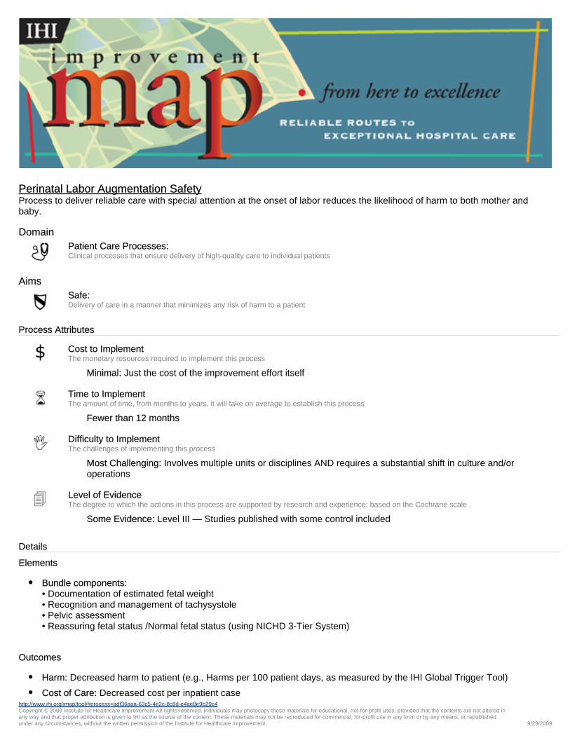
Perinatal Labor Augmentation SafetyProcess to deliver reliable care with special attention at the onset of labor reduces the likelihood of harm to both mother andbaby.
DomainPatient Care Processes:Clinical processes that ensure delivery of high-quality care to individual patients
AimsSafe:Delivery of care in a manner that minimizes any risk of harm to a patient
Process Attributes
Cost to ImplementThe monetary resources required to implement this process
Minimal: Just the cost of the improvement effort itself
Time to ImplementThe amount of time, from months to years, it will take on average to establish this process
Fewer than 12 months
Difficulty to ImplementThe challenges of implementing this process
Most Challenging: Involves multiple units or disciplines AND requires a substantial shift in culture and/oroperations
Level of EvidenceThe degree to which the actions in this process are supported by research and experience; based on the Cochrane scale
Some Evidence: Level III — Studies published with some control included
Details
Elements
● Bundle components:• Documentation of estimated fetal weight• Recognition and management of tachysystole• Pelvic assessment• Reassuring fetal status /Normal fetal status (using NICHD 3-Tier System)
Outcomes
● Harm: Decreased harm to patient (e.g., Harms per 100 patient days, as measured by the IHI Global Trigger Tool)● Cost of Care: Decreased cost per inpatient case
http://www.ihi.org/imap/tool/#process=adf36aaa-63c5-4e2c-8c8d-e4ae8e9b29c4Copyright © 2009 Institute for Healthcare Improvement All rights reserved. Individuals may photocopy these materials for educational, not-for-profit uses, provided that the contents are not altered inany way and that proper attribution is given to IHI as the source of the content. These materials may not be reproduced for commercial, for-profit use in any form or by any means, or republishedunder any circumstances, without the written permission of the Institute for Healthcare Improvement. 9/29/2009

Service Lines and Critical Functions
● Obstetrics
Key Measures
● All components of the Perinatal Augmentation Bundle completed• Numerator: Percent of patients where all components of the Perinatal Augmentation Bundle were completed• Denominator: Number of records reviewed
● Percent change in rate of mothers transferred to higher level of care
● Percent change in rate of newborns admitted to neonatal intensive care
Reasons and Implications
Importance for Patients and FamiliesWhen mothers in labor require medicines to strengthen labor, it is important that those medicines are used safely. This reducesthe chance of harm for both mother and baby and means that separating mother from infant after delivery is less likely.
Requirement, Standards, Policies, and Guidelines
● National Priorities Partnership (NPP)Safety
● The Joint Commission (TJC)National Patient Safety Goal 3: High Alert Medications
Financial Implications
● • Expense reduction can occur due to decreased length of stay of mother in labor and delivery, and infant in neonatalintensive care. • Expense increases can occur due to greater throughput in labor and delivery (e.g., nursing time, length ofstay). • Revenue reduction can occur due to the decreased length of stay for mothers and babies. • Overall, a positivereturn on investment is reported in the literature because hospitals that have increased the reliability of these componentshave also reported a decrease in medical malpractice set-asides and overall expenses.
Prerequisites
• Collaboration between nurses and obstetrical care providers in the labor and delivery unit• Acceptance of standard algorithms for treatment
Resources
Additional Resources
● American College of Obstetricians and Gynecologists (ACOG)Labor augmentation
● Map of Medicine• Caesarean Section• Term Labour
IHI.org Resources
● Improvement Map Discussion BoardsJoin the Improvement Map Discussion Groups to help IHI build dynamic communities of learning and support. Posequestions, offer new ideas, describe your improvement success stories and experiences, and share tips with your peers fortaking full advantage of all the Improvement Map has to offer.
● Reducing Birth-Related Trauma and Liability ExposureLehigh Valley Hospital and Health Network (Allentown, Pennsylvania, USA) reduces birth-related trauma through their workas part of a Perinatal Innovation Community.
● Improving Safety Within the Birth CenterIncreased reliability in the Birth Center makes perinatal care safer at HealthPartners Regions Hospital (St. Paul, Minnesota,
http://www.ihi.org/imap/tool/#process=adf36aaa-63c5-4e2c-8c8d-e4ae8e9b29c4Copyright © 2009 Institute for Healthcare Improvement All rights reserved. Individuals may photocopy these materials for educational, not-for-profit uses, provided that the contents are not altered inany way and that proper attribution is given to IHI as the source of the content. These materials may not be reproduced for commercial, for-profit use in any form or by any means, or republishedunder any circumstances, without the written permission of the Institute for Healthcare Improvement. 9/29/2009

USA).
Information Compiled By
• Peter Cherouny, MD, IHI Faculty• Randall Morgan, MD, IHI Faculty
http://www.ihi.org/imap/tool/#process=adf36aaa-63c5-4e2c-8c8d-e4ae8e9b29c4Copyright © 2009 Institute for Healthcare Improvement All rights reserved. Individuals may photocopy these materials for educational, not-for-profit uses, provided that the contents are not altered inany way and that proper attribution is given to IHI as the source of the content. These materials may not be reproduced for commercial, for-profit use in any form or by any means, or republishedunder any circumstances, without the written permission of the Institute for Healthcare Improvement. 9/29/2009

Richard Berkowitz, MD, FACOGCo-Chair
Peter Bernstein, MD, MPH, FACOGCo-Chair
Traci Burgess, MD, FACOGElisa Burns, MD, FACOGEdward Denious, MD, FACOGLawrence Dolkart, MD, FACOGDena Goffman, MD, FACOGAmos Grunebaum, MD, FACOGVictor Klein, MD, FACOGDenise Lester, MD, FACOGAbraham Lichtmacher, MD, FACOGPaul Ogburn, MD, FACOGMichael Scalzone, MD, MHCM, FACOGJoseph Sclafani, MD, FACOGHeather Shannon, CNM, NP, MSAlexander Shilkrut, MD, FACOGJohn Vullo, DO, FACOGBrian Wagner, MD, FACOGRichard Waldman, MD, FACOGStanley Zinberg, MD, MS, FACOG
StaffDonna Montalto, MPPKelly Gilchrist
American Congress of Obstetricians & Gynecologists (ACOG), District II152 Washington Ave, Suite 300Albany, New York 12210
Phone: 518-436-3461 • Fax: 518-426-4728E-mail: [email protected]: www.acogny.org
ACOG District II Patient Safety & Quality Improvement Committee