Oxidative stress increases 1-deoxysphingolipid levels in ...
11
Zurich Open Repository and Archive University of Zurich Main Library Strickhofstrasse 39 CH-8057 Zurich www.zora.uzh.ch Year: 2021 Oxidative stress increases 1-deoxysphingolipid levels in chronic kidney disease Gui, Ting ; Li, Yunlun ; Zhang, Shijun ; Alecu, Irina ; Chen, Qingfa ; Zhao, Ying ; Hornemann, Thorsten ; Kullak-Ublick, Gerd A ; Gai, Zhibo Abstract: Chronic kidney disease (CKD) leads to deep changes in lipid metabolism and obvious dyslipi- demia. The dysregulation of lipid metabolism in turn results in CKD progression and the complications of cardiovascular diseases. To obtain a profound insight into the associated dyslipidemia in CKD, we performed lipidomic analysis to measure lipid metabolites in the serum from a rat 5/6 nephrectomy (5/6 Nx) model of CKD as well as in the serum from CKD patients. HK-2 cells were also used to examine oxidative stress-induced sphingolipid changes. Totally 182 lipid species were identifed in 5/6 Nx rats. We found glycerolipids, total free fatty acids, and sphingolipids levels were signifcantly upregulated in 5/6 Nx rats. The atypical sphingolipids, 1-deoxysphingolipids, were signifcantly altered in both CKD animals and human CKD patients. The levels of 1-deoxysphingolipids directly relevant to the level of oxidative stress in vivo and in vitro. These results demonstrate that 1-deoxysphingolipid levels are increased in CKD and this increase directly correlates with increased kidney oxidative stress. DOI: https://doi.org/10.1016/j.freeradbiomed.2021.01.011 Posted at the Zurich Open Repository and Archive, University of Zurich ZORA URL: https://doi.org/10.5167/uzh-199983 Journal Article Published Version The following work is licensed under a Creative Commons: Attribution-NonCommercial-NoDerivatives 4.0 International (CC BY-NC-ND 4.0) License. Originally published at: Gui, Ting; Li, Yunlun; Zhang, Shijun; Alecu, Irina; Chen, Qingfa; Zhao, Ying; Hornemann, Thorsten; Kullak-Ublick, Gerd A; Gai, Zhibo (2021). Oxidative stress increases 1-deoxysphingolipid levels in chronic kidney disease. Free Radical Biology Medicine, 164:139-148. DOI: https://doi.org/10.1016/j.freeradbiomed.2021.01.011
Transcript of Oxidative stress increases 1-deoxysphingolipid levels in ...

Zurich Open Repository andArchiveUniversity of ZurichMain LibraryStrickhofstrasse 39CH-8057 Zurichwww.zora.uzh.ch
Year: 2021
Oxidative stress increases 1-deoxysphingolipid levels in chronic kidneydisease
Gui, Ting ; Li, Yunlun ; Zhang, Shijun ; Alecu, Irina ; Chen, Qingfa ; Zhao, Ying ; Hornemann,Thorsten ; Kullak-Ublick, Gerd A ; Gai, Zhibo
Abstract: Chronic kidney disease (CKD) leads to deep changes in lipid metabolism and obvious dyslipi-demia. The dysregulation of lipid metabolism in turn results in CKD progression and the complicationsof cardiovascular diseases. To obtain a profound insight into the associated dyslipidemia in CKD, weperformed lipidomic analysis to measure lipid metabolites in the serum from a rat 5/6 nephrectomy (5/6Nx) model of CKD as well as in the serum from CKD patients. HK-2 cells were also used to examineoxidative stress-induced sphingolipid changes. Totally 182 lipid species were identified in 5/6 Nx rats. Wefound glycerolipids, total free fatty acids, and sphingolipids levels were significantly upregulated in 5/6 Nxrats. The atypical sphingolipids, 1-deoxysphingolipids, were significantly altered in both CKD animalsand human CKD patients. The levels of 1-deoxysphingolipids directly relevant to the level of oxidativestress in vivo and in vitro. These results demonstrate that 1-deoxysphingolipid levels are increased inCKD and this increase directly correlates with increased kidney oxidative stress.
DOI: https://doi.org/10.1016/j.freeradbiomed.2021.01.011
Posted at the Zurich Open Repository and Archive, University of ZurichZORA URL: https://doi.org/10.5167/uzh-199983Journal ArticlePublished Version
The following work is licensed under a Creative Commons: Attribution-NonCommercial-NoDerivatives4.0 International (CC BY-NC-ND 4.0) License.
Originally published at:Gui, Ting; Li, Yunlun; Zhang, Shijun; Alecu, Irina; Chen, Qingfa; Zhao, Ying; Hornemann, Thorsten;Kullak-Ublick, Gerd A; Gai, Zhibo (2021). Oxidative stress increases 1-deoxysphingolipid levels in chronickidney disease. Free Radical Biology Medicine, 164:139-148.DOI: https://doi.org/10.1016/j.freeradbiomed.2021.01.011

Free Radical Biology and Medicine 164 (2021) 139–148
Available online 12 January 20210891-5849/© 2021 The Authors. Published by Elsevier Inc. This is an open access article under the CC BY-NC-ND license(http://creativecommons.org/licenses/by-nc-nd/4.0/).
Original article Oxidative stress increases 1-deoxysphingolipid levels in chronic kidney disease Ting Gui a,1, Yunlun Li b,c,1, Shijun Zhang d,k, Irina Alecu e, f,g, Qingfa Chen h, Ying Zhao i, Thorsten Hornemann j, Gerd A. Kullak-Ublick k, l,**, Zhibo Gai a,b,k,*
a Key Laboratory of Traditional Chinese Medicine Classical Theory, Ministry of Education, Shandong University of Traditional Chinese Medicine, Jinan, 250355, PR China b Innovation Research Institute of Traditional Chinese Medicine, Shandong University of Traditional Chinese Medicine, Jinan, 250355, PR China c The Third Department of Cardiovascular Diseases, Affiliated Hospital of Shandong University of Traditional Chinese Medicine, Jinan, 250000, PR China d First Clinical Medical College, Shandong University of Traditional Chinese Medicine, Jinan, 250355, PR China e Neural Regeneration Laboratory, Department of Biochemistry, Microbiology and Immunology, Ottawa Institute of Systems Biology, Ottawa, ON, Canada f Department of Cellular and Molecular Medicine, UOttawa Brain and Mind Research Institute, Ottawa, ON, Canada g Department of Chemistry and Biomolecular Sciences, Centre for Catalysis and Research Innovation, University of Ottawa, Ottawa, ON, Canada h Institute for Tissue Engineering and Regenerative Medicine, Liaocheng University/Liaocheng People’s Hospital, Liaocheng, Shandong, PR China i Department of Basic Biology, Institute of Biological Sciences, Jining Medical University, Jining, PR China j Department of Clinical Chemistry, University Hospital Zurich, University of Zurich, Switzerland k Department of Clinical Pharmacology and Toxicology, University Hospital Zurich, University of Zurich, Zurich, Switzerland l Mechanistic Safety, CMO & Patient Safety, Global Drug Development, Novartis Pharma, Basel, Switzerland
A R T I C L E I N F O
Keywords: Chronic kidney disease 1-Deoxysphingolipid Free fatty acid MDA Oxidative stress
A B S T R A C T
Chronic kidney disease (CKD) leads to deep changes in lipid metabolism and obvious dyslipidemia. The dysre-gulation of lipid metabolism in turn results in CKD progression and the complications of cardiovascular diseases. To obtain a profound insight into the associated dyslipidemia in CKD, we performed lipidomic analysis to measure lipid metabolites in the serum from a rat 5/6 nephrectomy (5/6 Nx) model of CKD as well as in the serum from CKD patients. HK-2 cells were also used to examine oxidative stress-induced sphingolipid changes. Totally 182 lipid species were identified in 5/6 Nx rats. We found glycerolipids, total free fatty acids, and sphingolipids levels were significantly upregulated in 5/6 Nx rats. The atypical sphingolipids, 1-deoxysphingo-lipids, were significantly altered in both CKD animals and human CKD patients. The levels of 1-deoxysphingoli-pids directly relevant to the level of oxidative stress in vivo and in vitro. These results demonstrate that 1- deoxysphingolipid levels are increased in CKD and this increase directly correlates with increased kidney oxidative stress.
1. Introduction
As an important public health problem, chronic kidney disease (CKD) damages up to 13% adults in America [1] and worldwide [2]. The classic biomarker used in clinics to monitor kidney function is serum creatinine, which is used to estimate the glomerular filtration rate and
monitor urine output [3]. Creatinine is, however, insensitive for kidney injury, especially in the beginning process of disease [4], and provides no information on the cause and nature of renal injury, since the levels of creatinine are influenced by non-renal factors, including age, sex, nutritional status, infection, and medications [5,6]. Large efforts have been put into identification of new markers that are more specific to
Abbreviations: CKD, Chronic kidney disease; LDL, low density lipoprotein; PAS, Periodic acid-Schiff; PS, phosphatidylserine; FA, fatty acids; TG, triglycerides; Cer, ceramide; PG, phosphatidylglycerol; CerG, hexosylceramide; PI, phosphatidylinositol; PC, phosphatidylcholine; PE, phosphatidylethanolamine; SM, sphingomyelin; C18SA, C18 sphinganine; C18SO, C18 sphingosine; doxSA, 1-deoxysphinganine; doxSO, 1-deoxysphingosine.
* Corresponding author. Key Laboratory of Traditional Chinese Medicine Classical Theory, Ministry of Education, Shandong University of Traditional Chinese Medicine, Jinan, 250355, PR China. ** Corresponding author. Department of Clinical Pharmacology and Toxicology, University Hospital Zurich, University of Zurich, Zurich, Switzerland.
E-mail addresses: [email protected] (G.A. Kullak-Ublick), [email protected] (Z. Gai). 1 These authors contributed equally to this work.
Contents lists available at ScienceDirect
Free Radical Biology and Medicine journal homepage: www.elsevier.com/locate/freeradbiomed
https://doi.org/10.1016/j.freeradbiomed.2021.01.011 Received 13 June 2020; Received in revised form 22 December 2020; Accepted 6 January 2021

Free Radical Biology and Medicine 164 (2021) 139–148
140
renal tubular oxidative stress or can predict ‘‘subclinical’’ renal damage before serum creatinine elevation rather than assessing glomerular filtration as an endpoint.
Lipids are small molecules representing products or intermediates of metabolic processes. Lipids play significant roles in cytotoxicity, signal transduction, and energy production. The balance of lipids intake and production, transport and excretion maintains the concentrations of various lipids in body fluids and cells. Kidney plays a central role in all these processes, which provides a theoretical basis for the study of lip-idomics in nephrology [7]. Recently, increasing researches indicated that serum lipid profile and lipid metabolism altered markedly during CKD pathogenesis of in vivo and in vitro [8,9] and linked them to enhanced reactive oxygen species production [10,11]. Lipidomics has been used to determine the lipid distribution of low density lipoprotein (LDL), which demonstrated that triacylglycerides are significantly increased, and plasmenylethanolamines, cholesterol sulfate, phospha-tidylcholines, ceramides, and sulfatides are significantly reduced in the advanced CKD patients [12].
Sphingolipids are a class of structurally highly diverse lipids that are fundamental components of eukaryotic cell membranes. Sphingolipid de novo synthesis starts with the formation of the long chain sphingoid base, which is the characteristic backbone of all sphingolipids. This first and rate-limiting step is catalyzed by the enzyme serine- palmitoyltransferase (SPT), which typically conjugates L-serine and palmitoyl-CoA [13]. Moreover, SPT shows an alternative activity with L-alanine, which results in the formation of an atypical category of 1-deoxysphingolipids [14]. Due to the lack of C1-hydroxyl group of sphinganine, 1-deoxysphingolipid can neither form complexes (sphin-gomyelins and glycosphingolipids) at its head group, nor proceed for further degradation [15]. Abnormal 1-deoxysphinglipid levels are found to be associated with mutations in SPT [16], certain metabolic condi-tions [17–19] or reductions of cytochrome P450 enzymes [20]. How-ever, the changes of serum 1-deoxysphingolipids in CKD, and the underlying mechanism is still unknown.
In this study, we performed a common procedure to construct Sprague-Dawley rats of 5/6 Nx-induced CKD model to assess the changes of the associated serum sphingolipid levels. To this end, we analyzed serum sphingolipid levels, renal function, and renal oxidative stress markers in 5/6 Nx rats. In addition kidney tissue and serum sphingolipids were examined in CKD patients. Both animal and clinical data indicate that oxidative stress marker malondialdehyde (MDA) co- relates with the increase of 1-deoxysphingolipids in CKD. By using free fatty acid (FFA)-albumin overload-induced oxidative stress models in vivo and in vitro, oxidative stress is shown to mediate the increase of 1-deoxysphingolipids, independent from SPT-related de novo synthesis.
2. Materials and methods
2.1. Animals
Male Sprague-Dawley rats (200–220 g per rat) from Vital River Laboratories (Beijing, China) were randomly divided into 5/6 ne-phrectomy (5/6 Nx) group and normal control sham group. Each group of rats were placed in the animal facility with a 12:12-h day and night cycle. The animals were allowed ad libitum to regular food (AIN93 M, Keao Xieli feed co. LTD, Beijing China) and water. As described previ-ously [21], animals from CKD group received a two-stage procedure of 5/6 nephrectomy with lateral incision to expose the retroperitoneal kidney. The animals from sham-operated group received the same incision and kidney exposure, while the kidney was kept intact. For the lipidomics analysis, 5 animals per group were used. In another experi-ment, at least 6 animals were used in each group to confirm lipidomics results and for further analysis. All rats were sacrificed under anesthesia 8 weeks after the second stage of surgery and kidneys were harvested. Half of the kidney from each animal was snap frozen in liquid nitrogen and stored at − 80 ◦C for RNA extraction. The other half was fixed with
formalin for histology. For FFA-induced kidney damage experiment, a mild albumin overload kidney damage model was used according to previous studies [11,22–25], with modifications. Briefly, a 30% BSA/-saline solution was made. For palmitate-loaded BSA, 150 μmolar palmitate acid was bound to 50 ml of 30% BSA to make a 1 mM BSA-FFA stock. Aliquots were stored at 4 ◦C until the time of injection. Groups of male Sprague-Dawley rats (200–220 g per rat) were randomly received i.p. injection with palmitate-loaded BSA (BSA-FFA, resolved in saline) or fatty acid-free BSA (vehicle, resolved in saline) at a dosage 1g BSA/200g body weight for 1 week.
2.2. Serum and urine samples measurement
For 24-h urine collection, rats were allowed with free access to food and water. Urinary H2O2 levels and urinary creatinine concentrations were measured with the Amplex Red H2O2 assay kit (A12214, Invi-trogen) and the creatinine assay kit (ab65340, Abcam), respectively. 12- hour fasted rats were used to obtain serum samples from their tail blood. Serum triglyceride, cholesterol, and malondialdehyde (MDA) levels were measured using the triglyceride assay kit (ETGA-200, EnzyChrom, Aachen, Germany), the Amplex Red cholesterol assay kit (A12216, Invitrogen), and the Lipid Peroxidation (MDA) Assay kit (ab118970, Abcam), respectively.
2.3. Renal pathology assessment and immunostaining
In order to assess renal pathology, kidneys were embedded in paraffin wax following overnight fixation in 10% neutral buffer formalin. Fixed kidneys were sectioned into 4-μm-thick slices using a microtome (Leica, RM2235). Then Periodic acid-Schiff (PAS) staining was performed to determine the mesangial area according to the man-ufacturer’s instruction. Masson’s trichrome staining and Methenamine Silver (MS) staining were performed to assess the fibrotic area using standard protocols. Six rats were analyzed per group.
For immunostaining analysis, de-waxed paraffin sections (4-μm- thick) were hydrated, microwaved for 8–15 min in 10 mM sodium cit-rate (pH 6.0) for antigen retrieval, and then immunostained with a rabbit antibody against 4-hydroxynonenal (4-HNE, ab46545, Abcam). Immunolabeled sections were then incubated with goat anti-rabbit second antibody conjugated to horseradish peroxidase and treated with the EnVision+ diaminobenzidine kit (DAB, Dako, Glostrup, Denmark) using standard protocols.
2.4. Lipid analysis
The lipidomic analysis was performed as described previously [26, 27]. Quality criteria for the identified lipid metabolites were linearity R2>0.9 and CV<20%. For analysis of the sphingoid base profile, the lipids were acid/base hydrolyzed to remove the headgroups and N-linked fatty acid as described earlier [20,28]. Briefly, acid/-base–hydrolyzed lipids were derivatized with ο-phthalaldehyde (OPA) by redissolving in 75 μl of 56.7% MeOH, 33.3% EtOH, 10% H2O and 5 μL OPA working solution (990 μl boric acid [3%] + 10 μl OPA [50 mg/ml in EtOH] + 0.5 μl 2-mercaptoethanol). For liquid chromatographic sepa-ration of the sphingoid and deoxysphingoid base backbones, a C18 column was used (Uptispere 120 Å, 5 μm, 125 × 2 mm, Interchim, Montluçon, France) coupled to a Transcend UPLC pump (Thermo, Reinach, BL, Switzerland). An atmospheric pressure chemical ionization source (APCI) was used to ionize the sphingoid and deoxysphingoid bases, which were then detected using a Q-Exactive hybrid quadrupole Orbitrap mass spectrometer (Thermo, Reinach, BL, Switzerland) run in full scan mode. The MS parameters used were as follows: scan range of m/z 120–1200, mass resolution of 140000, automatic gain control (ACG) target of 3.00E+06 and max injection time of 512 ms.
T. Gui et al.

Free Radical Biology and Medicine 164 (2021) 139–148
141
2.5. Patients
Kidney samples were collected in the Affiliated Hospital of Shandong University of Traditional Chinese Medicine. This study included sixty- three patients with CKD, as diagnosed according to KDIGO Clinical Practice Guideline for the Evaluation and Management of CKD. The exclusion criteria were: (a) liver, thyroid, or cardiovascular (previous myocardial infarction, heart failure) diseases; (b) type 2 diabetes mel-litus history; (c) high body mass index (more than 40 kg/m2); and (d) medication intake known to affect the test parameters. The weight change of participants was less than 5 kg during the last 6 months. Samples from 31 gender- and age-matched healthy volunteers were used as normal controls. Clinical and demographic data of included patients, such as gender, age, creatinine, and cystatin C are shown in Table 1.
2.6. Cell culture
HK-2 cells were maintained in K-SFM medium (17005-042, GIBCO) supplied with bovine pituitary extract (0.05 mg/ml, provided with the K-SFM kit) and human recombinant epidermal growth factor (5 ng/ml, provided with the K-SFM kit), plated in culture at 37 ◦C, 5% CO2. For the free fatty acid (FFA) treatment, cells were first starved in 0.2% FBS/ DMEM for 12h, cells were incubated by the addition of palmitate acid- bound BSA (100 μM PA) for 24 hours. BSA was used as the vehicle. FFA-containing medium was prepared by the conjugation of palmitate acid with FFA-free BSA, as previously described [29].
2.7. Cell staining
FFA-treated HK-2 cells were fixed in 4% paraformaldehyde in PBS, and blocked with 1% BSA/PBS for 30 min following with 0.1% Triton X- 100 treatment for 15 min. Then, the cells were incubated with primary antibody at 4 ◦C overnight. The cells were washed and incubated with secondary antibody for 1 h in the dark. After being washed, the cells were mounted with DAPI (Vector Laboratories) and visualized under a fluorescent microscope (Leica DMI6000B). For intracellular ROS detection, either plasmids encoding redox-sensitive GFR (roGFP plasmid #49435, AddGene) or CellROX green (Life Technologies) were used as previously described [30,31].
2.8. Isolation of RNA from kidney tissue and cells and quantification of transcript levels
Total RNA was extracted with Trizol (Invitrogen, Waltham, MA). Two micrograms of total RNA were reverse transcribed using random primers and Superscript II reverse transcriptase (Invitrogen). First- strand cDNA was used as the template for real-time PCR analysis with primers (Applied Biosystems, CA) and TaqMan master mix. Data were calculated and expressed relative to levels of RNA for the housekeeping gene β-actin.
2.9. Statistics
Data are expressed as mean ± S.D. Statistical significance of differ-ences between groups was assessed by Student’s t-test. PCA and heatmap
visualization were performed using Metaboanalyst software. Correlation analysis was done using the concentrations of 1-deoxysphingolipids and the concentration of clinical biochemistry markers including the renal function parameters creatinine and cystatin c. GraphPad was used to perform statistical analyses.
2.10. Study approval
Animal protocols and experiments abided by the laws of animal protection and were approved by the local institutional animal com-mittee of Shandong University of Traditional Chinese Medicine, China. The research from human samples was performed according to the Declaration of Helsinki guidelines regarding ethical principles for medical research involving human subjects. Written informed consent was provided by each patient participated in the research. The study protocol was approved by the Scientific Ethical Committee of Affiliated Hospital of Shandong University of Traditional Chinese Medicine, China (license number SDU2017045).
3. Results
3.1. General data and histological analysis of a 5/6 Nx-induced chronic kidney disease rat model
Serum samples collected from rats from the 5/6 Nx group or sham group were first analyzed for creatinine and BUN content, the standard markers currently used to assess CKD. Significant, general increases in serum creatinine (Fig. 1 A) and BUN (Fig. 1 B) were noted in the 5/6 Nx group, indicating glomerular filtration damage. Histological examina-tion of kidney tissue from 5/6 Nx rats showed clear signs of kidney damage in all individual, including glomeruli sclerosis, and interstitial fibrosis (Supplementary Fig. 1).
3.2. Lipid abnormalities in the 5/6 nx rat model
Next, lipidomic profiling of serum was performed. The PCA score plot showed a clear separation between 5/6 Nx and sham (Fig. 1C). 182 (10 down + 172 up) lipids passed the cut-off (1.5-fold change, p < 0.02, and FDR<0.05) were identified (Fig. 1 D, and Supplementary Table S1 and Supplementary Table S2). Furthermore, there were also significant differences in lipid subclass totals in the CKD group compared to the sham group (Fig. 1 E). Lipids that were significantly increased belong to four lipid classes: free fatty acids (FFA), triglycerides (TG), sphingolipids (including ceramides, Cer; hexosylceramides, CerG), and phosphati-dylglycerol (PG). These results confirm that there are profound distur-bance of fatty acid and triglyceride metabolism in rats with CKD, as previously reported in CKD patients and animal models [32,33]. Vali-dation of serum FFA and TG confirmed increased levels of both (Fig. 1 F and G). The increase of sphingolipids in serum of 5/6 Nx rats was particularly of note. To examine changes in the sphingoid base profile, a targeted liquid chromatography-mass spectrometry analysis of the total sphingoid- and 1-deoxysphingoid base composition was performed (Fig. 2). Extracted sphingolipids were subjected to a serial acid and base hydrolysis to release the N-linked acyl and O-linked headgroup struc-tures. Levels of canonical C18 sphingolipids represented as C18 sphin-ganine (C18SA) (Fig. 2 A) and C18 sphingosine (C18SO) (Fig. 2 B), were identical between the two groups. However, the levels of the atypical 1-deoxysphingolipids, 1-deoxysphinganine (doxSA) (Fig. 2C) and 1-deoxysphingosine (doxSO) (Fig. 2 D), were significantly increased in the serum of 5/6 Nx rats compared to the sham group.
3.3. Correlation between 1-deoxysphingolipid levels and oxidative stress in clinical samples
We next investigated whether there is a correlation between serum 1- deoxysphinganine and clinical parameters in human CKD patients. To
Table 1 Clinical and demographic data of included patients.
Normal (n = 31) CKD (n = 63) P value Age (years) 46.39 ± 3.231 52.06 ± 1.472 0.0729 Gender (M/F) 15/16 30/33 >0.99 Creatinine (μmol/L) 63.52 ± 2.126 143.2 ± 5.030 <0.0001 Cystatin C (mg/L) 0.8697 ± 0.0261 1.615 ± 0.04073 <0.0001
Data are means ± SD. Abbreviations: M, male; F, female; CKD, chronic kidney disease.
T. Gui et al.

Free Radical Biology and Medicine 164 (2021) 139–148
142
distinguish the lipid changes in CKD caused from systemic disorders, patients with amyloidosis, diabetes, systemic lupus erythematosus, etc. were excluded from the study. Table 1 shows the clinical and de-mographic data of patients participated in this study. No significant differences were found in age and gender between patients with CKD
and healthy controls. Serum creatinine and cystatin C were significantly higher in patients with CKD compared to the healthy controls. The kidneys from CKD patients showed signs of glomerulisclerosis, fibrosis and oxidative stress (Fig. 3 A). Blood levels of MDA, C18SA, doxSA, and doxSO were also increased in the CKD group (Fig. 3 B).
Fig. 1. Lipidomics analysis of serum lipids from sham and 5/6 Nx rats. (A) Serum creatinine and (B) serum BUN from sham and 5/6 Nx groups. n = 6/group. Data are mean ± SD, student’s t-test. (C) Principal component analysis (PCA) score plot of lipidomics profiles of the two groups (n = 5/group). (D) Volcano plot identified lipid species passing the cut-off (1.5-fold change and p < 0.02). (E) Heatmap of clustered lipids from sham and 5/6 Nx rats. Serum levels of (F) fatty acids (FA) and (G) triglycerides (TG) from different groups. n = 6/group. Data are means ± SD, student’s t-test. PS, phosphatidylserine; FA, fatty acids; TG, triglycerides; Cer, ceramide; PG, phosphatidylglycerol; CerG, hexosylceramide; PI, phosphatidylinositol; PC, phosphatidylcholine; PE, phosphatidylethanolamine; SM, sphingomyelin.
Fig. 2. Sphingoid base profile of total serum sphingolipids. Quantitative analysis of serum (A) C18SA, (B) C18SO, (C) doxSA, and (D) doxSO from sham and 5/6 Nx groups. The profile reflects the total sphingoid base composition of serum sphingolipids measured after hydrolysis. C18SA, C18 sphinganine; C18SO, C18 sphin-gosine; doxSA, 1-deoxysphinganine; doxSO, 1-deoxysphingosine. n = 6/group. Data are means ± SD, student’s t-test.
T. Gui et al.

Free Radical Biology and Medicine 164 (2021) 139–148
143
The relationships between serum concentrations of C18- sphingolipids or 1-deoxysphingolipids and the oxidative stress marker MDA and glomerular filtration markers (creatinine and cystatin C) in healthy subjects and CKD patients are illustrated in Table 2. Serum C18 sphingolipids were not co-related with either MDA or glomerular filtration markers (Table 2 and Fig. 4 A and B). Both 1-deoxysphinganine and 1-deoxysphingosine showed significant positive correlations with MDA in CKD patients but not in healthy subjects (Table 2 and Fig. 4 C and D). There was a significant negative correlation between serum 1- deoxysphinganine and Cystatin C (Table 2). There were no significant
negative correlations between serum 1-deoxyshingolipids with creati-nine. Interestingly, when the creatinine levels were translated into eGFR, serum 1-deoxysphingosine was positively correlated with eGFR levels in CKD patients. This indicates a positive relationship between the extent of oxidative stress and serum 1-deoxysphingolipid levels, but not with canonical C18-sphingolipids in CKD patients.
Fig. 3. CKD patients show higher oxidative stress and increased 1-deoxysphingolipid levels. (A) Representative images showing (a and d) Methenamine Silver (MS) staining, (b and e) Periodic acid–Schiff (PAS) staining and (c and f) immunostaining for 4-HNE on renal biopsies from (a, c, and e) normal and (b, d, and f) CKD patients (scale bar 20 μm for MS staining and PAS staining; scale bar 100 μm for 4-HNE staining). (g to i) Quantitative analysis of (g) MS staining, (h) PAS staining, and (i) immunostaining for 4-HNE. (B to F) Violin plot analysis showing the mean and variance differences in serum (B) MDA, (C) C18SA, (D) C18SO, (E) doxSA, and (F) doxSO between healthy subjects and CKD patients. C18SA, C18 sphinganine; C18SO, C18 sphingosine; doxSA, 1-deoxysphinganine; doxSO, 1-deoxysphingosine. Data are means ± SD, student’s t-test.
Table 2 1-deoxysphingolipids significantly associated with serum oxidative stress marker MDA.
C18SA dependent variable C18SO dependent variable doxSA dependent variable doxSO dependent variable R p value r p value r p value r p value
Creatinine -0.2007 0.1148 -0.2659 0.1772 -0.2183 0.0856 -0.2157 0.0896 Cystatin C -0.0452 0.7227 0.0215 0.8662 -0.2623 0.0362 -0.2434 0.0526 eGFR 0.1316 0.3039 0.2184 0.0855 0.1975 0.1207 0.2822 0.0251 MDA 0.0316 0.8024 -0.0300 0.8139 0.2619 0.0183 0.2383 0.0290
Data are means ± SD. Abbreviations: eGFR, estimated glomerular filtration rate; MDA, malondialdehyde.
T. Gui et al.

Free Radical Biology and Medicine 164 (2021) 139–148
144
3.4. Increased serum 1-deoxysphingolipid levels are a marker of FFA- induced tubule oxidative stress in a chronic kidney disease rat model
Since fatty acids are the preferred energy source for proximal tubular cells, excessive fatty acid supply induces oxidative stress and mito-chondrial dysfunction, which result in reduced fatty acid oxidation and abnormal lipid metabolism [8]. In human CKD and an animal model of 5/6 Nx-induced CKD, increased urinary albumin and albumin-bound FFA may increase the exposure of FFA to proximal tubules and result
in tubular damage, oxidative stress, and mitochondrial dysfunction [11, 34,35]. To evaluate if increased 1-deoxysphingolipid levels are a result of increased FFA exposure and subsequent abnormal lipid oxidation and oxidative stress in proximal tubules, rats were i.p. injected with BSA-bound FFA. Urinary H2O2 and serum levels of MDA were signifi-cantly increased by 1.5 to 2-fold in BSA-FFA compared to vehicle lit-termates (Fig. 5 A and B). A significant increase of lipid peroxidation was detected in BSA-FFA rats as examined by immunostaining for 4-HNE, which was closely associated with the severity of proximal tubule
Fig. 4. 1-Deoxysphingolipid levels correlated with oxidative stress marker MDA. Correlation analysis between serum MDA and serum (A) C18SA, (B) C18SO, (C) doxSA, and (D) doxSO in CKD patients. C18SA, C18 sphinganine; C18SO, C18 sphingosine; doxSA, 1-deoxysphinganine; doxSO, 1-deoxysphingosine.
Fig. 5. FFA overload in vivo induces oxidative stress and results in increased serum levels of 1-deoxysphingolipids. Quantitative analysis of (A) urinary H2O2 and (B) serum MDA from rats after vehicle (BSA) and FFA (BSA-bound FFA) injections. n = 6/group. Data are means ± SD, student’s t-test. Representative images showing (C) immunostaining for 4-HNE on renal sections from (a) vehicle and (b) FFA groups (scale bar 50 μm). (c) Quantitative analysis of immunostaining for 4-HNE. (D and E) Quantitative analysis of serum (D) C18 sphingoid bases, and (E) deoxysphingoid bases from vehicle and FFA groups. C18SA, C18 sphinganine; C18SO, C18 sphingosine; doxSA, 1-deoxysphinganine; doxSO, 1-deoxysphingosine. n = 6/group. Data are means ± SD, student’s t-test. (F and G) mRNA level of (F) SPTLC2 and (G) SPTLC3 in the kidney of rats with different treatments. n = 6/group. Data are means ± SD, student’s t-test.
T. Gui et al.
Free Radical Biology and Medicine 164 (2021) 139–148
145
oxidative stress (Fig. 5C). There was also a resulting increase in both 1-deoxysphinganine and 1-deoxysphingosine but not of the canonical sphingolipids (C18SA and C18SO) in the serum of BSA-FFA group (Fig. 5 D and E). In addition, mRNA expression of SPTLC2 (Serine Palmitoyl-transferase Long Chain Base Subunit 2) and SPTLC3 (Serine Palmitoyl-transferase Long Chain Base Subunit 3), the two main sphingolipid de novo synthesis enzymes, were not changed between treatment groups (Fig. 5 F and G), indicating that increased 1-deoxysphingolipids are not a direct conversion from fatty acids.
3.5. Oxidative stress induces 1-deoxysphingolipid levels in vitro
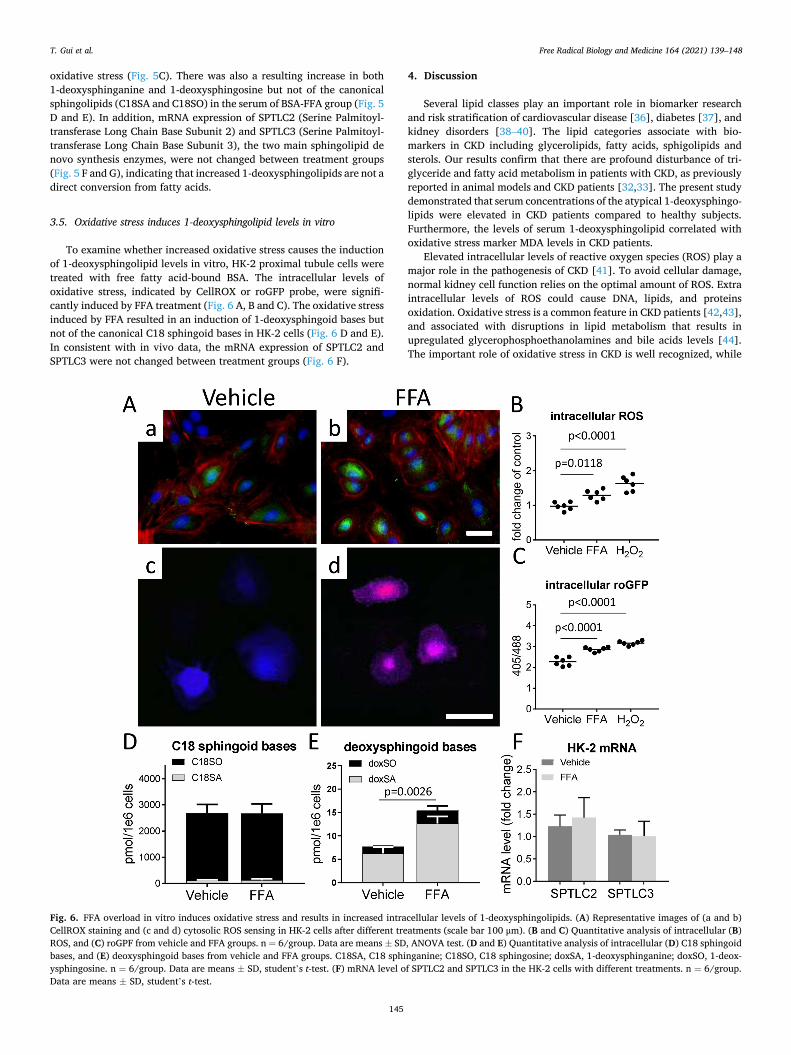
To examine whether increased oxidative stress causes the induction of 1-deoxysphingolipid levels in vitro, HK-2 proximal tubule cells were treated with free fatty acid-bound BSA. The intracellular levels of oxidative stress, indicated by CellROX or roGFP probe, were signifi-cantly induced by FFA treatment (Fig. 6 A, B and C). The oxidative stress induced by FFA resulted in an induction of 1-deoxysphingoid bases but not of the canonical C18 sphingoid bases in HK-2 cells (Fig. 6 D and E). In consistent with in vivo data, the mRNA expression of SPTLC2 and SPTLC3 were not changed between treatment groups (Fig. 6 F).
4. Discussion
Several lipid classes play an important role in biomarker research and risk stratification of cardiovascular disease [36], diabetes [37], and kidney disorders [38–40]. The lipid categories associate with bio-markers in CKD including glycerolipids, fatty acids, sphigolipids and sterols. Our results confirm that there are profound disturbance of tri-glyceride and fatty acid metabolism in patients with CKD, as previously reported in animal models and CKD patients [32,33]. The present study demonstrated that serum concentrations of the atypical 1-deoxysphingo-lipids were elevated in CKD patients compared to healthy subjects. Furthermore, the levels of serum 1-deoxysphingolipid correlated with oxidative stress marker MDA levels in CKD patients.
Elevated intracellular levels of reactive oxygen species (ROS) play a major role in the pathogenesis of CKD [41]. To avoid cellular damage, normal kidney cell function relies on the optimal amount of ROS. Extra intracellular levels of ROS could cause DNA, lipids, and proteins oxidation. Oxidative stress is a common feature in CKD patients [42,43], and associated with disruptions in lipid metabolism that results in upregulated glycerophosphoethanolamines and bile acids levels [44]. The important role of oxidative stress in CKD is well recognized, while
Fig. 6. FFA overload in vitro induces oxidative stress and results in increased intracellular levels of 1-deoxysphingolipids. (A) Representative images of (a and b) CellROX staining and (c and d) cytosolic ROS sensing in HK-2 cells after different treatments (scale bar 100 μm). (B and C) Quantitative analysis of intracellular (B) ROS, and (C) roGPF from vehicle and FFA groups. n = 6/group. Data are means ± SD, ANOVA test. (D and E) Quantitative analysis of intracellular (D) C18 sphingoid bases, and (E) deoxysphingoid bases from vehicle and FFA groups. C18SA, C18 sphinganine; C18SO, C18 sphingosine; doxSA, 1-deoxysphinganine; doxSO, 1-deox-ysphingosine. n = 6/group. Data are means ± SD, student’s t-test. (F) mRNA level of SPTLC2 and SPTLC3 in the HK-2 cells with different treatments. n = 6/group. Data are means ± SD, student’s t-test.
T. Gui et al.

Free Radical Biology and Medicine 164 (2021) 139–148
146
there are limited clinical tools to monitor oxidative stress. Levels of the oxidative stress markers methionine sulfoxide, methionine sulfoxide-to-methionine ratio [45,46], and malonedialdehyde [47] have already been analyzed by metabolomics. It is very difficult for quanti-tative analysis of oxidative stress in order to meet the special conditions for pre-analysis. Oxidative stress in CKD was demonstrated to be asso-ciated with marked alteration of serum concentrations of different lipids [32,48]. In the present study we found that FFA-induced oxidative stress results in increased 1-deoxysphingolipid levels both in vivo and in vitro. Furthermore, the correlation between 1-deoxysphingolipid levels and oxidative stress marker, MDA, was significant in serum from CKD pa-tients. Given the nature that 1-deoxysphingolipids are hardly degraded in the body once it is synthesized as it can not be phosphorylated by sphingosine kinase and degraded by S1P lyase, serum 1-deoxysphingoli-pids could be used as a useful marker for kidney oxidative stress in CKD patients.
A number of uremic toxins (including fatty acids) are found to be increased in CKD and are potent oxidative stress inducers through impairing mitochondrial function [49–51]. Considering the large num-ber of uremic toxins, 1-deoxysphingolipids could contribute partially to oxidative stress, as examined by recent publications [28,52]. However, several key points indicate that the increases of 1-deoxyphingolipid levels in CKD are independent to oxidative stress: (1) Because of the absence of the C1 hydroxyl group, 1-deoxysphingolipids can neither be converted to sphingomyelin through sphingomyelin phosphodiesterase (SMase), nor be degraded through sphingosine kinase (SPHK) [53]. Although increase of oxidants regulate enzyme activities of SMase [54, 55] and SPHK [56,57] and their metabolites [58], such enzymatic pathways are not related to 1-deoxysphingolipid production; (2) A number of reports suggest that lipid accumulation in kidney could be harmful and is referred to as lipotoxicity [59]. It is still under debate whether lipids per se are toxic, but it is clear that intra-renal lipid accumulation can cause characteristics of renal oxidative stress status [60]. The reason why 1-deoxysphingolipids are increased in CKD could be due to the dyslipidemia-induced oxidative stress and defective mitochondrial fatty acid oxidation which oftenly occurs in CKD [61,62], resulting in an accumulation of intracellular fatty acids; (3) Among patients with diabetic nephropathy, hypertensive nephropathy, and chronic nephritis, serum levels of serine and glycine are decreased compared with healthy controls [63]. These results indicate that serine deficiency increases lipid accumulation in ESRD patients. Serine defi-ciency is also linked with altered glutathione metabolism and mito-chondrial dysfunction (oxidative stress), as well as increased 1-deoxysphingolipid de novo synthesis [64]; (4) In parallel, reduced expression of cytochrome P450 genes and gene products (i.e., reduced mRNA and protein, or reduced protein with no change in mRNA) in several animal models of CKD were observed [65]. Although the precise mechanism(s) of the down-regulation of CYP genes in these CKD models remains unknown, ex vivo studies showed that uremic human serum obtained from patients with end-stage renal disease led to a decrease in protein expression and activity for all the major xenobiotic-metabolizing CYPs. Such decreased cytochrome enzymes may result in reduced degradation of 1-deoxysphingolipids, which was demonstrated in our previous study in NAFLD [20,28]; Taken together, dyslipidemia-induces oxidative stress and in parallel de novo synthesis of 1-deoxysphingoli-pids due to fatty acid accumulation and serine deficiency which is often seen in CKD. Furthermore, decreased cytochrome P450 enzymes may result in less degradation of 1-deoxysphingolipids and accelerate the progression of CKD through the direct lipotoxic effects of 1-deoxy-sphingolipids on renal cells.
The reason for increased circulating 1-deoxysphigolipids in CKD still remains unclear. When an uncontrolled influx of free fatty acids were flowing into the proximal tubules present in massive proteinuria, FFA not only promote oxidative stress and nephrotoxicity, but could be also transported to the ER where they could be used for de novo 1-deoxy-sphingolipid synthesis [66,67], which may in turn increase levels of
1-deoxysphingolipids in the blood. Dyslipidemic patients benefit from the 1-deoxyshingolipids-lowering effects of fenofibrate treatment [68], which is also commonly used in the treatment against CKD. Several recent clinical studies, including subanalyses of the FIELD (the Fenofi-brate Intervention and Event Lowering in Diabetes Study) and DAIS (Diabetes Atherosclerosis Intervention Study) trials, support the urinary albumin-lowering effects of fenofibrate [69–71]. However, as fibrates can be associated with a rise in blood creatinine levels, their effect on oxidative stress-related renal dysfunction is less obvious. Furthermore, although C18 sphingolipids and 1-deoxysphingolipids could both be de novo synthesized from palmitate acid, changes of 1-deoxysphingolipid levels seem to be independent to C18 sphingolipids and de novo syn-thesis through SPTs, because (1) myriocin, a potent inhibitor of sphin-golipid de novo synthesis, has no effects on 1-deoxysphingolipid de novo synthesis [72]; and (2) FXR, a nuclear receptor which regulates hepa-tocyte lipid homeostasis, induces 1-deoxysphingolipids degradation but does not change C18 sphingolipid levels [73]. Great efforts are still needed to further determine the exact molecular signaling mechanisms underlying the elevation of circulating 1-deoxysphingolipid levels.
The relations between glomerular filtration and increased serum 1- deoxysphingolipid levels are still not clear. In present study, doxSA is negatively correlated with Cystatin C, however, doxSO is positively correlated with eGFR. Since sphingolipids are constituents of lipopro-teins which are too large to pass the glomeruli [74], the elimination way of sphingolipids could be different to Cystatin C, which makes it difficult to directly compare the relationships between those parameters. Furthermore, since eGFR also depends on gender, age and ethnics, the observed corrections could also be indirect. Further studies regarding to CKD stages, real GFR (assessed from clearance measurements), and other factors, such as gender and age, need to be done.
Collectively, our experimental model indicates that the increased serum level of 1-deoxysphingolipids reflects a stress, such as FFA over-load or protein-bound FFA-induced oxidative stress, on the proximal tubules. The clinical results support this hypothesis and show that serum 1-deoxysphingolipids might be useful clinical markers for CKD. These results provide a novel possibility for applying for the clinical implica-tions of sphingolipid levels in the oxidative stress status of CKD.
Declaration of competing interest
The authors declare no conflict of interest.
Acknowledgments
None.
Appendix A. Supplementary data
Supplementary data to this article can be found online at https://doi. org/10.1016/j.freeradbiomed.2021.01.011.
Author contributions
Conceptualization, T.G. and Z.G.; methodology, T.G. and Z.G; vali-dation, T.G., I.A., Q.C. and S.Z.; formal analysis, T.G. and I.A.; investi-gation, T.G., I.A., Y.Z., Y.L. and S.Z.; resources, Y.L. and Z.G.; data curation, T.G. and Z.G.; writing—original draft preparation, T.G. and Z. G.; writing—review and editing, Z.G., T.H. and G.K-U; supervision, G.K- U; project administration, Y.L.; funding acquisition, Y.L. T.H. All authors have read and agreed to the published version of the manuscript.
Funding
This research was funded by the National Natural Science Founda-tion of China (81774242, 81974566) and Major Basic Research Projects of Shandong Natural Science Foundation (ZR2018ZC1157) (YL); by
T. Gui et al.

Free Radical Biology and Medicine 164 (2021) 139–148
147
Supporting Fund for Teachers’ research of Jining Medical University (JYFC2018KJ046) (YZ); by Swiss National Foundation SNF (Project 31003A_153390 and 31003A_179371) (TH).
References [1] J. Coresh, E. Selvin, L.A. Stevens, J. Manzi, J.W. Kusek, P. Eggers, F. Van Lente, A.
S. Levey, Prevalence of chronic kidney disease in the United States, Jama 298 (17) (2007) 2038–2047.
[2] M. Ruiz-Ortega, S. Rayego-Mateos, S. Lamas, A. Ortiz, R.R. Rodrigues-Diez, Targeting the progression of chronic kidney disease, Nat. Rev. Nephrol. 16 (5) (2020) 269–288.
[3] C.M. Helgason, A new view of anterior choroidal artery territory infarction, J. Neurol. 235 (7) (1988) 387–391.
[4] R.D. Perrone, N.E. Madias, A.S. Levey, Serum creatinine as an index of renal function: new insights into old concepts, Clin. Chem. 38 (10) (1992) 1933–1953.
[5] J. Westhuyzen, Z.H. Endre, G. Reece, D.M. Reith, D. Saltissi, T.J. Morgan, Measurement of tubular enzymuria facilitates early detection of acute renal impairment in the intensive care unit, Nephrol. Dial. Transplant. 18 (3) (2003) 543–551, official publication of the European Dialysis and Transplant Association - European Renal Association.
[6] W.K. Han, V. Bailly, R. Abichandani, R. Thadhani, J.V. Bonventre, Kidney Injury Molecule-1 (KIM-1): a novel biomarker for human renal proximal tubule injury, Kidney Int. 62 (1) (2002) 237–244.
[7] Y.Y. Zhao, X.L. Cheng, R.C. Lin, Lipidomics applications for discovering biomarkers of diseases in clinical chemistry, Int. Rev. Cell Mol. Biol. 313 (2014) 1–26.
[8] H.M. Kang, S.H. Ahn, P. Choi, Y.A. Ko, S.H. Han, F. Chinga, A.S. Park, J. Tao, K. Sharma, J. Pullman, E.P. Bottinger, I.J. Goldberg, K. Susztak, Defective fatty acid oxidation in renal tubular epithelial cells has a key role in kidney fibrosis development, Nat. Med. 21 (1) (2015) 37–46.
[9] Z.H. Zhang, H. Chen, N.D. Vaziri, J.R. Mao, L. Zhang, X. Bai, Y.Y. Zhao, Metabolomic signatures of chronic kidney disease of diverse etiologies in the rats and humans, J. Proteome Res. 15 (10) (2016) 3802–3812.
[10] A. Kamijo, T. Sugaya, A. Hikawa, M. Okada, F. Okumura, M. Yamanouchi, A. Honda, M. Okabe, T. Fujino, Y. Hirata, M. Omata, R. Kaneko, H. Fujii, A. Fukamizu, K. Kimura, Urinary excretion of fatty acid-binding protein reflects stress overload on the proximal tubules, Am. J. Pathol. 165 (4) (2004) 1243–1255.
[11] A. Kamijo, K. Kimura, T. Sugaya, M. Yamanouchi, H. Hase, T. Kaneko, Y. Hirata, A. Goto, T. Fujita, M. Omata, Urinary free fatty acids bound to albumin aggravate tubulointerstitial damage, Kidney Int. 62 (5) (2002) 1628–1637.
[12] A. Reis, A. Rudnitskaya, P. Chariyavilaskul, N. Dhaun, V. Melville, J. Goddard, D. J. Webb, A.R. Pitt, C.M. Spickett, Top-down lipidomics of low density lipoprotein reveal altered lipid profiles in advanced chronic kidney disease, J. Lipid Res. 56 (2) (2015) 413–422.
[13] C. Ziv, S. Malitsky, A. Othman, S. Ben-Dor, Y. Wei, S. Zheng, A. Aharoni, T. Hornemann, A. Vardi, Viral serine palmitoyltransferase induces metabolic switch in sphingolipid biosynthesis and is required for infection of a marine alga, Proc. Natl. Acad. Sci. U. S. A. 113 (13) (2016) E1907–E1916.
[14] A. Penno, M.M. Reilly, H. Houlden, M. Laura, K. Rentsch, V. Niederkofler, E. T. Stoeckli, G. Nicholson, F. Eichler, R.H. Brown, A. von Eckardstein, T. Hornemann, Hereditary sensory neuropathy type 1 is caused by the accumulation of two neurotoxic sphingolipids, J. Biol. Chem. 285 (15) (2010) 11178–11187.
[15] M.A. Lone, T. Santos, I. Alecu, L.C. Silva, T. Hornemann, 1-Deoxysphingolipids, Biochim. Biophys. Acta Mol. Cell Biol. Lipids 1864 (4) (2019) 512–521.
[16] A. Penno, M.M. Reilly, H. Houlden, M. Laura, K. Rentsch, V. Niederkofler, E. T. Stoeckli, G. Nicholson, F. Eichler, R.H. Brown Jr., A. von Eckardstein, T. Hornemann, Hereditary sensory neuropathy type 1 is caused by the accumulation of two neurotoxic sphingolipids, J. Biol. Chem. 285 (15) (2010) 11178–11187.
[17] Y. Yoshimine, H. Uto, K. Kumagai, S. Mawatari, S. Arima, R. Ibusuki, K. Mera, T. Nosaki, S. Kanmura, M. Numata, T. Tamai, A. Moriuchi, H. Tsubouchi, A. Ido, Hepatic expression of the Sptlc3 subunit of serine palmitoyltransferase is associated with the development of hepatocellular carcinoma in a mouse model of nonalcoholic steatohepatitis, Oncol. Rep. 33 (4) (2015) 1657–1666.
[18] R.A. Zuellig, T. Hornemann, A. Othman, A.B. Hehl, H. Bode, T. Güntert, O. O. Ogunshola, E. Saponara, K. Grabliauskaite, J.-H. Jang, U. Ungethuem, Y. Wei, A. von Eckardstein, R. Graf, S. Sonda, Deoxysphingolipids, novel biomarkers for type 2 diabetes, are cytotoxic for insulin-producing cells, Diabetes 63 (4) (2014) 1326–1339.
[19] A. Othman, M.F. Rütti, D. Ernst, C.H. Saely, P. Rein, H. Drexel, C. Porretta- Serapiglia, G. Lauria, R. Bianchi, A. von Eckardstein, T. Hornemann, Plasma deoxysphingolipids: a novel class of biomarkers for the metabolic syndrome? Diabetologia 55 (2) (2012) 421–431.
[20] Z. Gai, T. Gui, I. Alecu, M.A. Lone, T. Hornemann, Q. Chen, M. Visentin, C. Hiller, S. Hausler, G.A. Kullak-Ublick, Farnesoid X receptor activation induces the degradation of hepatotoxic 1-deoxysphingolipids in non-alcoholic fatty liver disease, Liver Int. : Off. J. Int. Assoc.e Stud. Liver 40 (4) (2019) 844–859.
[21] Z. Gai, L. Chu, C. Hiller, D. Arsenijevic, C.A. Penno, J.P. Montani, A. Odermatt, G. A. Kullak-Ublick, Effect of chronic renal failure on the hepatic, intestinal, and renal expression of bile acid transporters, Am. J. Physiol. Ren. Physiol. 306 (1) (2014) F130–F137.
[22] A.A. Eddy, H. Kim, J. Lopez-Guisa, T. Oda, P.D. Soloway, Interstitial fibrosis in mice with overload proteinuria: deficiency of TIMP-1 is not protective, Kidney Int. 58 (2) (2000) 618–628.
[23] Y. Takagaki, S. Shi, M. Katoh, M. Kitada, K. Kanasaki, D. Koya, Dipeptidyl peptidase-4 plays a pathogenic role in BSA-induced kidney injury in diabetic mice, Sci. Rep. 9 (1) (2019) 7519.
[24] D.A. Ishola Jr., D.M. van der Giezen, B. Hahnel, R. Goldschmeding, W. Kriz, H. A. Koomans, J.A. Joles, In mice, proteinuria and renal inflammatory responses to albumin overload are strain-dependent, Nephrol. Dial. Transplant. 21 (3) (2006) 591–597, official publication of the European Dialysis and Transplant Association - European Renal Association.
[25] M.E. Thomas, N.J. Brunskill, K.P. Harris, E. Bailey, J.H. Pringle, P.N. Furness, J. Walls, Proteinuria induces tubular cell turnover: a potential mechanism for tubular atrophy, Kidney Int. 55 (3) (1999) 890–898.
[26] R.M. Pellegrino, A. Di Veroli, A. Valeri, L. Goracci, G. Cruciani, LC/MS lipid profiling from human serum: a new method for global lipid extraction, Anal. Bioanal. Chem. 406 (30) (2014) 7937–7948.
[27] M. Narvaez-Rivas, Q. Zhang, Comprehensive untargeted lipidomic analysis using core-shell C30 particle column and high field orbitrap mass spectrometer, J. Chromatogr., A 1440 (2016) 123–134.
[28] I. Alecu, A. Othman, A. Penno, E.M. Saied, C. Arenz, A. von Eckardstein, T. Hornemann, Cytotoxic 1-deoxysphingolipids are metabolized by a cytochrome P450-dependent pathway, J. Lipid Res. 58 (1) (2017) 60–71.
[29] Z. Gai, T. Gui, C. Hiller, G.A. Kullak-Ublick, Farnesoid X receptor protects against kidney injury in uninephrectomized obese mice, J. Biol. Chem. 291 (5) (2016) 2397–2411.
[30] G.B. Waypa, J.D. Marks, R. Guzy, P.T. Mungai, J. Schriewer, D. Dokic, P. T. Schumacker, Hypoxia triggers subcellular compartmental redox signaling in vascular smooth muscle cells, Circ. Res. 106 (3) (2010) 526–535.
[31] Z. Gai, E. Krajnc, S.L. Samodelov, M. Visentin, G.A. Kullak-Ublick, Obeticholic acid ameliorates valproic acid-induced hepatic steatosis and oxidative stress, Mol. Pharmacol. 97 (5) (2020) 314–323.
[32] D.Q. Chen, H. Chen, L. Chen, N.D. Vaziri, M. Wang, X.R. Li, Y.Y. Zhao, The link between phenotype and fatty acid metabolism in advanced chronic kidney disease, Nephrol. Dial. Transplant. 32 (7) (2017) 1154–1166, official publication of the European Dialysis and Transplant Association - European Renal Association.
[33] H. Chen, L. Chen, D. Liu, D.Q. Chen, N.D. Vaziri, X.Y. Yu, L. Zhang, W. Su, X. Bai, Y. Y. Zhao, Combined clinical phenotype and lipidomic analysis reveals the impact of chronic kidney disease on lipid metabolism, J. Proteome Res. 16 (4) (2017) 1566–1578.
[34] M.E. Thomas, K.P. Harris, J. Walls, P.N. Furness, N.J. Brunskill, Fatty acids exacerbate tubulointerstitial injury in protein-overload proteinuria, Am. J. Physiol. Ren. Physiol. 283 (4) (2002) F640–F647.
[35] M. Arici, R. Chana, A. Lewington, J. Brown, N.J. Brunskill, Stimulation of proximal tubular cell apoptosis by albumin-bound fatty acids mediated by peroxisome proliferator activated receptor-gamma, J. Am. Soc. Nephrol. : JASN (J. Am. Soc. Nephrol.) 14 (1) (2003) 17–27.
[36] P. Wurtz, A.S. Havulinna, P. Soininen, T. Tynkkynen, D. Prieto-Merino, T. Tillin, A. Ghorbani, A. Artati, Q. Wang, M. Tiainen, A.J. Kangas, J. Kettunen, J. Kaikkonen, V. Mikkila, A. Jula, M. Kahonen, T. Lehtimaki, D.A. Lawlor, T. R. Gaunt, A.D. Hughes, N. Sattar, T. Illig, J. Adamski, T.J. Wang, M. Perola, S. Ripatti, R.S. Vasan, O.T. Raitakari, R.E. Gerszten, J.P. Casas, N. Chaturvedi, M. Ala-Korpela, V. Salomaa, Metabolite profiling and cardiovascular event risk: a prospective study of 3 population-based cohorts, Circulation 131 (9) (2015) 774–785.
[37] R. Wang-Sattler, Z. Yu, C. Herder, A.C. Messias, A. Floegel, Y. He, K. Heim, M. Campillos, C. Holzapfel, B. Thorand, H. Grallert, T. Xu, E. Bader, C. Huth, K. Mittelstrass, A. Doring, C. Meisinger, C. Gieger, C. Prehn, W. Roemisch-Margl, M. Carstensen, L. Xie, H. Yamanaka-Okumura, G. Xing, U. Ceglarek, J. Thiery, G. Giani, H. Lickert, X. Lin, Y. Li, H. Boeing, H.G. Joost, M.H. de Angelis, W. Rathmann, K. Suhre, H. Prokisch, A. Peters, T. Meitinger, M. Roden, H. E. Wichmann, T. Pischon, J. Adamski, T. Illig, Novel biomarkers for pre-diabetes identified by metabolomics, Mol. Syst. Biol. 8 (2012) 615.
[38] O.N. Goek, C. Prehn, P. Sekula, W. Romisch-Margl, A. Doring, C. Gieger, M. Heier, W. Koenig, R. Wang-Sattler, T. Illig, K. Suhre, J. Adamski, A. Kottgen, C. Meisinger, Metabolites associate with kidney function decline and incident chronic kidney disease in the general population, Nephrol. Dial. Transplant. 28 (8) (2013) 2131–2138, official publication of the European Dialysis and Transplant Association - European Renal Association.
[39] E. Albrecht, M. Waldenberger, J. Krumsiek, A.M. Evans, U. Jeratsch, M. Breier, J. Adamski, W. Koenig, S. Zeilinger, C. Fuchs, N. Klopp, F.J. Theis, H.E. Wichmann, K. Suhre, T. Illig, K. Strauch, A. Peters, C. Gieger, G. Kastenmuller, A. Doering, C. Meisinger, Metabolite profiling reveals new insights into the regulation of serum urate in humans, Metabolomics, Off. J. Metabol. Soc. 10 (1) (2014) 141–151.
[40] O.N. Goek, A. Doring, C. Gieger, M. Heier, W. Koenig, C. Prehn, W. Romisch-Margl, R. Wang-Sattler, T. Illig, K. Suhre, P. Sekula, G. Zhai, J. Adamski, A. Kottgen, C. Meisinger, Serum metabolite concentrations and decreased GFR in the general population, Am. J. Kidney Dis. : Off. J. Nat. Kidney Found. 60 (2) (2012) 197–206.
[41] M. Irazabal, V. Torres, Reactive oxygen species and redox signaling in chronic kidney disease, Cells 9 (6) (2020).
[42] N.D. Vaziri, Oxidative stress in uremia: nature, mechanisms, and potential consequences, Semin. Nephrol. 24 (5) (2004) 469–473.
[43] F. Locatelli, B. Canaud, K.U. Eckardt, P. Stenvinkel, C. Wanner, C. Zoccali, Oxidative stress in end-stage renal disease: an emerging threat to patient outcome, Nephrol. Dial. Transplant. 18 (7) (2003) 1272–1280, official publication of the European Dialysis and Transplant Association - European Renal Association.
T. Gui et al.

Free Radical Biology and Medicine 164 (2021) 139–148
148
[44] H. Chen, G. Cao, D.Q. Chen, M. Wang, N.D. Vaziri, Z.H. Zhang, J.R. Mao, X. Bai, Y. Y. Zhao, Metabolomics insights into activated redox signaling and lipid metabolism dysfunction in chronic kidney disease progression, Redox Biol. 10 (2016) 168–178.
[45] R. Mashima, T. Nakanishi-Ueda, Y. Yamamoto, Simultaneous determination of methionine sulfoxide and methionine in blood plasma using gas chromatography- mass spectrometry, Anal. Biochem. 313 (1) (2003) 28–33.
[46] M. Breier, S. Wahl, C. Prehn, M. Fugmann, U. Ferrari, M. Weise, F. Banning, J. Seissler, H. Grallert, J. Adamski, A. Lechner, Targeted metabolomics identifies reliable and stable metabolites in human serum and plasma samples, PloS One 9 (2) (2014), e89728.
[47] I. Fonseca, H. Reguengo, M. Almeida, L. Dias, L.S. Martins, S. Pedroso, J. Santos, L. Lobato, A.C. Henriques, D. Mendonca, Oxidative stress in kidney transplantation: malondialdehyde is an early predictive marker of graft dysfunction, Transplantation 97 (10) (2014) 1058–1065.
[48] Y.Y. Zhao, H.L. Wang, X.L. Cheng, F. Wei, X. Bai, R.C. Lin, N.D. Vaziri, Metabolomics analysis reveals the association between lipid abnormalities and oxidative stress, inflammation, fibrosis, and Nrf2 dysfunction in aristolochic acid- induced nephropathy, Sci. Rep. 5 (2015) 12936.
[49] T. Niwa, Role of indoxyl sulfate in the progression of chronic kidney disease and cardiovascular disease: experimental and clinical effects of oral sorbent AST-120, Ther. Apher. Dial. 15 (2) (2011) 120–124.
[50] M. Yisireyili, H. Shimizu, S. Saito, A. Enomoto, F. Nishijima, T. Niwa, Indoxyl sulfate promotes cardiac fibrosis with enhanced oxidative stress in hypertensive rats, Life Sci. 92 (24–26) (2013) 1180–1185.
[51] S. Schmidt, T.H. Westhoff, P. Krauser, W. Zidek, M. van der Giet, The uraemic toxin phenylacetic acid increases the formation of reactive oxygen species in vascular smooth muscle cells, Nephrol. Dial. Transplant. 23 (1) (2008) 65–71, official publication of the European Dialysis and Transplant Association - European Renal Association.
[52] M.A. Lauterbach, V. Saavedra, M.S.J. Mangan, A. Penno, C. Thiele, E. Latz, L. Kuerschner, 1-Deoxysphingolipids cause autophagosome and lysosome accumulation and trigger NLRP3 inflammasome activation, Autophagy 24 (2020) 1–15.
[53] A.C. Carreira, T.C. Santos, M.A. Lone, E. Zupancic, E. Lloyd-Evans, R.F.M. de Almeida, T. Hornemann, L.C. Silva, Mammalian sphingoid bases: biophysical, physiological and pathological properties, Prog. Lipid Res. (2019), 100995.
[54] S.S. Castillo, M. Levy, J.V. Thaikoottathil, T. Goldkorn, Reactive nitrogen and oxygen species activate different sphingomyelinases to induce apoptosis in airway epithelial cells, Exp. Cell Res. 313 (12) (2007) 2680–2686.
[55] A. Huwiler, J. Pfeilschifter, H. van den Bosch, Nitric oxide donors induce stress signaling via ceramide formation in rat renal mesangial cells, J. Biol. Chem. 274 (11) (1999) 7190–7195.
[56] I. Ader, L. Brizuela, P. Bouquerel, B. Malavaud, O. Cuvillier, Sphingosine kinase 1: a new modulator of hypoxia inducible factor 1alpha during hypoxia in human cancer cells, Canc. Res. 68 (20) (2008) 8635–8642.
[57] C. Cinq-Frais, C. Coatrieux, M.-H. Grazide, Y.A. Hannun, A. Negre-Salvayre, R. Salvayre, N. Auge, A signaling cascade mediated by ceramide, src and PDGFRβ
coordinates the activation of the redox-sensitive neutral sphingomyelinase-2 and sphingosine kinase-1, Biochim. Biophys. Acta 1831 (8) (2013) 1344–1356.
[58] K. Geoffroy, L. Troncy, N. Wiernsperger, M. Lagarde, S. El Bawab, Glomerular proliferation during early stages of diabetic nephropathy is associated with local increase of sphingosine-1-phosphate levels, FEBS Lett. 579 (5) (2005) 1249–1254.
[59] X.Z. Ruan, Z. Varghese, J.F. Moorhead, An update on the lipid nephrotoxicity hypothesis, Nat. Rev. Nephrol. 5 (12) (2009) 713–721.
[60] I.M. Wahba, R.H. Mak, Obesity and obesity-initiated metabolic syndrome: mechanistic links to chronic kidney disease, Clin. J. Am. Soc. Nephrol. 2 (3) (2007) 550–562.
[61] M. Jiang, M. Bai, J. Lei, Y. Xie, S. Xu, Z. Jia, A. Zhang, Mitochondrial dysfunction and the AKI-to-CKD transition, Am. J. Physiol. Ren. Physiol. 319 (6) (2020) F1105–F1116.
[62] K. Takemura, H. Nishi, R. Inagi, Mitochondrial dysfunction in kidney disease and uremic sarcopenia, Front. Physiol. 11 (2020) 565023.
[63] L. Zeng, Y. Yu, X. Cai, S. Xie, J. Chen, L. Zhong, Y. Zhang, Differences in serum amino acid phenotypes Among patients with diabetic nephropathy, hypertensive nephropathy, and chronic nephritis, Med. Sci. Mon. Int. Med. J. Exp. Clin. Res. 25 (2019) 7235–7242.
[64] C.R. Ferreira, S.M.I. Goorden, A. Soldatos, H.M. Byers, J.M.M. Ghauharali-van der Vlugt, F.S. Beers-Stet, C. Groden, C.D. van Karnebeek, W.A. Gahl, F.M. Vaz, X. Jiang, H.J. Vernon, Deoxysphingolipid precursors indicate abnormal sphingolipid metabolism in individuals with primary and secondary disturbances of serine availability, Mol. Genet. Metabol. 124 (3) (2018) 204–209.
[65] T.D. Nolin, J. Naud, F.A. Leblond, V. Pichette, Emerging evidence of the impact of kidney disease on drug metabolism and transport, Clin. Pharmacol. Ther. 83 (6) (2008) 898–903.
[66] R. Pralhada Rao, N. Vaidyanathan, M. Rengasamy, A. Mammen Oommen, N. Somaiya, M.R. Jagannath, Sphingolipid metabolic pathway: an overview of major roles played in human diseases, J. Lipids 2013 (2013) 178910.
[67] C.R. Gault, L.M. Obeid, Y.A. Hannun, An overview of sphingolipid metabolism: from synthesis to breakdown, Adv. Exp. Med. Biol. 688 (2010) 1–23.
[68] A. Othman, R. Benghozi, I. Alecu, Y. Wei, E. Niesor, A. von Eckardstein, T. Hornemann, Fenofibrate lowers atypical sphingolipids in plasma of dyslipidemic patients: a novel approach for treating diabetic neuropathy? J. Clin. Lipidol. 9 (4) (2015) 568–575.
[69] T.M. Davis, R. Ting, J.D. Best, M.W. Donoghoe, P.L. Drury, D.R. Sullivan, A. J. Jenkins, R.L. O’Connell, M.J. Whiting, P.P. Glasziou, R.J. Simes, Y.A. Kesaniemi, V.J. Gebski, R.S. Scott, A.C. Keech, I. Fenofibrate, i. Event, Lowering in diabetes study, effects of fenofibrate on renal function in patients with type 2 diabetes mellitus: the fenofibrate intervention and event lowering in diabetes (FIELD) study, Diabetologia 54 (2) (2011) 280–290.
[70] J.C. Ansquer, C. Foucher, S. Rattier, M.R. Taskinen, G. Steiner, D. Investigators, Fenofibrate reduces progression to microalbuminuria over 3 years in a placebo- controlled study in type 2 diabetes: results from the Diabetes Atherosclerosis Intervention Study (DAIS), Am. J. Kidney Dis. : Off. J. Nat. Kidney Found. 45 (3) (2005) 485–493.
[71] A.S. Group, H.N. Ginsberg, M.B. Elam, L.C. Lovato, J.R. Crouse 3rd, L.A. Leiter, P. Linz, W.T. Friedewald, J.B. Buse, H.C. Gerstein, J. Probstfield, R.H. Grimm, F. Ismail-Beigi, J.T. Bigger, D.C. Goff Jr., W.C. Cushman, D.G. Simons-Morton, R. P. Byington, Effects of combination lipid therapy in type 2 diabetes mellitus, N. Engl. J. Med. 362 (17) (2010) 1563–1574.
[72] R.A. Zuellig, T. Hornemann, A. Othman, A.B. Hehl, H. Bode, T. Güntert, O. O. Ogunshola, E. Saponara, K. Grabliauskaite, J.H. Jang, U. Ungethuem, Y. Wei, A. v. Eckardstein, R. Graf, S. Sonda, Deoxysphingolipids, novel biomarkers for type 2 diabetes, are cytotoxic for insulin-producing cells, Diabetes 63 (4) (2014) 1326–1339.
[73] Z. Gai, T. Gui, I. Alecu, M.A. Lone, T. Hornemann, Q. Chen, M. Visentin, C. Hiller, S. Hausler, G.A. Kullak-Ublick, Farnesoid X receptor activation induces the degradation of hepatotoxic 1-deoxysphingolipids in non-alcoholic fatty liver disease, Liver Int. : Off. J. Int. Assoc.e Stud. Liver 40 (4) (2020) 844–859.
[74] M. Bertea, M.F. Rutti, A. Othman, J. Marti-Jaun, M. Hersberger, A. von Eckardstein, T. Hornemann, Deoxysphingoid bases as plasma markers in diabetes mellitus, Lipids Health Dis. 9 (2010) 84.
T. Gui et al.