Oxidative Degradation of Cardiotoxic Anticancer Anthracyclines to Phthalic Acids
13
Oxidative Degradation of Cardiotoxic Anticancer Anthracyclines to Phthalic Acids NOVEL FUNCTION FOR FERRYLMYOGLOBIN* Received for publication, June 20, 2003, and in revised form, October 17, 2003 Published, JBC Papers in Press, November 21, 2003, DOI 10.1074/jbc.M306568200 Antonella Cartoni‡§, Pierantonio Menna§¶, Emanuela Salvatorelli§¶, Daniela Braghiroli, Rossella Giampietro¶, Fabio Animati‡, Andrea Urbani¶, Piero Del Boccio¶, and Giorgio Minotti¶** From the ‡Department of Chemistry, Menarini Ricerche, 00040 Pomezia, Rome, G. d’Annunzio University School of Pharmacy, ¶G. d’Annunzio University School of Medicine, and “Centro Studi sull’ Invecchiamento” (Ce.S.I), 66013 Chieti, Italy We show that the pseudoperoxidase activity of fer- rylmyoglobin (Mb IV ) promotes oxidative degradation of doxorubicin (DOX), an anticancer anthracycline known to induce severe cardiotoxicity. Mb IV , formed in vitro by reacting horse heart Mb III with H 2 O 2 , caused disappear- ance of the spectrum of DOX at 477 nm and appearance of UV-absorbing chromophores that indicated opening and degradation of its tetracyclic ring. Electron spray ionization mass spectrometry analyses of DOX/Mb IV ul- trafiltrates showed that DOX degradation resulted in formation of 3-methoxyphthalic acid, the product of ox- idative modifications of its methoxy-substituted ring D. Other methoxy-substituted anthracyclines similarly re- leased 3-methoxyphthalic acid after oxidation by Mb IV , whereas demethoxy analogs released simple phthalic acid. Kinetic and stoichiometric analyses of reactions between DOX and Mb III /H 2 O 2 or hemin/H 2 O 2 showed that the porphyrin radical of Mb IV -compound I and the iron-oxo moiety of Mb IV -compound II were sequentially involved in oxidizing DOX; however, oxidation by com- pound I formed more 3-methoxyphthalic acid than oxi- dation by compound II. Sizeable amounts of 3-me- thoxyphthalic acid were formed in the heart of mice treated with DOX, in human myocardial biopsies ex- posed to DOX in vitro, and in human cardiac cytosol that oxidized DOX after activation of its endogenous myoglo- bin by H 2 O 2 . Importantly, H9c2 cardiomyocytes were damaged by low concentrations of DOX but could toler- ate concentrations of 3-methoxyphthalic acid higher than those measured in murine or human myocardium. These results unravel a novel function for Mb IV in the oxidative degradation of anthracyclines to phthalic ac- ids and suggest that this may serve a salvage pathway against cardiotoxicity. Myoglobin (Mb) 1 has been implicated as a potential catalyst of cardiac damage induced by increased formation of hydrogen peroxide (H 2 O 2 ). Under normal conditions the majority of Mb is found in its oxygenated form (MbO 2 ), which interacts slowly with H 2 O 2 (k 20.8 s 1 M 1 ); however, both deoxy-Mb II and metmyoglobin (Mb III ) react rapidly with H 2 O 2 (k 3.6 10 3 and 3.4 10 4 s 1 M 1 , respectively) (1, 2). An ideal setting for reactions between Mb and H 2 O 2 has therefore been identified in cardiac ischemia-reperfusion, a condition characterized by conversion of Mb II O 2 to Mb III /deoxy-Mb II during ischemia and by formation of H 2 O 2 during blood reflow (3, 4). Hydrogen peroxide causes two-equivalent oxidation of Mb III to ferrylmyo- globin (Mb IV ), a hypervalent species that oxidizes polyunsatu- rated fatty acids and several other biomolecules in a fashion similar to that described for the compound I or II of peroxi- dases. The reaction sequence through which Mb IV is gener- ated from Mb III has been considered as shown in Reactions 1 and 2, Por-Mb III -globin H 2 O 2 3 Por -Mb IV -globin H 2 O REACTION 1 Por -Mb IV -globin 3 Por-Mb IV -globin REACTION 2 In Reaction 1, H 2 O 2 converts Mb III to a compound I-like species in which both oxidizing equivalents are retained in the heme pocket, one in the form of a long lived iron-oxo moiety (Fe IV O) and the other in the form of a transient porphyrin -cation radical (Por ) (5). In Reaction 2, the porphyrin radical dissipates in the globin, causing formation of amino acid radi- cals while leaving the heme moiety in a Fe IV O form similar to compound II (5– 8). We developed an interest in possible reactions between Mb and doxorubicin (DOX), an anticancer anthracycline which ex- hibits activity against several tumors but also causes severe cardiotoxicity. The rationale for investigating DOX-Mb inter- actions was offered by several considerations. On the one hand, cyclic reduction-oxidation of a quinone moiety in the tetracyclic ring of DOX (Fig. 1) generates H 2 O 2 in excess of the detoxifying capacity of cardiomyocytes (9 –11). On the other hand, DOX causes a 4-fold stimulation of the autoxidation of Mb II O 2 to Mb III (12) and inhibits Mb III reductases that would regenerate * This work was supported by Associazione Italiana Ricerca sul Can- cro, MURST COFIN 2001 and 2002, FIRB RBNE 014HJ3-002, and “Center of Excellence on Aging at the University of Chieti” (to G. M.). The costs of publication of this article were defrayed in part by the payment of page charges. This article must therefore be hereby marked “advertisement” in accordance with 18 U.S.C. Section 1734 solely to indicate this fact. § These authors contributed equally to this work. ** To whom correspondence should be addressed: G. d’Annunzio Uni- versity School of Medicine, Centro Studi sull’Invecchiamento, Rm. 412, Via dei Vestini, 66013 Chieti, Italy. Tel.: 39-0871-541391; Fax: 39-0871- 541480; E-mail: [email protected]. 1 The abbreviations used are: Mb, myoglobin; DOX, doxorubicin; Mb III , metmyoglobin; Mb IV , ferrylmyoglobin; H 2 O 2 , hydrogen peroxide; HRP, horseradish peroxidase; LPO, lactoperoxidase; DNR, daunorubi- cin; IDA, idarubicin; ABTS, 2,2-diazinobis(3-ethylbenzothiazoline-6- sulfonic acid); ESI-MS, electron spray ionization-mass spectrometry; TIC, total ion count; C. V., cone voltage; M, molecular ion; MTT, 1-(4,5- dimethylthiazol-2-yl)-3,5-diphenylformazan; HPLC, high pressure liq- uid chromatography. THE JOURNAL OF BIOLOGICAL CHEMISTRY Vol. 279, No. 7, Issue of February 13, pp. 5088 –5099, 2004 © 2004 by The American Society for Biochemistry and Molecular Biology, Inc. Printed in U.S.A. This paper is available on line at http://www.jbc.org 5088 by guest on April 12, 2019 http://www.jbc.org/ Downloaded from
Transcript of Oxidative Degradation of Cardiotoxic Anticancer Anthracyclines to Phthalic Acids

Oxidative Degradation of Cardiotoxic Anticancer Anthracyclines toPhthalic AcidsNOVEL FUNCTION FOR FERRYLMYOGLOBIN*
Received for publication, June 20, 2003, and in revised form, October 17, 2003Published, JBC Papers in Press, November 21, 2003, DOI 10.1074/jbc.M306568200
Antonella Cartoni‡§, Pierantonio Menna§¶, Emanuela Salvatorelli§¶, Daniela Braghiroli�,Rossella Giampietro¶, Fabio Animati‡, Andrea Urbani¶, Piero Del Boccio¶,and Giorgio Minotti¶**From the ‡Department of Chemistry, Menarini Ricerche, 00040 Pomezia, Rome, �G. d’Annunzio University Schoolof Pharmacy, ¶G. d’Annunzio University School of Medicine, and “Centro Studi sull’ Invecchiamento” (Ce.S.I),66013 Chieti, Italy
We show that the pseudoperoxidase activity of fer-rylmyoglobin (MbIV) promotes oxidative degradation ofdoxorubicin (DOX), an anticancer anthracycline knownto induce severe cardiotoxicity. MbIV, formed in vitro byreacting horse heart MbIII with H2O2, caused disappear-ance of the spectrum of DOX at 477 nm and appearanceof UV-absorbing chromophores that indicated openingand degradation of its tetracyclic ring. Electron sprayionization mass spectrometry analyses of DOX/MbIV ul-trafiltrates showed that DOX degradation resulted information of 3-methoxyphthalic acid, the product of ox-idative modifications of its methoxy-substituted ring D.Other methoxy-substituted anthracyclines similarly re-leased 3-methoxyphthalic acid after oxidation by MbIV,whereas demethoxy analogs released simple phthalicacid. Kinetic and stoichiometric analyses of reactionsbetween DOX and MbIII/H2O2 or hemin/H2O2 showedthat the porphyrin radical of MbIV-compound I and theiron-oxo moiety of MbIV-compound II were sequentiallyinvolved in oxidizing DOX; however, oxidation by com-pound I formed more 3-methoxyphthalic acid than oxi-dation by compound II. Sizeable amounts of 3-me-thoxyphthalic acid were formed in the heart of micetreated with DOX, in human myocardial biopsies ex-posed to DOX in vitro, and in human cardiac cytosol thatoxidized DOX after activation of its endogenous myoglo-bin by H2O2. Importantly, H9c2 cardiomyocytes weredamaged by low concentrations of DOX but could toler-ate concentrations of 3-methoxyphthalic acid higherthan those measured in murine or human myocardium.These results unravel a novel function for MbIV in theoxidative degradation of anthracyclines to phthalic ac-ids and suggest that this may serve a salvage pathwayagainst cardiotoxicity.
Myoglobin (Mb)1 has been implicated as a potential catalystof cardiac damage induced by increased formation of hydrogen
peroxide (H2O2). Under normal conditions the majority of Mb isfound in its oxygenated form (MbO2), which interacts slowlywith H2O2 (k � 20.8 s�1 M�1); however, both deoxy-MbII andmetmyoglobin (MbIII) react rapidly with H2O2 (k � 3.6 � 103
and 3.4 � 104 s�1 M�1, respectively) (1, 2). An ideal setting forreactions between Mb and H2O2 has therefore been identifiedin cardiac ischemia-reperfusion, a condition characterized byconversion of MbIIO2 to MbIII/deoxy-MbII during ischemia andby formation of H2O2 during blood reflow (3, 4). Hydrogenperoxide causes two-equivalent oxidation of MbIII to ferrylmyo-globin (MbIV), a hypervalent species that oxidizes polyunsatu-rated fatty acids and several other biomolecules in a fashionsimilar to that described for the compound I or II of peroxi-dases. The reaction sequence through which MbIV is gener-ated from MbIII has been considered as shown in Reactions 1and 2,
Por-MbIII-globin � H2O2 3 Por�-MbIV-globin � H2O
REACTION 1
Por�-MbIV-globin 3 Por-MbIV-globin�
REACTION 2
In Reaction 1, H2O2 converts MbIII to a compound I-likespecies in which both oxidizing equivalents are retained in theheme pocket, one in the form of a long lived iron-oxo moiety(FeIV�O) and the other in the form of a transient porphyrin�-cation radical (Por�) (5). In Reaction 2, the porphyrin radicaldissipates in the globin, causing formation of amino acid radi-cals while leaving the heme moiety in a FeIV�O form similar tocompound II (5–8).
We developed an interest in possible reactions between Mband doxorubicin (DOX), an anticancer anthracycline which ex-hibits activity against several tumors but also causes severecardiotoxicity. The rationale for investigating DOX-Mb inter-actions was offered by several considerations. On the one hand,cyclic reduction-oxidation of a quinone moiety in the tetracyclicring of DOX (Fig. 1) generates H2O2 in excess of the detoxifyingcapacity of cardiomyocytes (9–11). On the other hand, DOXcauses a 4-fold stimulation of the autoxidation of MbIIO2 toMbIII (12) and inhibits MbIII reductases that would regenerate
* This work was supported by Associazione Italiana Ricerca sul Can-cro, MURST COFIN 2001 and 2002, FIRB RBNE 014HJ3-002, and“Center of Excellence on Aging at the University of Chieti” (to G. M.).The costs of publication of this article were defrayed in part by thepayment of page charges. This article must therefore be hereby marked“advertisement” in accordance with 18 U.S.C. Section 1734 solely toindicate this fact.
§ These authors contributed equally to this work.** To whom correspondence should be addressed: G. d’Annunzio Uni-
versity School of Medicine, Centro Studi sull’Invecchiamento, Rm. 412,Via dei Vestini, 66013 Chieti, Italy. Tel.: 39-0871-541391; Fax: 39-0871-541480; E-mail: [email protected].
1 The abbreviations used are: Mb, myoglobin; DOX, doxorubicin;
MbIII, metmyoglobin; MbIV, ferrylmyoglobin; H2O2, hydrogen peroxide;HRP, horseradish peroxidase; LPO, lactoperoxidase; DNR, daunorubi-cin; IDA, idarubicin; ABTS, 2,2�-diazinobis(3-ethylbenzothiazoline-6-sulfonic acid); ESI-MS, electron spray ionization-mass spectrometry;TIC, total ion count; C. V., cone voltage; M, molecular ion; MTT, 1-(4,5-dimethylthiazol-2-yl)-3,5-diphenylformazan; HPLC, high pressure liq-uid chromatography.
THE JOURNAL OF BIOLOGICAL CHEMISTRY Vol. 279, No. 7, Issue of February 13, pp. 5088–5099, 2004© 2004 by The American Society for Biochemistry and Molecular Biology, Inc. Printed in U.S.A.
This paper is available on line at http://www.jbc.org5088
by guest on April 12, 2019
http://ww
w.jbc.org/
Dow
nloaded from

MbIIO2 (13). Thus, several factors seem to enable DOX topromote reactions between MbIII and H2O2, possibly exposingcardiomyocytes to lipid peroxidation or other forms of oxidativeinjury induced by MbIV. In contrast to these premises, however,we found that DOX inhibited lipid peroxidation induced byMbIV in reconstituted chemical models (14). We demonstratedthat the “antioxidant” effect of DOX was due to its ability toreduce MbIV back to MbIII, a process mediated by a hydroqui-none in juxtaposition to the quinone (cf. Fig. 1) (14). We alsonoticed that electron transfer from DOX to MbIV was accompa-nied by a loss of the optical and fluorescent properties of theanthracycline, as if DOX underwent opening and degradationof its tetracyclic ring (14). These results provided unexpectedevidence that DOX could both favor MbIV formation and pre-vent oxidative damage by serving itself as a suicide substratefor MbIV.
Attempts to identify the product(s) of MbIV-dependent DOXdegradation were unsuccessful (14), and similar problems wereencountered by other investigators when DOX was oxidizedwith horseradish peroxidase, microperoxidase 11 (product ofthe proteolytic digestion of cytochrome c), or �NO2 radicalsgenerated through lactoperoxidase/NO2
� or myeloperoxidase/NO2
� (15–18). Lack of information about the nature of degra-dation products precludes an appraisal of the mechanisms andconsequences of MbIV-anthracycline interactions. Therefore,we designed experiments to elucidate how MbIV oxidizes DOXand to identify the product(s) of DOX degradation. Moreover,we characterized the possible role of degradation product(s) inanthracycline-induced cardiotoxicity.
EXPERIMENTAL PROCEDURES
Chemicals—Doxorubicin, daunorubicin (DNR), and 4-demethoxy-daunorubicin (idarubicin, IDA) were obtained through the courtesy ofPharmacia-Upjohn, Milan, Italy. Thymol-free bovine liver catalase,horse heart MbIII, hemin, type VI-A horseradish peroxidase (HRP),bovine milk lactoperoxidase (LPO), 2,2�-diazinobis(3-ethylbenzothiazo-line-6-sulfonic acid) (ABTS), naphthazarin (5,8-dihydroxy-1,4-naphtho-quinone), and all other chemicals were from Sigma. 3-Methoxyphthalicacid was synthesized by oxidation of 2,3-dimethylanisole with a large
excess of potassium permanganate (19) and purified by crystallizationfrom water.
MbIV Formation—MbIV was formed by reacting MbIII with a 2-foldexcess of H2O2. As shown previously (14), this H2O2:MbIII ratio givescomplete oxidation of MbIII to MbIV while also avoiding confoundingfactors due to the possible release of iron from the heme pocket. MbIII
was quantitated by assuming �630 nm � 3.5 mM�1 cm�1; MbIV wasquantitated according to the formula: MbIV (mM) � ((249 � A550 nm) �(367 � A630 nm)) (20). Unless otherwise indicated, the experiments werecarried out at 37 °C in 0.3 M NaCl, carefully adjusted to pH 7.0. Thiswas done because we noticed that common buffers like phosphate orTris or Hepes accelerated a spontaneous decay of MbIV to MbIII; buffersare also known to participate in anthracycline redox reactions (21).Although unbuffered, the pH of incubations did not vary throughout theexperiment.
Spectrophotometric Assays for MbIV-dependent DOX Degradation—After correction of MbIII absorbance, known amounts of DOX wereadded, and the spectrum of the anthracycline at 477 nm was recordedimmediately. Next, H2O2 was added, and the disappearance of thespectrum of DOX was monitored at regular times. Similar settings wereadopted when hemin was used in place of MbIII; in the latter case 10 mM
stock solutions of hemin were prepared in 100 mM NaOH and dilutedappropriately with 50 mM phosphate buffer to obtain 1 mM workingsolutions.
Spectrophotometric Assays for DOX-dependent MbIV Reduction—Inthe experiments described in the preceding section, involving sequen-tial additions of MbIII and DOX and H2O2, stepwise H2O2-dependentoxidation of MbIII to MbIV/FeIV�O and DOX-dependent reduction ofMbIV/FeIV�O to MbIII were monitored at 426 or 410 nm, respectively(6). In other experiments, MbIII was added, and its spectrum (peaks at502 and 630 nm) was recorded. Next, H2O2 was added, and the spec-trum of MbIV/FeIV�O (peaks at 546 and 586 nm) was taken at regulartimes until DOX was included. Doxorubicin-dependent MbIV/FeIV�Oreduction and MbIII regeneration were eventually monitored as disap-pearance of peaks at 546 and 586 nm or reappearance of peaks at 502and 630 nm, respectively.
HPLC-UV-ESI(�) MS Analysis of DOX Degradation—Varyingamounts of DOX were incubated with equimolar MbIII and a 2-foldexcess of H2O2. After 10 min the reaction mixtures were added withcatalase (2600 units) to decompose any residual H2O2 and ultrafilteredthrough YM 10 Centricon® (Millipore Corp., Bedford, MA). Myoglobin-free ultrafiltrates were injected without further manipulations into aBioSys 510 Liquid Chromatography System (Beckman Instruments,Fullerton, CA) equipped with a (1 � 20 cm) Ultrogel Ac34 column
FIG. 1. Structures of DOX and ana-logs used in this study. The arrows in-dicate structural differences betweenDNR or IDA and DOX (DNR, presence ofa side chain methyl terminus in place of aprimary alcohol; IDA, same as DNR andlack of a methoxy residue in ring D).
Ferrylmyoglobin, Anthracyclines, and Phthalates 5089
by guest on April 12, 2019
http://ww
w.jbc.org/
Dow
nloaded from

(BioSepra SA, Villeneuve la Garenne Cedex, France) equilibrated with2.5 mM NaCl, 2.5 mM CH3COONH4. Samples were eluted with the samemedium at the flow rate of 0.5 ml/min. UV-absorbing fractions werepooled, passed through Anotop 25® filters (Merck), and concentrated byevaporation. After suspension in a minimum volume of CH3OH, thesample was laid into a Waters 2790 separation module (alliance HT)equipped with a Waters 996 photodiode array and a ZMD single qua-drupole mass spectrometer (Micromass, UK) as detectors in series.Reversed phase HPLC was performed using a LiChrospher® (Merck)analytical column (100 RP-18 (5 �m), 150 � 4 mm) and a mobile phasecomposed of (H2O � 0.1% trifluoroacetic acid):(CH3CN � 0.1% triflu-oroacetic acid) at an initial ratio of 80:20, which was decreased to 30:70in 15 min and returned to initial conditions in 20 min, at the flow rateof 0.5 ml/min. After chromatography and photodiode array detection(210–600 nm), mass spectra were acquired at low (unity) resolution inseries at a rate of 300 atomic mass unit/s in compressed centroid mode.Standard ESI ion source, operated in positive ion mode, was used withcapillary voltage of 3.25 kV and cone voltage (C.V.) of 20 or 50 V toobtain mass spectra under low or high energy conditions. The sourceand desolvation temperatures were 100 and 200 °C, respectively. Ni-trogen was used as the nebulizer gas at a flow rate of 59 liters/h and asthe desolvation gas at a flow rate of 430 liters/h.
Liquid Chromatography-Tandem Mass Spectrometry—BecauseHPLC-UV-ESI(�) MS identified 3-methoxyphthalic acid (molecularmass � 196 daltons) as a possible product of MbIV-dependent DOXdegradation, we used liquid chromatography-tandem mass spectrome-try to further characterize DOX/MbIV ultrafiltrates in comparison withauthentic 3-methoxyphthalic acid. Reversed phase HPLC was carriedout using a Waters Alliance 2795 separation module and an AgilentHypersil ODS® column (2.1 � 250 mm; C18 5 �m, 120 Å). Ten microlitersamples were eluted at a flow rate of 200 �l/min with (H2O � 0.2%HCOOH) � (10% CH3CN � 0.2% HCOOH). After 2 min CH3CN wasincreased linearly to reach 70% in 6 min and was then maintainedisocratically for 4 more min. The HPLC system was directly coupledwith a Micromass Quattro Ultima™ triple quadrupole mass spectrom-eter equipped with a Z-Spray ESI source, operated in negative ion modeto improve sensitivity. Analytical conditions were optimized with directinfusion of standard solutions of 4.5 �M 3-methoxyphthalic acid, dis-solved in H2O�CH3CN (50:50, v/v), 0.2% HCOOH, and eluted at a flowrate of 5 �l/min. The product ion scan mode for tandem MS was used.The mass spectrometer monitored the deprotonated molecule [M � H]�
of 3-methoxyphthalic acid at m/z 195 via the first quadrupole filter, andcollision-induced dissociation was performed at the second quadrupole(collision gas, argon, at 2.4e-3 mbar, and collision energy at 8 eV). Thedaughters of m/z 195 were monitored via the third quadrupole in themass range of 50–300 atomic mass units. Spectra were acquired in thecontinuous mode. The capillary and cone voltages were set at �3 kVand �40 V, respectively. The source and desolvation temperatures wereset at 110 and 400 °C, respectively, and the desolvation gas (N2) flowwas set at 600 liters/h. Cone gas (N2) was not used, whereas thenebulizer gas (N2) was left at its maximum flow.
Other HPLC Assays for Anthracyclines and Phthalates—Anthracy-cline/MbIII/H2O2 incubations were extracted with a 4-fold excess ofCHCl3/CH3OH (1:1) and subjected to 10 min of low speed centrifuga-tion. Next, 20 �l of the methanolic phase were analyzed for 3-me-thoxyphthalic acid or simple phthalic acid by reversed phase HPLCusing an HP 1100 system (Hewlett-Packard Co., Palo Alto, CA). Chro-matography was performed using a Hewlett-Packard Zorbax CN col-umn (250 � 4.6 mm, 5 �m) operated at 25 °C. Samples were eluted atthe flow rate of 1.5 ml/min by using a 15-min linear gradient from 50mM NaH2PO4 to CH3CN, 25 mM NaH2PO4, all adjusted to pH 4.0 withorthophosphoric acid and filtered through a 0.22-�m membrane (Milli-pore Corp.). Phthalates were detected at 300 nm by on-line diode arrayspectrometer. Identification was obtained by co-elution with authenticstandards and ESI(�)-MS analysis of peak fractions; quantification wasobtained against standard curves prepared with known amounts ofphthalates subjected to the same extraction and chromatographic pro-cedures. Retention times of 3-methoxyphthalic acid or simple phthalicacid were 4.1 and 4.7 min, respectively. Anthracyclines were detectedby diode array and fluorescence spectroscopy (excitation at 470 nm,emission at 550 nm); retention times of DOX, DNR, IDA, and naphtha-zarin were 12.1, 13.8, 14.3, or 14.5 min, respectively. Quantification wasobtained against appropriate standard curves of each anthracycline.
3-Methoxyphthalic Acid Formation in DOX-treated Mice—Two- tothree-month-old male Balb/c mice, weighing 20–30 g, were treated withDOX (10 mg/kg, intravenously) and sacrificed after 4 or 24 h. Heart,liver, and kidneys were removed, and 0.2-g aliquots were homogenizedin ice-cold 0.3 M NaCl. Next, the homogenates were added with 5 ml of
ice-cold acetone, incubated at �20 °C for 30 min, and centrifuged toprecipitate proteins. The supernatants were vacuum-dried, cleared ofgross particulates by low speed centrifugation in CH3OH, and assayedfor DOX and 3-methoxyphthalic acid by HPLC. Fluorescent anthracy-cline metabolites, like the C-13 secondary alcohol metabolite and hy-droxy- or deoxyaglycones, were also measured using appropriate stand-ards (retention times: 10.9, 12.5, and 13.8 min, respectively).
3-Methoxyphthalic Acid Formation in Human Myocardium—Smallmyocardial samples were taken from the lateral aspect of excluded rightatrium of patients undergoing aorto-coronary bypass grafting. All spec-imens were routinely disposed of by the surgeons during cannulationprocedures; therefore, patients were not subjected to any unjustified orethically unacceptable loss of tissue (22). Thin myocardial strips (�0.1g) were dissected and placed in 5 ml of Krebs bicarbonate buffer in thepresence of 1–10 �M DOX. After 4 h at 37 °C the strips were removed,washed extensively in ice-cold 0.3 M NaCl, and extracted for HPLCanalysis of DOX and fluorescent metabolites or 3-methoxyphthalic acidas described in the preceding section. In other experiments, pools of10–15 anonymous biopsies were processed by sequential homogeniza-tion, 20-min centrifugations at 16,000 and 25,000 � g, and 90-minultracentrifugation at 105,000 � g. The supernatant (cytosol) was usedimmediately or after precipitation with 65% ammonium sulfate, a pro-cedure known to remove Mb by “salting out” (4, 14). Mb-containing orMb-depleted cytosol (referred to as Mb� or Mb� cytosol, respectively)was eventually dialyzed against two 1-liter changes of 0.3 M NaCl, 1 mM
EDTA to remove adventitious iron and other low molecular weightcontaminants, and then against two 1-liter changes of 0.3 M NaCl toremove EDTA-iron complexes (23). Based on nephelometric and spec-tral assays by us and others (24), the myoglobin content of right atriumhomogenates and Mb� or Mb� cytosol was determined to be 1.3 and 2.9or 0.6 nmol/mg of protein, respectively. Whole homogenates and Mb� orMb� cytosol were eventually reconstituted with known amounts of DOXand H2O2 and assayed for anthracycline degradation and 3-methoxyph-thalic acid formation by HPLC.
Experiments with Isolated Cardiomyocytes—The embryonic ratheart-derived cell line H9c2 (American Type Culture Collection (CRL1446)) was grown in Dulbecco’s modified Eagle’s medium as describedpreviously (25). Subconfluent cells were seeded in quadruplicate in24-well plates (�105 cells/well) and incubated overnight in the absenceor presence of increasing amounts of DOX or 3-methoxyphthalic acid. Atthe end of treatment, cell viability was measured by 1-(4,5-dimethyl-thiazol-2-yl)-3,5-diphenylformazan (MTT) conversion assay as an indi-cator of mitochondrial function (Sigma kit) (26)
Other Conditions and Assays—Proteins were assayed by the bicin-choninic acid method (27). The peroxidative activity of HRP, LPO, MbIII
and hemin was determined by monitoring oxidation of ABTS to itscation radical ABTS�� (�660 nm � 12 � 103 M�1 cm�1) (16). All values aregiven as means � S.E.; where indicated, data were analyzed by un-paired Student’s t test, and differences were considered significantwhen p was � 0.05. Other conditions are indicated in the legends tofigures and tables.
RESULTS
MbIV-dependent DOX Degradation, Role of Compound I andII—Incubation of DOX with H2O2 and MbIII resulted in atime-dependent decay of the spectrum of the anthracycline andconcomitant development of absorbance at 350 nm (Fig. 2A).These spectral changes were similar to those observed by oth-ers when monitoring reactions of DOX with H2O2 and HRP orlactoperoxidase (16); under those conditions the loss of absorb-ance at 477 nm was attributed to degradation of the anthra-quinone chromophore, whereas the increase of absorbance at350 nm was attributed to accumulation of degradation prod-uct(s). In our study putative product(s) of anthracycline degra-dation were found to exhibit more complex characteristics. Infact, difference spectra obtained by subtracting the initial scanof DOX/MbIII mixtures to the scans obtained at regular timesafter H2O2 addition revealed that the increase in absorbance at350 nm was accompanied by additional peaks at �210, 242,and 280 nm, possibly reflecting the formation and spectraloverlapping of a complex mixture of degradation products (Fig.2B). Doxorubicin degradation increased with the ratio of MbIV
to DOX, near-to-complete degradation occurring at MbIV:DOXratios around unity (Fig. 2C). MbIV was less effective than HRP
Ferrylmyoglobin, Anthracyclines, and Phthalates5090
by guest on April 12, 2019
http://ww
w.jbc.org/
Dow
nloaded from

or LPO at oxidizing a typical peroxidatic substrate like ABTS,its kcat/km for formation of ABTS�� being �2 orders of magni-tude lower than that determined for either peroxidase (1.8 �0.2 � 103 for MbIV versus 7 � 0.4 � 105 or 1.1 � 0.3 � 105 M�1
s�1, respectively; n � 3). However, MbIV was more effectivethan HRP or LPO at degrading DOX, its kcat/km for DOXdisappearance being �1 order of magnitude higher than thatdetermined for either peroxidase (1.1 � 0.2 � 103 versus 0.9 �0.1 � 102 or 3 � 0.2 � 102 M�1 s�1, respectively; n � 3). Thus,a pseudoperoxidase like MbIV was more effective than authen-tic peroxidases at inducing oxidative degradation of DOX.
Incubation of DOX with H2O2 and MbIII was also accompa-nied by appearance of peaks at 426 and 410 nm. The peak at426 nm developed immediately after the addition of H2O2; itreflected oxidation of MbIII with H2O2 and consequent forma-tion of FeIV�O (6). The peak at 410 nm developed later duringthe course of the reaction and eventually replaced the peak at426 nm; it reflected reduction of FeIV�O back to MbIII (6).These results suggested that DOX degraded by oxidizing withFeIV�O/compound II, but the kinetics of DOX degradation didnot match the kinetics of FeIV�O reduction/MbIII regeneration.In fact, the absorbance of DOX at 477 nm decreased immedi-ately after mixing MbIII with H2O2 and dropped to �60% itsinitial level in �4 min, i.e. the time when FeIV�O was at itsmaximum detectable level (Fig. 3, A and B). Based upon thesefindings, we characterized whether DOX degradation was ini-tiated by a compound I involving FeIV�O and a porphyrinradical. To obtain this information, we incubated DOX withH2O2/hemin, an established source of FeIV�O/porphyrin radi-cal (28). Preliminary experiments showed that hemin was lesseffective than equimolar MbIII at catalyzing ABTS oxidation inthe presence of H2O2, even when H2O2:hemin ratios were muchhigher than H2O2:MbIII ratios (units of peroxidative activities(nmol ABTS��/nmol heme/min): 0.02 for 20:1 H2O2-hemin ver-sus 0.4 for 2:1 H2O2-MbIII). Despite its reduced peroxidativeactivity, hemin/H2O2 was quite effective at degrading DOX,giving the same spectral changes obtained with MbIII/H2O2
(loss of absorbance at 477 and formation of multiple peaks at�210, 242, 280, and 350 nm; Fig. 4A and inset). Doxorubicindegradation actually occurred much faster with hemin-H2O2
than with MbIII-H2O2. This was observed when DOX was in-cubated with equimolar MbIII or hemin, and anthracyclinedegradation was normalized to peroxidative activities (Fig. 4B).It was also observed when the concentrations of H2O2 and MbIII
or hemin were adjusted to achieve similar peroxidative activi-ties in the incubations (Fig. 4C). The anthracycline-degradingactivity of hemin/H2O2 could not be attributed to a denatur-ation of the heme pocket and consequent release of redox-activeiron due to the high H2O2:hemin ratios used in this study; infact, DOX underwent degradation also in the presence of EDTA(cf. Fig. 4, B and C). Thus, DOX oxidative degradation wasinitiated by the porphyrin radical of compound I, followed bythe action of FeIV�O/compound II.
HPLC-UV-ESI(�)-MS Characterizations of DOX Degrada-tion Products—Previous ESI and MALDI mass spectral anal-yses of organic extracts derived from peroxidase-anthracyclineincubations did not reveal anything but reduced intensity ordisappearance of molecular ions attributable to intact anthra-cyclines or fragments generated under high energy conditions;unambiguous evidence for newly formed degradation productswas not obtained (16). In attempting characterization of prod-ucts of MbIV-dependent DOX degradation, we therefore consid-ered that it was necessary to avoid organic extractions or otherextensive manipulations that might have caused a loss of prod-ucts with unknown polarity and partitioning in laboratory sol-vents. We developed a flow chart in which MbIV/DOX incuba-tions were subjected to ultrafiltration in place of organicextractions. Next, the ultrafiltrates were subjected to gel per-meation under low ionic strength conditions (2.5 mM NaCl, 2.5mM CH3COONH4), a procedure needed to reduce the concen-tration of NaCl in the samples and to avoid interferences ofNa� clusters with ESI-MS analyses (cf. “Experimental Proce-dures”).2 This procedure enabled us to isolate a gross fractionthat eluted before DOX, possibly due to micelle formationand/or gel electrostatic repulsions, and exhibited the same re-tention time when the eluant was monitored at 210, 242, 280,or 350 nm (i.e. the UV peaks of MbIV- or H2O2/hemin-degraded
2 In preparing for analyses of DOX degradation product(s), we ac-quired ESI(�)-MS spectra of DOX standards subjected to the sameultrafiltration/gel permeation procedure and dissolved in a (1:1) H2O:CH3CN mixture containing 2 mM CH3COONH4. Low energy mass spec-trum showed a quasi-molecular ion at m/z 544 [M � H]�, whereas inthe high energy mass spectrum several fragments were observed: m/z415 (aglycone), 397 (aglycone without one water molecule), 379 (agly-cone with aromatized ring A), 361 (probably aromatized aglycone with-out �OH in the side chain), 321 (aromatized aglycone without sidechain), 148 (protonated sugar), and 130 (sugar without �OH in ano-meric position) (see also Refs. 16, 29, and 30).
FIG. 2. MbIV-dependent DOX degra-dation. A, incubations (1 ml final vol-ume) contained 3 �M MbIII in 0.3 M NaCl,pH 7.0, 37 °C. After correction for MbIII
absorbance, 5 �M DOX was added, and itsspectrum was recorded immediately.Next, 6 �M H2O2 was added, and spectrawere recorded every minute. All sampleswere corrected for background absorb-ance of reference cuvettes lacking DOX.B, difference spectra obtained by sub-tracting the spectrum of unchanged DOXto the scans recorded at different timesafter H2O2 addition; the boldface trace isthe difference spectrum recorded 1 minafter H2O2 addition. C, increasing MbIV:DOX ratios were obtained by incubating 5�M DOX with 0.1–10 �M MbIII, alwaysreacted with a 2-fold excess of H2O2. Val-ues are means � S.E. of three determina-tions; values without vertical bars haveS.E. within the symbols.
Ferrylmyoglobin, Anthracyclines, and Phthalates 5091
by guest on April 12, 2019
http://ww
w.jbc.org/
Dow
nloaded from

DOX). The amplitude of this peak increased with the amount ofDOX that had been reacted with MbIII/H2O2, further indicatingthat it derived from MbIV-degraded DOX (Fig. 5A). Reversedphase HPLC resolved this material into several peaks, asshown in the chromatogram obtained at �210–600 nm (Fig. 5B).Among these peaks only that corresponding to a retention timeof 5.02 min gave also a significant total ion count (TIC), whichwas detected at a retention time of 5.21 min in the ESI-positive
mass spectra acquisition (Fig. 5C). The slight peak-to-peakdelay (from 5.02 to 5.21 min) was due to the dead volumebetween photodiode array and mass spectrometer.
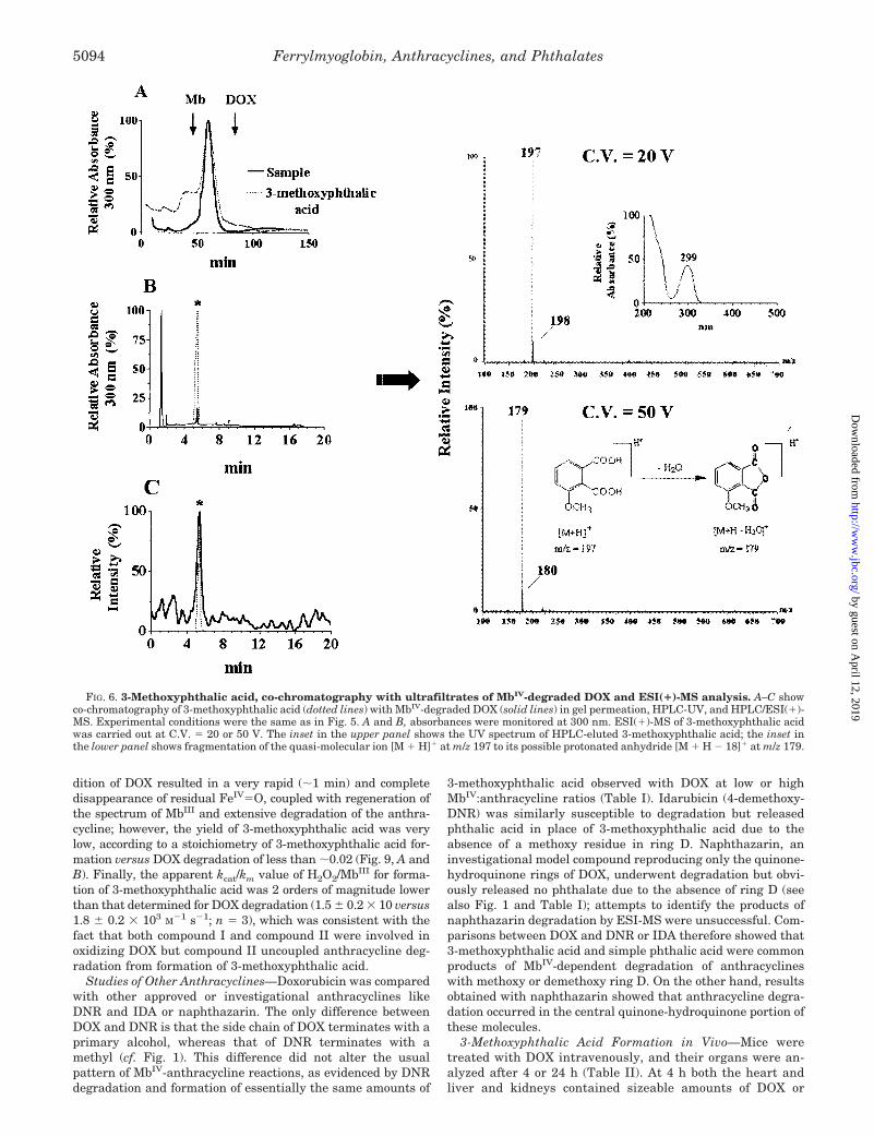
Because mass spectra acquisition is fundamental for charac-terizing molecules, these conditions gave us an opportunity toidentify only one component of the complex material analyzed.As also shown in Fig. 5, the UV spectrum of the unknowncompound was characterized by a single maximum at 299 nmand no absorbance over 350 nm. Under low energy conditions(C.V. � 20 V) the mass spectrum displayed a quasi-molecularion [M � H]� at m/z 197, indicating a possible molecular massof 196 daltons for the molecule; under high energy conditions(C.V. � 50 V) a main fragment at m/z 179 was recorded,possibly indicating the loss of water [M � H � 18]� from thequasi-molecular ion. Based upon these mass spectra, and tak-ing the structure of DOX into account, we tentatively assignedthe unknown molecule to 3-methoxyphthalic acid, product ofoxidative modifications of ring D of DOX. Indirect support tosuch conclusion was also offered by the striking similaritybetween the UV spectrum of the unknown molecule and thatreported for 3-methoxyphthalic acid in ethanol (�max � 298 nm)(31). Moreover, (i) authentic 3-methoxyphthalic acid co-chromatographed with the ultrafiltrates of MbIV/DOX in gelpermeation while also giving the same retention time in HPLC/ESI(�)-MS; (ii) the UV spectrum of HPLC-eluted 3-methoxyph-thalic acid exhibited the same �max � 299 nm of the spectrumof the unknown compound; (iii) the low energy mass spectrumof 3-methoxyphthalic acid gave the expected quasi-molecularion [M � H]� at m/z 197, and the high energy mass spectrumgave a fragment at m/z 179 (Fig. 6). This fragment ion, similarto that observed when recording a mass spectrum of the un-known compound under high energy, was therefore consistentwith H2O elimination from protonated 3-methoxyphthalic acidand with the possible formation of its protonated anhydride(see also Fig. 6).
LC-ESI(�) MS/MS of 3-Methoxyphthalic Acid and MbIV-de-graded DOX—To strengthen identification of 3-methoxyph-thalic acid as a product of DOX degradation, we characterizeda more detailed fragmentation pattern of DOX/MbIV ultrafil-trates vis a vis authentic 3-methoxyphthalic acid. For thispurpose we used LC-ESI MS/MS in product ion scan mode.Under the analytical conditions adopted in our study (cf. “Ex-perimental Procedures”), the negative ion mode proved to be
FIG. 3. Time courses of FeIV�O formation, MbIII regeneration,and DOX degradation. Incubations and spectral analyses were asdescribed in Fig. 2A, and absorbances at 410, 426 (A), and 477 nm (B)were monitored simultaneously.
FIG. 4. Hemin-dependent DOX deg-radation. A, incubations (1 ml final vol-ume) contained DOX and hemin (both 50�M); reactions were started by addingH2O2 (1 mM), and spectra were taken ev-ery 90 s. The inset shows spectra obtainedby subtracting the spectrum of un-changed DOX to the scans recorded atdifferent times after H2O2 addition; theboldface trace is the difference spectrumrecorded 90 s after H2O2 addition. B, in-cubations contained 25 �M DOX andequimolar MbIII or hemin; reactions werestarted by adding H2O2 at 2 or 20:1 ratiosto MbIII or hemin, respectively, and ratesof DOX degradation were normalized toperoxidative activities. C, incubationscontained 50 �M DOX and either 2.5 �M
MbIII or 50 �M hemin, corresponding to 1unit of peroxidative activity/ml; reactionswere started by adding H2O2 at 2 or 20:1ratios to MbIII or hemin, respectively.Where indicated incubations contained0.1 mM EDTA.
Ferrylmyoglobin, Anthracyclines, and Phthalates5092
by guest on April 12, 2019
http://ww
w.jbc.org/
Dow
nloaded from

more sensitive and convenient than the positive mode for per-forming these experiments. Fig. 7A shows the TIC chromato-gram of authentic 3-methoxyphthalic acid, characterized by apeak at 9.6 min, and Fig. 7B shows the product ion scanspectrum corresponding to that peak. The spectrum consistedof an ion at m/z � 195, corresponding to the deprotonatedmolecule [M � H]� of 3-methoxyphthalic acid in the ESI(�)mode, and two more ions at m/z � 151 and 107. The latterprobably indicated, respectively, the loss of one [M � H � 44]�
or both [M � H � 88]� carboxyl groups of 3-methoxyphthalicacid (see also Fig. 7B, inset). The ultrafiltrates of DOX/MbIV
incubations gave the same TIC chromatogram of 3-methoxyph-thalic acid (Fig. 7C) as well as the same MS/MS fragmentationpattern with ions at m/z � 195, 151, and 107 (Fig. 7D).
Stoichiometries and Time Courses—HPLC analyses of DOX/MbIII/H2O2 incubations showed that 3-methoxyphthalic acidformation closely paralleled DOX degradation induced by in-creasing levels of MbIV (Fig. 8A).
Doxorubicin degradation and 3-methoxyphthalic acid forma-tion occurred in a time-dependent manner (Fig. 8B), but thestoichiometry of 3-methoxyphthalic acid formation versus DOXdegradation decreased from �0.5 to � 0.12 in 3–4 min (Fig.8C). This was not attributable to DOX depletion or secondary
metabolization of 3-methoxyphthalic; in fact, �60–70% of DOXwas still available for further degradation after 3–4 min reac-tion between MbIII and H2O2, and 3-methoxyphthalic acid wasstable to prolonged incubation with MbIII or H2O2 or MbIII/H2O2 (cf. Fig. 8, B and C, and inset). Data analysis showed thatthe stoichiometry decreased due to an �4-fold increase of DOXdegradation that was not accompanied by an increased forma-tion of 3-methoxyphthalic acid. Because these changes oc-curred 3–4 min after mixing MbIII with H2O2, which is the timewhen the anthracycline began reacting with FeIV�O, we con-sidered that compound II was highly effective at oxidizing DOXbut generated several product(s) other than 3-methoxyphthalicacid, eventually decreasing the stoichiometry of 3-methoxyph-thalic acid formation versus DOX degradation. This possibilitywas anticipated by the fact that hemin/H2O2 (a surrogate ofcompound I) allowed DOX oxidation and 3-methoxyphthalicacid formation to proceed stoichiometrically coupled for severalmore minutes than did MbIII/H2O2 (a source of both compoundI and II) (not shown). Further evidence was obtained by addingDOX 1 h after mixing MbIII with H2O2, a time when bothporphyrin and globin radicals had decayed (2, 32, 33), whereasthe long lived FeIV�O was still present at �50% its initialabsorbance (Fig. 9A). Under these defined conditions, the ad-
FIG. 5. Gel permeation, HPLC, and HPLC/ESI(�)-MS of DOX/MbIV ultrafiltrates. Incubations (1 ml final volume) contained DOX(100–500 �M) and equimolar MbIII, always reacted with a 2-fold excess of H2O2. After 10 min, reactions mixtures were added with catalase (2600units) and ultrafiltered. A shows gel permeation of ultrafiltrates derived from MbIV-degraded DOX (100 �M (line a), 200 �M (line b), or 500 �M (linec)); the upper arrows indicate the retention times of MbIII or DOX standards chromatographed under comparable conditions. B shows HPLC ofgel-filtered fraction c in A, and C shows the corresponding HPLC/ESI(�)-MS chromatogram. ESI(�)-MS of the HPLC fraction eluted at 5.21 minwas carried out at C.V. � 20 or 50 V; the inset in the upper panel shows the UV spectrum associated with the HPLC fraction eluted at 5.02 min.
Ferrylmyoglobin, Anthracyclines, and Phthalates 5093
by guest on April 12, 2019
http://ww
w.jbc.org/
Dow
nloaded from
dition of DOX resulted in a very rapid (�1 min) and completedisappearance of residual FeIV�O, coupled with regeneration ofthe spectrum of MbIII and extensive degradation of the anthra-cycline; however, the yield of 3-methoxyphthalic acid was verylow, according to a stoichiometry of 3-methoxyphthalic acid for-mation versus DOX degradation of less than �0.02 (Fig. 9, A andB). Finally, the apparent kcat/km value of H2O2/MbIII for forma-tion of 3-methoxyphthalic acid was 2 orders of magnitude lowerthan that determined for DOX degradation (1.5 � 0.2 � 10 versus1.8 � 0.2 � 103 M�1 s�1; n � 3), which was consistent with thefact that both compound I and compound II were involved inoxidizing DOX but compound II uncoupled anthracycline deg-radation from formation of 3-methoxyphthalic acid.
Studies of Other Anthracyclines—Doxorubicin was comparedwith other approved or investigational anthracyclines likeDNR and IDA or naphthazarin. The only difference betweenDOX and DNR is that the side chain of DOX terminates with aprimary alcohol, whereas that of DNR terminates with amethyl (cf. Fig. 1). This difference did not alter the usualpattern of MbIV-anthracycline reactions, as evidenced by DNRdegradation and formation of essentially the same amounts of
3-methoxyphthalic acid observed with DOX at low or highMbIV:anthracycline ratios (Table I). Idarubicin (4-demethoxy-DNR) was similarly susceptible to degradation but releasedphthalic acid in place of 3-methoxyphthalic acid due to theabsence of a methoxy residue in ring D. Naphthazarin, aninvestigational model compound reproducing only the quinone-hydroquinone rings of DOX, underwent degradation but obvi-ously released no phthalate due to the absence of ring D (seealso Fig. 1 and Table I); attempts to identify the products ofnaphthazarin degradation by ESI-MS were unsuccessful. Com-parisons between DOX and DNR or IDA therefore showed that3-methoxyphthalic acid and simple phthalic acid were commonproducts of MbIV-dependent degradation of anthracyclineswith methoxy or demethoxy ring D. On the other hand, resultsobtained with naphthazarin showed that anthracycline degra-dation occurred in the central quinone-hydroquinone portion ofthese molecules.
3-Methoxyphthalic Acid Formation in Vivo—Mice weretreated with DOX intravenously, and their organs were an-alyzed after 4 or 24 h (Table II). At 4 h both the heart andliver and kidneys contained sizeable amounts of DOX or
FIG. 6. 3-Methoxyphthalic acid, co-chromatography with ultrafiltrates of MbIV-degraded DOX and ESI(�)-MS analysis. A–C showco-chromatography of 3-methoxyphthalic acid (dotted lines) with MbIV-degraded DOX (solid lines) in gel permeation, HPLC-UV, and HPLC/ESI(�)-MS. Experimental conditions were the same as in Fig. 5. A and B, absorbances were monitored at 300 nm. ESI(�)-MS of 3-methoxyphthalic acidwas carried out at C.V. � 20 or 50 V. The inset in the upper panel shows the UV spectrum of HPLC-eluted 3-methoxyphthalic acid; the inset inthe lower panel shows fragmentation of the quasi-molecular ion [M � H]� at m/z 197 to its possible protonated anhydride [M � H � 18]� at m/z 179.
Ferrylmyoglobin, Anthracyclines, and Phthalates5094
by guest on April 12, 2019
http://ww
w.jbc.org/
Dow
nloaded from

metabolites (like the C-13 secondary alcohol metabolite andhydroxy- or deoxyaglycones), which retained the same fluo-rescence of unchanged DOX due to the presence of an intacttetracyclic ring in their molecule. The values of DOX and/orfluorescent metabolites decreased, to a variable extent, at24 h due to a tissue clearance of both parent drug and itsmetabolites. Different results were obtained in regard to3-methoxyphthalic acid. As also shown in Table II, 3-me-thoxyphthalic acid was found in the heart at 4 h and in-creased further at 24 h, reaching levels that were higher thanthose of DOX and fluorescent metabolites. The liver con-tained 3-methoxyphthalic acid only at 24 h, but its levels wereonly �30% of those found in the heart at the same post-treatmenttime. Kidneys did not contain 3-methoxyphthalic acid at either 4or 24 h. These results showed that (i) 3-methoxyphthalic acid wasformed in vivo after DOX administration; (ii) the heart was moreactive than other organs in degrading DOX to 3-methoxyphthalicacid; and (iii) there were conditions when the cardiac levels of3-methoxyphthalic acid exceeded those of unchanged DOX or itsmetabolites.
3-Methoxyphthalic Acid Formation in Human Myocardium—Human myocardial strips were exposed to 1 or 10 �M DOX,concentrations found in the plasma of patients after slow orbolus infusions of the drug (34). At the end of the experimentsthe strips contained very low amounts of residual unchanged
DOX but contained sizeable amounts of fluorescent metabolitesand 3-methoxyphthalic acid. At 1 �M DOX intramyocardiallevels of 3-methoxyphthalic acid were higher than those offluorescent metabolites; at 10 �M DOX the levels of fluorescentmetabolites increased and exceeded those of 3-methoxyphthalicacid, but the latter remained several times higher than un-changed DOX (Table III). By having demonstrated that 3-me-thoxyphthalic was an important component of the metabolicfate of DOX in human myocardium, we performed experimentsto confirm that it was formed by H2O2-activated myoglobin.Three lines of evidence showed that this was the case. First,incubation of a whole homogenate of human myocardium withDOX and H2O2 resulted in anthracycline degradation and3-methoxyphthalic acid formation, but these processes becamemore evident when H2O2 was added to cytosol, i.e. the subcel-lular fraction containing myoglobin. Second, treatment of cy-tosol with 65% ammonium sulfate removed �80% of myoglobinby salting out and gave an Mb� cytosol that was essentiallyinactive at degrading DOX or generating 3-methoxyphthalicacid upon incubation with H2O2. Finally, Mb� cytosol regainedactivity in DOX degradation and 3-methoxyphthalic acid for-mation after reconstitution with its salted out myoglobin (seeFig. 10, A–D, and legend).
Studies with Isolated Cardiomyocytes—We have shown pre-viously (25) that the embryonic rat heart-derived cell line H9c2
FIG. 7. LC-ESI(�) MS/MS of authentic 3-methoxyphthalic acid and DOX/MbIV ultrafiltrates. All conditions were as described under“Experimental Procedures.” A shows the TIC chromatogram of authentic 3-methoxyphthalic acid, with a peak at 9.6 min, and B shows the production scan spectrum corresponding to that peak. The fragmentation pattern (inset) probably indicates the loss of one or both carboxyl groups. C andD show that similar results were obtained with an ultrafiltrate derived from DOX/MbIV incubations.
Ferrylmyoglobin, Anthracyclines, and Phthalates 5095
by guest on April 12, 2019
http://ww
w.jbc.org/
Dow
nloaded from

offers a convenient and reproducible model for evaluating car-diotoxicity induced by DOX or related compounds in vitro.Therefore, H9c2 cardiomyocytes were exposed to 0.01–10 �M
3-methoxyphthalic acid to see whether it caused increased ordecreased toxicity compared with equimolar DOX. Inasmuch ascardiac tissue has a density very similar to that of water (1g/ml) (14), the range of concentrations of 3-methoxyphthalicacid in the incubation medium was broad enough to includeand exceed those detected in mouse heart (1–3 nmol/g, cf. TableII) or in human myocardial strips (0.37–0.44 nmol/g, cf. TableIII). As shown in Fig. 11, 3-methoxyphthalic acid never reducedcardiomyocyte viability in the MTT assay; in contrast, a signif-icant loss of viability occurred if cardiomyocytes were exposed
to �1 �M DOX. Thus, 3-methoxyphthalic acid was essentiallynon-toxic compared with DOX.
DISCUSSION
We have shown that MbIV, a pseudoperoxidase, is moreeffective than authentic peroxidases at promoting the oxidativedegradation of DOX, exhibiting a kcat/km 1 order of magnitudehigher than that of HRP or LPO. Time course analyses ofFeIV�O formation and decay versus DOX degradation, andexperiments conducted with hemin/H2O2 as a source of porphy-rin radical/FeIV�O, indicate that DOX is oxidized by the por-phyrin radical of a compound I-like species and then by theFeIV�O moiety of a compound II-like species (cf. Figs. 2–4).
FIG. 8. Stoichiometries and timecourses of DOX degradation and3-methoxyphthalic acid formation. A,increasing MbIV:DOX ratios were ob-tained by incubating 100 �M DOX with 10�M to 0.8 mM MbIII, always reacted with a2-fold excess of H2O2. After 10 min DOXand 3-methoxyphthalic acid were meas-ured by HPLC as described under “Exper-imental Procedures.” B, DOX and MbIII
(both 100 �M) were incubated with 200 �M
H2O2. Reaction mixtures were analyzedat regular times for DOX degradation(initial DOX-residual DOX) and 3-me-thoxy phthalic acid formation. C showstime-dependent decrease of the stoichi-ometry of 3-methoxyphthalic acid versusDOX degradation. Values were deter-mined based on data in B; the inset showsthe recovery of 3-methoxyphthalic acid(100 �M) after 10 min of incubation withequimolar MbIII and/or 200 �M H2O2. Allvalues were means � S.E. of threeexperiments.
Ferrylmyoglobin, Anthracyclines, and Phthalates5096
by guest on April 12, 2019
http://ww
w.jbc.org/
Dow
nloaded from

The ease with which the porphyrin radical of compound Idissipates in the globin clearly calls attention also on a possibleinvolvement of amino acid radicals centered at Tyr103 or Trp14,for example (4–7). In this regard we have data showing thattryptophan radicals, generated by oxidizing tryptophan withHRP, lack reactivity toward DOX; tyrosine radicals, generatedthrough the same procedure, did degrade DOX but acetylationof myoglobin tyrosine residues did not decrease the kcat/km ofMbIII/H2O2 for degradation of DOX (not shown). These reason-ings do not completely rule out a possible role for globin-cen-tered reactive species in oxidizing DOX. Free radicals areknown to transfer from one site of the globin to another, andchemical modifications or site-directed mutagenesis of the pri-mary sites of free radical formation may not prevent the for-mation of other radicals on nearby residues (32). Moreover,MbIV may display an additional radical signal that is not cen-tered at Tyr or Trp and has been tentatively assigned to oxi-dation of an unidentified aromatic amino acid in close proxim-ity to the heme pocket (32). The possible role of globin radicals
as additional determinants of DOX oxidation therefore remainsa matter of consideration.
Chromatographic, UV, and mass spectrometry analyses pro-vide novel evidence that a biologic oxidant like MbIV degradesDOX to 3-methoxyphthalic acid, product of oxidative modifica-tions of the methoxy-substituted ring D (cf. Figs. 1 and 5–7).Biochemical evidence for the formation of oxidized ring D prod-ucts was also offered by the fact that methoxy or demethoxyanalogs, like DNR or IDA, similarly released 3-methoxyph-thalic acid or simple phthalic acid after exposure to MbIV (cf.Table I). Previously, the formation of 3-methoxyphthalic acid orother products of ring D oxidation (like e.g. 3-methoxysalycilicacid) was only observed under artificial conditions such aspermanganate oxidation of investigational anthraquinones(35) or riboflavin-mediated photooxidation of DOX (36).
TABLE IAnthracycline degradation and phthalic acids formation, comparisons
between DOX and analogsDrugs were 100 �M, and MbIV:drug ratios (0.5/2:1) were obtained by
incubation with 50-200 �M MbIII always reacted with 2-fold H2O2. After10 min, incubations were assayed by HPLC for drug degradation (ini-tial-residual) and phthalates formation. Values were means � S.E. ofthree experiments. —, not detectable.
Drug MbIV:drug Degradation 3-Methoxyphthalicacid
Phthalicacid
�M
DOX 0.5:1 53 � 7 3.8 � 0.8 —2:1 98 � 2 16 � 1 —
DNR 0.5:1 40 � 4 3.6 � 0.7 —2:1 98 � 1 13 � 1.5 —
IDA 0.5:1 62 � 9 — 14 � 1.52:1 99 � 1 — 23 � 2
Naphthazarin 0.5:1 83 � 20 — —2:1 97 � 2 — —
TABLE IILevels of DOX, fluorescent metabolites, and 3-methoxyphthalic acid in
organs of mice treated with DOXOther conditions are as described under “Experimental Procedures.”
—, not detectable.
DOX Fluorescentmetabolitesa
3-Methoxyphthalicacid
nmol/g
Heart4 h 8.1 � 0.5 0.3 � 0.05 1.7 � 0.124 h 1.9 � 0.2b 0.2 � 0.1 3.0 � 0.3c
Liver4 h 6.1 � 0.8 0.4 � 0.1 —24 h 1.3 � 0.2b 0.1 � 0.02d 1.0 � 0.2
Kidney4 h 12.6 � 1 1.9 � 0.3 —24 h 9.1 � 0.8e 0.2 � 0.1f —
a Sum of C-13 secondary alcohol metabolite and hydroxy- or deoxy-aglycones. Values were means � S.E. (n � 9).
b p � 0.01 or � 0.05 versus 4 h.c p � 0.025 versus 4 h and p � 0.05 versus DOX 24 h.d p � 0.01 versus 4 h.e p � 0.05 versus 4 h.f p � 0.01 versus 4 h.
TABLE IIILevels of DOX and fluorescent metabolites versus 3-methoxyphthalic
in human myocardial strips exposed to 1 or 10 �M DOX
DOX UnchangedDOX
Fluorescentmetabolitesa
3-Methoxyphthalicacid
�M nmol/g
1 0.02 � 0.01 0.27 � 0.04 0.44 � 0.0610 0.04 � 0.01 1.2 � 0.2b 0.37 � 0.05
a Sum of C-13 secondary alcohol metabolite and hydroxy- or deoxya-glycones. Other conditions were as described under “Experimental Pro-cedures.”
b Values were means of � SE (n � 4). p � 0.01 versus 1 �M.
FIG. 9. DOX degradation and 3-methoxyphthalic acid forma-tion. Results were obtained by adding DOX 1 h after mixing MbIII withH2O2. A, MbIII (50 �M) was reacted with H2O2 (100 �M) to form MbIV/FeIV�O (peaks at 546 and 586 nm). Spectra were taken at 4 and 10 minand then every 10 min until 60 min (solid lines). At 60 min DOX (50 �M)was added, and spectra were taken every min for 10 consecutive min(dotted lines). The boldface trace is the spectrum of unreacted MbIII. B,experimental conditions were as in A, except that MbIII and H2O2 were100 and 200 �M, respectively. After 60 min, DOX (100 �M) was added,and aliquots were taken every min and assayed for DOX degradation(initial DOX � residual DOX) or 3-methoxyphthalic acid formation.Values were means � S.E. of three determinations.
Ferrylmyoglobin, Anthracyclines, and Phthalates 5097
by guest on April 12, 2019
http://ww
w.jbc.org/
Dow
nloaded from

The complete sequence of events leading to the formation of3-methoxyphthalic acid cannot be envisaged at this time asMbIV-DOX interactions released other products that were de-tected by HPLC but could not be characterized by ESI(�) MS(cf. Fig. 5, B and C).3 The formation of such unknown productsseemed to occur primarily during oxidation of DOX by com-pound II, characterized by a stoichiometry of 3-methoxyph-thalic acid formation versus DOX degradation much lower thanthat determined for oxidation of DOX by compound I (cf. Fig.8C). Nevertheless, there are at least two mechanisms that mayhelp to anticipate how DOX releases 3-methoxyphthalic acidafter oxidation by MbIV. As mentioned, anthracycline degrada-tion by MbIV or peroxidases is preceded by one-electron oxida-tion of the hydroquinone in juxtaposition to the quinone (14,16). This reaction generates a semiquinone whose dispropor-tionation regenerates a hydroquinone while also forming adiquinone (16). Because diquinones are rather unstable com-pounds that decompose spontaneously (16), oxidation of DOXfrom its hydroquinone-quinone form to a diquinone form mayrepresent a plausible mechanism to explain how MbIV pro-motes opening and degradation of the planar anthracyclinering. In keeping with this concept, we found that also naphtha-zarin, a two-ring membered compound reproducing the qui-
none-hydroquinone portion of anthracyclines, was degraded byMbIV (cf. Table I); in contrast, one ring-membered hydroquino-nes like simple hydroquinone or 2,5-di-tert-butylhydroquinonewere not degraded by MbIV but converted quantitatively to thecorresponding stable quinones (not shown). Another mecha-nism to be taken into account when considering the carboxyl-ation of ring D in the form of 3-methoxyphthalic acid may beinferred from the ability of lignin peroxidase to oxidativelydegrade 3,4-dimethoxybenzyl methyl ether to muconic acid(37). These reactions suggest that peroxidases can cleave aro-matic rings and that secondary reactions, mediated by oxygenactivation/addition, can result in formation of carboxylic acidderivatives (37).
Doxorubicin has long been known to undergo one-electronreduction of its quinone, two-electron reduction of its side chainC-13 carbonyl group, or reductive cleavage of the glycosidicbond linking its tetracyclic ring with an amino sugar (cf. Fig. 1).While forming semiquinone free radicals and secondary alcoholor deoxyaglycone derivatives that have been implicated to ex-plain, at least in part, the cardiotoxic properties of DOX (25, 38,39), these reductive processes do not induce any important orirreversible loss of the optical and fluorescent properties ofunchanged DOX. However, the administration of DOX to hu-mans and laboratory animals was shown to generate also non-fluorescent compounds that were tentatively attributed to ox-idative degradation rather than reductive biotransformation ofthe drug (40, 41). Our results indicate that 3-methoxyphthalicacid is formed not only in reconstituted chemical systems butalso in the heart of mice treated with DOX or in human myo-cardial biopsies exposed to concentrations of DOX similar tothose found in the plasma of patients (cf. Tables II and III). Inmice, the heart generates considerably more 3-methoxyph-thalic acid than organs like liver or kidneys, as one wouldexpect if MbIV were more effective than other peroxidases atdegrading DOX. Comparisons between human heart homoge-nate and Mb� or Mb� cytosol also confirmed that cytosol serveda preferred site of DOX degradation and 3-methoxyphthalicacid formation, and that both processes could be abolished orreactivated by removing or reintroducing myoglobin (cf. Fig.10). Our results therefore identify MbIV and 3-methoxyphthalicacid among the long sought catalyst(s) and product(s) of an-thracycline oxidation in biologic systems.
3 In re-attempting characterization of other products of DOX degra-dation products by ESI(�)-MS, we occasionally detected ions at m/z �203 or 237, 242, and 265. However, further characterizations and un-ambiguous assignment of these ions were not possible.
FIG. 10. DOX degradation and 3-me-thoxyphthalic acid formation in hu-man myocardium; role of myoglobin.Incubations (200 �l final volume) con-tained 15 �M DOX and 3 mg of protein/mlof human heart whole homogenate (1.3nmol of MbIII/mg of protein), Mb� cytosol(2.9 nmol of MbIII/mg of protein), or Mb�
cytosol (0.6 nmol of MbIII/mg of protein).Reactions were started by adding increas-ing amounts of H2O2, as indicated. After10 min incubations were assayed for DOX(A) or 3-methoxyphthalic acid (B). C andD, DOX degradation (initial DOX-resid-ual DOX) and 3-methoxyphthalic acid for-mation were measured in incubationscontaining 500 �M H2O2 and 3 mg of pro-tein/ml of Mb� cytosol, Mb� cytosol, orMb� cytosol reconstituted with its saltedout myoglobin (2.3 nmol of MbIII/mg ofprotein, dialyzed just prior to experi-ments to remove ammonium sulfate). Val-ues were means � S.E. of triplicateexperiments.
FIG. 11. Toxicity of DOX or 3-methoxyphthalic acid to H9c2cardiomyocytes. Toxicity was evaluated as the percentage of viablecells (MTT assay) after an overnight exposure to DOX or 3-methoxyph-thalic acid. Values were means � S.E. of four separate determinations.
Ferrylmyoglobin, Anthracyclines, and Phthalates5098
by guest on April 12, 2019
http://ww
w.jbc.org/
Dow
nloaded from

Experiments with DOX-treated mice or human myocardialstrips exposed to DOX show that there may be conditions whenthe cardiac levels of 3-methoxyphthalic acid exceed those ofunchanged DOX or fluorescent metabolites (cf. the heart ofmice 24 h after DOX administration or myocardial strips ex-posed to 1 �M DOX) (cf. Tables II and III). These observationssuggest that 3-methoxyphthalic acid might be an importantdeterminant of the mode of action of anthracyclines and offereda rationale to see how it compared with DOX in inducingtoxicity to cardiomyocytes. We demonstrate that concentra-tions of 3-methoxyphthalic acid equal to or several times higherthan those found in whole cardiac tissues lack toxicity to iso-lated cardiomyocytes; under comparable conditions, however,low concentrations of DOX are highly toxic to cardiomyocytes(cf. Fig. 11). Although the biologic action(s) of other currentlyunknown degradation products cannot be disregarded, theseresults raise the possibility that the pseudoperoxidase activityof MbIV may serve an important mechanism to diminish cellu-lar levels of anthracyclines and to divert them from formationof toxic reduced metabolites toward formation of non-toxic ox-idized products.
In summary, we have shown the following. (i) MbIV is a verygood catalyst of anthracycline degradation. (ii) 3-Methoxyph-thalic acid and simple phthalic acid are common products of theoxidative degradation of methoxy-substituted or demethoxyanalogs like DOX and DNR or IDA, respectively. (iii) Thecompounds I and II of MbIV are sequentially involved in oxi-dizing DOX, although with different stoichiometries of 3-me-thoxyphthalic acid formation versus anthracycline degrada-tion. (iv) 3-Methoxyphthalic acid is an abundant product ofDOX degradation in the heart of laboratory animals or inhuman myocardium. (v) 3-Methoxyphthalic acid does not in-duce toxicity to cardiomyocytes. These results unravel novelfunctions for MbIV and pose mechanism-based foundations tosee whether reactions of MbIV with anthracyclines may beexploited to improve cardiac tolerability of these otherwiseuseful agents.
Acknowledgments—We thank Professor Gaetano Cairo (Institute ofGeneral Pathology, University of Milan) and Dr. Alessandro Mauro(Department of Chemistry, Menarini Ricerche S.pA.) for assistanceduring some phases of this work. We also thank Professor Antonio M.Calafiore and Giovanni Liberi (Department of Cardiac Surgery, G.d’Annunzio University School of Medicine, Chieti) for providing humanmyocardium samples.
REFERENCES
1. Yusa, K., and Shikama, K. (1987) Biochemistry 26, 6684–66882. Galaris, D., and Korantzopoulos, P. (1997) Free Radic. Biol. Med. 22, 657–6673. Arduini, A., Eddy, L., and Hochstein, P. (1990) Free Radic. Biol. Med. 9,
511–5134. Gunther, M. R., Sampath, V., and Caughey, W. S. (1999) Free Radic. Biol. Med.
26, 1388–13955. Egawa, T., Shimada, H., and Ishimura, Y. (2000) J. Biol. Chem. 275,
34858–348666. Rao, S. I., Wilks, A., Hamberg, M., and Ortiz de Montellano, P. R. (1994)
J. Biol. Chem. 269, 7210–72167. DeGray, J. A., Gunther, M. R., Tschirret-Guth, R., Ortiz de Montellano, P. R.,
and Mason, R. P. (1997) J. Biol. Chem. 272, 2359–23628. Gunther, M. R., Sturgeon, B. E., and Mason, R. P. (2000) Free Radic. Biol. Med.
28, 709–7199. Doroshow, J. H., and Davies, K. J. (1986) J. Biol. Chem. 261, 3068–3074
10. Minotti, G., Cairo, G., and Monti, E. (1999) FASEB J. 13, 199–21211. Kalyanaraman, B., Joseph, J., Kalivendi, S., Wang, S., Konorev, E., and
Kotamraju, S. (2002) Mol. Cell. Biochem. 234, 119–12412. Trost, L. C., and Wallace, K. B. (1994) Biochem. Biophys. Res. Commun. 204,
30–3713. Taylor, D., and Hochstein, P. (1978) Biochem. Pharmacol. 27, 2079–208214. Menna, P., Salvatorelli, E., Giampietro, R., Liberi, G., Teodori, G., Calafiore,
A. M., and Minotti, G. (2002) Chem. Res. Toxicol. 15, 1179–118915. Miura, T., Muraoka, S., and Fujimoto, Y. (2000) Biochem. Pharmacol. 60,
95–9916. Reszka, K. J., McCormick, M. L., and Britigan, B. E. (2001) Biochemistry 40,
15349–1536117. Reszka, K. J., McCormick, M. L., and Britigan, B. E. (2003) Free Radic. Biol.
Med. 35, 78–9318. Reszka, K. J., and Britigan, L. H. (2002) Free Radic. Biol. Med. 33, (suppl.) 44319. Wentzel, B. B., Donners, M. P. J., Alsters, P. L., Feiters, M. C., and Nolte,
R. J. M. (2000) Tetrahedron 56, 7797–780320. Mordente, A., Santini, S., Miggiano, G. A. D., Martorana, G. E., Petitti, T.,
Minotti, G., and Giardina, B. (1994) J. Biol. Chem. 269, 27394–2740021. Taatjes, D. J., Gaudiano, G., Resing, K., and Koch, T. H. (1997) J. Med. Chem.
40, 1276–128622. Minotti, G., Saponiero, A., Licata, S., Menna, P., Calafiore, A. M., Teodori, G.,
and Gianni, L. (2001) Clin. Cancer Res. 7, 1511–151523. Minotti, G., Cavaliere, A. F., Mordente, A., Rossi, M., Schiavello, R.,
Zamparelli, R., and Possati, G. F. (1995) J. Clin. Investig. 95, 1595–160524. Swaanenburg, J. C., Visser-VanBrummen, P. J., DeJongste, M. J., and Tie-
bosch, A. T. (2001) Am. J. Clin. Pathol. 115, 770–77725. Minotti, G., Ronchi, R., Salvatorelli, E., Menna, P., and Cairo, G. (2001) Cancer
Res. 61, 8422–842826. Mosmann, T. (1983) J. Immunol. Methods 65, 55–6327. Stoscheck, C. M. (1990) Methods Enzymol. 182, 50–6828. Van der Zee, J., Barr, D. P., and Mason, R. P. (1996) Free Radic. Biol. Med. 20,
199–20629. Bloom, J., Lehman, P., Israel, M., Rosario, O., and Korfmacher, W. A. (1992) J.
Anal. Toxicol. 16, 223–22730. Arcamone, F., Cassinelli, G., Franceschi, G., Penco, S., Pol, C., Redaelli, S., and
Selva, A. (1972) International Symposium on Adriamycin, pp. 10–22,Springer-Verlag, Berlin
31. Santavy, F., Hruban, L., Simanek, V., and Walterova, D. (1970) Collect. Czech.Chem. Commun. 35, 2418–2443
32. Giulivi, C., and Cadenas, E. (1998) Free Radic. Biol. Med. 24, 269–27933. Baron, C. P., Skibsted, L. H., and Andersen, H. J. (2000) Free Radic. Biol. Med.
28, 549–55834. Gianni, L., Vigano, L., Locatelli, A., Capri, G., Giani, A., Tarenzi, E., and
Bonadonna, G. (1997) J. Clin. Oncol. 15, 1906–191535. Alexander, J., Bhatia, A. V., Mitscher, L. A., Omoto, S., and Suzuki, T. (1980)
J. Org. Chem. 45, 20–2436. Ramu, A., Mehta, M. M., Liu, J., Turyan, I., and Aleksic, A. (2000) Cancer
Chemother. Pharmacol. 46, 449–45837. Schmidt, H. W., Haemmerli, S. D., Schoemaker, H. E., and Leisola, M. S. A.
(1989) Biochemistry 28, 1776–178338. Akman, S. A., Forrest, G., Chu, F. F., Esworthy, R. S., and Doroshow, J. H.
(1990) Cancer Res. 50, 1397–140239. Gille, L., and Nohl, H. (1997) Free Radic. Biol. Med. 23, 775–78240. Alberts, D. S., Bachur, N. R., and Holtzman, J. L. (1971) Clin. Pharmacol.
Ther. 12, 96–10441. Arcamone, F., Lazzati, M., Vicario, G. P., and Zini, G. (1984) Cancer Che-
mother. Pharmacol. 12, 157–166
Ferrylmyoglobin, Anthracyclines, and Phthalates 5099
by guest on April 12, 2019
http://ww
w.jbc.org/
Dow
nloaded from

MinottiRossella Giampietro, Fabio Animati, Andrea Urbani, Piero Del Boccio and Giorgio Antonella Cartoni, Pierantonio Menna, Emanuela Salvatorelli, Daniela Braghiroli,
NOVEL FUNCTION FOR FERRYLMYOGLOBINOxidative Degradation of Cardiotoxic Anticancer Anthracyclines to Phthalic Acids:
doi: 10.1074/jbc.M306568200 originally published online November 21, 20032004, 279:5088-5099.J. Biol. Chem.
10.1074/jbc.M306568200Access the most updated version of this article at doi:
Alerts:
When a correction for this article is posted•
When this article is cited•
to choose from all of JBC's e-mail alertsClick here
http://www.jbc.org/content/279/7/5088.full.html#ref-list-1
This article cites 40 references, 9 of which can be accessed free at
by guest on April 12, 2019
http://ww
w.jbc.org/
Dow
nloaded from