Overview of Controlled Release Mechanisms
26
Chapter 2 Overview of Controlled Release Mechanisms Ronald A. Siegel and Michael J. Rathbone Abstract Controlled release systems have been developed to improve the temporal and spatial presentation of drug in the body, to protect drug from physiological degradation or elimination, to improve patient compliance, and to enhance quality control in manufacturing of drug products. When designing controlled-release systems, it is important to identify and understand particular mechanisms involved in the release process. Often, more than one mechanism is involved at a given time or different mechanisms may dominate at different stages of the drug delivery process. This chapter begins with several vignettes, each highlighting a mode of controlled drug delivery and identifying associated mechanisms. An introductory description of several of the mechanisms follows. Details regarding these mechanisms are provided in subsequent chapters. 2.1 Introduction Controlled-release systems are designed to enhance drug therapy. There are several motivations for developing controlled-release systems, which may depend on the drug of interest. Controlled release systems have been devised to enable superior control of drug exposure over time, to assist drug in crossing physiological barriers, to shield drug from premature elimination, and to shepherd drug to the desired site of action while minimizing drug exposure elsewhere in the body. Controlled release systems R.A. Siegel (*) Departments of Pharmaceutics and Biomedical Engineering, University of Minnesota, Minneapolis, MN 55419, USA Department of Pharmaceutics WDH 9-177, University of Minnesota, 308 Harvard St. S.E., Minneapolis, MN 55455, USA e-mail: [email protected] M.J. Rathbone School of Pharmacy, Griffith University, Southport, QLD 4222, Australia J. Siepmann et al. (eds.), Fundamentals and Applications of Controlled Release Drug Delivery, Advances in Delivery Science and Technology, DOI 10.1007/978-1-4614-0881-9_2, # Controlled Release Society 2012 19
Transcript of Overview of Controlled Release Mechanisms
Chapter 2
Overview of Controlled Release Mechanisms
Ronald A. Siegel and Michael J. Rathbone
Abstract Controlled release systems have been developed to improve the temporal
and spatial presentation of drug in the body, to protect drug from physiological
degradation or elimination, to improve patient compliance, and to enhance quality
control in manufacturing of drug products. When designing controlled-release
systems, it is important to identify and understand particular mechanisms involved
in the release process. Often, more than one mechanism is involved at a given time
or different mechanisms may dominate at different stages of the drug delivery
process. This chapter begins with several vignettes, each highlighting a mode
of controlled drug delivery and identifying associated mechanisms. An introductory
description of several of the mechanisms follows. Details regarding these
mechanisms are provided in subsequent chapters.
2.1 Introduction
Controlled-release systems are designed to enhance drug therapy. There are several
motivations for developing controlled-release systems, whichmay depend on the drug
of interest. Controlled release systems have been devised to enable superior control of
drug exposure over time, to assist drug in crossing physiological barriers, to shield
drug from premature elimination, and to shepherd drug to the desired site of action
while minimizing drug exposure elsewhere in the body. Controlled release systems
R.A. Siegel (*)
Departments of Pharmaceutics and Biomedical Engineering, University of Minnesota,
Minneapolis, MN 55419, USA
Department of Pharmaceutics WDH 9-177, University of Minnesota, 308 Harvard St. S.E.,
Minneapolis, MN 55455, USA
e-mail: [email protected]
M.J. Rathbone
School of Pharmacy, Griffith University, Southport, QLD 4222, Australia
J. Siepmann et al. (eds.), Fundamentals and Applications of Controlled ReleaseDrug Delivery, Advances in Delivery Science and Technology,
DOI 10.1007/978-1-4614-0881-9_2, # Controlled Release Society 2012
19
may also increase patient compliance by reducing frequency of administration,
and may add commercial value to marketed drugs by extending patent protection.
Finally, use of controlled release technologymay reduce variability of performance of
drug products. The latter aspect is increasingly important given the current emphasis
on “quality by design” by regulatory agencies such as FDA.
The mechanisms used to achieve these goals are diverse and complex, and
depend on the particular application. In fact, several mechanisms may operate
simultaneously or at different stages of a delivery process. An understanding of
these mechanisms is important when designing and manufacturing controlled-
release systems, and in identifying potential failure modes. Delineation of mecha-
nism is also important in intellectual property prosecution and quality assurance/
quality control.
This chapter starts with a series of vignettes illustrating mechanisms and
their interplay in particular controlled release systems. Essentials of individual
mechanisms are then outlined. More elaborate descriptions are deferred to later
chapters.
2.2 Vignettes
2.2.1 Zero Order Oral Delivery
Zero order, or constant rate release of drug is desirable in order to minimize swings
in drug concentration in the blood. Such excursions, which may lead to periods of
underexposure or overexposure, are particularly likely to occur for drugs that are
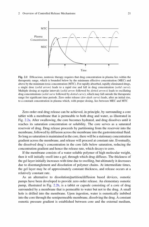
rapidly absorbed and rapidly eliminated. Figure 2.1 illustrates the plasma concen-
tration profile over time for such drugs when administered from rapid-release
dosage forms. A rapid increase in concentration is followed by a rapid decrease,
and little time is spent inside the so-called therapeutic range, which is bounded
below by a minimum effective concentration (MEC) and above by a minimum
toxic concentration (MTC) (see also Figs. 1.9 and 1.10). Frequent repetitive dosing
is required to maintain concentration within these limits, and compliance and
control are difficult.
Dosage forms that prolong release can maintain drug concentration within
the therapeutic range for extended periods and minimize episodes of underexposure
or toxicity. A well designed system displays a narrow, predictable residence time
distribution in the gastrointestinal (GI) tract, and releases drug by a controlled
mechanism. As shown in Fig. 2.1, zero order release leads, in principle, to the best
control of plasma concentration. Such control leads to constant drug effect, provided
the drug’s pharmacokinetic and pharmacodynamic properties, including absorption,
distribution, metabolism, and excretion (ADME), and its pharmacodynamic
properties relating plasma concentration to drug effect, are stationary. While this
proviso is believed to apply to most drugs, there are notable exceptions, as detailed
in Chap. 13.
20 R.A. Siegel and M.J. Rathbone
Zero order oral drug release can be achieved, in principle, by surrounding a core
tablet with a membrane that is permeable to both drug and water, as illustrated in
Fig. 2.2a. After swallowing, the core becomes hydrated, and drug dissolves until it
reaches its saturation concentration or solubility. The core serves as a saturated
reservoir of drug. Drug release proceeds by partitioning from the reservoir into the
membrane, followed by diffusion across the membrane into the gastrointestinal fluid.
So long as saturation ismaintained in the core, there will be a stationary concentration
gradient across the membrane, and release will proceed at constant rate. Eventually,
the dissolved drug’s concentration in the core falls below saturation, reducing the
concentration gradient and hence the release rate, which decays to zero.
If the membrane consists of a water-soluble polymer of high molecular weight,
then it will initially swell into a gel, through which drug diffuses. The thickness of
the gel layer initially increases with time due to swelling, but ultimately it decreases
due to disentanglement and dissolution of polymer chains. At intermediate times,
the gel layer may be of approximately constant thickness, and release occurs at a
relatively constant rate.
As an alternative to dissolution/partition/diffusion based devices, osmotic
pumps have been developed to provide zero order release. An elementary osmotic
pump, illustrated in Fig. 2.2b, is a tablet or capsule consisting of a core of drug
surrounded by a membrane that is permeable to water but not to the drug. A small
hole is drilled into the membrane. Upon ingestion, water is osmotically imbibed
into the core through the semipermeable membrane, dissolving the drug. A constant
osmotic pressure gradient is established between core and the external medium,
Time
MTC
MEC
Plasma Concentration
Fig. 2.1 Efficacious, nontoxic therapy requires that drug concentration in plasma lies within the
therapeutic range, which is bounded below by the minimum effective concentration (MEC) and
above by the minimum toxic concentration (MTC). For rapidly absorbed, rapidly eliminated drugs,
a single dose (solid arrow) leads to a rapid rise and fall in drug concentration (solid curve).Multiple dosing at regular intervals (solid arrow followed by dotted arrows) leads to oscillating
drug concentrations (solid curve followed by dotted curve), which may fall outside the therapeutic
range for significant time periods. Zero order release (dot–dash curve) leads, after an initial rise,
to a constant concentration in plasma which, with proper dosing, lies between MEC and MTC
2 Overview of Controlled Release Mechanisms 21
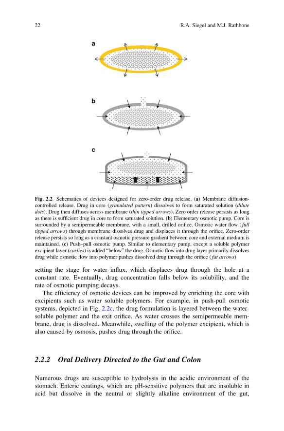
setting the stage for water influx, which displaces drug through the hole at a
constant rate. Eventually, drug concentration falls below its solubility, and the
rate of osmotic pumping decays.
The efficiency of osmotic devices can be improved by enriching the core with
excipients such as water soluble polymers. For example, in push-pull osmotic
systems, depicted in Fig. 2.2c, the drug formulation is layered between the water-
soluble polymer and the exit orifice. As water crosses the semipermeable mem-
brane, drug is dissolved. Meanwhile, swelling of the polymer excipient, which is
also caused by osmosis, pushes drug through the orifice.
2.2.2 Oral Delivery Directed to the Gut and Colon
Numerous drugs are susceptible to hydrolysis in the acidic environment of the
stomach. Enteric coatings, which are pH-sensitive polymers that are insoluble in
acid but dissolve in the neutral or slightly alkaline environment of the gut,
Fig. 2.2 Schematics of devices designed for zero-order drug release. (a) Membrane diffusion-
controlled release. Drug in core (granulated pattern) dissolves to form saturated solution (dilutedots). Drug then diffuses across membrane (thin tipped arrows). Zero order release persists as long
as there is sufficient drug in core to form saturated solution. (b) Elementary osmotic pump. Core is
surrounded by a semipermeable membrane, with a small, drilled orifice. Osmotic water flow ( fulltipped arrows) through membrane dissolves drug and displaces it through the orifice. Zero-order
release persists so long as a constant osmotic pressure gradient between core and external medium is
maintained. (c) Push–pull osmotic pump. Similar to elementary pump, except a soluble polymer
excipient layer (curlies) is added “below” the drug. Osmotic flow into drug layer primarily dissolves
drug while osmotic flow into polymer pushes dissolved drug through the orifice ( fat arrows)
22 R.A. Siegel and M.J. Rathbone
are designed to protect drug as it passes through the stomach. If the molecular
weight of the coating polymer is relatively low, then it will dissolve and drug will
be released rapidly. If the molecular weight of the polymer is high enough,
however, it will swell into a gel layer that controls drug release as above. Passage
of the dosage form through the stomach to the small intestine affects the time
required following ingestion to activate swelling and diffusion.
Certain drugs are more efficacious when released in the colon. The colon is rich
in bacterial azoreductases, which cleave polymers with azoaromatic crosslinks.
By encapsulating drug in such polymers, colon-specific drug delivery can be
achieved. Further encapsulation by a rapidly dissolving enteric coating would
permit colon-specific delivery of acid-labile drugs. The enteric coating is first
stripped off upon entering the gut, but drug is released only when the internal
polymer is degraded by the azoreductases in the colon.
2.2.3 Oral Delivery of Polypeptides
Polypeptides, including proteins, are extremely challenging to deliver orally.
Problems include acid lability, susceptibility to peptidases and proteases in the
stomach and gut, and limited absorption due to high molecular weight and charge.
Most protein bioavailabilities, measured as fraction absorbed into the systemic
circulation, hover around or below 1%. Reliable, efficient delivery of polypeptides,
if possible, will have enormous payoffs.
Let us assume that acid lability can be handled by an enteric coating layer and
that the polypeptide is incorporated into micro- or nanoparticles that are designed to
adhere to the gut wall. The particles release their payload into the wall or are taken
up by endocytosis into enterocytes. While encapsulated in the particles, the poly-
peptide molecules are protected from attack by enzymes. By these means, it is
postulated that bioavailability will be improved.
2.2.4 Delivery of Drugs Through the Skin
Numerous drugs are problematic for oral delivery due to their low solubility and
susceptibility to first pass metabolism in the liver. For such drugs, alternative ports
of entry are of interest, and practically every available body surface and orifice has
been considered. Since the skin is readily accessible and has a large surface area,
transdermal drug delivery has been the subject of much research and product
development.
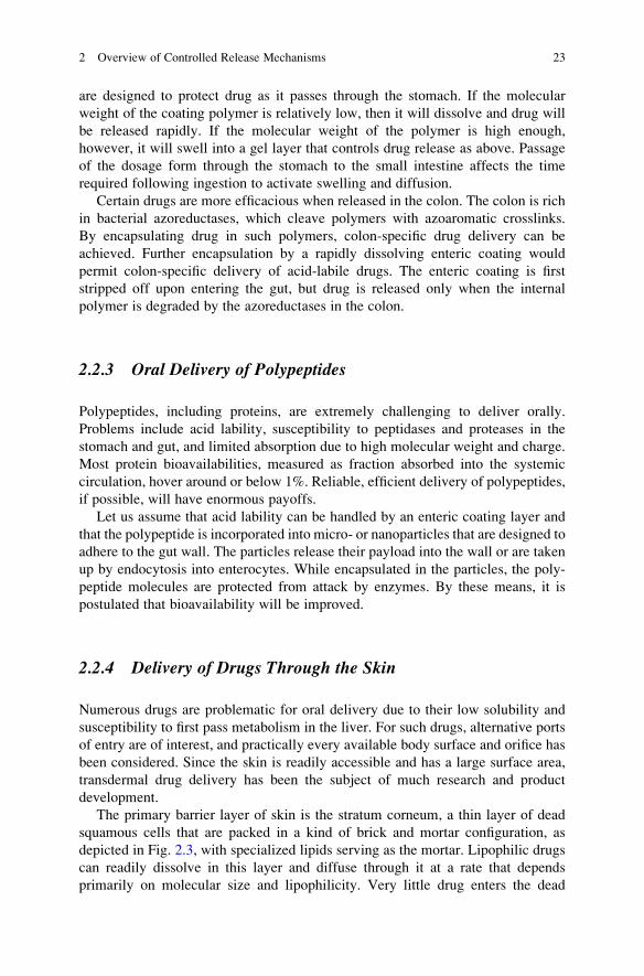
The primary barrier layer of skin is the stratum corneum, a thin layer of dead
squamous cells that are packed in a kind of brick and mortar configuration, as
depicted in Fig. 2.3, with specialized lipids serving as the mortar. Lipophilic drugs
can readily dissolve in this layer and diffuse through it at a rate that depends
primarily on molecular size and lipophilicity. Very little drug enters the dead
2 Overview of Controlled Release Mechanisms 23
cells, and the lipid pathways for diffusion are marked by numerous detours. After
passing through the stratum corneum, drug encounters the more hydrophilic, viable
epidermis and dermis, before being absorbed in capillaries perfusing the dermis.
Drug that is absorbed through the skin is not susceptible to first pass metabolism by
gut and liver, although some metabolism may occur in the skin itself.
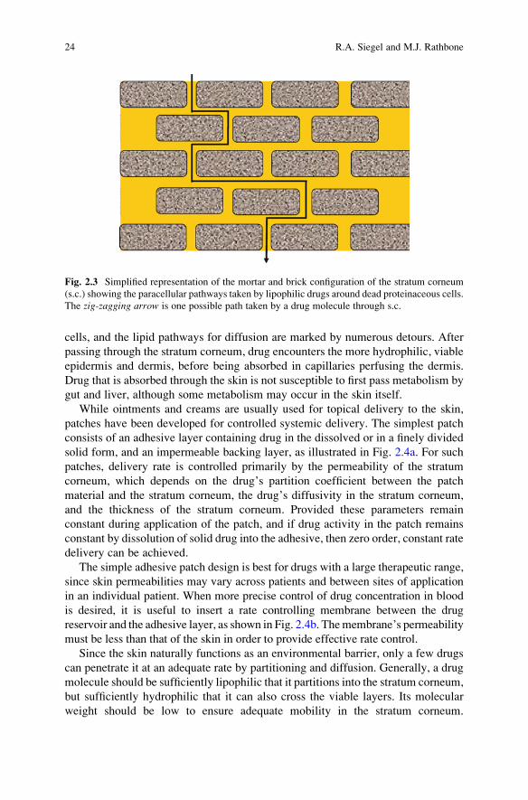
While ointments and creams are usually used for topical delivery to the skin,
patches have been developed for controlled systemic delivery. The simplest patch
consists of an adhesive layer containing drug in the dissolved or in a finely divided
solid form, and an impermeable backing layer, as illustrated in Fig. 2.4a. For such
patches, delivery rate is controlled primarily by the permeability of the stratum
corneum, which depends on the drug’s partition coefficient between the patch
material and the stratum corneum, the drug’s diffusivity in the stratum corneum,
and the thickness of the stratum corneum. Provided these parameters remain
constant during application of the patch, and if drug activity in the patch remains
constant by dissolution of solid drug into the adhesive, then zero order, constant rate
delivery can be achieved.
The simple adhesive patch design is best for drugs with a large therapeutic range,
since skin permeabilities may vary across patients and between sites of application
in an individual patient. When more precise control of drug concentration in blood
is desired, it is useful to insert a rate controlling membrane between the drug
reservoir and the adhesive layer, as shown in Fig. 2.4b. Themembrane’s permeability
must be less than that of the skin in order to provide effective rate control.
Since the skin naturally functions as an environmental barrier, only a few drugs
can penetrate it at an adequate rate by partitioning and diffusion. Generally, a drug
molecule should be sufficiently lipophilic that it partitions into the stratum corneum,
but sufficiently hydrophilic that it can also cross the viable layers. Its molecular
weight should be low to ensure adequate mobility in the stratum corneum.
Fig. 2.3 Simplified representation of the mortar and brick configuration of the stratum corneum
(s.c.) showing the paracellular pathways taken by lipophilic drugs around dead proteinaceous cells.
The zig-zagging arrow is one possible path taken by a drug molecule through s.c.
24 R.A. Siegel and M.J. Rathbone
Finally, the drug’s potency and pharmacokinetic properties should be such that
delivery through the skin places drug concentration in plasma within the therapeutic
range. While the rate of delivery can be increased by using larger patches, there are
practical size limitations.
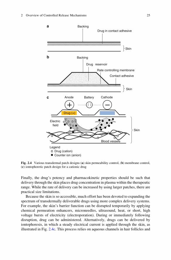
Because the skin is so accessible, much effort has been devoted to expanding the
spectrum of transdermally deliverable drugs using more complex delivery systems.
For example, the skin’s barrier function can be disrupted temporarily by applying
chemical permeation enhancers, microneedles, ultrasound, heat, or short, high
voltage bursts of electricity (electroporation). During or immediately following
disruption, drug can be administered. Alternatively, drugs can be delivered by
iontophoresis, in which a steady electrical current is applied through the skin, as
illustrated in Fig. 2.4c. This process relies on aqueous channels in hair follicles and
BackingDrug in contact adhesive
Skin
Backing
Drug reservoir
Rate controlling membrane
Contact adhesive
Skin
Skin
Electric field
Battery
Drug (+)
Blood vessels
Anode Cathode
LegendDrug (cation)Counter ion (anion)
a
b
c
Fig. 2.4 Various transdermal patch designs (a) skin permeability control, (b) membrane control,
(c) iontophoretic patch design for a cationic drug
2 Overview of Controlled Release Mechanisms 25
sweat glands, or new channels formed by the current. Charged drug molecules are
driven through these channels by a like-charged electrode while uncharged drug
molecules are delivered through the channels by electroosmotic convection.
Ideas discussed in this vignette may apply to drug delivery across other well
perfused epithelia, including the rectum, vagina, scrotum, cornea and sclera, and
the buccal and nasal mucosae.
2.2.5 Depot Delivery of Reproductive Hormones
While the introduction of daily oral steroid contraceptives in the mid-twentieth
century was a breakthrough with historic medical and social consequences, it is
recognized that there is substantial room for improvement. Daily oral dosing can
lead to incomplete compliance and effectiveness, so other routes have been studied.
For example, a transdermal, patch-based contraceptive system that delivers its
payload over 1 week has appeared on the market (ORTHO EVRA®), as has an
insertable vaginal ring that releases drug over three weeks (NuvaRing®).
The Norplant® system was introduced in the 1980s to provide five years contin-
uous release of levonorgestrel. Drug is incorporated into silicone capsules that are
placed under the skin in a routine clinical procedure. Release is mediated by slow
diffusion through the silicone matrix. Because the silicone capsules do not degrade,
they must be retrieved after they are spent. An alternative biodegradable implant
called Capronor was investigated but was not marketed.
Besides steroid hormones, analogs of luteinizing hormone-releasing hormone
(LHRH) have been developed. LHRH is the master hormone that is secreted
rhythmically in the hypothalamus, and activates numerous hormones on the repro-
ductive axis. Both LHRH agonists and antagonists have been developed as
contraceptives, and they also have been used to treat disorders, such as endometri-
osis, vaginal bleeding due to fibroids, precocious puberty, and prostate cancer.
When these analogs are delivered continuously, they interfere with the rhythmic
signaling by endogenous LHRH. Because they are extremely potent, they can be
injected as a slow-release depot. In one system, Leupron Depot1, leuprolide acetate
is formulated into biodegradable polymer microspheres, which degrade and
release drug over three months. In this system, drug release is controlled by
diffusion through a pore network whose structure evolves as the polymer degrades.
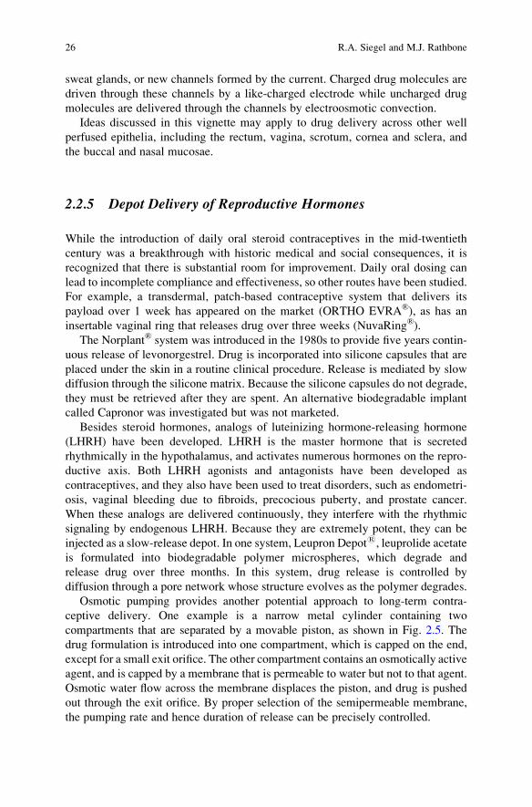
Osmotic pumping provides another potential approach to long-term contra-
ceptive delivery. One example is a narrow metal cylinder containing two
compartments that are separated by a movable piston, as shown in Fig. 2.5. The
drug formulation is introduced into one compartment, which is capped on the end,
except for a small exit orifice. The other compartment contains an osmotically active
agent, and is capped by a membrane that is permeable to water but not to that agent.
Osmotic water flow across the membrane displaces the piston, and drug is pushed
out through the exit orifice. By proper selection of the semipermeable membrane,
the pumping rate and hence duration of release can be precisely controlled.
26 R.A. Siegel and M.J. Rathbone
Complementary to contraception is fertility therapy. Patients with lesions that
suppress LHRH secretion can be treated with rhythmic intravenous injections of
LHRH, delivered from an externally worn, programmed pump through a catheter.
This mode is best for short term needs, such as induction of fertility, but it is less
desirable when the need is long term, as in the treatment of arrested puberty. Since
LHRH is exceptionally potent, each dose is very small, so the possibility of an
implantable rhythmic dosing device is intriguing. Such devices may ameliorate the
inconvenience associated with intravenous delivery. One approach under consider-
ation is a controlled-release microchip, into which thousands of microwells are
machined. Each well is filled with a single dose of LHRH and sealed by a thin gold
membrane that is addressably connected to a current source. Under the control of a
microprocessor, individual membranes are ruptured with a current pulse and their
encapsulated doses are released. By proper programming, any sequence of release
pulses can be programmed into the system.
2.2.6 Regional Drug Delivery
Thus far, we have discussed scenarios in which drug enters the systemic circulation
after release. Drug then distributes according to its relative affinities to all tissues,
and only a small fraction is present at or near the target site. Drug toxicity and side
effects are often associated with accumulation in tissues not associated with the
target. In regional (sometimes called local or topical) delivery, drug is administered
directly to the target tissues. Under proper conditions, regional delivery should
permit substantially reduced drug dosing to reach the desired effect, with reduced
exposure of other tissues to the drug.
Regional delivery is potentially most effective when drug is not transferred
substantially from the target tissue to the systemic circulation due to anatomic
or physiological barriers, or when systemic drug is rapidly eliminated. Traditional
examples include topical drugs, inhalation based asthma therapies, and chemo-
therapies directed by drug pumps to tumors. The release of chemotherapeutic agents
from polymer disks implanted next to brain tumors provides another example, as
does insulin delivery to the peritoneal cavity, which drains through the hepatic portal
vein into the liver, a primary target for insulin.
Fig. 2.5 Implantable cylindrical osmotic pump with piston. Water flows through semipermeable
membrane at left into a chamber containing osmotic excipient (curlies), displacing piston, which inturn pushes drug formulation (dots) out through the orifice at right
2 Overview of Controlled Release Mechanisms 27
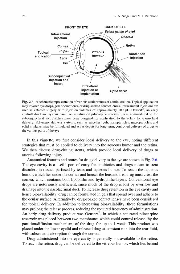
In this vignette, we first consider local delivery to the eye, noting different
strategies that must be applied to delivery into the aqueous humor and the retina.
We then discuss drug-eluting stents, which provide local delivery of drugs to
arteries following injury.
Anatomical features and routes for drug delivery to the eye are shown in Fig. 2.6.
The eye cavity is a useful port of entry for antibiotics and drugs meant to treat
disorders in tissues perfused by tears and aqueous humor. To reach the aqueous
humor, which lies under the cornea and houses the lens and iris, drug must cross the
cornea, which contains both lipophilic and hydrophilic layers. Conventional eye
drops are notoriously inefficient, since much of the drop is lost by overflow and
drainage into the nasolacrimal duct. To increase drug retention in the eye cavity and
hence bioavailability, drug can be formulated in gels that spread over and adhere to
the ocular surface. Alternatively, drug-soaked contact lenses have been considered
for topical delivery. In addition to increasing bioavailability, these formulations
may prolong the release process, reducing the required frequency of administration.
An early drug delivery product was Ocusert®, in which a saturated pilocarpine
reservoir was placed between two membranes which could control release, by the
partition/diffusion mechanism, of the drug for up to 1 week. This product was
placed under the lower eyelid and released drug at constant rate into the tear fluid,
with subsequent absorption through the cornea.
Drug administered into the eye cavity is generally not available to the retina.
To reach the retina, drug can be delivered to the vitreous humor, which lies behind
Vitreous humour
Topicalapplication
Subconjuctivalinjection and
insert Intravitrealinjection orimplantation
Subtendoninjection
Intracameralinjection
Lens
Pupil
Cornea
Iris
Retina
Choroid
Optic nerve
FRONT OF EYE BACK OF EYESclera (white of eye)
Fig. 2.6 A schematic representation of various ocular routes of administration. Topical application
may involve eye drops, gels or ointments, or drug-soaked contact lenses. Intracameral injections are
used in cataract surgery with injection volumes of approximately 100 mL. Ocusert®, an early
controlled-release system based on a saturated pilocarpine reservoir, was administered to the
subconjunctival sac. Patches have been designed for application to the sclera for transscleral
delivery. Polymeric delivery systems, such as micelles, gels, nanoparticles, microparticles, and
solid implants, may be formulated and act as depots for long-term, controlled delivery of drugs to
the various parts of the eye
28 R.A. Siegel and M.J. Rathbone
the lens. The vitreous is a viscous gel that slowly circulates, providing convective
transport of drug to the retinal surface. Several schemes have been investigated.
The ocular sclera (white) provides a large surface area, and patches have been
devised for transscleral delivery into the vitreous. A problem arises because the
sclera is heavily perfused by choroidal blood vessels, which remove drug before it
reaches the vitreous by diffusion. Alternatively, solid implants that slowly release
the drug can be injected or placed surgically into the vitreous. Presentation of drug to
the retina, then, depends both on the rate of release by diffusion and rate of
convection to the retinal surface.
Drug eluting stents, discussed in Chap. 14, have recently been developed to
prevent restenosis or reclosing of coronary arteries following angioplasty and
stenting procedures in response to heart attacks. Restenosis is an inflammatory
response to these procedures, and involves the growth of arterial smooth muscle
cells over the stents. To arrest such growth, small amounts of anti-inflammatory and
antiproliferative drugs are coated onto the stents and are released directly into the
adjacent arterial tissue by dissolution, partitioning, and diffusion. Because the dose
is so small and targeting is so precise, it is possible to prevent restenosis without
releasing detectable amounts of drug into the systemic circulation and other tissues.
2.2.7 Nanoparticulate Targeting of Drugs to Specific Tissues
Besides improving systemic bioavailability and the temporal and regional patterns
of drug release and absorption, controlled release systems have been developed to
alter the residence time of circulating drug. In these systems, drug is incorporated in
nanocarriers that have access to the whole systemic circulation, but are cleared less
rapidly than free drug. The nanocarriers can be regarded as circulating drug depots.
Nanocarriers may also have favorable distribution properties into target tissues and
away from tissues associated with toxic side effects. Examples of nanocarriers
include microemulsions, liposomes, dendrimers, block polymer micelles, solid
lipid and polymer nanoparticles, and soluble polymers with drug attached on side
chains by biodegradable linkages.
At the nano level, it is also possible to incorporate targeting ligands that permit
particles to bind preferentially to specific cell types and promote the uptake and
drug release into those cells. It has been suggested that cellular processes that rely
on multivalent attachment, including particle uptake, can be modulated by drug/
nanoparticle composites by suitable placement of multiple-targeting ligands on
particle surfaces.
Design of nanoparticulate drug delivery systems must take into account normal
physiological scavenging processes that remove small foreign objects from the
blood. Special coatings, such as poly(ethylene oxide)s, are used for this purpose.
Suitably coated nanoparticulates exhibit reduced opsonization and clearance by
the reticuloendothelial system. Renal clearance is avoided when nanoparticulates
are larger than glomerular pores. Hence, circulating half-lives of nanoparticulates and
2 Overview of Controlled Release Mechanisms 29
their associated drugs are prolonged. Furthermore, coated nanoparticles and their
associated drug are largely restricted to the vascular space, in contrast to free drug
which may have much a larger volume of distribution. It should be noted, however,
that if drug is released from the nanoparticle into systemic circulation, as opposed to
a specific target site, it will possess the same pharmacokinetic properties as otherwise
administered free drug.
It is believed that nanoparticulate delivery systems may be very useful in treating
some cancers due to the enhanced permeation and retention (EPR) effect. Com-
pared to normal tissues, tumors have leaky capillaries with large fenestrations in the
capillary walls that permit the passage of nanoparticulates. Drug loaded into the
nanoparticulates is, therefore, relatively more accessible to tumor tissues compared
to tissues associated with toxic side effects.
2.3 Survey of Mechanisms
The previous vignettes highlighted several controlled-release mechanisms, including
dissolution, partitioning, diffusion, osmosis, swelling, erosion, and targeting. Basic
principles associated with these mechanisms are presented in this section.
2.3.1 Dissolution
Most drug molecules form crystals at room temperature. In fact, they may take on
various crystal forms (polymorphs) or form crystal hydrates, depending on their
processing conditions. In some cases drug particles can be processed into an
amorphous, glassy form. These forms have differing thermodynamic stabilities,
and interconversion between solid forms can occur during storage and after admin-
istration. Dissolution involves transfer of drug from its solid phase to the
surrounding medium, which may be water, polymer, or tissue. The solubility of
drug in a medium, CS;medium, is defined as the concentration of drug in the medium
at saturation, i.e., in equilibrium with the solid form. Higher concentrations of drug
are thermodynamically unstable, and with time drug crystallizes out of solution
until its concentration equals CS;medium. Useful rules of thumb are that CS;medium
decreases with increasing melting point of the drug and increases with increasing
chemical compatibility of drug with the surrounding medium.
While solubility is a thermodynamic property of a drug and a medium, the
dissolution rate is a kinetic property. Dissolution rate increases with solubility
and decreases with drug particle size. As discussed below, dissolution rate is
commonly controlled by diffusion.
30 R.A. Siegel and M.J. Rathbone
2.3.2 Partitioning
During drug delivery, drug molecules often encounter an interface between two
materials or phases. The partition coefficient is a measure of the relative affinity for
drug between the two phases, and is roughly given by the ratio of drug solubilities in
the two phases. At the interface, the partition coefficient prescribes the relative
frequency that a molecule moves into one medium compared to the other.
As an example, recall that drugs of high lipid solubility are suitable for entry into
the stratum corneum. However, if the drug is not sufficiently water soluble, i.e., its
lipid/water partition coefficient is too high, it will not partition efficiently into the
viable epidermis, and drug will be detained in the stratum corneum. Absorption into
capillaries might then occur at an unacceptably low rate.
As a second example, block copolymer micelles are formulated with hydropho-
bic cores and hydrophilic coronas, hence they are soluble in blood. Hydrophobic
drugs preferentially partition into the core, where they are retained for extended
periods of time. Pharmacokinetic characteristics of such drugs, i.e., clearance and
volume of distribution, reflect those of the micelles, leading to longer retention in
the circulation and preferred distribution into tumors due to the EPR effect.
2.3.3 Diffusion
Diffusion is a very important component of many controlled-release systems, hence
we devote considerable space in this chapter to it. More details about diffusion-
controlled drug delivery systems are provided in Chaps. 6 and 9.
2.3.3.1 Molecular Basis
All molecules constantly undergo random collisions with other molecules. As a
result, molecules execute thermal or Brownian motion. At any step, the direction of
motion of a molecule is random, and it repeatedly changes due to collisions with
other molecules. Over time, the displacement of the molecule from its point of
origin is the result of a multitude of such random steps. Macroscopically, the
independent random walks taken by large number of drug molecules lead them
from regions of higher concentration to regions of lower concentration. Thus
diffusion of a substance occurs down its concentration gradient.
The theory of random walks shows that the average (actually, root mean
squared) distance that molecules travel by diffusion is proportional to the squareroot of time, i.e., average distance traveled � ffiffiffiffiffi
Dtp
, whereD (cm2/s) is the diffusion
coefficient, or diffusivity, and t is time (s). The diffusion coefficient is a measure of
the molecule’s mobility in the medium. Conversely, the typical time required to
diffuse over a particular distance is proportional to the square of that distance and
2 Overview of Controlled Release Mechanisms 31
inversely proportional to the diffusion coefficient. Thus, while diffusion is an
efficient means of mass transport over short distances, its effectiveness decreases
over longer distances.
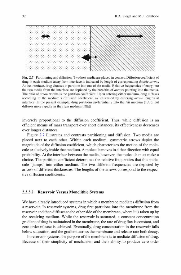
Figure 2.7 illustrates and contrasts partitioning and diffusion. Two media are
placed next to each other. Within each medium, symmetric arrows depict the
magnitude of the diffusion coefficient, which characterizes the motion of the mole-
cule exclusively inside that medium. Amolecule moves in either direction with equal
probability. At the interface between the media, however, the molecule must make a
choice. The partition coefficient determines the relative frequencies that this mole-
cule “jumps” into either medium. The two different frequencies are depicted by
arrows of different thicknesses. The lengths of the arrows correspond to the respec-
tive diffusion coefficients.
2.3.3.2 Reservoir Versus Monolithic Systems
We have already introduced systems in which a membrane mediates diffusion from
a reservoir. In reservoir systems, drug first partitions into the membrane from the
reservoir and then diffuses to the other side of the membrane, where it is taken up by
the receiving medium. While the reservoir is saturated, a constant concentration
gradient of drug is maintained in the membrane, the rate of drug flux is constant, and
zero order release is achieved. Eventually, drug concentration in the reservoir falls
below saturation, and the gradient across the membrane and release rate both decay.
In reservoir systems, the purpose of the membrane is to mediate diffusion of drug.
Because of their simplicity of mechanism and their ability to produce zero order
Fig. 2.7 Partitioning and diffusion. Two host media are placed in contact. Diffusion coefficient of
drug in each medium away from interface is indicated by length of corresponding double arrow.At the interface, drug chooses to partition into one of the media. Relative frequencies of entry into
the two media from the interface are depicted by the breadths of arrows pointing into the media.
The ratio of arrow widths is the partition coefficient. Upon entering either medium, drug diffuses
according to the medium’s diffusion coefficient, as illustrated by differing arrow lengths at
interface. In the present example, drug partitions preferentially into the left medium ( ), but
diffuses more rapidly in the right medium ( )
32 R.A. Siegel and M.J. Rathbone
release, reservoir systems would seem to be highly advantageous. However, reservoir
systems can be difficult to fabricate reliably. Pinhole defects and cracks in the
membrane can lead to dose dumping. These problems are avoided in monolithic
systems, in which drug is loaded directly into a polymer, which now acts as both a
storage medium and a mediator of diffusion.
Drug is typically loaded uniformly into monolithic devices, and release is
controlled by diffusion through the monolith’s matrix material or through aqueous
pores. Monolithic devices typically exhibit an initial burst of release from the
surface. With passing time, release rate decreases as drug that is deeper inside the
monolith must diffuse to the surface, since it has farther to travel, and the quadratic
relation between distance and time becomes important. This effect occurs in planar
monoliths, but it is even more prominent with cylinders or spheres, as the amount of
drug available decreases with distance from the surface. This geometric factor can be
substantially reversed using specially coated wedge, cone, or hemisphere monoliths
to provide near-zero-order release, but such devices are not easy to fabricate.
2.3.3.3 Factors Affecting Diffusivity
The diffusivity, D, depends on the molecule and the medium. For a hard spherical
molecule in a liquid solvent, the Stokes-Einstein equation prescribesD ¼ kBT=6pa�,where a is the molecule’s radius, � is the solvent’s viscosity, kB is Boltzmann’s
constant, and T is absolute (Kelvin) temperature. This relation confirms the intui-
tion that large molecules should diffuse more slowly than small ones and that
diffusion should be slowed in viscous liquids. The factor kBT accounts for the
intensity of thermal agitation, which drives Brownian motion.
In typical polymeric controlled release systems, the polymer matrix does not
flow like a liquid, and bulk viscosity is not the correct parameter to use in predicting
mobility of drug. The matrix may possess, however, a “microviscosity” that is
related to molecular mobility. Free volume theory provides a useful picture that
accounts for both bulk and microviscosity. While it may be natural to think of a
polymer matrix as a static solid, it is actually a dynamic fluctuating structure, and
D may be thought of as a measure of the degree that these fluctuations accommo-
date random motion of the diffusing molecule. In free volume theory, each drug,
solvent, and polymer molecule contains an impenetrable core that is surrounded by
nanovoids, called free volume. Thermal motions cause the size of voids to fluctuate.
Occasionally, a void becomes large enough for a diffusing molecule to move into
or through it. Clearly, if this mechanism is operative, then the diffusion coefficient
will decrease sharply with increasing molecular radius and when the matrix’s
density increases upon cooling. At a critical density, often associated with the
medium’s glass transition temperature, Tg, free volume becomes so sparse that
the diffusion coefficient drops by several orders of magnitude.
In addition to temperature, the free volume of a polymer matrix depends on its
composition. For homogeneous materials, free volume increases as the difference
2 Overview of Controlled Release Mechanisms 33
between ambient temperature and Tg increases. Copolymerization and blending can
lead to matrices with suitably averaged free volumes and mobility properties. Free
volume can also be increased substantially by sorption of small molecules, such as
water. Thus, a glassy dry polymer can be converted to the rubbery state by sorption
of a small amount of water, substantially increasing the mobility of drug molecules
in the polymer.
Besides the glass transition, polymers can form crystalline domains which
exclude drug molecules and obstruct diffusion. The propensity to crystallize
depends on the polymer’s melting point and its stereoregularity. Random
copolymers generally do not form crystalline domains. Crystallization can be
mediated by the polymer backbone or by the side chains, especially when the latter
are long.
For a molecule diffusing through a water-swollen hydrogel, diffusivity of drug is
affected by the viscosity of thewater space and also by obstructions placed in the drug
molecule’s path by the hydrogel chains. Many models of diffusion in hydrogels,
therefore, combine elements of Stokes–Einstein and free volume theories. In this
case, the size of water-filled spaces between hydrogel chains is assumed to fluctuate,
making room for movement of the diffusing drug molecule. The characteristic
distance between points of chain crossings in the hydrogel is called the correlation
length, and the ratio ofmolecular radius of drug to the correlation length is considered
to be the primary structural parameter governing the drug’s diffusion coefficient in
the hydrogel.
2.3.3.4 Heterogeneous Systems
Thus far, we have discussed diffusion mediated systems in which the medium is a
uniform polymer matrix or hydrogel. Local matrix fluctuations were assumed to
control the rate of diffusion. In more heterogeneous media, other factors also
become important.
We have already noted that the presence of dead cell bodies in the stratum
corneum increases the effective path length for drugs diffusing through skin
lipids. We have also seen that crystalline domains in a polymer can obstruct and
retard diffusion. More generally, diffusion of drug through a heterogeneous
medium depends on the solubility and diffusivity of drug in the different material
domains of the medium, and the geometric manner in which the domains are
dispersed.
For example, consider a polymer blend or block structure, where one component
has a much higher drug solubility than the other. If the “drug-philic” domains
comprise a discrete phase dispersed in a “drug-phobic” continuous phase, then the
disconnected phases will retain drug and retard its release, by analogy to affinity
chromatography. If on the other hand the drug-philic domain is continuous, then
release will be controlled by diffusion through the continuous phase, but will be
retarded by detours around the drug-phobic domains.
34 R.A. Siegel and M.J. Rathbone
Porous systems are often encountered in controlled release. Empty pores can be
introduced into a matrix during fabrication to serve as pathways for drug diffusion
through water that enters the pores. Alternatively, solid drug or excipient particles
can be introduced into a polymer, and pores form around the particles. Also drug
and excipient may precipitate from a polymer solution during solvent removal,
again resulting in a porous amalgam of drug and polymer. The pores then act as
both depots for drug storage and as conduits for diffusion. Pore structure and
connectivity may have a profound effect on release by diffusion, as is discussed
in Chap. 9.
2.3.3.5 Diffusion Affects Dissolution
We conclude this section with a discussion of dissolution and diffusion in drug
delivery. Dissolution occurs when the solvating medium surrounding a solid drug
particle is not saturated. This process involves two steps. First, drug must dissociate
from the surface of the particle and surround itself with solvent. Second, the newly
solvated drug must diffuse away from the surface. The first process is usually more
rapid than the second, unless the drug is extremely insoluble. Thus, the drug is very
close to its saturation concentration in the immediate vicinity of the particle.
A concentration gradient is, therefore, established between the particle/medium
interface and the “bulk” of the medium, and diffusion controls the rate that drug
flows down this gradient. In drug delivery systems containing solid drug particles,
both CS;medium and D are therefore important determinants of release rate.
In an important class of drug delivery systems discussed in Chap. 6, solid drug
particles are incorporated into a monolithic matrix. Release of drug occurs by
dissolution followed by diffusion through the matrix. Particles at the surface
dissolve quickly, leading to a burst. Particles further inside dissolve more slowly,
since dissolution rate is controlled by diffusion through the matrix. At intermediate
times, a moving front is observed, separating a central core containing solid drug
from a periphery containing completely dissolved drug. Because the diffusion
distance from the front to the monolith’s surface increases with time, the march
of this front slows down as the release process proceeds, and the rate of release
decreases with time.
2.3.4 Osmosis
Osmosis is a dramatic phenomenon that occurs when a membrane that is permeable
to water but not to particular solutes, called osmolytes, separates aqueous solutions
of the osmolytes. Water flows through the semipermeable membrane in an effort to
equalize concentrations of the impermeable solutes on both sides of the membrane.
In most cases of interest, water flow occurs by diffusion through the semipermeable
membrane. However, the nature of water transport may differ from that discussed
2 Overview of Controlled Release Mechanisms 35
above for drugs. First, it should be emphasized that there tends to be a lot of water on
both sides of the membrane, and flux of water through the membrane is determined
by the difference in chemical potentials of water on the two sides, not simply the
concentration gradient of water. These chemical potentials may depend on both
concentrations of the osmolytes and the thermodynamic compatibility of water with
the osmolytes. When the osmolytes are small molecules, such as salts, osmotic
pressure is reasonably accounted for osmolyte concentrations according to van’t
Hoff’s law, but when the osmolytes are polymers, osmotic pressure is determined
jointly by polymer concentration and polymer/water compatibility. Second, when
themembrane is adequately hydrated, water molecules are in contact with each other
and neighboring molecules’ motions are correlated. The Brownian mode of diffu-
sion discussed for drug molecules is then replaced by the so-called collective mode.
The rate of osmotic flow across a unit area of the membrane is determined
by the concentration and nature of osmolytes on both sides of the membrane,
temperature, and the hydraulic permeability of the membrane, which can be deter-
mined by measuring water flow when a hydrostatic pressure is applied across the
membrane. Osmotic flow is reduced when the membrane is partially permeable
to the osmolytes. As water flows into a device containing osmolytes, it dilutes
the osmolytes, lowering the osmotic pressure, unless new osmolytes are introduced,
for example, by dissolution.
We have already described osmotic pumps in which water invasion across
a membrane displaces drug through an orifice. Another way to use osmosis is to
coat individual drug particles with semipermeable polymers. After release from a
capsule, these particles are exposed to gastric fluid. Water crosses the polymer
coatings and dissolves the drug, leading to a gradient in solute concentration that
drives even more water inside. To accommodate, the coating must expand, and wall
stresses are developed. With sufficient osmotic driving force, the coating ruptures,
releasing the drug. Using different coating thicknesses, particles can be programmed
to burst at different times. The original time release capsules were based on
this principle.
A variation of the elementary osmotic pump theme involves particles or tablets
that are coated with a semipermeable polymer membrane which includes sparsely
but well-distributed aqueous pores. These pores can be created by excipients
blended into the membrane, which dissolve upon exposure to water. Here, water
flows across the semipermeable parts of the membrane and displaces dissolved drug
inside through the aqueous pores into the release medium.
2.3.5 Swelling
Swelling refers to the uptake of water by a polymer system, with increase in volume.
Swelling is often a prelude to polymer dissolution. However, swelling may
occur without dissolution if water and the polymer are insufficiently compatible,
if polymer chain length is sufficiently large, or if crosslinks are introduced to form
36 R.A. Siegel and M.J. Rathbone

a polymer network. Swollen polymer networks or hydrogels reviewed in Chap. 4
may imbibe many times their weight in water.
The swelling process is analogous to osmosis, since water enters the polymer
relatively rapidly, while dissolution of polymer into water, if it occurs, is compara-
tively slow because of the need for polymer chains to disentangle. The extent
of swelling depends on the compatibility of water with the polymer material, i.e.
the polymer’s hydrophilicity, and on the density of crosslinks between polymer
chains, if present. Hydrophobic polymers, reviewed in Chap. 3, imbibe very little
water and hence do not swell significantly.
Swelling is a mechanism by which release of otherwise confined drug is
activated. If swelling is rapid, then drug diffusion through the swollen polymer is
the controlling process for drug release. If swelling is relatively slow, then it can be
the process controlling the rate of drug release. A more detailed description of
swelling controlled systems is given in Chap. 7.
Swelling controlled release systems are typically glassy polymers at room and
body temperatures. Water uptake is initially resisted by the glass, but eventually it
makes its way into the free volume at the surface. The glassy polymer at the surface
relaxes to a configuration that ismore compatiblewithwater, and swells. This permits
water to intrude even further, and a moving front is often observed separating a
swollen outer layer from a dry inner core. Usually, swelling is accompanied by a
glass-to-rubber transition. If drug is trapped inside the glass, it will be liberated when
the polymer swells, and if it can diffuse through the softened matrix faster than water
can invade, then the release process is swelling controlled. Swelling dynamics are
often complex, and a variety of temporal release patterns are observed under swelling
control. Under proper conditions, swelling, dissolution of polymer chains, and drug
release may occur simultaneously, further contributing to complexity.
Swelling in a polymer may be induced or accelerated by drugs or other additives,
which act as effective osmolytes, drawing water into the polymer. By proper
selection of polymer, it is also possible to induce swelling by changes in external
parameters, such as temperature and pH, which may occur, for example, upon
ingestion. Reversible swelling and shrinking of hydrogels can also be induced by
alternating these parameters with concomitant on/off patterns of drug release.
2.3.6 Erosion and Degradation
Erodible anddegradable drug delivery systems are popular, particularly for implantable
or injectable therapies, since they do not require retrieval after drug is fully released.
Presently, the most common erodible systems are based on poly(lactic acid) or poly
(lactic acid-co-glycolic acid), although systems based on poly(e-vinyl caprolactone),poly(ortho esters), polyanhydrides, polyphosphates, poly(phosphazenes), and pseudo-
poly(amino acids) have also been utilized or studied. Important characteristics of
erodible systems are their mechanism and kinetics of erosion. Erosion products must
be nontoxic and excretable or resorbable. Principles and applications of erodible
2 Overview of Controlled Release Mechanisms 37
systems are elaborated in Chaps. 5, 8, and 10. In this section, we call attention to two
limits of behavior in erodible systems, namely, bulk erosion and surface erosion.
Erosion of polymer monoliths occurs when components of the release medium,
especially water, attack covalent bonds in the polymer matrix. For hydrolytically
labile bonds, availability of water is an important determinant of local erosion rate.
Hydrolysis of bonds may also be acid or base catalyzed, and if so depends on local
concentration of proton donors and acceptors. For PLA and PLGA and other
polyesters or polyamides, acidic protons are provided by chain ends; hence, con-
centration of acid protons is inversely proportional to chain length.
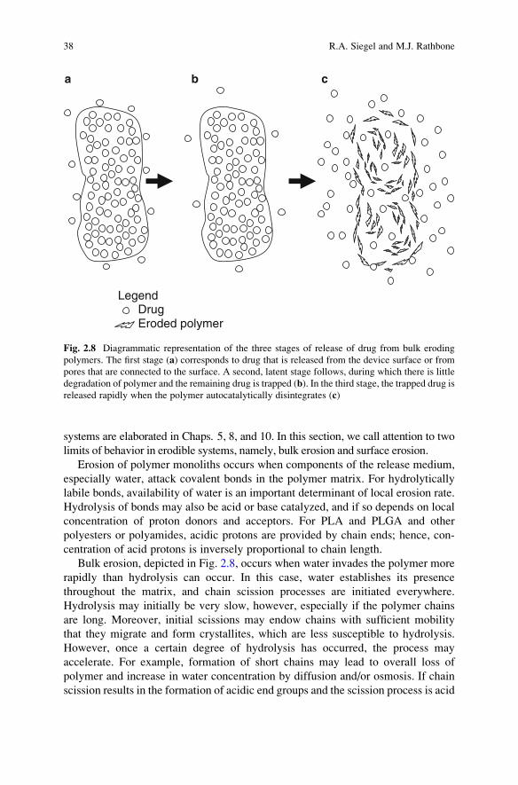
Bulk erosion, depicted in Fig. 2.8, occurs when water invades the polymer more
rapidly than hydrolysis can occur. In this case, water establishes its presence
throughout the matrix, and chain scission processes are initiated everywhere.
Hydrolysis may initially be very slow, however, especially if the polymer chains
are long. Moreover, initial scissions may endow chains with sufficient mobility
that they migrate and form crystallites, which are less susceptible to hydrolysis.
However, once a certain degree of hydrolysis has occurred, the process may
accelerate. For example, formation of short chains may lead to overall loss of
polymer and increase in water concentration by diffusion and/or osmosis. If chain
scission results in the formation of acidic end groups and the scission process is acid
Legend Drug Eroded polymer
a b c
Fig. 2.8 Diagrammatic representation of the three stages of release of drug from bulk eroding
polymers. The first stage (a) corresponds to drug that is released from the device surface or from
pores that are connected to the surface. A second, latent stage follows, during which there is little
degradation of polymer and the remaining drug is trapped (b). In the third stage, the trapped drug isreleased rapidly when the polymer autocatalytically disintegrates (c)
38 R.A. Siegel and M.J. Rathbone

catalyzed, then erosion will be autocatalytic. Thus, bulk erosion may exhibit a
sustained quiescent phase, followed by rapid disintegration of the matrix. Prior to
disintegration, the dimensions of the device remain relatively constant.
Release of drug from bulk eroding polymers typically exhibits three stages.
The first stage corresponds to drug that is released from the device surface
or from pores that are connected to the surface. A second, latent stage follows,
during which there is little degradation of polymer, and the remaining drug is
trapped. In the third stage, the trapped drug is released rapidly when the polymer
disintegrates.
Surface erosion, illustrated in Fig. 2.9, occurs either when water invasion is slow
or hydrolysis is rapid. For example, polyanhydrides are exceptionally hydrophobic,
and the hydrolytically labile anhydride bonds are protected from exposure to water
in the interior of the polymer matrix. Thus, hydrolysis with accompanying drug
release only occurs at or close to the surface.
A hallmark of surface erosion is that device dimensions decrease with time.
If the device is formulated as a slab, then release will be approximately zero order,
since each time interval will correspond to the erosion of a layer of polymer and
release of drug incorporated in that layer. Erosion rate of cylinders and spheres
decreases with time, however, due to reduction in exposed surface area. In princi-
ple, drug release correlates with erosion.
Legend
Drug Surface eroding polymer
Fig. 2.9 Diagrammatic representation of surface erosion. Drug trapped in the outer layers of
the delivery system is released into the surrounding media following erosion of the surface of the
polymer. Remaining drug is trapped in the delivery system; however, as time progresses,
the polymeric devise erodes from the surface inward and reduces in size, eventually resulting in
all drug being released
2 Overview of Controlled Release Mechanisms 39
While the idealized mechanisms underlying bulk and surface erosion-controlled
release are simple, practical systems exhibit extra complexity. Pure surface erosion
is almost impossible to achieve, and diffusion of drug out of a matrix may occur
ahead of erosion. The drug itself may draw in water, and osmotic stresses (due
also to small chain fragments) in the polymer can lead to fracture and uneven
penetration. In bulk eroding systems, degradation may even occur more rapidly in
the interior of the device due to accumulation of autocatalytic erosion products
while leaching of these products leads to slower erosion at the surface. When this is
true, thicker matrices may erode more rapidly than thinner matrices.
This section has focused on erosion as a means for controlling drug release.
However, it is also possible to program polymer degradation to occur after
drug release is more or less complete. For example, hydrogels with degradable
crosslinks have been prepared for release of proteins. As these crosslinks degrade,
the hydrogel first swells, and then it eventually disintegrates when too few
crosslinks are left to maintain the polymer network. Eventually, only primary
polymer chains remain, and these are either excreted or resorbed. If degradation
is slow, then release is controlled by protein diffusion through the swollen hydrogel
network. If degradation of crosslinks is relatively rapid, then the swelling state
of the network may change during the release process, and a complex interplay
between swelling and diffusion will determine release kinetics.
Finally, we note that water need not be the only agent causing polymer degrada-
tion. Incorporating enzyme-labile chains or crosslinkers into a polymer network
renders it susceptible to enzymatic degradation. For example, collagen and fibrin
gels are specifically degraded in the presence of collagenase and plasmin, respec-
tively. Enzyme-labile peptide fragments of collagen and fibrin can be incorporated
into other hydrogels, yielding similar, enzyme specific degradation patterns.
Enzyme-mediated degradation exhibits either surface- or bulk mediated erosion
features, depending on the ability of enzyme to diffuse into the network and the
reactivity of enzyme with the labile components of the network. Such enzyme-
degradable systems may be useful in tissue engineering applications, reviewed in
Chap. 17, as degradation of a hydrogel may be desirable with growth of tissue,
which is signaled by local release of enzyme by cells.
Besides enzymes, small molecules can trigger erosion by cleaving polymer
chains or crosslinks. For example, reducing agents can degrade polymers that include
disulfide bonds. Since small molecules readily diffuse in even moderately swollen
networks, bulk erosion is expected to predominate.
2.3.7 Regional Delivery and Targeting
The benefit of a drug can be greatly enhanced if it can be targeted to its preferred
site of action and kept away from sites associated with toxicity. Localization can
occur at the organ, tissue, cellular, and subcellular compartment or organelle level.
Direct administration at or near the site of action has already been discussed, with
40 R.A. Siegel and M.J. Rathbone
examples provided by systems designed for drug delivery to the eye and coronary
arteries. Direct injection of drug carriers into solid tumors or wound sites provides
another example. As a third example, the growth, integration, and vascularization
of surgically implanted tissue engineered constructs (Chap. 17) may require the
localized and well-timed release of growth and angiogenesis factors.
We have also discussed nanocarriers that distribute preferentially in tumors
by the EPR effect. To further specify delivery at the cellular level, it is necessary
to coat the carrier surface with ligands that bind to specific cell surface features,
such as polysaccharides or receptor proteins. Antibodies raised against antigens
expressed at the cell surface are the most obvious targeting ligands, but in recent
years peptide ligands have been designed based on other known interactions
between cell surface receptors and both soluble and extracellular matrix proteins.
Since tumor cells express multiple drug resistance transporters, release of drug
from the carrier at the cell surface may not result in increased drug uptake in target
cells. The drug/nanocarrier combination is likely to be more effective if it can be
brought into the cell by active processes, such as coated pit-mediated endocytosis.
Once in the cell, the drug needs to dissociate from the carrier and exit the endosome,
in either order. Further targeting of drug to an organellemay require that an organelle-
specific “address label” be conjugated to the drug. For example, gene and protein
delivery to the nucleus may require that a nuclear localization sequence be conju-
gated to the active biomolecule in order for the latter to be able to penetrate through
nuclear pores.
Targeting systems are the subject of Chaps. 10–12 and further examples are
provided in Chaps. 14–16.
2.4 Concluding Remarks
This chapter has illustrated a variety of controlled release strategies and underlying
mechanisms. We emphasize that several mechanisms may be at play in a particular
controlled release system, especially when more than one stage is involved. We also
have reviewed methods to achieve the various goals of controlled release, including
improved temporal presentation, drug protection, and localization of drug at the
preferred site of action.
This chapter and this book are written from the perspective that controlled
release adds substantial value to a drug. However, it should be recognized that
development of a controlled-release product can be expensive. For many drugs, the
extra expense may not be warranted on purely therapeutic grounds, although
developers may pursue controlled release formulations for marketing, quality
control, and regulatory reasons. Drugs with a relatively narrow therapeutic range,
drugs that are eliminated rapidly from the body, drugs whose efficacy would be
enhanced by targeting, and drugs that are susceptible to physiological degradation
before absorption are probably the best candidates for controlled release.
2 Overview of Controlled Release Mechanisms 41
Paradoxically, molecular entities that possess these attributes are often screened out
early in the discovery and development stages. With improved understanding of
controlled-release mechanisms and improved development of technologies, it may
be possible to increase the number of bioactive molecules that can be developed
fully into drug products [1–35].
References
1. Amidon GL, Lee PI, Topp EM (eds) (2000) Transport processes in pharmaceutical systems.
Marcel Dekker, New York
2. Amiji M (ed) (2005) Polymeric gene delivery. CRC, Boca Raton, FL
3. Arshady R (ed) (2003) Biodegradable polymers. Citus Ltd, London
4. Baker RW (1987) Controlled release of biologically active agents. Wiley, New York
5. Chasin M, Langer R (eds) (1990) Biodegradable polymers as drug delivery systems. Marcel
Dekker, New York
6. Fang J, Nakamura H, Maeda H (2011) The EPR effect: unique features of tumor blood vessels
for drug delivery, factors involved, and limitations and augmentation of the effect. Adv Drug
Deliv Rev 63(3):136–151
7. Flynn GL, Yalkowski SH, Roseman TJ (1974) Mass transfer phenomena and models: theoret-
ical concepts. J Pharm Sci 63:479–510
8. Friend DR (ed) (1992) Oral colon specific drug delivery. CRC, Boca Raton, FL
9. Grassi M, Grassi G, Lapasin R, Colombo I (eds) (2007) Understanding drug release and
absorption mechanisms. CRC, Boca Raton, FL
10. Guy RH, Hadgraft J (eds) (2003) Transdermal drug delivery systems: second edition, revised
and expanded. Marcel Dekker, New York
11. Hillery AM, Lloyd AW, Swarbrick J (2001) Drug delivery and targeting for pharmacists and
pharmaceutical scientists. Taylor & Francis, Oxford
12. Kim C-J (1999) Controlled release dosage form design. Technomic, Lancaster, PA
13. Kydonieus A (ed) (1993) Treatise on controlled release. Marcel Dekker, New York
14. Langer R (1990) New methods in drug delivery. Science 249:1527–1533
15. Langer R (1998) Drug delivery and targeting. Nature 392:5–10
16. Langer R, Peppas NA (1981) Present and future applications of biomaterials in controlled drug
delivery systems. Biomaterials 2:201–214
17. Langer R, Peppas NA (2003) Advances in biomaterials, drug delivery, and bionanotechnology.
AIChE J 49:2990–3006
18. Lee VHL (ed) (1991) Peptide and protein drug delivery. CRC, Boca Raton, FL
19. Mahato R (ed) (2005) Biomaterials for delivery and targeting of proteins and nucleic acids.
CRC, Boca Raton, FL
20. Mathiowitz E (ed) (1999) Encyclopedia of controlled drug delivery. Wiley, New York
21. Mathiowitz E, Chickering DE, Lehr C-M (eds) (1999) Bioadhesive drug delivery systems.
Marcel Dekker, New York
22. Mitra AK (ed) (2003) Ophthalmic drug delivery systems. Marcel Dekker, New York
23. Ottenbrite R, Kim SW (eds) (2001) Polymeric drugs and drug delivery systems. CRC, Boca
Raton, FL
24. Park K (ed) (1997) Controlled drug delivery. Challenges and strategies. American Chemical
Society, Washington, DC
25. Park K, Shalaby WSW, Park K (1993) Biodegradable hydrogels for drug delivery. Technomic,
Lancaster, PA
26. Peppas NA (ed) (1987) Hydrogels in medicine and pharmacy. CRC, Boca Raton, PA
42 R.A. Siegel and M.J. Rathbone
27. Peppas NA, Hilt JZ, Khademhosseini A, Langer R (2006) Hydrogels in biology and medicine:
from molecular principles to bionanotechnology. Adv Mater 18:1345–1380
28. Ranade VV, Hollinger MA (eds) (2003) Drug delivery systems. CRC, Boca Raton, FL
29. Rathbone MJ, Hadgraft J, Roberts MS (eds) (2003) Modified-release drug delivery technology.
Marcel Dekker, New York
30. Saltzman WM (2001) Drug delivery: engineering principles for drug therapy. Oxford Univer-
sity Press, New York
31. Santini JT, Cima MJ, Langer R (1999) A controlled release microchip. Nature 397:335–338
32. Santus G, Baker RW (1995) Osmotic drug delivery: a review of the patent literature. J Contr
Rel 35:1–21
33. Tanquary AC, Lacey RE (eds) (1974) Controlled release of biologically active agents. Plenum,
New York
34. Uchegbu IF, Schatzlein AG (eds) (2008) Polymers in drug delivery. Taylor and Francis, Boca
Raton, FL
35. Wise DL (ed) (2000) Handbook of pharmaceutical controlled release technology. Marcel
Dekker, New York
2 Overview of Controlled Release Mechanisms 43

http://www.springer.com/978-1-4614-0880-2