2013 Annual Report: UCSF Medical Center and UCSF Benioff Children's Hospital
Upload
nguyenlienCategory
view
221download
0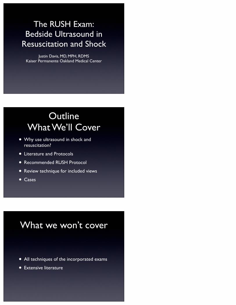
The RUSH Exam:Bedside Ultrasound in
Resuscitation and ShockJustin Davis, MD, MPH, RDMS
Kaiser Permanente Oakland Medical Center
OutlineWhat We’ll Cover
• Why use ultrasound in shock and resuscitation?
• Literature and Protocols
• Recommended RUSH Protocol
• Review technique for included views
• Cases
What we won’t cover
• All techniques of the incorporated exams
• Extensive literature
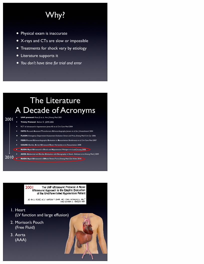
Why?
• Physical exam is inaccurate
• X-rays and CTs are slow or impossible
• Treatments for shock vary by etiology
• Literature supports it
• You don’t have time for trial and error
The LiteratureA Decade of Acronyms• UHP protocol: Rose JS et al, Am J Emerg Med 2001
• Trinity Protocol: Bahner D, JDMS 2002
• RCT of ultrasound in hypotension: Jones AE et al, Crit Care Med 2004
• FATE: Focused Assessed Transthoracic Echocardiography: Jensen et al, Eur J Anaesthesiol 2004
• FLASH: Emergency Department Assessment Evolution: Simon and Price, Emerg Med Crit Car 2006
• FEER: Focused Echocardiographic Evaluation in Resuscitation: Breitkreutz et al, Crit Care Med 2007
• CAUSE: Cardiac Arrest Ultrasound Exam: Hernandez et al, Resuscitation 2008
• RUSH: Rapid Ultrasound in Shock and Hypotension: Weingart et al, emCrit.org 2008,
• ACES: Abdominal and Cardiac Evaluation with Sonography in Shock : Atkinson et al, Emerg Med J 2009
• RUSH: Rapid Ultrasound in SHock: Perera P et al, Emerg Med Clin N Am 2010
2001
2010
1. Heart(LV function and large effusion)
2. Morison’s Pouch(Free Fluid)
3. Aorta(AAA)
The UHP Ultrasound Protocol: A Novel Ultrasound Approach to the Empiric Evaluation
of the Undifferentiated Hypotensive Patient
JOHN S. ROSE, MD,* AARON E. BAIR, MD,* DIKU MANDAVIA, MD, t AND DONNA J. KINSER, MD*
This report describes a novel sonographic protocol for the evaluation of the undifferentiated hypotensive patient. This protocol combines com- ponents of 3 sonographic applications: free fluid, cardiac, and abdomi- nal aorta into a single protocol. We believe this protocol and its under- lying principles should be a routine part of the empiric evaluation of the patient with undifferentiated hypotension or pulseless electrical activity. (Am J Emerg Med 2001;19:299-302. Copyright © 2001 by W.B. Saunders Company)
Many critical conditions in emergency medicine involve
the use of empiric protocols or techniques to facilitate the
detection of reversible and time-dependent conditions. Car-
ing for a patient with an unknown cause of hypotension can
be one of the most challenging situations in emergency
medicine. We describe the use of novel focused, goal-
directed ultrasound protocol as a part of the empiric evalu-
ation of the patient with hypotension of uncertain origin. We
have termed this sonographic evaluation the undifferenti-
ated hypotensive patient (UHP) ultrasound protocol. The
UHP protocol uses components of 3 accepted emergency
department (ED) ultrasound applications: free fluid evalua-
tion, qualitative cardiac evaluation, and abdominal aorta
evaluation. The rationale for the UHP protocol is to facili-
tate the rapid and systematic evaluation of reversible causes
of hypotension when the clinical history is limited or un-
known. We describe 3 actual cases where the UHP protocol
was pivotal in the emergency evaluation of an undifferen-
tiated hypotensive patient. A description and discussion of
the protocol follow the case presentations. We believe this
sonographic approach to be an important addition to the role
of emergency ultrasound for the practicing emergency phy-
sician.
CASE 1
A 70-year old woman is brought to the ED for evaluation
of syncope after being found on the floor by family mem-
From the * Division of Emergency Medicine, University of Califor- nia, Davis Medical Center, Sacramento, CA and tDepartment of Emergency Medicine, University of Southern California, Los Ange- les, CA.
Manuscript received December 20, 2000, accepted December 30, 2000.
Address reprint requests to John S. Rose, MD, Assistant Profes- sor of Medicine, Division of Emergency Medicine, University of California Davis, 2315 Stockton BIvd, PSSB #2100, Sacramento, CA 95817. E-maih [email protected]
Key Words: Ultrasound, hypotension, resuscitation. Copyright © 2001 by W.B. Saunders Company 0735-6757/01/1904-0012535.00/0 doi:l 0.1053/ajem.2001.24481
bets. The patient had complained earlier in the evening of a
"stomach ache" and gone to bed early. Family members
remarked to the paramedics that they heard a crash in the
woman's room and immediately went to investigate where
she was found on the floor. At the time of arrival in the ED
her blood pressure was 80/palpation, heart rate of 120
beats/min, respiratory rate of 30 breaths/min. Her pulse
oximetry was 100% on high flow oxygen. The only past
medical history available was hypotension for which she
took a single unknown prescription medication. On exami-
nation she was mumbling and disoriented. There was no
gross evidence of trauma. Her chest was clear to ausculta-
tion and her heart is regular without murmurs. Her abdomen
was obese and soft without apparent masses. Mild tender-
ness, without peritoneal signs, was noted in the midepigas-
trium. Initial standard resuscitative measures included crys-
talloid infusion. An electrocardiogram (ECG) was obtained
and was normal. While awaiting the return of the portable
chest x-ray machine, the UHP ultrasound protocol was
performed as a routine component of her hypotension eval-
uation. The hepatorenal interface view showed grossly nor-
mal anatomy without evidence of free intraperitoneal fluid.
The cardiac view revealed normal cardiac activity without
pericardial effusion. Evaluation of the aorta revealed a
6-centimeter aneurysm with associated intraluminal clot.
The vascular surgeon on call was immediately notified and
the patient was taken directly to the operating room where
her aorta was successfully repaired. The total time in the ED
was less than 20 minutes.
CASE 2
A 40-year old woman with a significant prior history of
systemic lupus erythematosus (SLE) and recurrent pulmo-
nary embolus arrived in the ED with the chief complaint of
shortness of breath. She stated that her symptoms had been
progressive over the past several days and associated with a
mild diffuse chest "tightness." Her review of systems was
notable for an absence of fever, peripheral edema, and
cough. On initial examination she was speaking in full
sentences and had normal vital signs including a room air
oxygen saturation of 99%. Her chest was clear and her heart
regular in rate and rhythm. A chest radiograph revealed mild
cardiomegaly and clear lung fields. Her ECG was within
normal limits with the exception of questionably low volt-
age. She had a precipitous drop in her blood pressure and on
re-evaluation was in obvious distress. She was cool and
diaphoretic complaining of shortness of breath. Although
the initial impression was a recurrent pulmonary embolism,
the UHP protocol was performed as part of the evaluation of
299
2001
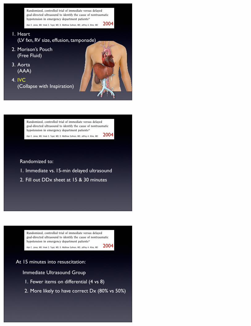
1. Heart(LV fxn, RV size, effusion, tamponade)
2. Morison’s Pouch(Free Fluid)
3. Aorta(AAA)
4. IVC(Collapse with Inspiration)
Randomized, controlled trial of immediate versus delayedgoal-directed ultrasound to identify the cause of nontraumatichypotension in emergency department patients*
Alan E. Jones, MD; Vivek S. Tayal, MD; D. Matthew Sullivan, MD; Jeffrey A. Kline, MD
Prior research has suggestedthat emergency department(ED) patients with symptom-atic hypotension in the ab-
sence of trauma have a high mortalityrate. Jones et al. (1) found that symptom-atic patients with a systolic blood pres-sure !100 mm Hg measured during am-bulance transport had an in-hospitalmortality rate of 25%. Moore et al. (2)found an 18% in-hospital mortality ratein 50 consecutive ED patients presentingwith nontraumatic, symptomatic hypo-tension. In the latter study, emergencyphysicians accurately determined final
etiology of hypotension in only 24% ofpatients (2).
Ultrasound has emerged as a usefuldiagnostic tool for a variety of emergentsituations, and both its availability andincorporation into emergency medicinepractice are increasing (3). The diag-nostic utility of ultrasound in patientswith nontraumatic, undifferentiatedhypotension has not been systemati-cally evaluated. The hypothesis of thepresent study was that the results of anemergency physician performed, goal-directed ultrasound protocol would sig-nificantly narrow the number of poten-tial viable diagnoses of patients withnontraumatic, symptomatic, undiffer-entiated hypotension and would signif-icantly improve physician accuracy inidentifying the correct diagnosis ofnontraumatic, symptomatic, undiffer-entiated hypotension.
MATERIALS AND METHODS
Patients were enrolled from July 2002through September 2003 in the ED at CarolinasMedical Center, an urban 800-bed teaching hos-
pital with "100,000 patient visits per year. Ex-plicit criteria for enrollment included the follow-ing: a) age "17 yrs; b) written agreement of twoindependent physician observers on the pres-ence of the first measured vital signs consistentwith shock (systolic blood pressure !100 mmHg or shock index (pulse rate/systolic bloodpressure) "1.0); and c) a minimum of both onesign and one symptom listed in Table 1, re-corded by each observer independently andblinded to the other observers’ observations. Ex-clusions included a) either observer found nosymptom or sign in Table 1; b) history of “lowblood pressure” reported by the patient or dis-covered from chart review; c) cardiopulmonaryresuscitation, defibrillation, or advanced cardiaclife support medications before enrollment; d)history of significant trauma to the chest orabdomen in the previous 24 hrs; e) a 12-leadelectrocardiogram diagnostic of acute myocar-dial infarction; f) presence of an obvious cause ofshock that would mandate immediate specifictreatment (active gastrointestinal bleeding,known drug overdose, external hemorrhage); g)referral from another hospital with a knowndiagnosis; h) development of signs and symp-toms of shock in the ED after the results ofdiagnostic testing (radiographic imaging andlaboratory results) were known to the treating
*See also p. 1798.From the Department of Emergency Medicine,
Carolinas Medical Center, Charlotte, NC.Presented at the annual meeting of the Society for
Academic Emergency Medicine, Orlando, FL, May2004.
Address requests for reprints to: Jeffrey A. Kline,MD, 1000 Blythe Boulevard, Charlotte, NC 28203.E-mail: [email protected]
Copyright © 2004 by the Society of Critical CareMedicine and Lippincott Williams & Wilkins
DOI: 10.1097/01.CCM.0000133017.34137.82
Objective: We examined a physician-performed, goal-directedultrasound protocol for the emergency department managementof nontraumatic, symptomatic, undifferentiated hypotension.
Design: Randomized, controlled trial of immediate vs. delayedultrasound.
Setting: Urban, tertiary emergency department, census >100,000.Patients: Nontrauma emergency department patients, aged
>17 yrs, and initial emergency department vital signs consistentwith shock (systolic blood pressure <100 mm Hg or shock index>1.0), and agreement of two independent observers for at leastone sign and symptom of inadequate tissue perfusion.
Interventions: Group 1 (immediate ultrasound) received standardcare plus goal-directed ultrasound at time 0. Group 2 (delayedultrasound) received standard care for 15 mins and goal-directedultrasound with standard care between 15 and 30 mins after time 0.
Measurements and Main Results: Outcomes included the number of
viable physician diagnoses at 15 mins and the rank of their likelihood ofoccurrence at both 15 and 30 mins. One hundred eighty-four patientswere included. Group 1 (n ! 88) had a smaller median number of viablediagnoses at 15 mins (median ! 4) than did group 2 (n ! 96, median! 9, Mann-Whitney U test, p < .0001). Physicians indicated the correctfinal diagnosis as most likely among their viable diagnosis list at 15 minsin 80% (95% confidence interval, 70–87%) of group 1 subjects vs.50% (95% confidence interval, 40–60%) in group 2, difference of 30%(95% confidence interval, 16–42%).
Conclusions: Incorporation of a goal-directed ultrasound pro-tocol in the evaluation of nontraumatic, symptomatic, undifferen-tiated hypotension in adult patients results in fewer viable diag-nostic etiologies and a more accurate physician impression offinal diagnosis. (Crit Care Med 2004; 32:1703–1708)
KEY WORDS: hypotension; shock; ultrasound; diagnosis; mortal-ity; clinical trial
1703Crit Care Med 2004 Vol. 32, No. 8
2004
Randomized to:
1. Immediate vs. 15-min delayed ultrasound
2. Fill out DDx sheet at 15 & 30 minutes
Randomized, controlled trial of immediate versus delayedgoal-directed ultrasound to identify the cause of nontraumatichypotension in emergency department patients*
Alan E. Jones, MD; Vivek S. Tayal, MD; D. Matthew Sullivan, MD; Jeffrey A. Kline, MD
Prior research has suggestedthat emergency department(ED) patients with symptom-atic hypotension in the ab-
sence of trauma have a high mortalityrate. Jones et al. (1) found that symptom-atic patients with a systolic blood pres-sure !100 mm Hg measured during am-bulance transport had an in-hospitalmortality rate of 25%. Moore et al. (2)found an 18% in-hospital mortality ratein 50 consecutive ED patients presentingwith nontraumatic, symptomatic hypo-tension. In the latter study, emergencyphysicians accurately determined final
etiology of hypotension in only 24% ofpatients (2).
Ultrasound has emerged as a usefuldiagnostic tool for a variety of emergentsituations, and both its availability andincorporation into emergency medicinepractice are increasing (3). The diag-nostic utility of ultrasound in patientswith nontraumatic, undifferentiatedhypotension has not been systemati-cally evaluated. The hypothesis of thepresent study was that the results of anemergency physician performed, goal-directed ultrasound protocol would sig-nificantly narrow the number of poten-tial viable diagnoses of patients withnontraumatic, symptomatic, undiffer-entiated hypotension and would signif-icantly improve physician accuracy inidentifying the correct diagnosis ofnontraumatic, symptomatic, undiffer-entiated hypotension.
MATERIALS AND METHODS
Patients were enrolled from July 2002through September 2003 in the ED at CarolinasMedical Center, an urban 800-bed teaching hos-
pital with "100,000 patient visits per year. Ex-plicit criteria for enrollment included the follow-ing: a) age "17 yrs; b) written agreement of twoindependent physician observers on the pres-ence of the first measured vital signs consistentwith shock (systolic blood pressure !100 mmHg or shock index (pulse rate/systolic bloodpressure) "1.0); and c) a minimum of both onesign and one symptom listed in Table 1, re-corded by each observer independently andblinded to the other observers’ observations. Ex-clusions included a) either observer found nosymptom or sign in Table 1; b) history of “lowblood pressure” reported by the patient or dis-covered from chart review; c) cardiopulmonaryresuscitation, defibrillation, or advanced cardiaclife support medications before enrollment; d)history of significant trauma to the chest orabdomen in the previous 24 hrs; e) a 12-leadelectrocardiogram diagnostic of acute myocar-dial infarction; f) presence of an obvious cause ofshock that would mandate immediate specifictreatment (active gastrointestinal bleeding,known drug overdose, external hemorrhage); g)referral from another hospital with a knowndiagnosis; h) development of signs and symp-toms of shock in the ED after the results ofdiagnostic testing (radiographic imaging andlaboratory results) were known to the treating
*See also p. 1798.From the Department of Emergency Medicine,
Carolinas Medical Center, Charlotte, NC.Presented at the annual meeting of the Society for
Academic Emergency Medicine, Orlando, FL, May2004.
Address requests for reprints to: Jeffrey A. Kline,MD, 1000 Blythe Boulevard, Charlotte, NC 28203.E-mail: [email protected]
Copyright © 2004 by the Society of Critical CareMedicine and Lippincott Williams & Wilkins
DOI: 10.1097/01.CCM.0000133017.34137.82
Objective: We examined a physician-performed, goal-directedultrasound protocol for the emergency department managementof nontraumatic, symptomatic, undifferentiated hypotension.
Design: Randomized, controlled trial of immediate vs. delayedultrasound.
Setting: Urban, tertiary emergency department, census >100,000.Patients: Nontrauma emergency department patients, aged
>17 yrs, and initial emergency department vital signs consistentwith shock (systolic blood pressure <100 mm Hg or shock index>1.0), and agreement of two independent observers for at leastone sign and symptom of inadequate tissue perfusion.
Interventions: Group 1 (immediate ultrasound) received standardcare plus goal-directed ultrasound at time 0. Group 2 (delayedultrasound) received standard care for 15 mins and goal-directedultrasound with standard care between 15 and 30 mins after time 0.
Measurements and Main Results: Outcomes included the number of
viable physician diagnoses at 15 mins and the rank of their likelihood ofoccurrence at both 15 and 30 mins. One hundred eighty-four patientswere included. Group 1 (n ! 88) had a smaller median number of viablediagnoses at 15 mins (median ! 4) than did group 2 (n ! 96, median! 9, Mann-Whitney U test, p < .0001). Physicians indicated the correctfinal diagnosis as most likely among their viable diagnosis list at 15 minsin 80% (95% confidence interval, 70–87%) of group 1 subjects vs.50% (95% confidence interval, 40–60%) in group 2, difference of 30%(95% confidence interval, 16–42%).
Conclusions: Incorporation of a goal-directed ultrasound pro-tocol in the evaluation of nontraumatic, symptomatic, undifferen-tiated hypotension in adult patients results in fewer viable diag-nostic etiologies and a more accurate physician impression offinal diagnosis. (Crit Care Med 2004; 32:1703–1708)
KEY WORDS: hypotension; shock; ultrasound; diagnosis; mortal-ity; clinical trial
1703Crit Care Med 2004 Vol. 32, No. 8
2004
Immediate Ultrasound Group
1. Fewer items on differential (4 vs 8)
2. More likely to have correct Dx (80% vs 50%)
Randomized, controlled trial of immediate versus delayedgoal-directed ultrasound to identify the cause of nontraumatichypotension in emergency department patients*
Alan E. Jones, MD; Vivek S. Tayal, MD; D. Matthew Sullivan, MD; Jeffrey A. Kline, MD
Prior research has suggestedthat emergency department(ED) patients with symptom-atic hypotension in the ab-
sence of trauma have a high mortalityrate. Jones et al. (1) found that symptom-atic patients with a systolic blood pres-sure !100 mm Hg measured during am-bulance transport had an in-hospitalmortality rate of 25%. Moore et al. (2)found an 18% in-hospital mortality ratein 50 consecutive ED patients presentingwith nontraumatic, symptomatic hypo-tension. In the latter study, emergencyphysicians accurately determined final
etiology of hypotension in only 24% ofpatients (2).
Ultrasound has emerged as a usefuldiagnostic tool for a variety of emergentsituations, and both its availability andincorporation into emergency medicinepractice are increasing (3). The diag-nostic utility of ultrasound in patientswith nontraumatic, undifferentiatedhypotension has not been systemati-cally evaluated. The hypothesis of thepresent study was that the results of anemergency physician performed, goal-directed ultrasound protocol would sig-nificantly narrow the number of poten-tial viable diagnoses of patients withnontraumatic, symptomatic, undiffer-entiated hypotension and would signif-icantly improve physician accuracy inidentifying the correct diagnosis ofnontraumatic, symptomatic, undiffer-entiated hypotension.
MATERIALS AND METHODS
Patients were enrolled from July 2002through September 2003 in the ED at CarolinasMedical Center, an urban 800-bed teaching hos-
pital with "100,000 patient visits per year. Ex-plicit criteria for enrollment included the follow-ing: a) age "17 yrs; b) written agreement of twoindependent physician observers on the pres-ence of the first measured vital signs consistentwith shock (systolic blood pressure !100 mmHg or shock index (pulse rate/systolic bloodpressure) "1.0); and c) a minimum of both onesign and one symptom listed in Table 1, re-corded by each observer independently andblinded to the other observers’ observations. Ex-clusions included a) either observer found nosymptom or sign in Table 1; b) history of “lowblood pressure” reported by the patient or dis-covered from chart review; c) cardiopulmonaryresuscitation, defibrillation, or advanced cardiaclife support medications before enrollment; d)history of significant trauma to the chest orabdomen in the previous 24 hrs; e) a 12-leadelectrocardiogram diagnostic of acute myocar-dial infarction; f) presence of an obvious cause ofshock that would mandate immediate specifictreatment (active gastrointestinal bleeding,known drug overdose, external hemorrhage); g)referral from another hospital with a knowndiagnosis; h) development of signs and symp-toms of shock in the ED after the results ofdiagnostic testing (radiographic imaging andlaboratory results) were known to the treating
*See also p. 1798.From the Department of Emergency Medicine,
Carolinas Medical Center, Charlotte, NC.Presented at the annual meeting of the Society for
Academic Emergency Medicine, Orlando, FL, May2004.
Address requests for reprints to: Jeffrey A. Kline,MD, 1000 Blythe Boulevard, Charlotte, NC 28203.E-mail: [email protected]
Copyright © 2004 by the Society of Critical CareMedicine and Lippincott Williams & Wilkins
DOI: 10.1097/01.CCM.0000133017.34137.82
Objective: We examined a physician-performed, goal-directedultrasound protocol for the emergency department managementof nontraumatic, symptomatic, undifferentiated hypotension.
Design: Randomized, controlled trial of immediate vs. delayedultrasound.
Setting: Urban, tertiary emergency department, census >100,000.Patients: Nontrauma emergency department patients, aged
>17 yrs, and initial emergency department vital signs consistentwith shock (systolic blood pressure <100 mm Hg or shock index>1.0), and agreement of two independent observers for at leastone sign and symptom of inadequate tissue perfusion.
Interventions: Group 1 (immediate ultrasound) received standardcare plus goal-directed ultrasound at time 0. Group 2 (delayedultrasound) received standard care for 15 mins and goal-directedultrasound with standard care between 15 and 30 mins after time 0.
Measurements and Main Results: Outcomes included the number of
viable physician diagnoses at 15 mins and the rank of their likelihood ofoccurrence at both 15 and 30 mins. One hundred eighty-four patientswere included. Group 1 (n ! 88) had a smaller median number of viablediagnoses at 15 mins (median ! 4) than did group 2 (n ! 96, median! 9, Mann-Whitney U test, p < .0001). Physicians indicated the correctfinal diagnosis as most likely among their viable diagnosis list at 15 minsin 80% (95% confidence interval, 70–87%) of group 1 subjects vs.50% (95% confidence interval, 40–60%) in group 2, difference of 30%(95% confidence interval, 16–42%).
Conclusions: Incorporation of a goal-directed ultrasound pro-tocol in the evaluation of nontraumatic, symptomatic, undifferen-tiated hypotension in adult patients results in fewer viable diag-nostic etiologies and a more accurate physician impression offinal diagnosis. (Crit Care Med 2004; 32:1703–1708)
KEY WORDS: hypotension; shock; ultrasound; diagnosis; mortal-ity; clinical trial
1703Crit Care Med 2004 Vol. 32, No. 8
2004
At 15 minutes into resuscitation:
• H eart
• I VC
• M orison’s
• A orta
• P neumothorax
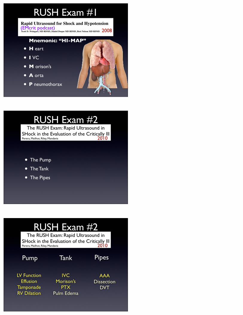
RUSH Exam #1
Home
Deep Dive
Severe Sepsis
Hypothermia
Procedures
Airway
Preox & Reox
DSI
Central Lines
Set-Up CVP
Webtext
EM Crit Care
About Emergency Critical Care
Critical Care Fellowship FAQ
About
About the Author
FAQ
Favorites
Subscription Options
Archives
Contact
EMCrit Blog - Emergency Department Critical Care
You are Here: EMCrit.org » Rapid Ultrasound for Shock and Hypotension
Rapid Ultrasound for Shock and Hypotension
Like Sign Up to see what your friends like.
Scott D. Weingart, MD RDMS, Daniel Duque MD RDMS, Bret Nelson MD RDMS
Hear the Lecture
It is now the standard of care to perform focused assessment using sonography for trauma (FAST)
early in the evaluation of a sick trauma patient. There seems to be far less urgency to use ultrasound to
evaluate the medical patient with hypotension or signs of shock. We believe that part of the reason for
this discrepancy is the lack of an accepted way to refer to the exam and a standardized sequencing. In
Get all the EMCrit Goodness as soon as it is published! Sign up for Email Updates
2008
Mnemonic: “HI-MAP”
(EMcrit podcast)
RUSH Exam #2The RUSH Exam: Rapid Ultrasound in
SHock in the Evaluation of the Critically Ill Perera, Mailhot, Riley, Mandavia
• The Pump
• The Tank
• The Pipes
2010
RUSH Exam #2The RUSH Exam: Rapid Ultrasound in
SHock in the Evaluation of the Critically Ill Perera, Mailhot, Riley, Mandavia 2010
LV FunctionEffusion
TamponadeRV Dilation
IVCMorison’s
PTXPulm Edema
AAADissection
DVT
Pump Tank Pipes
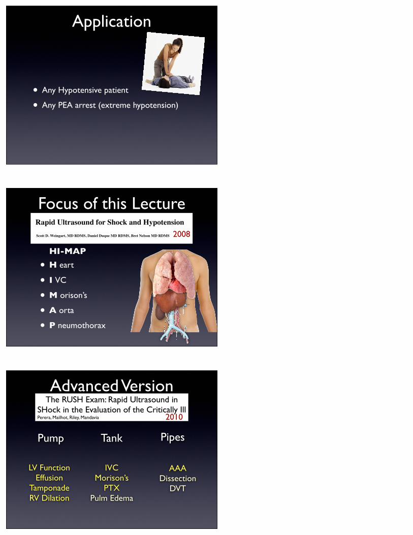
Application
• Any Hypotensive patient
• Any PEA arrest (extreme hypotension)
• H eart
• I VC
• M orison’s
• A orta
• P neumothorax
Focus of this Lecture
Home
Deep Dive
Severe Sepsis
Hypothermia
Procedures
Airway
Preox & Reox
DSI
Central Lines
Set-Up CVP
Webtext
EM Crit Care
About Emergency Critical Care
Critical Care Fellowship FAQ
About
About the Author
FAQ
Favorites
Subscription Options
Archives
Contact
EMCrit Blog - Emergency Department Critical Care
You are Here: EMCrit.org » Rapid Ultrasound for Shock and Hypotension
Rapid Ultrasound for Shock and Hypotension
Like Sign Up to see what your friends like.
Scott D. Weingart, MD RDMS, Daniel Duque MD RDMS, Bret Nelson MD RDMS
Hear the Lecture
It is now the standard of care to perform focused assessment using sonography for trauma (FAST)
early in the evaluation of a sick trauma patient. There seems to be far less urgency to use ultrasound to
evaluate the medical patient with hypotension or signs of shock. We believe that part of the reason for
this discrepancy is the lack of an accepted way to refer to the exam and a standardized sequencing. In
Get all the EMCrit Goodness as soon as it is published! Sign up for Email Updates
2008
HI-MAP
Advanced VersionThe RUSH Exam: Rapid Ultrasound in
SHock in the Evaluation of the Critically Ill Perera, Mailhot, Riley, Mandavia 2010
LV FunctionEffusion
TamponadeRV Dilation
IVCMorison’s
PTXPulm Edema
AAADissection
DVT
Pump Tank Pipes
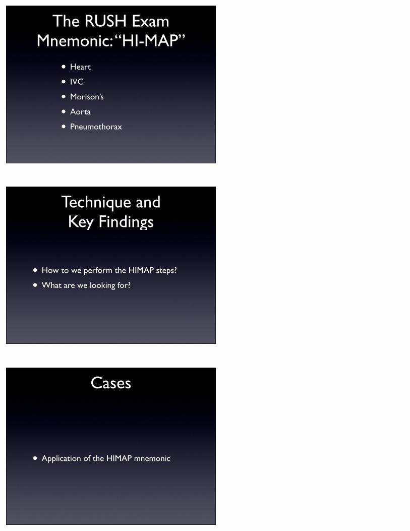
The RUSH ExamMnemonic: “HI-MAP”
• Heart
• IVC
• Morison’s
• Aorta
• Pneumothorax
Technique andKey Findings
• How to we perform the HIMAP steps?
• What are we looking for?
Cases
• Application of the HIMAP mnemonic
Cases
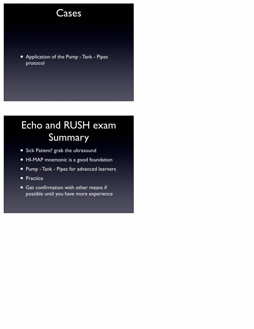
• Application of the Pump - Tank - Pipes protocol
Echo and RUSH examSummary
• Sick Patient? grab the ultrasound
• HI-MAP mnemonic is a good foundation
• Pump - Tank - Pipes for advanced learners
• Practice
• Get confirmation with other means if possible until you have more experience