Our Experience Using MobileDaRt Evolution MX8 k type
5
RAD No.86 (2019.8) 1. Hospital Overview Shiroyama Hospital (Fig. 1) is located in Habikino City in southern Osaka, in an area well-loved by history mania . The hospital stands in the Habikino Hills overlooking the Furuichi Kofungun (a cluster of tumuli), now a World Cultural Heritage Site, and the nearest train station is Furuichi Station on the Kintetsu Minami Osaka Line, an area that played host to the clash between Date Masamune and Sanada Yukimura in the famous Battle of Domyoji/ Konda during the Summer Campaign of the Siege of Osaka. Shiroyama Hospital was first established in 1978 with 156 beds on a site adjacent to Furuichi Station. In 1989 it was expanded to 299 beds, and in 2006 the hospital moved to new facilities at its current site. Operating on the hospital motto of “Shiroyama Hospital Exists for the Patients,” we are a community-based, acute care-oriented core hospital that provides 24-hour medical care with 23 outpatient departments plus a critical care department that see 12,000 emergency patients and 140,000 outpatients each year. In 2018, Shiroyama Hospital also accepted the request to become a Disaster Medical Center for Habikino City. 2. Equipment Introduction story and Selection Although computed radiography (CR) was used in our previous mobile X-ray system, an increasing number of technical problems stemming from aging equipment and the departments of radiology and medical examination wanting to improve imaging work efficiency led to the acquisition of a new mobile X-ray system with a flat panel detector (FPD), rather than the previous CR system. The previous mobile X-ray CR system required the system to carry the same number of CR cassettes as the number of images taken and the aging equipment had become less maneuverable, making work difficult for radiology technologists. Our department of radiology has 24 clinical radiology technologists of whom 9 are women. Our primary requirement for selecting a mobile X-ray system was that the equipment can be used safely and comfortably by all our staff, including female and male technologists under 150 cm and over 180 cm in height. (1) The FPD is wireless and lightweight. (2) The system itself comes with a built-in viewing monitor that is as large as possible. (3) The system can access the hospital’s radiology information system (RIS) via remote desktop on site. (4) The system allows patient authentication with a wireless bar code reader. (5) Images can be viewed and sent to the image server immediately after acquisition. (6) There is a wireless hand switch for exposures. (7) As much storage space as possible for FPD device. (8) The system is quiet during both transport and operation. (9) The external design of the equipment does not cause concern to patients. Our Experience Using MobileDaRt Evolution MX8 k type Department of Radiology, Shiroyama Hospital Hisahiro Kitai Hisahiro Kitai, R.T. Fig.1 View of Shiroyama Hospital
Transcript of Our Experience Using MobileDaRt Evolution MX8 k type

RAD
No.86 (2019.8)
1. Hospital Overview
Shiroyama Hospital (Fig. 1) is located in Habikino City in southern Osaka, in an area well-loved by history mania . The hospital stands in the Habikino Hills overlooking the Furuichi Kofungun (a cluster of tumuli), now a World Cultural Heritage Site, and the nearest train station is Furuichi Station on the Kintetsu Minami Osaka Line, an area that played host to the clash between Date Masamune and Sanada Yukimura in the famous Battle of Domyoji/Konda during the Summer Campaign of the Siege of Osaka.Shiroyama Hospital was first established in 1978 with 156 beds on a site adjacent to Furuichi Station. In 1989 it was expanded to 299 beds, and in 2006 the hospital moved to new facilities at its current site.Operating on the hospital motto of “Shiroyama Hospital Exists for the Patients,” we are a community-based, acute care-oriented core hospital that provides 24-hour medical care with 23 outpatient departments plus a critical care department that see 12,000 emergency patients and 140,000 outpatients each year.In 2018, Shiroyama Hospital also accepted the request to become a Disaster Medical Center for Habikino City.
2. Equipment Introduction story and Selection
Although computed radiography (CR) was used in our previous mobile X-ray system, an increasing number of technical problems stemming from aging equipment and the departments of radiology and medical examination wanting to improve imaging work efficiency led to the acquisition of a new mobile X-ray system with a flat panel detector (FPD), rather than the previous CR system.The previous mobile X-ray CR system required the system to carry the same number of CR cassettes as the number of images taken and the aging equipment had become less maneuverable, making work difficult for radiology technologists.Our department of radiology has 24 clinical radiology technologists of whom 9 are women. Our primary requirement for selecting a mobile X-ray system was that the equipment can be used safely and comfortably by all our staff, including female and male technologists under 150 cm and over 180 cm in height.
(1) The FPD is wireless and lightweight.(2) The system itself comes with a built-in viewing
monitor that is as large as possible.(3) The system can access the hospital’s radiology
information system (RIS) via remote desktop on site.
(4) The system allows patient authentication with a wireless bar code reader.
(5) Images can be viewed and sent to the image server immediately after acquisition.
(6) There is a wireless hand switch for exposures.(7) As much storage space as possible for FPD
device.(8) The system is quiet during both transport and
operation.(9) The external design of the equipment does not
cause concern to patients.
Our Experience Using MobileDaRt Evolution MX8 k type
Department of Radiology, Shiroyama HospitalHisahiro Kitai
Hisahiro Kitai, R.T.
Fig.1 View of Shiroyama Hospital

No.86 (2019.8)
We found MobileDaRt Evolution MX8 k type (hereinafter referred to as MobileDaRt Evolution MX8) met all our desired specifications noted above and chose it as our new mobile X-ray system (Fig. 2).
3. Hospital Environment
The electronic medical records system (hereinafter referred to as hospital information system [HIS]) at Shiroyama Hospital is made by Software Service, Inc., our image server, in-hospital distributed image viewers, and image reading and reporting system is made by PSP Corporation, and our Radiology Information System [RIS] is made by Infocom Corporation. The hospital operates a wireless local area network (LAN) that allows order information on the RIS to be downloaded wirelessly by remote control and viewed on mobile systems, and after imaging allows the resulting images to be immediately transferred to the image server from anywhere in the hospital.
4. Workflow
We mainly use MobileDaRt Evolution MX8 in hospital wards but also sometimes use it in operating rooms and emergency rooms.The imaging workflow is shown below.
(1) A doctor orders a radiography at the ward via HIS.
(2) Order information and patient information are verified in the RIS terminal screen shown on the MobileDaRt Evolution MX8 built-in monitor and the system is transported to the radiography site.
(3) At the bedside of the radiography subject, the bar code on the patient’s wristband is read using the wireless bar code reader that comes with the system. Upon reading the bar code, patient information is identified in the RIS terminal screen from the radiography schedule patient list, and after checking patient information and order details, radiography request procedures are followed on the RIS terminal screen.
(4) The radiography screen is then activated and radiography is performed.
(5) After performing radiography, an image inspection is performed immediately using the built-in monitor, and after image processing and annotation are complete, the radiography images are transmitted to the image server over the wireless LAN.
(6) After using the RIS terminal screen shown on the built-in monitor to confirm that images successfully reached the image server, an imaging termination process (post-imaging processing) is implemented.
5. Our Experience Using MobileDaRt Evolution MX8
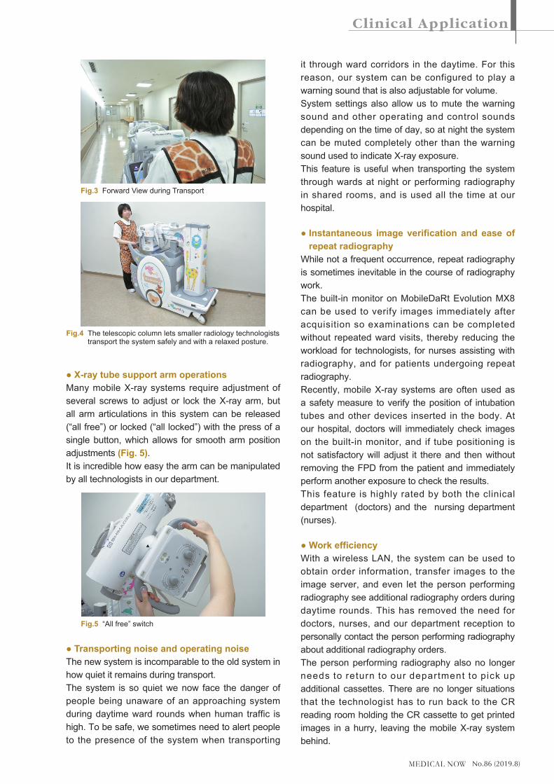
● Telescopic column and motor-assisted transportOur previous mobile X-ray system included a long, wide column that stood directly in front of the operator and obstructed their view during transport. The column forced even tall technologists to lean around the column and adopt an unnatural body position to see in front of the system when holding the transport handle, and to be very cautious when transporting the system.A distinguishing feature of MobileDaRt Evolution MX8 is its telescopic column that does not obstruct the technologist’s field of view during transport so even smaller technologists under 150 cm in height can transport the system safely through the hospital with a natural body posture (Fig.3, 4).The low center of gravity of the system combined with the smoothness of its motor-assisted drive mechanism allows all technologists, both men and women, to perform radiology rounds easily, safely, and without stress.
Fig.2 MobileDaRt Evolution MX8 k type (a) side view and (b) 19-inch touch panel viewing monitor.
(b)
(a)
No.86 (2019.8)
● X-ray tube support arm operationsMany mobile X-ray systems require adjustment of several screws to adjust or lock the X-ray arm, but all arm articulations in this system can be released (“all free”) or locked (“all locked”) with the press of a single button, which allows for smooth arm position adjustments (Fig. 5).It is incredible how easy the arm can be manipulated by all technologists in our department.
● Transporting noise and operating noiseThe new system is incomparable to the old system in how quiet it remains during transport.The system is so quiet we now face the danger of people being unaware of an approaching system during daytime ward rounds when human traffic is high. To be safe, we sometimes need to alert people to the presence of the system when transporting
it through ward corridors in the daytime. For this reason, our system can be configured to play a warning sound that is also adjustable for volume.System settings also allow us to mute the warning sound and other operating and control sounds depending on the time of day, so at night the system can be muted completely other than the warning sound used to indicate X-ray exposure.This feature is useful when transporting the system through wards at night or performing radiography in shared rooms, and is used all the time at our hospital.
● Instantaneous image verification and ease of repeat radiography
While not a frequent occurrence, repeat radiography is sometimes inevitable in the course of radiography work.The built-in monitor on MobileDaRt Evolution MX8 can be used to verify images immediately after acquisition so examinations can be completed without repeated ward visits, thereby reducing the workload for technologists, for nurses assisting with radiography, and for patients undergoing repeat radiography.Recently, mobile X-ray systems are often used as a safety measure to verify the position of intubation tubes and other devices inserted in the body. At our hospital, doctors will immediately check images on the built-in monitor, and if tube positioning is not satisfactory will adjust it there and then without removing the FPD from the patient and immediately perform another exposure to check the results.This feature is highly rated by both the clinical department (doctors) and the nursing department (nurses).
● Work efficiencyWith a wireless LAN, the system can be used to obtain order information, transfer images to the image server, and even let the person performing radiography see additional radiography orders during daytime rounds. This has removed the need for doctors, nurses, and our department reception to personally contact the person performing radiography about additional radiography orders.The person performing radiography also no longer needs to return to our department to pick up additional cassettes. There are no longer situations that the technologist has to run back to the CR reading room holding the CR cassette to get printed images in a hurry, leaving the mobile X-ray system behind.
Fig.5 “All free” switch
Fig.4 The telescopic column lets smaller radiology technologists transport the system safely and with a relaxed posture.
Fig.3 Forward View during Transport

No.86 (2019.8)
● Wireless hand switch (optional) (Fig. 6)The wireless hand switch can be used to increase the distance between the technologist and the patient when performing exposures and reduce exposure for the technologist.Previously, the hand switch cable sometimes required extra attention during radiography that involved patient assistance, but the wireless hand switch solves this problem and has proven to be extremely convenient.
● Wireless bar code reader (optional) (Fig. 7)Patients can be matched to a radiography order by reading the bar code on the wrist strap worn permanently by the patient. This helps not only to reduce work in preparation for imaging but also to prevent patient misidentification.
● Storage bin (Fig. 8)The system comes with a large amount of storage bin that is extremely useful not only for carrying grids, but also for wet wipes, plastic bags to protect FPDs, and other materials used during radiography.
● Decorated version (Fig. 9)Patients have told us that seeing the machine entering their room makes them nervous and afraid, as does seeing the large, mechanized device approaching them anywhere in the hospital.To alleviate this nervousness even just a little, the exterior of the system has been decorated with stickers of animals and dessert foods.Extending the system arm now reveals cute monkeys and also extends the giraffe’s neck.Matching this theme, the protective clothing worn by technologists operating the mobile X-ray system also has a giraffe pattern.Somewhat unexpectedly, the decorated system has been universally well-received by inpatients (including patients not being examined), patient
Fig.7 Wireless bar code reader
Fig.9 Decorated version
Fig.8 Storage space
Fig.6 Wireless hand switch

No.86 (2019.8)
attendants, visitors, and also other hospital staff who frequently smile when they see the decorated system.The decoration also attracts peoples’ attention during radiology rounds, though this is an acceptable side effect.It is perhaps worth noting that our hospital has no pediatric outpatient department or pediatric wards.
● Height-adjustable steering handle (optional) (Fig. 10)
Technologists of many different statures (male and female) perform mobile radiography work, so the ability to adjust the height of the steering handle (two levels) helps technologists who were otherwise forced to adopt an unnatural body position while moving the system, and therefore reduces the physical burden on technologists.
6. Summary
At Shiroyama Hospital, we have acquired the MobileDaRt Evolution MX8 k type (combined with a Konika Minolta CS-7) system made by Shimadzu Corporation. Changing from a CR system to an FPD system has allowed us not only to carry out radiology rounds with a single FPD but also to verify images immediately after radiography and send images to the image server from the bedside. These improvements make radiography work easier for our radiology technologists and bring substantial time
savings to mobile radiography tasks.Technologists can now complete their scheduled mobile radiography work and return to the radiology department earlier, leaving more time to deal with other tasks and thereby increasing the overall work efficiency of the radiology department.Before acquiring this system, our workflow after imaging involved scanning the imaging plate (IP) and sending the scanned image to the image server, which took around 10–15 minutes between imaging and being able to view the image on the HIS. With MobileDaRt Evolution MX8, images can now be viewed on the HIS within 1 minute of imaging, reducing significantly the time between imaging and image viewing and even improving the work efficiency of our doctors.On a full charge and assuming motor assistance is not used, in 2.5 hours of digital radiography (DR) we can acquire around 150 images at 100 kV and 4 mAs, which makes MobileDaRt Evolution MX8 extremely well-suited to a Disaster Medical Center like ours.
The irradiation field adjustment knob, irradiation field lamp switch, quick switches, and inclusion of multiple arm lock release buttons in different locations on the system also constantly remind us of the clever design details that have gone into this excellent mobile X-ray system.When it comes to Shimadzu, we would expect nothing less.
Fig.10 Driving handle height adjustment