Other Human Amoebae Entamoeba dispar/E. hartmanni: Non pathogenic; anatomically indistinguishable...
74
Other Human Amoebae • Entamoeba dispar/E. hartmanni: Non pathogenic; anatomically indistinguishable from E. histolytica.
-
Upload
abraham-ross -
Category
Documents
-
view
220 -
download
3
Transcript of Other Human Amoebae Entamoeba dispar/E. hartmanni: Non pathogenic; anatomically indistinguishable...
Other Human Amoebae
• Entamoeba dispar/E. hartmanni: Non pathogenic; anatomically indistinguishable from E. histolytica.

Entamoeba coli
Trophozoite Cyst Nucleus

Entamoeba coli• Life cycle and location identical to E. histolytica.

Entamoeba coli• Life cycle and location identical to E. histolytica.
• Most common endocommensal in people; has a worldwide distribution and 10-50% of the population can be infected in different parts of the world.

Entamoeba coli• Life cycle and location identical to E. histolytica.
• Most common endocommensal in people; has a worldwide distribution and 10-50% of the population can be infected in different parts of the world.
• Not pathogenic.

Entamoeba coli• Life cycle and location identical to E. histolytica.
• Most common endocommensal in people; has a worldwide distribution and 10-50% of the population can be infected in different parts of the world.
• Not pathogenic.
• Feeds on bacteria and any other cells available to it; does not invade tissue.
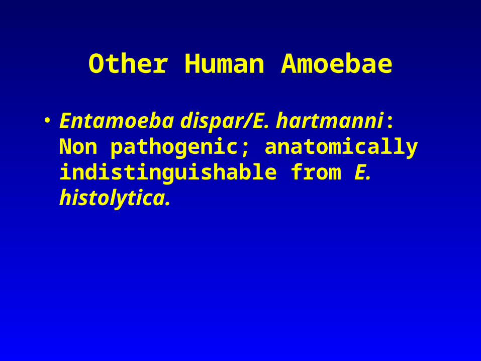
Other Hosts
Monkeys Apes
Pigs

10 m
Trophozoites 20-30 μm in diameter (15-50 µm)

10 m
A
DC
B

10 m
A
DC
B
E. coli
E. histolytica

C
10 m
Cysts 10-30 μm

Entamoeba gingivalis
Habitat: Mouth
Hosts: Humans, other primates, dogs and cats.
Prevalence is from 50 to 95%. Stage: Trophozoite, no cyst.

Entamoeba gingivalis
Trophozoite lives on the surface of teeth and gums. Feed on epithelial cells of the mouth, bacteria, food debris, and other cells available to them.

Entamoeba gingivalis Trophozoite lives on the surface of teeth and gums. Feed on epithelial cells of the mouth, bacteria, food debris, and other cells available to them.
Organisms are more common in persons with pyorrhea (gum disease) but they are not the cause of the condition.

Entamoeba gingivalis Trophozoite lives on the surface of teeth and gums. Feed on epithelial cells of the mouth, bacteria, food debris, and other cells available to them.
Organisms are more common in persons with pyorrhea (gum disease) but they are not the cause of the condition. Transmission mouth to mouth, droplet spray, or sharing eating utensils.
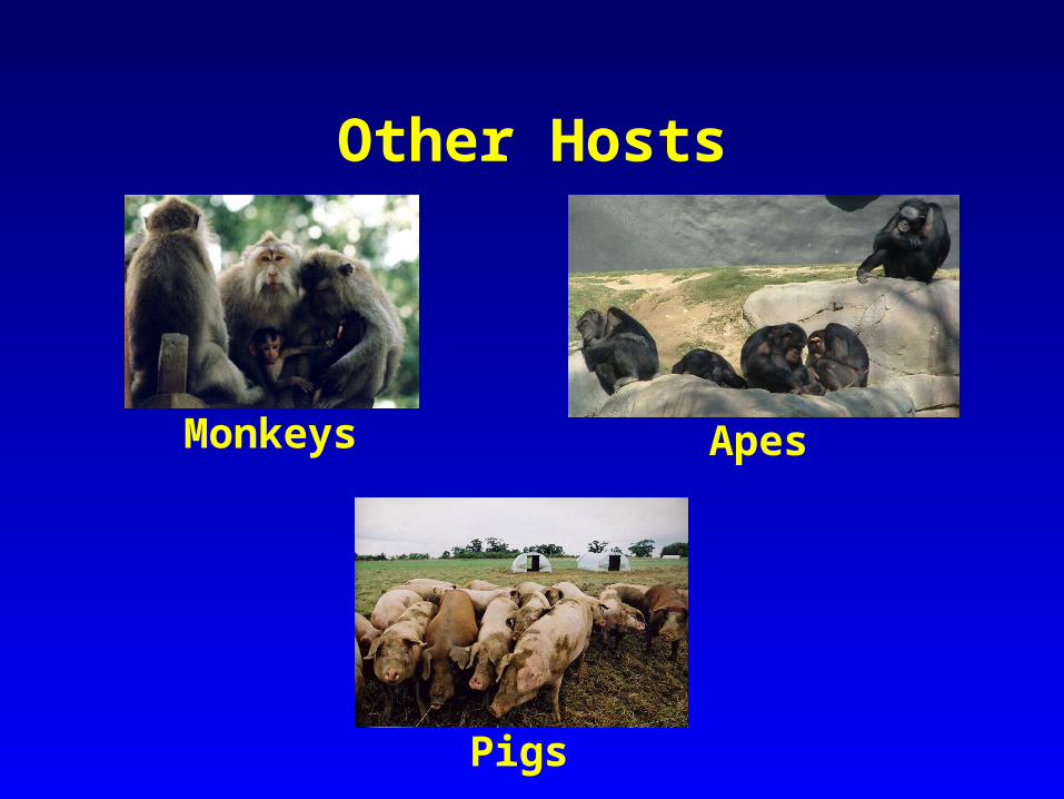
Endolimax nana “The dwarf internal slug”
Trophozoite Cyst Nucleus

Endolimax nana
• Second most common endocommensal of humans, worldwide distribution 30%.
Endolimax nana
• Second most common endocommensal of humans,
worldwide distribution 30%.
• Lives in the large intestine mainly near the cecum and feed on bacteria; non pathogenic.

Endolimax nana
• Second most common endocommensal of humans, worldwide distribution 30%.
• Lives in the large intestine mainly near the cecum and
feed on bacteria; non pathogenic.
• Also occurs in monkeys.
10 m
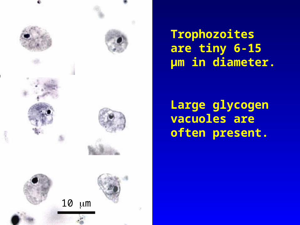
Trophozoites are tiny 6-15 μm in diameter.
Large glycogen vacuoles are often present.

10 m
Mature cyst is 5 – 14 μm in diameter; contains 4 nuclei; shape is round to elliptical
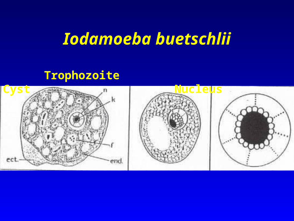
Iodamoeba buetschlii
Trophozoite Cyst Nucleus

Iodamoeba buetschlii• Not very common endocommensal in
people.

Iodamoeba buetschlii• Not very common endocommensal in people.
• Lives in the large intestine, predominantly in the cecal areas.
Iodamoeba buetschlii• Not very common endocommensal in people.
• Lives in the large intestine, predominantly in the cecal areas.
• Has a very high prevalence in pigs; 50% of pigs are infected with this amoeba in France and Egypt; pigs are probably its normal host.

10 m
Trophozoites are 9-14 μm long but may be as large as 20μm

10 m

10 m
E. nana
I. buetschlii

10 m
Cysts are 6-15 µm long and have a large glycogen vacuole.

Dientamoeba fragilis
Trophozoite No Cyst Nucleus

Dientamoeba fragilis
• LIFE CYCLE - it does not form cysts and trophozoites cannot survive passage through the small intestine.

Dientamoeba fragilis
• LIFE CYCLE - it does not form cysts and trophozoites cannot survive passage through the small intestine.
• Humans probably get infected by this endocommensal when they ingest pinworm eggs!

10 m
Trophozoites small 6-12 µm long; binucleated.

Histomonas meleagridis
Cosmopolitan parasite of Birds in the order Galiformes.
Causes a severe and often fatal disease called histomoniasis, “blackhead” in turkeys.
Only a trophozoite stage present; no cyst:
• trophozoite is irregular in shape
• may appear as an amoeboid form with pseudopodia or a flagellated form with a single flagellum
You are not responsible for all 9 topics for this parasite


Histomonas meleagridis Life Cycle
Transmission is within the egg of the cecal nematode of chickens and turkeys (Heterakis gallinarum)
You are not responsible for all 9 topics for this parasite
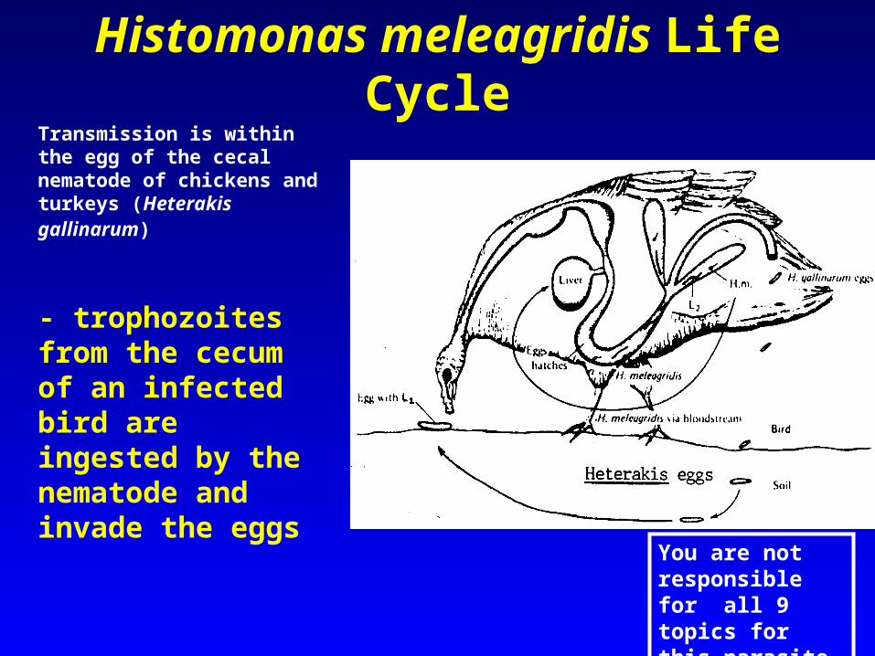
Histomonas meleagridis Life CycleTransmission is within the egg of the cecal nematode of chickens and turkeys (Heterakis gallinarum)
- trophozoites from the cecum of an infected bird are ingested by the nematode and invade the eggs
You are not responsible for all 9 topics for this parasite

Histomonas meleagridis Life CycleTransmission is within the egg of the cecal nematode of chickens and turkeys (Heterakis gallinarum)
-trophozoites from the cecum of an infected bird are ingested by the nematode and invade the eggs
- infected eggs of the nematode are released onto the soil where they are eaten by young birds during pecking activities
You are not responsible for all 9 topics for this parasite

Histomonas meleagridis Life CycleTransmission is within the egg of the cecal nematode of chickens and turkeys (Heterakis gallinarum)
- trophozoites from the cecum of an infected bird are ingested by the nematode and invade the eggs
- infected eggs of the nematode are released onto the soil where they are eaten by young birds during pecking activities
- as nematode eggs hatch in the small intestine, Histomonas trophozoites are released to invade the cecum.
You are not responsible for all 9 topics for this parasite

Pinpoint ulcersPinpoint ulcers

Habitat of trophozoites: Cecum
Pathology:
Histomonas meleagridis pathology
You are not responsible for all 9 topics for this parasite

Habitat of trophozoites: Cecum
Pathology:
Young turkeys are more susceptible to the infection than are chickens.
Mortality can reach 100% in young turkeys - millions of dollars worth of turkeys are lost to this parasite.
Histomonas meleagridis pathology

Symptoms
• Infected birds develop ruffled feathers, dark skin pigmentation, and hanging wings and tail

Look at Mr. Pro Diver!!!
Hello, The VISIBVILITY IS GREAT!!!!

This is Matt!! Holding a steering wheel of a sunken boat!! Melissa took the picture from too far away. Sorry Matt
Matt’s hose and his bubbles
Steering Wheel


Amoebic Meningitis

Naegleria fowleri
• Free-living in freshwater and soil including thermal pools; are bacteriophagous.
• They have even been isolated from bottled mineral water in Mexico.

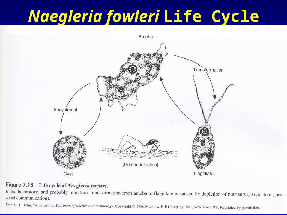
Naegleria fowleri Life Cycle

Naegleria fowleri Pathology
• After entering the nose and nasal cavities, the trophozoites migrate along the olfactory nerves, through the cribriform plate, and into the cranium.

Naegleria fowleri Pathology
• After entering the nose and nasal cavities, the trophozoites migrate along the olfactory nerves, through the cribriform plate, and into the cranium.
• Amoeboid trophozoites multiply rapidly by binary fission in the brain and cause rapid brain tissue destruction.

Naegleria fowleri Pathology
• After entering the nose and nasal cavities, the trophozoites migrate along the olfactory nerves, through the cribriform plate, and into the cranium.
• Amoeboid trophozoites multiply rapidly by binary fission in the brain and cause rapid brain tissue destruction.
• Symptoms include a headache, fever, neck rigidity, and mental confusion followed by coma and death.
• Death usually occurs from brain destruction.

Trophozoites are clustered around small vessels near the brain surface
Primary Amoebic Meningoencephalits (PAM)


Figure 1. A) Computed tomographic scan: note the right fronto-basal collection (arrow) with a midline shift right to left. B) Brain histology: three large clusters of amebic vegetative forms are seen (H-E stain, x 250). Inset: Positive indirect immunofluorescent analysis on tissue section with anti– Naegleria fowleri serum.

• Two boys, ages 7 and 9, in Tulsa, Oklahoma, died from rare parasite Saturday August 5, 2005 from infection with Naegleria fowleri.
Naegleria in Oklahoma

• Two boys, ages 7 and 9, in Tulsa, Oklahoma, die from rare parasite Saturday August 5, 2005 from infection with Naegleria fowleri.
• The two boys were not related, but both came to their doctors with symptoms of fever, hallucinations, and headaches, and despite medical care neither was able to survive the deadly infection.
• Of the 200 known cases of Naegleria infection in the past 40 years, only two people have survived. Only 24 infections were documented in the U.S. between 1989 and 2000. Six confirmed cases in Oklahoma.
Naegleria in Oklahoma

Acanthamoeba spp.
At least 5 species of Acanthamoeba have been identified in human tissues, this is one of the most common amebas in soil and freshwater.
Trophozoites occur only as amoeboid forms:

Life Cycle Stages
Free-living trophozoites and cysts occur in both the soil and freshwater.

Acanthamoeba spp.
These species cause 2 pathological effects:
1) Over 100 cases of granulomatous amebic meningoencephalitis caused by Acanthamoeba have been documented.

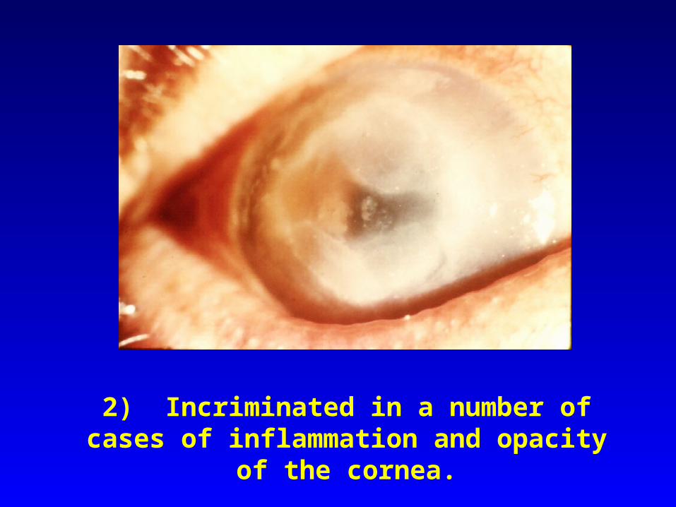
2) Incriminated in a number of cases of inflammation and opacity of the cornea.
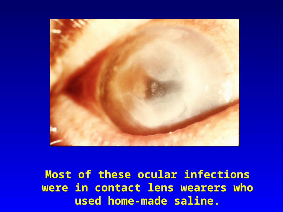
Most of these ocular infections were in contact lens wearers who used home-made saline.
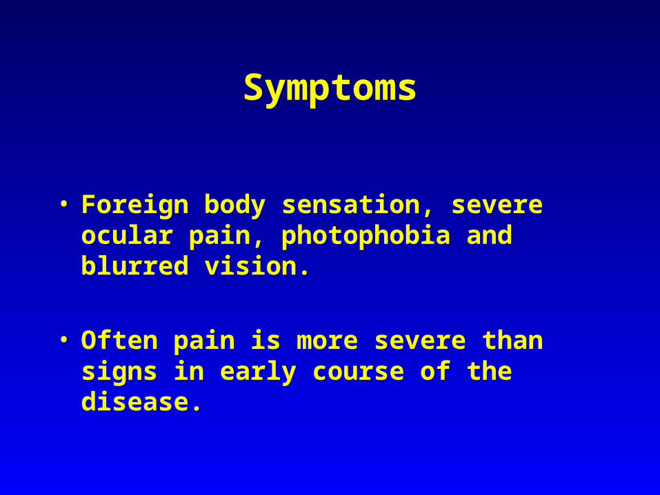
Symptoms
• Foreign body sensation, severe ocular pain, photophobia and blurred vision.
• Often pain is more severe than signs in early
course of the disease.

• Usually unilateral diffuse punctate epitheliopathy, dendritic epithelial lesion which may gradually progress to stromal infection associated with ring infiltrate formation.
Pathology

• Usually unilateral diffuse punctate epitheliopathy, dendritic epithelial lesion which may gradually progress to stromal infection associated with ring infiltrate formation.
• Enlarged corneal nerve (keratoneuritis) is
pathognomonic of the infection.
Pathology

• Usually unilateral diffuse punctate epitheliopathy, dendritic epithelial lesion which may gradually progress to stromal infection associated with ring infiltrate formation.
• Enlarged corneal nerve (keratoneuritis) is pathognomonic of the infection.
• Scleritis may be found in advanced cases.
Pathology

• Management: – Early diagnosis is a prognostic factor of a
successful outcome. – Topical anti-amoeba agents. – Penetrating keratoplasty in a severe
progressive keratitis.
Acanthamoeba spp.