
Osteomyelitis andinfectiveendocarditis · of of the diagnosis of osteomyelitis of the The...
6
Postgrad Med J (1994) 70, 885 - 890 A) The Fellowship of Postgraduate Medicine, 1994 Osteomyelitis and infective endocarditis M.E. Speechly-Dick and R.H. Swanton Department of Cardiology, The Middlesex Hospital, Mortimer Street, London WIN 8AA, UK Summary: Osteomyelitis is thought to occur as a complication of infectious endocarditis in as many as 6% of cases of endocarditis. We describe this association in three patients. Osteomyelitis may be difficult to diagnose in patients with endocarditis because symptoms such as fever, bone pain and stiffness are common to both illnesses, therefore physicians need to have a high index of suspicion to avoid missing this important complication. We recommend that patients with endocarditis and persistent or localized musculoskeletal symptoms should be investigated to exclude osteomyelitis. Plain radiographs can be normal in 50% of cases of osteomyelitis in the early stages or show only minor abnormalities, but bone scans are highly sensitive. We suggest that a bone scan is performed if radiography is unhelpful, since a diagnosis of osteomyelitis can effectively be excluded if the bone scan is normal. We advocate close follow-up of these patients with prolonged antibiotic treatment consisting of at least 6 weeks of intravenous therapy, and 3 months or longer of oral therapy. Introduction We describe three cases of osteomyelitis in patients with endocarditis, two due to Staphylococcus epidermidis and one due to Staphylococcus aureus. Musculoskeletal symptoms are very common in patients with endocarditis and this makes it difficult to determine which patients warrant investigation to exclude osteomyelitis. Up to 6% of all cases of endocarditis are complicated by osteomyelitis' and it occurs slightly more frequently in cases with Staphylococcus aureus endocarditis.2 There is no consensus of opinion on the length of treatment for this condition, and we review the literature and advocate a management regime. Case reports Case I A 64 year old man was admitted because of 3 weeks of lumbar backache, malaise and night sweats. Six years earlier he had undergone a homograft aortic valve replacement for severe aortic regurgitation due to rheumatic fever in childhood. He was pyrexial (40TC), with a tachycardia of 120/minute and a blood pressure of 180/70 mmHg. There were no stigmata of endocarditis. On auscul- tation there was a soft ejection systolic murmur at the left sternal edge radiating to the aortic area. There was a tender area in the right lower lumbar area. Neurological examination was normal. Investigations revealed a haemoglobin of 12.2 g/ dl, white cell count of 28 x 109/l (of which 86% were neutrophils), ESR 115 mm/hour, C-reactive protein (CRP) 190 mg/I, and mean cell volume (MCV) 104 fl. The chest radiograph was normal except for a mildly enlarged heart and sternal wires from previous surgery. The electrocardiogram showed sinus tachycardia, left ventricular hyper- trophy and a PR interval of 0.2 seconds. Echocar- diography revealed a normal aortic homograft with no evidence of aortic regurgitation or vegeta- tions. A diagnosis of probable infective endocarditis was made and treatment was started with intra- venous gentamicin (60 mg 12 hourly) and high dose benzyl penicillin (1.2 g 4 hourly). Blood cultures grew a penicillin-resistant Staphylococcus epider- midis from all bottles on day 3. The antibiotic therapy was changed to flucloxacillin and gen- tamicin. Thoracolumbar spine radiographs on day 2 showed extensive degenerative disease in the thoracic and lumbar spine, which was especially marked at the level of L2-L3 with anterior bridg- ing osteophytes. The temperature settled and the ESR fell to 56 but he still complained of severe back pain so a bone scan was performed (Figure 1). This sug- gested infection of the lumbar vertebrae L2 and L3. This was confirmed by magnetic resonance imaging of the thoracolumbar spine, which showed evi- Correspondence: M.E. Speechly-Dick, B.Sc., M.R.C.P., Department of Academic and Clinical Cardiology, University College London Hospitals, Grafton Way, London WC1E 6DB, UK. Accepted: 20 June 1994 by copyright. on December 31, 2019 by guest. Protected http://pmj.bmj.com/ Postgrad Med J: first published as 10.1136/pgmj.70.830.885 on 1 December 1994. Downloaded from
Transcript of Osteomyelitis andinfectiveendocarditis · of of the diagnosis of osteomyelitis of the The...
Postgrad Med J (1994) 70, 885 - 890 A) The Fellowship of Postgraduate Medicine, 1994
Osteomyelitis and infective endocarditis
M.E. Speechly-Dick and R.H. Swanton
Department ofCardiology, The Middlesex Hospital, Mortimer Street, London WIN 8AA, UK
Summary: Osteomyelitis is thought to occur as a complication ofinfectious endocarditis in as many as6% of cases of endocarditis. We describe this association in three patients. Osteomyelitis may be difficultto diagnose in patients with endocarditis because symptoms such as fever, bone pain and stiffness arecommon to both illnesses, therefore physicians need to have a high index of suspicion to avoid missing thisimportant complication. We recommend that patients with endocarditis and persistent or localizedmusculoskeletal symptoms should be investigated to exclude osteomyelitis. Plain radiographs can benormal in 50% of cases of osteomyelitis in the early stages or show only minor abnormalities, but bonescans are highly sensitive. We suggest that a bone scan is performed if radiography is unhelpful, since adiagnosis of osteomyelitis can effectively be excluded if the bone scan is normal. We advocate closefollow-up of these patients with prolonged antibiotic treatment consisting of at least 6 weeks ofintravenous therapy, and 3 months or longer of oral therapy.
Introduction
We describe three cases of osteomyelitis in patientswith endocarditis, two due to Staphylococcusepidermidis and one due to Staphylococcus aureus.Musculoskeletal symptoms are very common inpatients with endocarditis and this makes it difficultto determine which patients warrant investigationto exclude osteomyelitis. Up to 6% of all cases ofendocarditis are complicated by osteomyelitis' andit occurs slightly more frequently in cases withStaphylococcus aureus endocarditis.2 There is noconsensus ofopinion on the length oftreatment forthis condition, and we review the literature andadvocate a management regime.
Case reports
Case I
A 64 year old man was admitted because of 3 weeksof lumbar backache, malaise and night sweats. Sixyears earlier he had undergone a homograft aorticvalve replacement for severe aortic regurgitationdue to rheumatic fever in childhood.He was pyrexial (40TC), with a tachycardia of
120/minute and a blood pressure of 180/70 mmHg.There were no stigmata ofendocarditis. On auscul-
tation there was a soft ejection systolic murmur atthe left sternal edge radiating to the aortic area.There was a tender area in the right lower lumbararea. Neurological examination was normal.
Investigations revealed a haemoglobin of 12.2 g/dl, white cell count of 28 x 109/l (of which 86%were neutrophils), ESR 115 mm/hour, C-reactiveprotein (CRP) 190 mg/I, and mean cell volume(MCV) 104 fl. The chest radiograph was normalexcept for a mildly enlarged heart and sternal wiresfrom previous surgery. The electrocardiogramshowed sinus tachycardia, left ventricular hyper-trophy and a PR interval of 0.2 seconds. Echocar-diography revealed a normal aortic homograftwith no evidence of aortic regurgitation or vegeta-tions.A diagnosis of probable infective endocarditis
was made and treatment was started with intra-venous gentamicin (60 mg 12 hourly) and high dosebenzyl penicillin (1.2 g 4 hourly). Blood culturesgrew a penicillin-resistant Staphylococcus epider-midis from all bottles on day 3. The antibiotictherapy was changed to flucloxacillin and gen-tamicin. Thoracolumbar spine radiographs on day2 showed extensive degenerative disease in thethoracic and lumbar spine, which was especiallymarked at the level of L2-L3 with anterior bridg-ing osteophytes.The temperature settled and the ESR fell to 56
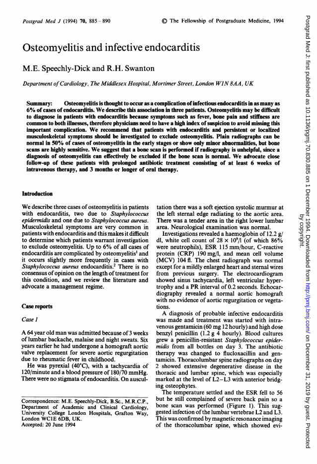
but he still complained of severe back pain so abone scan was performed (Figure 1). This sug-gested infection ofthe lumbar vertebrae L2 and L3.This was confirmed by magnetic resonance imagingof the thoracolumbar spine, which showed evi-
Correspondence: M.E. Speechly-Dick, B.Sc., M.R.C.P.,Department of Academic and Clinical Cardiology,University College London Hospitals, Grafton Way,London WC1E 6DB, UK.Accepted: 20 June 1994
by copyright. on D
ecember 31, 2019 by guest. P
rotectedhttp://pm
j.bmj.com
/P
ostgrad Med J: first published as 10.1136/pgm
j.70.830.885 on 1 Decem
ber 1994. Dow
nloaded from
886 M.E. SPEECHLY-DICK & R.H. SWANTON
Figure 1 Bone scan showing increased concentration of tracer at the level of the second lumbar vertebra (L2), whichwould be compatible with neoplasia or osteomyelitis.
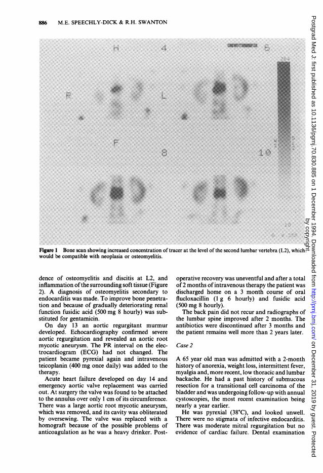
dence of osteomyelitis and discitis at L2, andinflammation ofthe surrounding soft tissue (Figure2). A diagnosis of osteomyelitis secondary toendocarditis was made. To improve bone penetra-tion and because of gradually deteriorating renalfunction fusidic acid (500 mg 8 hourly) was sub-stituted for gentamicin.On day 13 an aortic regurgitant murmur
developed. Echocardiography confirmed severeaortic regurgitation and revealed an aortic rootmycotic aneurysm. The PR interval on the elec-trocardiogram (ECG) had not changed. Thepatient became pyrexial again and intravenousteicoplanin (400 mg once daily) was added to thetherapy.Acute heart failure developed on day 14 and
emergency aortic valve replacement was carriedout. At surgery the valve was found to be attachedto the annulus over only 1 cm of its circumference.There was a large aortic root mycotic aneurysm,which was removed, and its cavity was obliteratedby oversewing. The valve was replaced with ahomograft because of the possible problems ofanticoagulation as he was a heavy drinker. Post-
operative recovery was uneventful and after a totalof 2 months ofintravenous therapy the patient wasdischarged home on a 3 month course of oralflucloxacillin (1 g 6 hourly) and fusidic acid(500 mg 8 hourly).The back pain did not recur and radiographs of
the lumbar spine improved after 2 months. Theantibiotics were discontinued after 3 months andthe patient remains well more than 2 years later.
Case 2
A 65 year old man was admitted with a 2-monthhistory of anorexia, weight loss, intermittent fever,myalgia and, more recent, low thoracic and lumbarbackache. He had a past history of submucousresection for a transitional cell carcinoma of thebladder and was undergoing follow-up with annualcystoscopies, the most recent examination beingnearly a year earlier.He was pyrexial (38°C), and looked unwell.
There were no stigmata of infective endocarditis.There was moderate mitral regurgitation but noevidence of cardiac failure. Dental examination
by copyright. on D
ecember 31, 2019 by guest. P
rotectedhttp://pm
j.bmj.com
/P
ostgrad Med J: first published as 10.1136/pgm
j.70.830.885 on 1 Decem
ber 1994. Dow
nloaded from
OSTEOMYELITIS AND ENDOCARDITIS 887
Figure 2 Magnetic resonance imaging scan showingdiscitis ofthe second lumbar vertebra (L2) with surround-ing soft-tissue inflammation indicative of osteomyelitis.
revealed an infected left lower molar (there was nohistory of recent dental treatment). Neurologicalexamination was normal, and the spine was mobileand non-tender.
Investigations revealed a haemoglobin of 10.2 g/dl, a white cell count of 8.4 x 10⁹/1 and an ESR of50 mm/hour. The chest radiograph showed car-
diomegaly with clear lung fields. Echocardio-graphy confirmed a dilated left ventricle withvolume overload, moderate mitral regurgitationbut no vegetations on the mitral valve. Bloodcultures grew Staphylococcus epidermidis, whichwas fully sensitive to penicillin.A diagnosis of probable endocarditis had been
made on admission and treatment was started withintravenous penicillin (1.2 g, 4 hourly) and gen-tamicin (80 mg, 12 hourly). When the blood cultureresults were available, intravenous flucloxacillin(500 mg, 6 hourly) was substituted for penicillin.The infected tooth was extracted with antibioticcover.The backache did not improve and a lateral
thoracic spine radiograph was performed, whichshowed slight wedging ofthe 10th thoracic vertebra(T10). A subsequent bone scan revealed denseuptake of isotope at T1O, raising the possibility ofeither malignant infiltration or infection. In view ofthe history, a diagnosis of osteomyelitis secondaryto infective endocarditis was made. The paingradually improved with bed rest and antibiotics. Arepeat lateral thoracic spine radiograph 6 weekslater showed wedging of three vertebral bodies butintact disc spaces with appearances more sugges-tive of osteoporotic collapse.
A total of 6 weeks of intravenous antibiotics anda further 2 months of oral flucloxacillin (500 mg 6hourly) and probenicid (500 mg 6 hourly) weregiven. Post-discharge follow-up was close tomonitor the resolution of the endocarditis andosteomyelitis, and to follow-up the mitral regur-gitation.The mitral regurgitation gradually deteriorated
and 9 months later the patient underwent a mitralvalve replacement. There was no evidence of arecurrence of endocarditis. Repeat thoracic spineradiographs showed gradual improvement overseveral months. The patient remains well 3 yearslater.
Case 3
A 41 year old man was admitted to anotherhospital with a 2 month history of malaise, weightloss, night sweats and diarrhoea. He was known tohave a bicuspid aortic valve but had had dentaltreatment 3 months earlier without antibiotic pro-phylaxis. He had a history of a suppurating skinwound 2 months before. One week before admis-sion he had noticed black spots on his scalp, fingersand toes. Blood cultures grew Staphylococcusaureus, and echocardiography showed a biscupidaortic valve with vegetations on the anterior valveleaflet and a dilated aortic root. A diagnosis ofinfective endocarditis was made, but despite intra-venous treatment with vancomycin and ciprofloxa-cin, his condition continued to deteriorate haemo-dynamically. He was then transferred to a car-diology unit with cardiothoracic surgery facilities.On admission he was pyrexial with splinter
haemorrhages and Osler's nodes, mild splenomeg-aly and early cardiac failure. Investigation revealeda haemoglobin of 10.4 g/dl, white cell count of10.7 x 109 and ESR of 92 mm/hour. Echocardio-graphy revealed that a fistula between the aorticroot and the right ventricle had developed, andthere was severe aortic regurgitation. The antibio-tics were changed to a combination of teicoplanin(800 mg 12 hourly) and gentamicin (80 mg 8hourly). Aortic valve replacement (AVR) with aStarr-Edwards prosthesis and patch repair of thefistula were carried out. Five days postoperativelycomplete heart block developed and a permanentWI pacemaker was inserted. He became apyrexialon antibiotics and made steady improvement. Twoweeks after surgery he developed atrial flutter andunderwent cardioversion. His renal function start-ed to deteriorate and therefore gentamicin wasdiscontinued and rifampicin was added. Threeweeks after the initial surgery, he developedworsening cardiac failure. Echocardiography re-vealed that the fistula between the aortic root andright ventricle was patent again, and that the patchhad come away. Surgical repair was attempted for
by copyright. on D
ecember 31, 2019 by guest. P
rotectedhttp://pm
j.bmj.com
/P
ostgrad Med J: first published as 10.1136/pgm
j.70.830.885 on 1 Decem
ber 1994. Dow
nloaded from
888 M.E. SPEECHLY-DICK & R.H. SWANTON
a second time and the prosthesis was also replaced.Active infection was seen around the site of thepatch closure. Two days postoperatively cardiactamponade developed requiring emergency aspira-tion. A Hickman line was inserted for the adminis-tration of long-term antibiotics.The patient had 6 further weeks of intravenous
therapy with teicoplanin following the secondoperation and steadily improved. He gained weightand participated in active physiotherapy. He com-plained of mild lumbar back pain 5 weeks aftersurgery, but there were no abnormal findings onclinical examination or on X-ray and the painresolved with mild analgesics. He was dischargedhome with very close follow-up. Unfortunately hewas readmitted with a pyrexia of 40'C and severeback pain 2 weeks later. Samples were taken forculture and he was restarted on intravenous teico-planin. Spinal radiographs revealed reduced heightand increased lucency of the 9th thoracic vertebra(T9) indicating probable osteomyelitis and this wasconfirmed by a bone scan. Blood culturesconfirmed a recrudescence of S. aureus andechocardiography showed probable vegetations onthe aortic valve, and a fistula between the aorticroot and right ventricle. The patient developedsevere acute pulmonary oedema and could not beresuscitated from a cardiac arrest. Post-mortemexamination showed that T9 was soft and sur-rounded by purulent exudate. There was activeinfection in the heart but no abscesses in the liver orspleen.
Discussion
Musculoskeletal symptoms are common in infec-tious endocarditis and this makes it difficult for theclinician to select those patients who need inves-tigation for osteomyelitis. Four papers specificallyexamined the incidence of musculoskeletal symp-toms in large groups of patients with endocarditis.Thomas et al.3 described musculoskeletal manifes-tations in 32 of 108 (29.6%) patients treated forinfectious endocarditis. These symptoms includedmyalgia, articular pain, aseptic arthritis, low backpain and osteomyelitis. Thirteen of these patients(12%) had spinal musculoskeletal symptoms andthere were four cases of osteomyelitis among them.Patients with endocarditis and musculoskeletalsymptoms did not differ from those without symp-toms in terms ofcause of infection, cardiac history,age, sex, or coexistent clinical signs. Churchill et al.Idescribed an incidence of44% for musculoskeletalsymptoms in a survey of 192 patients with endocar-ditis and in 27% these symptoms were the presen-ting feature. Osteomyelitis was found to be thecause of back pain in 6% of the 192 patients; asimilar result to that in the study by Thomas et al.3
Another large retrospective study by Meyers etal.4 of the musculoskeletal manifestations ofendocarditis examined the records of 180 cases ofproven endocarditis and found an incidence of28% ofmusculoskeletal symptoms, but they foundno cases of osteomyelitis. However, there was onepatient with disc-space narrowing and anotherwith a paravertebral abscess, and it is possible thatthese patients had undiagnosed osteomyelitis. Itwould have been difficult to diagnose osteomyelitisin retrospect if patients were not investigated for itat the time. Mansur et al.S also examined retrospec-tively the incidence ofall complications ofendocar-ditis in 300 patients who presented between 1978and 1986, and found no diagnoses ofosteomyelitis.
Therefore musculoskeletal symptoms, which canbe a prominent complaint in anyone who is unwell,are acommon feature in patients with endocarditis.The seriousness ofthe illness and the high incidenceof musculoskeletal symptoms can mean that thepossibility of osteomyelitis is overlooked. Osteo-myelitis probably occurs with a frequency ofapproximately 6%.1 It is a well-recognized comp-lication of endocarditis, and is thought to be dueeither to microembolism of bacteria or haemato-genous spread. Invasive organisms such as S.aureus are thought to result in a higher incidence ofosteomyelitis and, for example, this diagnosis wasmade in 9% of 35 patients with native valvecoagulase-negative staphylococcus.2 Mansur et al.'found that streptococci were the culprit organismfor endocarditis in 49% of 300 cases, but that therewas also a high incidence of S. aureus (20%) and S.epidermidis (5%), and all of these organisms arecapable of causing osteomyelitis.Back pain in the context of positive blood
cultures should be taken seriously and thephysician should investigate any patient with thesefindings thoroughly to exclude osteomyelitis. S.epidermidis is the most frequent cause of positiveblood cultures in hospital but only 6.3% of theseare true bacteraemia.6 However, we should notdismiss coagulase-negative staphylococci in bloodcultures as a contaminant, especially if more thanone sample is positive because S. aureus and S.epidermidis account for 60-90% of cases ofisolated osteomyelitis7 and approximately 30% ofcases of infective endocarditis.5
Fever, back pain and stiffness are the majorsymptoms of osteomyelitis and, as describedabove, these symptoms are also very common inendocarditis. Ifthe back pain is persistent or severe,it would seem sensible to investigate the patient forosteomyelitis. It takes between 2 and 8 weeks fromthe onset of osteomyelitis before there are anyobvious abnormalities on a plain radiograph.7Even then, in 50% of cases the radiographs arenormal early on or show changes consistent withdegenerative arthritis only, the rest are abnormal
by copyright. on D
ecember 31, 2019 by guest. P
rotectedhttp://pm
j.bmj.com
/P
ostgrad Med J: first published as 10.1136/pgm
j.70.830.885 on 1 Decem
ber 1994. Dow
nloaded from
OSTEOMYELITIS AND ENDOCARDITIS 889
and show narrowing of the disc space, sclerosis,erosion of the endplates and destruction of thevertebral bodies.8 Computerized tomography (CT)may also be normal in the early stages or maydisclose a reduction in disc density, an early sign ofosteomyelitis.8 Magnetic resonance imaging mayreveal abnormalities earlier than either plainradiography or CT scanning, but is not alwaysavailable. However, radionuclide bone scans witheither gallium or technetium have a very highsensitivity and are the diagnostic procedure ofchoice if plain radiographs have been unhelpful.8'9Radionuclide scanning can provide evidence ofbone infection within 48 hours of the infection; theradionuclide concentrates avidly at sites of in-creased blood flow and bone turnover. Therefore,the differential diagnosis of osteomyelitis is ofmetastatic malignancy, where lesions are usuallymultiple and scattered through the axial skeleton,arthritides and Paget's disease, which have typicaldistributions, and trauma, which can be differen-tiated by the history.'" The diagnosis of osteomyel-itis is made from the typical picture of increasedradionuclide uptake of the affected area on thebone scan in association with an appropriateclinical history. Adatepe et al.9 state that a normalbone scan excludes a diagnosis ofosteomyelitis. Werecommend that any patient in whom there is areasonable suspicion of osteomyelitis should have abone scan.
Prolonged treatment with antibiotics is neededto sterilize a bony focus, which may be a potentialsite for reinfection in a patient with an abnormalvalve. Acute haematogenous osteomyelitis can besuccessfully treated with antibiotics alone unless alate diagnosis has been made and extensive bonenecrosis has already occurred, in which case thedead bone must be surgically removed because itharbours microorganisms and is difficult tosterilize.'Recommended antibiotic regimes range in total
length from 6 weeks to 6 months. Waldvogel et al.7suggest that at least 6 weeks of parenteral
antibiotics be given to cases of isolated osteomyel-itis without endocarditis and even with this regimethey experienced a relapse rate of one case in 20.They advocate surgical debridement where there isextensive destruction of the vertebral bodies withsequestra and abscess formation and, of course, ifthere are signs of spinal cord compression. Barhamet al."1 described a case of streptococcal endocar-ditis and osteomyelitis, which resolved after 5weeks of intravenous antibiotics followed by 3weeks of oral amoxycillin and erythromycin. Acase of S. epidermidis endocarditis resulting inosteomyelitis at the level T9-TI0 was cured after acourse of4 weeks ofhigh-dose intravenous fluclox-acillin followed by 5 months of oral amoxycillin.'2In our experience of S. epidermidis endocarditisand osteomyelitis, successful cure can be achievedif treatment is continued for at least 3 months. Forexample, case 1 had a total of 8 weeks of intra-venous therapy followed by 3 months of oralflucloxacillin and fusidic acid, and case 2 had 6weeks of intravenous therapy and 2 months of oralflucloxacillin with probenecid. Case 3 had 10 weeksofintravenous antibiotics prior to discharge but nolong-term treatment. Unfortunately, osteomyelitiswas diagnosed late in case 3 and a recrudescence ofendocarditis, most likely due to local reinfectionalthough possibly from the bony focus, provedfatal.
In conclusion, we recommend that a diagnosis ofosteomyelitis is considered in any patient withinfective endocarditis and back pain, especially ifthe pain is persistent, severe or localized, and thatphysicians should have a high index of suspicionfor this condition. These patients should be inves-tigated with plain radiography and a bone scan, ifthe radiograph is normal. We also advocate thatthe treatment regime consists of at least 6 weeks ofintravenous antibiotics, and 3 months or longer ofoutpatient oral therapy with close follow-up.Patients should be encouraged to keep a tempera-ture chart at home and any recurrence of fever orback pain should, of course, be investigated.
References
1. Churchill, M.A., Geraci, J.E. & Hunder, G.G. Mus-culoskeletal manifestations of bacterial endocarditis. AnnIntern Med 1977, 87: 754-759.
2. Etienne, J. & Eykyn, S.J. Increase in native valve endocarditiscaused by coagulase negative staphylococci: an Anglo-French clinical and microbiological study. Br Heart J 1990,64: 381-384.
3. Thomas, P., Allal, J., Bontoux, D. et al. Rheumatologicalmanifestations of infective endocarditis. Ann Rheum Dis1984, 43: 716-720.
4. Meyers, O.L. & Commerford, P.J. Musculoskeletal manifes-tations of bacterial endocarditis. Ann Rheum Dis 1977, 36:517-519.
5. Mansur, A.J., Grinberg, M., Lemos de la Luz, P. & Bellotti,G. The complications of infective endocarditis. Arch InternMed 1992, 152: 2428-2432.
6. Arber, N, Militianu, A., Ben-Yehuda, A. et al. Native valveStaphylococcus epidermidis endocarditis: report of sevencases and review of the literature. Am J Med 1991, 90:758-762.
7. Waldvogel, F.A. & Vasey, H. Osteomyelitis: the past decade.N Engl J Med 1980, 7: 360-368.
8. Demers, C., Tremblay, M. & Lacourciere, Y. Acute vertebralosteomyelitis complicating Streptococcus sanguis endocar-ditis. Ann Rheum Dis 1988, 47: 333-336.
by copyright. on D
ecember 31, 2019 by guest. P
rotectedhttp://pm
j.bmj.com
/P
ostgrad Med J: first published as 10.1136/pgm
j.70.830.885 on 1 Decem
ber 1994. Dow
nloaded from
890 M.E. SPEECHLY-DICK & R.H. SWANTON
9. Adatepe, M.H., Powell, O.M., Isaacs, G.H., Nichols, K. &Cefola, R. Hematogenous pyogenic vertebral osteomyelitis:diagnostic value of radionuclide bone imaging. J Nucl Med1986, 11: 1680-1685.
10. Sutton, D. Textbook of Radiology and Imaging, 5th edn.Churchill Livingstone, Edinburgh, 1993, pp. 46-62.
11. Barham, N.J., Flint, E.J. & Mifsud, R.P. Osteomyelitiscomplicating Streptococcus milleri endocarditis. PostgradMed J 1990, 66: 314-315.
12. Gosbell, I., Gottlieb, T. & Bradbury, R. Native valveendocarditis and vertebral osteomyelitis caused by Staphylo-coccus epidermidis. Med J Aust 1992, 156: 662.
by copyright. on D
ecember 31, 2019 by guest. P
rotectedhttp://pm
j.bmj.com
/P
ostgrad Med J: first published as 10.1136/pgm
j.70.830.885 on 1 Decem
ber 1994. Dow
nloaded from


