Osteogenic mandibular distraction in Nager s Syndrome ...
10
Vol. 1, No. 1 October-December 2013 pp 44-53 Revista Mexicana de Ortodoncia CASE REPORT www.medigraphic.org.mx Osteogenic mandibular distraction in Nager’s Syndrome. Case report Distracción osteogénica mandibular en síndrome de Nager. Reporte de un caso Nubia Yadira Prado Bernal,* Norma Villanueva, § Héctor Rincón II * Former resident maxillofacial surgery «Federico Gómez» Children’s Hospital, Mexico City. § Attending Maxillofacial Surgeon «Federico Gómez» Children’s Hospital, Mexico City. II Chief of Maxillofacial Surgery «Federico Gómez» Children’s Hospital, Mexico City. This article can be read in its full version in the following page: http://www.medigraphic.com/ortodoncia RESUMEN La distracción osteogénica es actualmente utilizada para el elonga- miento tisular, gracias a la neoformación ósea que ocurre durante la separación progresiva de los segmentos después de la cortico- tomia de los mismos. Se ha utilizado con excelentes resultados en anomalías dentofaciales especialmente en hipoplasias mandibulares severas. Reportamos un paciente con síndrome de Nager, disostosis acrofacial del tipo preaxial con hipoplasia mandibular severa y agene- sias dentales, quien fue tratado con distracción mandibular a través de corticotomías en ramas mandibulares y posterior manejo ortopé- dico funcional con aparatología tipo Spring Bite. Con la presentación de este caso podemos sugerir que el manejo de la distracción man- dibular debiera ser apoyado con el uso de aparatología miofuncional. Key words: Nager’s syndrome, postaxial acrofacial dysostosis, mandibular distraction, mandibular hypoplasia and oligodonthia. Palabras clave: Síndrome de Nager, acrofacial disostosis preaxial, distraccion mandibular, hipoplasia mandibular, oligodoncia. ABSTRACT Osteogenic distraction is used nowadays for tissuelengtheningdue to the bone formation that occurs during the progressive segment separation after corticotomy, being very useful in dentofacial anomalies, especially in severe hypoplasia. We present the case report of a patient with Nager’s syndrome, acrofacial dysostosis of the preaxial type, severe mandibular hypoplasia and oligodonthia who was treated by means of distraction osteogenesiswith ramus osteotomies to lengthen the mandible. He was treated with a Spring Bite-type orthopaedic appliance after the osteotomies. We conclude that treatment with mandibular distraction should be comprehensive and supported with the use of miofuncional advices. Nager’s syndrome was described for the ルrst time by Nager and De Reynier in 1948; there have been 100 cases reported in the literature up to now. 1-3 It belongs to the vast group of otofacialmandibular disostosis such as the Treacher Collins Syndrome, Nager’s Syndrome, Pierre Robin Anomaly, Hemifacial Microsomia, among others. These are malformations associated with a hypoplasia or agenesis of the earlobe and mandibular hypoplasia among other facial deformities which can be found isolated or in association with other malformations. 3,4 Nager’s syndrome is a preaxial acrofacial disostosis that consists in facial malformations associated with radial effects (absence of the radial or tibial axis- first metacarpian and first toe). 1-5 It has a recessive autosomal genetic pattern and an alteration of the 9q32 chromosome, 1q12q21 deletion with an average neonatal birth rate of 20%, growth delay of 10% and usually normal intelligence. 2-4 With craniofacial characteristics in 25% of the cases, it presents cygomatic and maxillaryhypoplasia, severe mandibular micrognathia, outwards and downwards palpebral ルssures, absence of the lower lid eyelashes, lower lid coloboma, broad nasal bridge, depressed tip of the nose, limited mandibular movements secondary to alterations in the mandibular ramus and the temporomandibular joint, macrostomia, cleft lip and palate, soft palate agenesis, short soft palate, high and narrow palate, dysplasic earlobes, atresia of theear meatus, conductive deafness, enamel hypoplasia and oligodonthia. In the muscular skeletal system, it is characterized by radial anomalies, 75% of them being radial hypoplasia or aplasia, sinostosis of the carpal bones, absence of the 5 th metacarpian, agenesis of toes and anomalies of pelvic limbs. Cardiovascular anomalies such as Fallot tetralogy www.medigraphic.org.mx Æ 4235 Wpkxgtukfcf Pcekqpcn Cwv„pqoc fg Ofizkeq. Hcewnvcf fg Qfqpvqnqi‡c0Vjku ku cp qrgp ceeguu ctvkeng wpfgt vjg EE D[/PE/PF nkegpug *jvvr<11etgcvkxgeqooqpu0qti1nkegpugu1d{/pe/pf16021+
Transcript of Osteogenic mandibular distraction in Nager s Syndrome ...

Vol. 1, No. 1 October-December 2013
pp 44-53
Revista Mexicana de Ortodoncia
CASE REPORT
www.medigraphic.org.mx
Osteogenic mandibular distraction in Nager’s Syndrome.
Case report
Distracción osteogénica mandibular en síndrome de Nager.
Reporte de un caso
Nubia Yadira Prado Bernal,* Norma Villanueva,§ Héctor RincónII
* Former resident maxillofacial surgery «Federico Gómez»
Children’s Hospital, Mexico City.§ Attending Maxillofacial Surgeon «Federico Gómez» Children’s
Hospital, Mexico City.II Chief of Maxillofacial Surgery «Federico Gómez» Children’s
Hospital, Mexico City.
This article can be read in its full version in the following page:http://www.medigraphic.com/ortodoncia
RESUMEN
La distracción osteogénica es actualmente utilizada para el elonga-
miento tisular, gracias a la neoformación ósea que ocurre durante
la separación progresiva de los segmentos después de la cortico-
tomia de los mismos. Se ha utilizado con excelentes resultados en
anomalías dentofaciales especialmente en hipoplasias mandibulares
severas. Reportamos un paciente con síndrome de Nager, disostosis
acrofacial del tipo preaxial con hipoplasia mandibular severa y agene-
sias dentales, quien fue tratado con distracción mandibular a través
de corticotomías en ramas mandibulares y posterior manejo ortopé-
dico funcional con aparatología tipo Spring Bite. Con la presentación
de este caso podemos sugerir que el manejo de la distracción man-
dibular debiera ser apoyado con el uso de aparatología miofuncional.
Key words: Nager’s syndrome, postaxial acrofacial dysostosis, mandibular distraction, mandibular hypoplasia and oligodonthia.
Palabras clave: Síndrome de Nager, acrofacial disostosis preaxial, distraccion mandibular, hipoplasia mandibular, oligodoncia.
ABSTRACT
Osteogenic distraction is used nowadays for tissuelengtheningdue
to the bone formation that occurs during the progressive segment
separation after corticotomy, being very useful in dentofacial
anomalies, especially in severe hypoplasia. We present the case
report of a patient with Nager’s syndrome, acrofacial dysostosis of
the preaxial type, severe mandibular hypoplasia and oligodonthia
who was treated by means of distraction osteogenesiswith ramus
osteotomies to lengthen the mandible. He was treated with a
Spring Bite-type orthopaedic appliance after the osteotomies.
We conclude that treatment with mandibular distraction should
be comprehensive and supported with the use of miofuncional
advices.
Nager’s syndrome was described for the ル rst time
by Nager and De Reynier in 1948; there have been
100 cases reported in the literature up to now.1-3
It belongs to the vast group of otofacialmandibular
disostosis such as the Treacher Collins Syndrome,
Nager’s Syndrome, Pierre Robin Anomaly, Hemifacial
Microsomia, among others. These are malformations
associated with a hypoplasia or agenesis of the
earlobe and mandibular hypoplasia among other
facial deformities which can be found isolated or in
association with other malformations.3,4
Nager’s syndrome is a preaxial acrofacial disostosis
that consists in facial malformations associated with
radial effects (absence of the radial or tibial axis-
first metacarpian and first toe).1-5 It has a recessive
autosomal genetic pattern and an alteration of the
9q32 chromosome, 1q12q21 deletion with an average
neonatal birth rate of 20%, growth delay of 10% and
usually normal intelligence.2-4
With craniofacial characteristics in 25% of the
cases, it presents cygomatic and maxillaryhypoplasia,
severe mandibular micrognathia, outwards and
downwards palpebral ル ssures, absence of the lower
lid eyelashes, lower lid coloboma, broad nasal
bridge, depressed tip of the nose, limited mandibular
movements secondary to alterations in the mandibular
ramus and the temporomandibular joint, macrostomia,
cleft lip and palate, soft palate agenesis, short soft
palate, high and narrow palate, dysplasic earlobes,
atresia of theear meatus, conductive deafness, enamel
hypoplasia and oligodonthia. In the muscular skeletal
system, it is characterized by radial anomalies, 75%
of them being radial hypoplasia or aplasia, sinostosis
of the carpal bones, absence of the 5th metacarpian,
agenesis of toes and anomalies of pelvic limbs.
Cardiovascular anomalies such as Fallot tetralogy
www.medigraphic.org.mx
Æ"4235"Wpkxgtukfcf"Pcekqpcn"Cwv„pqoc"fg"Ofizkeq."Hcewnvcf"fg"Qfqpvqnqi‡c0Vjku"ku"cp"qrgp"ceeguu"ctvkeng"wpfgt"vjg"EE"D[/PE/PF"nkegpug"*jvvr<11etgcvkxgeqooqpu0qti1nkegpugu1d{/pe/pf16021+""

Revista Mexicana de Ortodoncia 2013;1 (1): 44-53
45
www.medigraphic.org.mx
and/or ventricular septum defect may be present.1-5
It can also be associated with vesicoureteral reレ ux or
renal agenesis.3
Acral deformities associated with a facial disostosis
allow it to be differentiated from the Treacher Collins
syndrome, the Nager syndrome and other dysplasiasof
the 1st and 2nd facial arch (Table I).3,4
OSTEOGENIC DISTRACTION
Osteogenic distraction is a method for bone
lengthening that allows the correction of deformities
and bone deル ciencies with the subsequent correction
of the soft tissues6 by means of a distracting appliance.
It was first used by Dr. Codevilla in 1905, when
he performed femur osteotomies. This technique
remained forgotten for several decades until Dr.
Ilizarov in 1950 made it popular in the field of
trauma and orthopedics.8,9 Its clinical and systematic
application in craniofacial deformities began with
McCarthy in 19929-11mainly by using it in children with
hemifacial microsomias for mandibular distraction.11-14
Its indications have broadened for the correction
of facial asymmetries of diverse etiologies such as
severe maxillaryand mandibular retrognathias.9,10
Osteogenic distraction is a biological procedure
of new bone formation by applying constant traction
forces during a period of time. These forces are applied
with a distraction device on a bone area that has been
previously weakened by corticotomy.6-8 Thedistraction
device is an expansion screw that has been universally
graduated in such a way that every 360 degree turn
will provide a 0.5 mm18,19 movement; all this process
is under biological principles such as: vascular
preservation, adequatelatency period, distraction
rhythm and consolidation period. During this last phase
the objective is to keep the bone segment immobile to
achieve a correct organization and condensation of the
elements that will offer the characteristics of resistance
to the newly formed tissue.6,7,18
Physiologically, the process of distraction begins
when the loading stimulus is detected by the osteoblast
thus triggering a fast and continuous signaling cascade;
in this process the bone growth and differentiation is
established, the osteoblastic proliferation is followed
by cell differentiation and ル nally by the mineralization
of the extracellular matrix; also, speciル c factors have
been identiル ed associated with the beta 1 transforming
growth factor (TGFB-1), the insulinic growth factor( IGF-
1) and the E2 prostaglandin (PGE2). The created bone
gap is initially filled by fibrillar connective tissue with
collagen ル bers oriented parallel to the force vector of
the distraction forces.6-8
Once the tissue neoformation objective has been
accomplished, a tissue regeneration phase of this
newly formed tissue follows.18,19
The success of the distraction wil l depend
on numerous factors such as: small incisions,
preservation of the periostium and vascularity, latency
period without distraction of 5 to 7 days, an expansion
rhythm of 1mm once a day, a stabilization period or
consolidating phase of 8 to 12 weeks and finally, a
remodeling period.18
On this last period one can have more certainty
on the formation of new bone tissue as well as in the
histodistraction; in this stage the distraction appliance
may be removed. Equally important is to verify this
process by periodical imagetechniques to monitor the
correct function and evolution of the distraction.19-22
Difル culties have been found such as the distractor
selection, the determination of the direction of
the vector, the site for the osteotomy and patient
cooperation.23,24
The orthodontist should be present during the
complete process of study and treatment of these
patients, playing an important role in the diagnosis,
treatment planning and postsurgical management.
The wide variety of mandibular anomalies associated
with a syndromic deformity makes it difル cult to predict
the treatmentresult, in spite of the surgical, orthodontic
and physiotherapeutic management. Alsorelapse
appears to be inevitable and overcorrection may
not compensate central growthalterationsand poor
muscular function.Nevertheless, osteogenic distraction
has proved to be the most useful method for solving
breathing and swallowing problems in patients with
severe mandibular hypoplasia asides of improving
facial esthetics.9,10
In mandibular retrognathias there is a severe
hypoplasia of the mandibular ramus, body and
chin so by creating new bone in the posterior part
of the mandible (body and ramus) a more anterior
positioning of the mandible is obtained,however a
moreeffective chin is not always obtained.13, 14Once
growth has ended the convenience of performing only
a mentoplasty or mandibular osteotomies must be
reconsidered to achieve the esthetic results.
It is so that osteogenic distraction has quickly
become the treatment of choice in craniofacial
syndromes with severe mandibular deformities
because it is possible to perform during childhood
opposite to conventional treatments which may only be
performed upon completion of growth; this statement
has been controversial due to the fact that multiple
studies also report successful results by performing
mandibular osteotomies in children.8,19,25

Prado BNY et al. Osteogenic mandibular distraction in Nager's Syndrome
46
www.medigraphic.org.mx
Table I. Nager’s syndrome associated malformations compared with others with similar phenotype.
Malformation Propositus Sx Nager Sx Miller Sx. 1 and 2 brachial arch
Cranium yes 25 % yes 10% plagiocephalic
Zygomatic bone , maxillary and
mandibular hypoplasia
yes,
mandible
yes,
mandible
yes zygomatic,
mandibular
65% skeletal asymmetry Temporal, zy-
gomatic, ramus y condilar hypoplasia
Ectropion no no yes Blepharoptosis
Long philtrum yes no yes no
Inclined palpebral ル ssures yes 100% no Narrow palpebral ル ssures
35% epibulbar Tumors
20 % lower lid colobomaLack of eyelashes no 80% no
Lower lid coloboma no 50% yes
Broad nasal bridge with descend-
ed tip of the nose
yes constant no no
Mandibular Ankylosis no 25% no yes
Macrostomia no 20% no yes
Cleft palate no 60% no 15 %
Soft palate agenesis or partial
agenesis
no 60% no no
Short soft palate yes 60% constant 35% paralysis VF insufル ciency
High and narrow palate yes 60% constant no
Submucous cleft palate no 60% constant no
Biル d uvula no 60% constant no
Cleft lip no 10% constant no
Hypoplasia oligodonthia yes constant No no
Dysplasic earlobes yes 80% constant wine-
glass shape65 % dysmorphic
earlobes, microthia,
anothia, preauricular
appendicis o ル ssures
Hypoplasia, helix and tragus no constant constant
CAE Atresia yes 85% constant
Conductive deafness yes 85% constant 15%
Muscular skeletal system
Radial abnormalities yes 75% no 10%
Radial hypoplasia or aplasia no 25% no
Carpal bones synostosis no constant yes yes
Absence of the 5 metacarpian no no yes no
Toe agenesis no no constant no
Pelvic abnormalities yes constant yes
Growth delay yes 10 % no
Cardiovascular System 5 a 58 %
Fallot Tetralogy no frequent no 65%
Ventricular-septal defect no frequent no yes
Genitourinary System no frequent no no
Vesicular uretral reレ ux no frequent no yes
Renal agenesis no frequent no no
Central Nervous System Facial paralysis
Normal Intelligence yes yes no 5 a 15 % Mental deル ciency
Learning difル culties yes constant no yes
Cervical vertebrae fussion no no no 60%
Costal abnormalities no no no 30 %
Genetics 9q32 deletion
1q12-q21
5p del 6 q, trisomy 7 mosaicism, del 8q

Revista Mexicana de Ortodoncia 2013;1 (1): 44-53
47
www.medigraphic.org.mx
Este documento es elaborado por Medigraphic
Finally, inaddition to being controversial it is
difル cult to predict that a mandibular distraction during
childhood will definitely substitute an advancement
osteotomy in the adult.10,11,13
MANDIBULAR OSTEOTOMIES
Poswillo and Obwegeser in 1974 stated that
surgical trauma in children may alter the mandibular
functional matrix and interfere insubsequent facial
growth.26,27 However, Converse, Horowitz, Coccaro and
Woodsmith in 1973 recommended mandibularsurgery
in children pursuing the following objectives:
1. To improve mandibular symmetry by performing
bilateral osteotomies in the ramus during the mixed
dentition.
2. To provide maxil lary growth in response to
mandibular growth.
3. To provide an adequate height of the ramus by
using an interocclusal splint.
4. By expanding the facial skeleton early,the soft
tissues will respond adequately.26,27
In 1941 Converse and Rushton reported the first
mandibular osteotomies in children using horizontal
osteotomies performed superior to the inferior dental
Figure 1a. Initial phase. Notice the characteristic face of Nager’a Syndrome. 1b. Profile view. 1c. Post mandibular
distraction control 1 year later. 1d. Profile view 1 year later. 1e. Post mandibular distracion control 2 years later. 1f.
Profile view 2 years later.
a. c. e.
b. d. f.

Prado BNY et al. Osteogenic mandibular distraction in Nager's Syndrome
48
www.medigraphic.org.mx
nerve and placing interpositional iliac grafts after
the placement of interocclusal splints that increased
vertical dimension and thus the augmentation of the
mandibular ramus.26
Osborne supported the benefits of mandibular
osteotomies performed prior to six years of age. He
stated that an early surgery on the mandible provides
an opportunity for normal development of the maxilla
which is also affected by a hypoplasic mandible.26-28
In 1970 Delaire recommended mandibular surgeries
even at an earlier stage, between the ages of 4 to 6
elongating a short ramus with inverted L osteotomies
and placing a rib interpositional graft.26-29
CASE REPORT
We present an eight-year-old male patient
diagnosed with acrofacial dysplasia compatible with
Nager’s syndrome with characteristic phenotype and
the following relevant findings: inclined palpebral
fissures, broad nasal bridge, severe mandibular
hypoplasia, atresia of the acoustic meatus, conductive
deafness, protruded ear lobes, o l igodonthia,
excessively short soft palate, brachydactilia and pes
cavus (claw foot) (Figure 1).2,4,5
An interdisciplinary consult with our serviceis
performed for the evaluation and management of the
severe mandibular hypoplasia and the oral hypometria
of 20 mm.
A complete study of the case is performed with a
clinical facial esthetic analysis and lateral cefalometry
in which a severe mandibular hypoplasia with a
delayed growth is evident. Temporomandibular joint
pathology is discarded (Figure 2).
The patient enters a protocol for mandibular
osteogenic distraction in which placement of bilateral
extraoral mandibular distractors is suggested. A
distraction for a total mandibular advancement of 25
mm is planned (Figure 3).
SURGICAL TECHNIQUE
The procedure is begun under balanced general
anesthesia. The patient presented a difficult airway
so the use of nasoル broendoscopy was required for an
optimum nasotracheal intubation.
a.
b. c.
Figure 2.
Radiographic study where the
short mandibular, ramus and the
oligodonthia can be observed.
2a. Panoramic radiograph.
2b. PA radiograph. 2c. Lateral
cephalogram.

Revista Mexicana de Ortodoncia 2013;1 (1): 44-53
49
www.medigraphic.org.mx
Asepsis of the intervention area was performed and
sterile ル elds were placed.
Throughan intraoral approach, a 2 cm incision was
performedin the oblique line region, a mucoperiosteal
flap was dissected and ramus corticotomies were
marked using an oscillating saw (Stryker Corporation,
Kalamazoo, Mich. USA). On both sides of the
osteotomy two 2.4 x 30 mm bicortical intraosseous
screws (w. Lorenz-Bioment Microル xation HTX-Drive
JacksonvilleFL)were placed percutaneously and
attached to the external one-way distractor (25 mm.
Eby fix w. Lorenz-Bioment Microfixation HTX-Drive
Jacksonville FL). The same procedure is performed on
the other side (Figure 3).
The position of the screws was determined prior to
the surgical procedure taking underconsideration the
degree of mandibular shortening, the location of the
tooth germ and the prediction for mandibular growth
(Figure 3b).
The distraction process is begun on the 5th day
post-surgery, at a 1 mm per day rate for 21 days with a
consolidation period of 8 weeks.
Mandibular growth, occlusion and facial symmetry
were assessed and regular radiographic controls
were performed as well. Myofunctional therapy was
continued due to the oral hypometria an also the
management with excursive movements with a spring
bite-type appliance (Figures 4 and 5).
RESULTS
The final result was assessed at the endof the
distraction period, upon removal of the distractor,
1 year and 2 years after the procedure (Figure 1).
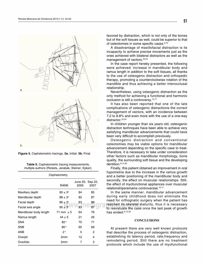
Cephalometric tracings were performed and they
showed an overall improvement of the mandibular
position. An important mandibular advancement was
obtained until a 4 mm overjet was reached (Figure 5).
A proル le improvement was accomplished, obtaining
a mandibular advancement of 20 mm and a 36 mm
mouth opening. Due to the use ofmyofunctional
appliances astimulus for mandibular growth was
evident and an adequatevertical dimension was
obtained. By doing so, horizontal growth was
accomplished despite the use of a one-way distraction
in the ramus and the fact that the patient presented
dental agenesias which prevented an adequate
interocclusal relationship (Figures 1e and f, 6).
Facial characteristics and interocclusal relationship
as well as the anterior guidance were satisfying,
contributing to the esthetic improvement of the patient
(Figures 1 c, d, f, and 6) (Table II).
a. b.
c. d.
Figure 3.
Surgical procedure. 3a. Corticotomy
of the ramus. 3b. Extraoral
distractors in position. 3c. PA
radiograph with full distraction.
3d. Panoramic radiograph before
distractors removal.

Prado BNY et al. Osteogenic mandibular distraction in Nager's Syndrome
50
www.medigraphic.org.mx
Figure 4.
Postsurgical radiographic control.
The patient remains under growth and development
control with the intention of overcorrecting his overjet,
stimulating mandibular growth and trying to create
more interarch space foreseeing the subsequent
rehabilitation by means of endoosseous dental
implants thus achieving a normal vertical dimension
and restoring masticatory function.
DISCUSSION
Mandibular hypoplasia is the most commonly found
dentofacial deformity.11,13,14
Severe mandibular def iciency maybe non-
syndromic, an isolated finding or a morphological
component of some dentofacial anomaly, in this case
speciル cally, Nager’s syndrome among others.1-5
Functional consequences of severe mandibular
hypoplasia includeairway obstruction, obstructive
sleep apnea, speech and feeding difル culties and many
times, lack of psychosocial adaptation.11,13,14
On the long term, children who are affected
by this condition may suffer a delay in growth,
cardiopulmonary changes, (pulmonary hypertension
and rightcardiac failure) and in some cases, death.9,10
For many years this dentofacial deformity has
been treated withosteotomies of the mandibular body
and ramus and interpositional graft placement with
acceptable results although some authors state that
such osteotomies may alter the functional matrix of the
mandible.26,17,28,29 Additionally, it is known that mandibular
advancements of more than 10 mm. with sagittal and
inverted L osteotomy techniques are unpredictable and
their long term stability may be compromised.25,30
Since its introduction, osteogenic distraction
hasprovedto possess many advantages for the
treatment of severe mandibular hypoplasias, especially
when it comes to patients with syndromic-type
dysgnathias in whom structural anatomy is altered18,19
and the magnitude of the corrective treatment is far
greater. In the same manner, the postsurgical stability
Revista Mexicana de Ortodoncia 2013;1 (1): 44-53
51
www.medigraphic.org.mx
Table II. Cephalometric tracing measurements,
multiple authors (Rickets, Jarabak, Steiner, Epker).
Cephalometry
RANK
June 20,
2005
Sep 22,
2007
Maxillary depth 90 ± 3o 84 85
Mandibular depth 88 ± 3o 82 87
Facial depth 86 ± 3o 83 86
Facial axis angle 90 ± 3o 83 87
Mandibular body length 71 mm ± 5 64 76
Ramus length 44 ± 5 21 26
SNA 82 o 70 71
SNB 80 o 65 69
ANB 2 o 5 2
Overjet 2mm 12 4
Overbite 2mm 7 3
a.
b.
Figure 5. Cephalometric tracings. 5a. Initial. 5b. Final.
favored by distraction, which is not only of the bones
but of the soft tissues as well, could be superior to that
of osteotomies in some speciル c cases.9,10
A disadvantage of maxillofacial distraction is its
incapacity to achieve precise movements just as the
ones achieved with bilateral distractors as well as the
management of vectors.23,24
In the case report hereby presented, the following
were achieved: increase in mandibular body and
ramus length in addition to the soft tissues, all thanks
to the use of osteogenic distraction and orthopedic
therapy, promoting a counterclockwise rotation of the
mandible and thus achieving a better interocclusal
relationship.
Nevertheless, using osteogenic distraction as the
only method for achieving a functional and harmonic
occlusion is still a controversy.15,17
It has also been reported that one of the late
complications of osteogenic distractionis the correct
management of vectors, with an incidence between
7.2 to 8.8% and even more with the use of a one-way
distractor.23,24
In children younger than six years old, osteogenic
distraction techniques have been able to achieve very
satisfying mandibular advancements that could have
been very difル cult to accomplish previously.
Osteogen ic d is t rac t ion and convent iona l
osteotomies may be viable options for mandibular
advancement depending on the speciル c case to treat.
Therefore, it is necessary to take under consideration
other factors such as mandibular morphology, bone
quality, the surrounding soft tissue and the developing
dentition.9-14,30
Finally, this patient obtained an improvement in oral
hypometria due to the increase in the ramus growth
and a better positioning of the mandibular body and
secondly, the effect on muscular relationships. Still,
the effect of myofunctional appliances over muscular
relationshipsremains controversial.15-17
In the same manner, mandibular advancement
during early childhood does not eliminate the
need for orthognatic surgery when the patient has
reached its skeletal maturity, thus it is necessary
to reevaluate the case once the last peak of growth
has ended.9,10,30
CONCLUSIONS
At present there are very well known protocols
that describe the process of osteogenic distraction,
establishing its latency period, rate,frequency and
remodeling period. Still there are no treatment
protocols which include the use of myofunctional

Prado BNY et al. Osteogenic mandibular distraction in Nager's Syndrome
52
www.medigraphic.org.mx
a. b.
c.
d.
Figure 6.
Final occlussion control. 6a y b.
Monomaxillary and bimaxillary
Spring bite. 6c. Overjet and overbite
appliances at an early stage after performing the
mandibular distractions.
This type of treatment must be considered in patients
with severe mandibular deficiencies, significant oral
hypometria, oligodonthia and lack of appropriate
interocclusal relationships thatwould allow an adequate
vertical dimension. All of these factors contribute to a
more difル cult direction of the vector, even more if we are
dealing with one-way distractors.23,24
In this specific case, the patient will remain under
growth and development control so that once the last
peak of growth has concluded we can reassess the
mandibular projection and determine the need for
mandibularadvancement osteotomies or just a sliding
mentoplasty for better chinprojection.Equally importantis
the need for an adequate interocclusal relationship so
that endoosseous dental implants can be placed.
ACKNOWLEDGEMENTS
To all of my teachers and a special recognition to
Dr. Juan Carlos López Noriega and Dr. Rafael Ruiz

Revista Mexicana de Ortodoncia 2013;1 (1): 44-53
53
www.medigraphic.org.mx
for their invaluable support during the entire process of
my professional education.
REFERENCES
1. Bowen P, Harley F. Mandibulo-facial dysostosis with limb
malformations (Nager’s acrofacial dysostosis). Birth Defects Orig
Art Ser. 1974; X(5): 109-115.
2. Zori R, Gray B, Bent-Williams A, Zackowski J. Preaxial acrofacial
dysostosis (Nager syndrome) associated with an inherited and
apparently balanced X;9 translocation: prenatal and postnatal
late replication studies. Am J Med Genet. 1993; 46: 379-383.
3. Gorlin RJ, Cohen M. Syndromes of the head and neck. 4ta Ed.
OXFORD, University Press; 2001.
4. Burglen L, Soupre V, Diner M. Dysplasies oto-mandibulaires :
génétique et nomenclature des formes syndromiques. Ann Chir
Plast Esthét. 2001; 46: 400-409.
5. Herrmann B, Karzon R, Molter D. Otologic and audiologic
features of Nager acrofacial dysostosis. International Journal of
Pediatric Otorhinolaryngology. 2005; 69: 1053-1059.
6. Swennen S, Dempf R. Cranio-Facial Distraction Osteogenesis:
A review of the literature. Part II Experimental Studies. Int J Oral
Maxillofac Surg. 2002; 31: 89-103.
7. Kessler P, Neukama F, Wilfang B. Effects Of Distraction On
Bony Regeneration. British J Oral Maxillofac Surg. 2005, 43:
392-398.
8. Milloro M. Peterson’s Principies of Oral and Maxillofacial
Surgery, 2da Ed, Bc Decker Inc. 2004.
9. Ruiz R, Turvey T, Costello B. Mandibular Distraction Osteogenesis
in Children. Oral Maxillofacial Surg Clin N Am. 2005; 17: 475-484.
10. Sancho MA, Parri F, Rivera F. Elongación ósea progresiva del área
máxilo-facial: Distracción mandibular. Cir Pediatr. 2000; 13: 167-169 .
11. Fuente del Campo A,Castro M, Yudovich M, Distracción
osteogénica de la mandíbula. Principios e indicaciones. Rev
Hosp Gral Dr. M Gea González Vol 3, No. 1 Enero-Marzo 2000:
Págs. 7-12.
12. Molina F. Combined Maxillary and Mandibular Distraction
Osteogenesis. Seminars in Orthodontics, Vol 5, No i (March), 1999:
pp 41-45.
13. Kisnisci R, Fowel S, Epker Bruce N. Distraction osteogenesis in
Silver Russell syndrome to expand the mandible. Am J Orthod
Dentofacial Orthop. 1999;116:25-30.
14. Friedrich R., Hermann F. Seven years clinical experience with
mandibular distraction in children. Journal of Cranio-Maxillofacial
Surgery. 1998; 26: 197-8.
15. Kevin O’Brien, Jean Wright. Effectiveness of early orthodontic
treatment with the Twin-block appliance: A multicenter,
randomized, controlled trial. Part 1: Dental and skeletal effects.
Am J Orthod Dentofacial Orthop. 2003; 124: 234-43.
16. Kumar A, Duggal R, Parkashc J. Skeletal and dentoalveolar
effects of Twinblock and bionator appliances in the treatment
of Class II malocclusion: A comparative study. Am J Orthod
Dentofacial Orthop. 2006;130:594-602.
17. Trenouth J. Ortopedia funcional de los maxilares con el
aparato Twin Block (Bloques Gemelos). Ortodoncia Clínica.
2001;4(2):86-93.
18. Suhr M, Kreusch Th. Technical considerations in distraction
osteogenesis. Int J Oral Maxillofac Surg. 2004; 33: 89–94.
19. Ulrich M,Kleinheinz J. Biomechanical and Clinical Implications
of Distraction Osteogenesis in Craniofacial Surgery. Journal of
Cranio-Maxillo-Facial Surgery. 2005; 32: 140-149.
20. Wiens J, Forte R, Wien J. The use of distraction osteogenesis
to treat hemifacial microsomia:A clinical report, J Prosthet Dent.
2003;89:11-4.
21. GR Swennen, C Euzler, F Schutser. Assement Of the Distraction
Regenerate Using Three Dimensional Quantitative Computer
Tomography. Int J Oral Maxillofac Surg. 2005; 34: 64-73.
22. Pcer F, Alavanse M, Wangerin H. Distraction Osteogenesis
of the Mandible: Evaluation Of Callus Distraction by Scan
Ultrasonography. Journal of Cranio-Maxillo- Facial Surgery. 2002;
30: 286-291.
23. Juson B, Milkhail L. Biomechanics of Mandibular distractor
Orientation: An animal Model Analysis, J Oral Maxillofacial Surg.
1999; 57: 952-962.
24. Demann E., Haug R, Do posicion And Soft Tissue Affect
Distraction Vector? An in Vitro Investigation. J Oral Maxillofacial
Surg. 2002; 60: 149-155.
25. Profル t WR, Turvey TA, Phillips C. Orthognathic surgery: a hierarchy
of stability. Int J Adult Orthod Orthogn Surg .1996;11:191–204.
26. Converse M, Mc Carthy J. Reconstructive Plastic Surgery, Vol 4,
2Ed, W.B Saunders. N.Y, 1977.
27. Kaban L, Padwa B, Mulliken J, Surgical correction of Mandibular
Hypoplasia in Hemifacial Microsomia. J Oral Maxillofacial Surg.
1998; 56: 628-638.
28. Posnik Jc. Surgical Correction Of Mandibular Hypoplasia In
Hemifacial Microsomia. J Oral Maxillofacial Surg. 1998; 56:
639-650.
29. Svensson B, Feldmann G, Rindler A. Early surgical-orthodontic
treatment of the mandibular hypoplasia in juvenile chronic arthritis.
Journal of Cranio-Maxillo- Facial Surgery. 1993; 21: 67-75.
30. Van Strijen PJ, Breuning KH, Becking A G. Cost, operation and
hospitalization times in distraction osteogenesis versus sagittal split
osteotomy Journal of Cranio-Maxillofacial Surgery. 2003; 31: 42–45.
Mailing address:
Nubia Yadira Prado Bernal
E-mail: [email protected]