
1.2 Structural and molecular formulae and molecular weight ...

OSMOTIC PRESSURE, MOLECULAR WEIGHT, AND STABILITY OF GLIADIN
BY NORVAL F. BURK*
(From the Department of Physical Chemistry in the Laboratories of Physiology, Harvard Medical School, Boston)
(Received for publication, January 20, 1938)
Hauggaard and Johnson (1) and Krejci and Svedberg (2) have shown, contrary to the opinion of Osborne, that gliadin is an in- homogeneous protein. To account for this inhomogeneity in gliadin and other proteins, S@rensen (3) has proposed a hypoth- esis that proteins in general are made up of reversibly dissoci- able systems of components. Among the proteins investigated by Sorensen and coworkers, gliadin is listed as one exhibiting a very high degree of dissociation tendency.
Burk and Greenberg (4) found that urea solutions bring about the dissociation of certain proteins and that, in most cases, disso- ciation into definite units of lower molecular weight appeared complete. Excelsin, for example, exhibits a molecular weight of 212,000 in aqueous salt solution and 35,700 in urea solution (5).
It seemed of interest to investigate the dissociation of gliadin by this method of comparative molecular weight estimations in different solvents, and the present work reports osmotic pres- sure measurements upon gliadin in alcohol and urea solutions, and also in glycerol and urethane solutions.
EXPERIMENTAL
The experiments in this work were carried out on several differ- ent samples of gliadin. Three samples were prepared by the author from wheat flour gluten by the method of Dill and Als- berg (6). Three other samples were kindly supplied by Dr. Edwin J. Cohn. Of these, one was prepared by Dr. Cohn in
* National Research Council Fellow in Biochemistry, 1929-31, during which time the experiments reported in this paper were carried out.
49
by guest on July 15, 2018http://w
ww
.jbc.org/D
ownloaded from
50 Osmotic Pressure of Gliadin
1917, one by Dr. Dill in 1925, and one was prepared at the Con- necticut Agricultural Experiment Station. Dr. C. L. A. Schmidt kindly supplied a sample prepared by Robertson.
The osmotic pressure measurements were carried out by the method used in previous work (4, 5, 7). Tests for protein in the outer solutions were negative, or indicated less than 1: 10,000, a negligible amount. The membranes used were prepared from alcohol-ether solutions of Schering’s celloidin or from a special preparation of guncotton. For alcohol solutions of gliadin some difficulty was encountered in preparing membranes completely impermeable to protein. By preparing a series of membranes and testing them out, certain ones were found which were truly semi- permeable (less than l:lO,OOO of protein in the outer solution after 1 to 2 weeks time), and these were used in the experiments reported.
Protein concentration was determined calorimetrically with the phenol reagent of Folin and Ciocalteu (8) by a procedure like that of Greenberg (9). Urea, at a final concentration of 6.66 M, was used to prevent the precipitation of gliadin by the phenol reagent. Lower concentrations of urea can be used for this purpose, how- ever. Protein standards were used in the calorimetric compari- son. For each preparation of gliadin used in the osmotic experi- ments, a corresponding standard was prepared. The data of Cohn and coworkers (10) were used in the preparation of aqueous solutions of phosphate and acetate buffers of a given pH value. The use of these data has been greatly facilitated by the tables and graphs prepared by Green (11). For the preparation of urea solutions of known pH, the dissociation-titration curves of these buffers in 6.66 M urea, as previously given (4) and slightly re- vised, were employed. Other details of experimental procedure not reported here will be found in previous papers (4, 5, 7).
Osmotic Pressure and Molecular Weight in Alcohol Xolution-A few osmotic pressure measurement+ were carried out upon gliadin in 50 and 60 per cent alcohol solutions, buffered by 0.02 M phos- phate. The pH of the buffer solutions in the absence of alcohol ranged from pH 6.4 to 6.6. This range probably coincides or lies close to the isoelectric region of gliadin,l and therefore the Donnan effect is probably small or completely eliminated.
1 In aqueous solution, gliadin is isoelectric at pH 6.5 (12); in 60 per cent alcohol solution, at pH 7.1 (I). When alcohol is added to a simple aqueous
by guest on July 15, 2018http://w
ww
.jbc.org/D
ownloaded from
N. F. Burk 51
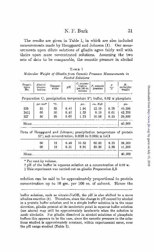
The results are given in Table I, in which are also included measurements made by Hauggaard and Johnson (1). Our meas- urements upon dilute solutions of gliadin agree fairly well with theirs upon more concentrated solutions. Assuming the two sets of data to be comparable, the osmotic pressure in alcohol
TABLE I Molecular Weight of Gliadin from Osmotic Pressure Measurements in
Alcohol Solutions
Preparation C, precipitation temperature 9”; buffer, 0.02 M phosphate
per cent’ “C. gm. cm. Hz0
326 50 25 6.4t 1.94 12.19 6.28 241f 60 30 6.41 1.53 9.19 6.01 327 50 25 s.st 1.73 10.98 6.35
Mean.......................................................
gm. 40 ) 300 42,700 39,800
40,900
Data of Hauggaard and Johnson; precipitation temperature of protein 10”; salt concentration, 0.0028 to 0.0034 M LiCl
1 ii j :: / I::; 1 ‘E 1 2 / ~:~~ z::: Mean....................................................... 40,400
* Per cent by volume. t pH of the buffer in aqueous solution at a concentration of 0.02 M.
$ This experiment was carried out on gliadin Preparation 2,B
solution can be said to be approximately proportional to protein concentration up to 10 gm. per 100 cc. of solvent. -Hence the
buffer solution, such as citrate-NaOH, the pH is also shifted to a more alkaline reaction (1). Therefore, since the change in pH caused by alcohol in a protein buffer solution and in a simple buffer solution is in the same direction, gliadin present at its isoelectric point in aqueous buffer solution (see above) may still be approximately isoelectric when the solution is made alcoholic. For gliadin dissolved in alcohol solutions of phosphate buffers this appears to be the case, since the osmotic pressure in the solu- tions studied is approximately constant, within experimental error, over the pH range studied (Table I).
by guest on July 15, 2018http://w
ww
.jbc.org/D
ownloaded from
52 Osmotic Pressure of Gliadin
mean molecular weight may be calculated from the van% Hoff- Morse equation,
M=RTC P
(1)
where M = molecular weight in gm. dry protein C = concentration in gm. per 100 cc. solvent P = osmotic pressure in cm. Hz0 of density 1
RT = gas constant X absolute temperature = 2.315 X lo6 (100 cc. X cm. Hz0 per gm. molecule) at 0”, 2.528 X lo5 at 25’, or 2.628 X IO5 at 30”
The mean molecular weight of gliadin in alcohol solution, ob- tained from our data, is 40,900 and from that of Hauggaard and Johnson, 40,400 (Table I). Slight changes in alcohol concentra- tion, temperature, or pH do not appear to affect the molecular weight appreciably.
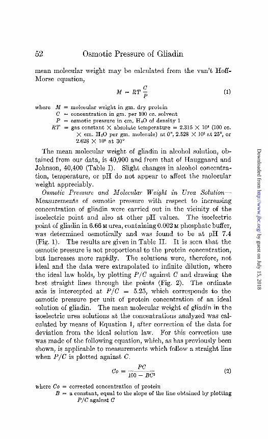
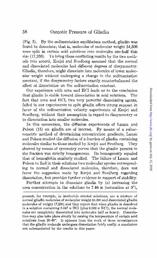
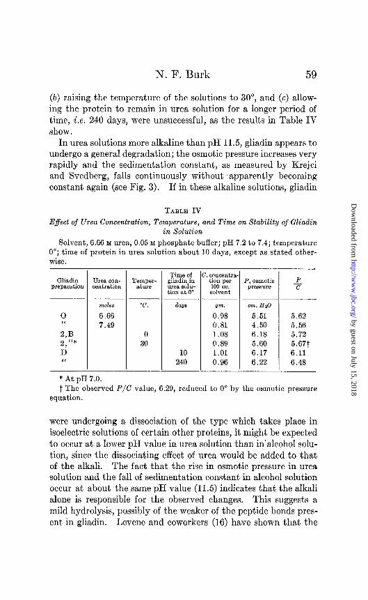
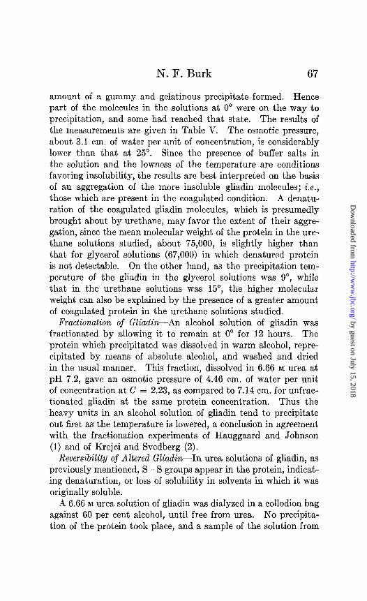
Osmotic Pressure and Molecular Weight in Urea Solution- Measurements of osmotic pressure with respect to increasing concentration of gliadin were carried out in the vicinity of the isoelectric point and also at other pH values. The isoelectric point of gliadin in 6.66 M urea, containing 0.002 M phosphate buffer, was determined osmotically and was found to be at pH 7.4 (Fig. 1). The results are given in Table II. It is seen that the osmotic pressure is not proportional to the protein concentration, but increases more rapidly. The solutions were, therefore, not ideal and the data were extrapolated to infinite dilution, where the ideal law holds, by plotting P/C against C and drawing the best straight lines through the points (Fig. 2). The ordinate axis is intercepted at P/C = 5.25, which corresponds to the osmotic pressure per unit of protein concentration of an ideal solution of gliadin. The mean molecular weight of gliadin in the isoelectric urea solutions at the concentrations analyzed was cal- culated by means of Equation 1, after correction of the data for deviation from the ideal solution law. For this correction use was made of the following equation, which, as has previously been shown, is applicable to measurements which follow a straight line when P/C is plotted against C.
co = PC 100 - BC~
(2)
where Co = corrected concentration of protein B = a constant, equal to the slope of the line obtained by plotting
P/C against C
by guest on July 15, 2018http://w
ww
.jbc.org/D
ownloaded from
N. F. Burk
a
E s
FIG. 1. Influence of the hydrogen ion activity on the osmotic pressure of gliadin. Location of the pH at which the osmotic pressure is a minimum. Ordinates, osmotic pressure in cm. of water per unit of concentration. Glycerol curve, solvent, 75 per cent glycerol, ,J = 0.02 in phosphate buffer; protein concentration, 0.93 to 1.10 gm. per 100 cc. of solvent; temperature 30”; gliadin Preparation 2,B, precipitation temperature 15”. Urea curve, solvent, 6.66 M urea, 0.002 M in phosphate buffer; protein concentration, 2.65 to 3.13 gm. per 100 cc. of solvent; temperature 0”; gliadin Preparation 2,D, precipitation temperature 5”.
FIG. 2. Relation between the osmotic pressure per unit of concentration and the concentration of gliadin at various pH values. The curve for pH 7.2 to 7.53 is for gliadin in approximately isoelectric solutions. Sol- vent, 6.66 M urea, 0.05 M in phosphate or acetate buffer; temperature 0”; gliadin Preparation D.
by guest on July 15, 2018http://w
ww
.jbc.org/D
ownloaded from
54 Osmotic Pressure of Gliadin
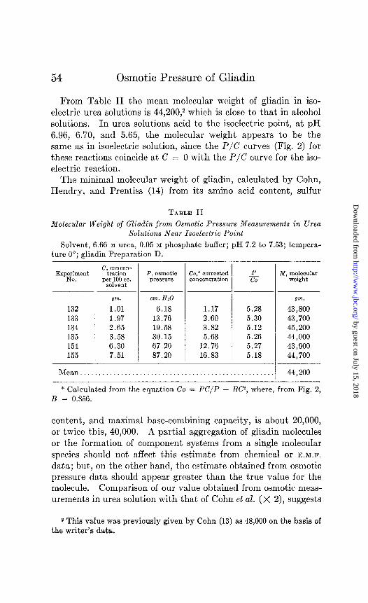
From Table II the mean molecular weight of gliadin in iso- electric urea solutions is 44,200,2 which is close to that in alcohol solutions. In urea solutions acid to the isoelectric point, at pH 6.96, 6.70, and 5.65, the molecular weight appears to be the same as in isoelectric solution, since the P/C curves (Fig. 2) for these reactions coincide at C = 0 with the P/C curve for the iso- electric reaction.
The minimal molecular weight of gliadin, calculated by Cohn, Hendry, and Prentiss (14) from its amino acid content, sulfur
TABLE II Molecular Weight of Gliadin from Osmotic Pressure Measurements in Urea
Solutions Near Isoelectric Point
Solvent, 6.66 M urea, 0.05 M phosphate buffer; pH 7.2 to 7.53; tempera- ture 0”; gliadin Preparation D.
132 133 134 135 154 155
P, osmotic Co,* corrected pX?SS”P? concentration
Qm. cm.HzO
1.01 6.18 1.97 13.76 2.65 19.58 3.58 30.15 6.30 67.20 7.51 87.20
- P co
1.17 5.28 2.60 5.30 3.82 5.12 5.63 5.26
12.76 5.27 16.83 5.18
-
-
M, molecular weight
gm.
43,800 43,700 45,200 44,000 43,900 44,700
Mean................................................... ,I 44,200
* Calculated from the equation Co = PC/P - BC2, where, from Fig. 2, B = 0.856.
content, and maximal base-combining capacity, is about 20,000, or twice this, 40,000. A partial aggregation of gliadin molecules or the formation of component systems from a single molecular species should not affect this estimate from chemical or E.M.F.
data; but, on the other hand, the estimate obtained from osmotic pressure data should appear greater than the true value for the molecule. Comparison of our value obtained from osmotic meas- urements in urea solution with that of Cohn et al. (X 2), suggests
2 This value was previously given by Cohn (13) as 48,000 on the basis of the writer’s data.
by guest on July 15, 2018http://w
ww
.jbc.org/D
ownloaded from
N. F. Burk 55
that the factor of association should be taken into consideration. Further experiments dealing with this point will be described later on in this paper.
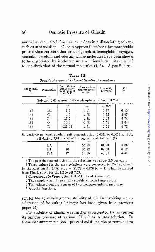
Osmotic Pressure of Diferent Gliadin Preparations-Dill and Alsberg prepared five samples of gliadin and found easily demon- strable differences in their physicochemical properties. Chief among these was the “critical peptization temperature” of gliadin. This is the temperature at which first signs of turbidity appear when an alcohol solution of gliadin is cooled slowly under given conditions. The precipitation temperature of Dill and Alsberg’s five preparations varied from 2-8.5”. The work of Hauggaard and Johnson also shows that the precipitation temperature may serve as a useful index figure for the characterization of gliadin preparations or fractions.
The precipitation temperature is in a way related to solubility, and it may be inferred that the higher the precipitation tempera- ture of a gliadin preparation the lower the solubility. In 57.2 per cent alcohol at 18”, Hauggaard and Johnson found solubilities of 22.3, 7.3, and 1.8 mg. of N per cc. for gliadin fractions at pre- cipitation temperatures of lo, lo”, and 17” respectively.
In order to determine whether osmotic pressure varies with precipitation temperature, pressure measurements were carried out in urea solution upon five samples of gliadin prepared by different investigators. The results are given in Table III where it is seen that the osmotic pressure in urea solution falls slightly as the precipitation temperature increases. Over the range of precipitation temperature studied by us, this same relationship holds also in alcohol solution, as the data of Hauggaard and John- son show (Table III). However, it appears from the data of Hauggaard and Johnson that below a certain precipitation tem- perature, about lo’, the osmotic pressure does not appreciably change, within experimental error, with this index figure. Hence on the basis of these data, the value for the mean molecular weight of gliadin obtained by us upon Preparation D (precipita- tion temperature 7.5”) can be said to represent that of well puri- fied gliadin and possibly is an approximation to the lower limit for the mean molecular weight of the protein.
Stability of Gliadin-The above measurements show that well purified gliadin has essentially the same molecular weight in its
by guest on July 15, 2018http://w
ww
.jbc.org/D
ownloaded from
56 Osmotic Pressure of Gliadin
normal solvent, alcohol-water, as it does in a dissociating solvent such as urea solution. Gliadin appears therefore a far more stable protein than certain other proteins, such as hemoglobin, myogen, amandin, excelsin, and edestin, whose molecules have been shown to be dissociated by isoelectric urea solutions into units one-half to one-sixth that of the normal molecules (4, 5). A possible rea-
TABLE III Osmotic Pressure of Different Gliadin Preparations
ExpELyt / Preparation / ig{ / tZ~~T$~. / p;fz$l:: 1 -$+
Solvent, 6.66 M urea, 0.05 M phosphate buffer, pH 7.2
“C. gm. G?n. Hz0
118 DS 7.5 1 .Ol 6.17 6.10 143 C 9.0 1.08 6.52 5.97 150 B 13.0 1.14 6.68 5.74 152 0 16.0 0.98 5.51 5.64 139 R <22.0$ 1.31 6.54 4.72
Solvent, 60 per cent alcohol, salt concentration, 0.0024 to 0.0033 M LiCI; pH 6.48 to 7.07; data11 of Hauggaard and Johnson (1)
11% 1 1.0.93 61.80 5.65 III 10 10.22 62.56 6.12 IV7 17 11.06 48.85 4.41
* The protein concentration in the solutions was about 2.5 per cent. -i These values for the urea solutions were corrected to P/C at C = 1
by the relationship (P/C) c _ 1 = (P/C) - 0.856 (C - l), which is derived from Fig. 2, curve for pH 7.2 to pH 7.53.
$ Corresponds to Preparation 2,B of Dill and Alsberg (6). 0 The sample was only partially soluble at room temperature. 11 The values given are a mean of two measurements in each case. 7 Gliadin fractions.
son for the relatively greater stability of gliadin involving a con- sideration of its sulfur linkages has been given in a previous
paper (5). The stability of gliadin was further investigated by measuring
its osmotic pressure at various pH values in urea solution. In these measurements, upon 1 per cent solutions, the pressure due to
by guest on July 15, 2018http://w
ww
.jbc.org/D
ownloaded from
N. F. Burk 57
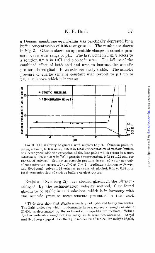
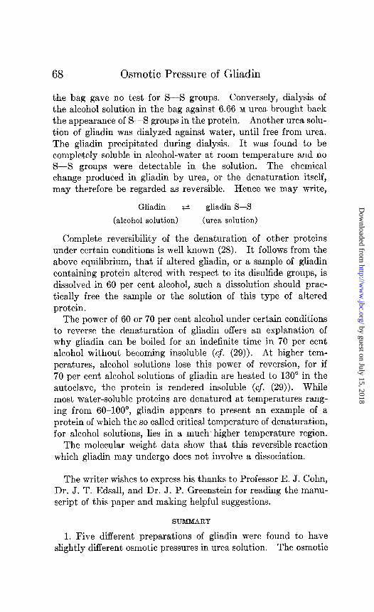
a Donnan membrane equilibrium was practically depressed by a buffer concentration of 0.05 M or greater. The results are shown in Fig. 3. Gliadin shows no appreciable change in osmotic pres- sure over a wide range of pH. The first point in Fig. 3 refers to a solution 0.2 M in HCI and 6.66 M in urea. The failure of the combined effect of both acid and urea to increase the osmotic pressure shows gliadin to be extraordinarily stable. The osmotic pressure of gliadin remains constant with respect to pH up to pH 11.5, above which it increases.
I I I I I
FIG. 3. The stability of gliadin with respect to pH. Osmotic pressure curve, solvent, 6.66 M urea, 0.05 M in total concentration of various buffers or electrolytes, with the exception of the first point which refers to a urea solution which is 0.2 M in HCl; protein concentration, 0.92 to 1.23 gm. per 100 cc. of solvent. Ordinates, osmotic pressure in cm. of water per unit of concentration, corrected to P/C at C = 1. Sedimentation curve (Krejci and Svedberg), solvent, 64 volumes per cent of alcohol, 0.01 to 0.23 M in total concentration of various buffers or electrolytes.
Krejci and Svedberg (2) have studied gliadin in the ultracen- trifuge.3 By the sedimentation velocity method, they found gliadin to be stable in acid soluDions, which is in harmony with the osmotic pressure measurements presented in this work
3 Their data show that gliadin is made up of light and heavy molecules. The light molecules which predominate have a molecular weight of about 26,000, as determined by the sedimentation equilibrium method. Values for the molecular weight of the heavy units were not obtained. Krejci and Svedberg suggest that the light molecules of molecular weight 26,000,
by guest on July 15, 2018http://w
ww
.jbc.org/D
ownloaded from
Osmotic Pressure of Gliadin
(Fig. 3). By the sedimentation equilibrium method, gliadin was found to dissociate; that is, molecules of molecular weight 34,500 were split in certain acid solutions into molecules one-half this size (17,250). To bring these conflicting results by the two meth- ods into accord, Krejci and Svedberg assumed that the normal and dissociated molecules had different degrees of dissymmetry. Gliadin, therefore, might dissociate into molecules of lower molec- ular weight without undergoing a change in the sedimentation constant, if the dissymmetry factors exactly counterbalanced the effect of dissociation on the sedimentation constant.
Our experience with urea and HCl leads us to the conclusion that gliadin is stable toward dissociation in acid solutions. The fact that urea and HCl, two very powerful dissociating agents, failed in our experiments to split gliadin offers strong support in favor of the sedimentation velocity experiments of Krejci and Svedberg, without their assumption in regard to dissymmetry or to dissociation into smaller molecules.
In this connection, the diffusion experiments of Lamm and Polson (15) on gliadin are of interest. By means of a refrac- tometric method of determining concentration gradients, Lamm and Polson studied the diffusion of a fraction of gliadin containing molecules similar to those studied by Krejci and Svedberg. They showed by means of symmetry curves that the gliadin present in the fraction was strictly homogeneous. Its homogeneity equaled that of hemoglobin similarly studied. The failure of Lamm and Polson to find in their solutions two molecular species correspond- ing to normal and dissociated molecules, therefore, does not favor the suggestion made by Krejci and Svedberg regarding dissociation, but provides further evidence in support of stability.
Further attempts to dissociate gliadin by (a) increasing the urea concentration in the solutions to 7.46 M (saturation at 0”),
present, for example, in isoelectric alcohol solutions, are a mixture of normal gliadin molecules of molecular weight 34,500 and dissociated gliadin molecules of weight 17,250; and they report that when gliadin is dissolved in a solution containing 0.047 M HCl (plus 0.008 N KC]), the normal mole- cules are completely dissociated into molecules half as heavy. Dissocia- tion may also take place simply by raising the temperature of certain acid solutions from 20-30”. It appears from the work of these investigators that the gliadin molecule undergoes dissociation fairly easily, a conclusion not substantiated by the results in this paper.
by guest on July 15, 2018http://w
ww
.jbc.org/D
ownloaded from
N. F. Burk 59
(b) raising the temperature of the solutions to 30”, and (c) allow- ing the protein to remain in urea solution for a longer period of time, i.e. 240 days, were unsuccessful, as the results in Table IV show.
In urea solutions more alkaline than pH 11.5, gliadin appears to undergo a general degradation; the osmotic pressure increases very rapidly and the sedimentation constant, as measured by Krejci and Svedberg, falls continuously without apparently becoming constant again (see Fig. 3). If in these alkaline solutions, gliadin
TABLE IV
E$ect of Urea Concentration, Temperature, and Time on Stability of Gliadin in Solution
Solvent, 6.66 M urea, 0.05 M phosphate buffer; pH 7.2 to 7.4; temperature 0”; time of protein in urea solution about 10 days, except as stated other- wise.
Time of c. concentra- Gliadin Urea con- Temper- gliadin in tion per P
preparation centration P, osmotio
ature urea solu- 100 cc. PR?SSUi-e tion at 0” solvent
-CT
moles “C. days om. cm.HzO
0 6.66 0.98 5.51 5.62 “ 7.49 0.81 4.50 5.56 2,B 0 1.08 6.18 5.72
2, I‘* 30 0.89 5.60 5.67t D 10 1.01 6.17 6.11 I‘ 240 0.96 6.22 6.48
* At pH 7.0. t The observed P/C value, 6.29, reduced to 0” by the osmotic pressure
equation.
were undergoing a dissociation of the type which takes place in isoelectric solutions of certain other proteins, it might be expected to occur at a lower pH value in urea solution than in’alcohol solu- tion, since the dissociating effect of urea would be added to that of the alkali. The fact that the rise in osmotic pressure in urea solution and the fall of sedimentation constant in alcohol solution occur at about the same pH value (11.5) indicates that the alkali alone is responsible for the observed changes. This suggests a mild hydrolysis, possibly of the weaker of the peptide bonds pres- ent in gliadin. Levene and coworkers (16) have shown that the
by guest on July 15, 2018http://w
ww
.jbc.org/D
ownloaded from
60 Osmotic Pressure of Gliadin
strength of the peptide bond depends on the nature of the amino acids which take part in its formation. Certain peptide bonds are easy and others are hard to hydrolyze. Cohn and Berggren (17) found that casein, after treatment with a mildly alkaline solution, possessed an acid- or base-combining capacity consider- ably greater than that of casein not so treated, again indicating that in certain proteins there are groups which hydrolyze very easily in alkaline solutions.
Osmotic Pressure and Molecular Weight in Glycerol Xdutions- In order to obtain information on the state of gliadin in a solvent in which it is not highly soluble, osmotic pressure measurements were carried out in glycerol solutions. In 75 per cent glycerol, buffered at the isoelectric reaction, gliadin is much less readily soluble than in buffered 60 per cent alcohol or buffered 6.66 M
urea. In the experiments reported, the solutions were prepared by gently rocking gliadin with 75 per cent glycerol (buffered with 0.02 M phosphate) for several hours, and then filtering the mix- ture. Dissolution became more and more incomplete when solu- tions of increasing gliadin concentration were prepared.
The isoelectric point of gliadin in glycerol solution was first determined (Fig. 1). In Fig. 1 the pH refers to that of t’he phos- phate buffer in the absence of glycerol. A phosphate buffer solu- tion of ionic strength 0.02, having about pH 6.77, was found to have a reaction of pH 6.78 in 75 per cent glycerol, as determined by the hydrogen electrode. The reaction at the minimum os- motic pressure in Fig. 1, at pR 6.5, may therefore be taken as the isoelectric point of gliadin in 75 per cent glycerol solution.
The results of osmotic pressure measurements upon glycerol solutions of increasing protein concentration, in the vicinity of the isoelectric point, are given in Table V, which shows that the os- motic pressure in glycerol solutions, unlike that in urea solutions, increases slightly less rapidly than the protein concentration. Ex- trapolation of P/C values to zero concentration yields an ideal pressure of 3.84 cm. of water per unit of protein concentration, corresponding to a mean molecular weight of 67,000 for gliadin in glycerol solution.
The facts that this figure is greater than that for the mean molecular weight in alcohol solution, that P/C decreases as the concentration increases, and that the protein shows a lower solu-
by guest on July 15, 2018http://w
ww
.jbc.org/D
ownloaded from
N. F. Burk
bility in the glycerol solvent, suggest that there is an aggrega- tion of gliadin molecules in glycerol solutions. The fact that the mean molecular weight in glycerol solutions is less than twice that in alcohol or urea solutions indicates that only part of the gliadin molecules (or units) present in these solvents undergo aggregation when dissolved in glycerol solution.
TABLE V Osmotic Pressure and Molecular Weight of Gliadin in Glycerol Solutions and
in Urethane Solutions
Exp~~yt 1 pH* j ~~~~gqj- 1 FQ&;;~ 1 $ 1 M,y$p
Solvent, 75 per cent glycerol, 0.02 M in phosphate buffer; temperature 30”; gliadin Preparation C, precipitation temperature 9”
6-m. cm.HzO gm.
0.00 0.00 334t 67,000 244 6.43 0.97 3.54 3.65 245 6.43 1.65 5.72 3.47 246 6.43 2.95 9.55 3.24 247 6.43 4.98 15.62 3.14
Solvent, 15 per cent urethane (1.68 M), 0.05~ phosphate buffer; temperature 0”; gliadin Preparation 2,B, precipitation temperature 15”
* Reaction of the buffer in aqueous solution in the absence of glycerol or urethane.
t Obtained by extrapolation of the first three measurements. $ Uncorrected for deviations from the ideal solution law.
Krejci and Svedberg (2) have shown that gliadin is an inhomo- geneous protein, consisting of light and heavy molecules. It has been found that the heavy molecules may be separated from the lighter ones by cooling an alcohol solution of gliadin to 0” and then to -11” (1, 2, 15). It is reasonable to suppose therefore that in glycerol solutions the heavy molecules (or units), because of their lower solubility, are the ones which are aggregating. It is well known in colloid chemistry that a low solubility of a solute
by guest on July 15, 2018http://w
ww
.jbc.org/D
ownloaded from
Osmotic Pressure of Gliadin
in a given solvent is one of the chief determining factors regulating aggregate formation. The soaps, for example, which are fairly soluble in alcohol, but less soluble in water, dissolve in the former solvent as single molecules but in the latter solvent as aggre- gates (18).
The solubility of a native protein may be decreased in two ways, by denaturation or by coagulation.4 While our knowledge of the mechanism of these two processes is still incomplete, Mirsky and Anson (21) have established that in denaturation a lowering of the solubility is accompanied by the appearance of -SH or S-S groups or both; and that in coagulation the loss of solubility is not accompanied by the appearance of such groups or additional groups (19). Qualitative tests5 for S-S groups were positive in urea solutions of gliadin and negative in glycerol and alcohol solutions. (Tests for -SH groups were negative in all solvents employed.) We shall assume on the basis of Mirsky and Anson’s work (21) that denatured gliadin is present in the former solutions,6 but not in the latter. If varying amounts of denatured
4 The term coagulation is employed throughout this paper in the sense used by Mirsky (19) and Mirsky and Pauling (20). It denotes an asso- ciation of molecules due to an interaction between them, as, for example, by hydrogen bond formation. Coagulated molecules, unlike flocculated molecules, may exist in solution. On the other hand, denaturation prob- ably involves no association of molecules, since the rate of denaturation follows the course of a monomolecular reaction, suggesting that the change lies within the molecule.
5 Sodium cyanide and nitroprusside (and dilute ammonia, if necessary) (22) or sodium sulfite and 18-phosphotungstic acid (and phosphate buffer at pH 7 to 9, or sodium carbonate) (23) were the reagents used to test the solutions for S-S groups. Tests for -SH groups were made with these same reagents in the absence of the reducing agents, sodium cyanide or sodium sulfite. The phosphotungstic acid test is not truly applicable in alcohol solutions, since this reagent precipitates the protein from such solutions.
6 While Mirsky and Anson show that in general denaturation of proteins is accompanied by the formation of S-S or -SH groups, it does not neces- sarily follow that an increase in the number of such groups in proteins can be used as a criterion of their denaturation, since the change in the sulfur linkages may be an intermediate reaction occurring prior to this process (cf. (24)). However, it makes but little difference in regard to the con- clusions drawn here, whether the alteration of gliadin by urea is termed denaturation or designated by some other term.
by guest on July 15, 2018http://w
ww
.jbc.org/D
ownloaded from
N. F. Burk 63
protein were present in our preparations, the osmotic pressure of each should be the same in urea solution, since all the native gli- adin would no doubt be completely transformed into denatured protein upon dissolution in concentrated urea solution (cf. (25) p. 127 and (21) p. 443). The fact that different preparations of gliadin had different osmotic pressures in urea solution is there- fore evidence that denatured gliadin was not significantly present in the samples of gliadin studied. The possibility of coagulated gliadin being present in gliadin samples or in gliadin solutions is considered in the next section.
Osmotic Pressure of Partially Coagulated G&din-The work of Mirsky (19) has shown that certain purified native proteins (myosin, for example), in solution or in the gel state, coagulate but do not denature when dehydrated by partial drying or freez- ing. Since freezing removes water in the form of ice, this process as well as that of drying brings about a concentration of protein. Coagulation, of the type referred to by Mirsky, may then be looked upon as a process which presumably occurs in highly con- centrated protein solutions, or in relatively concentrated protein gels, under certain conditions. This process of coagulation ap- pears to take place in those proteins which form gels easily, rather than in proteins which are thrown down as dense precipitates. As is well known (26), evaporation of an alcohol solution of gli- adin, when the alcohol concentration is kept at about 60 per cent, leads to no visible precipitation of the protein, but to a clear gel.
The following is quoted from Dill and Alsberg (6): “If dry gliadin is covered with 70 per cent alcohol, it becomes solvated, forming a concentrated, clear viscous solution below the larger part of the solvent. If this is then allowed to stand quietly for 2 or 3 days, part of the gliadin will be altered and will not be dis- solved in any concentration of alcohol at room temperature.” Gliadin also undergoes a decrease in solubility in contact with 75 to 85 per cent alcohol (6). On the other hand, little or no altera- tion takes place if dry gliadin stands for several days in contact with absolute alcohol, or is precipitated by a large excess of abso- lute alcohol from 70 per cent ethanol solution, under which condi- tions it separates as a dense, white precipitate.
A sample of gliadin was partially coagulated by allowing it to
by guest on July 15, 2018http://w
ww
.jbc.org/D
ownloaded from
64 Osmotic Pressure of Gliadin
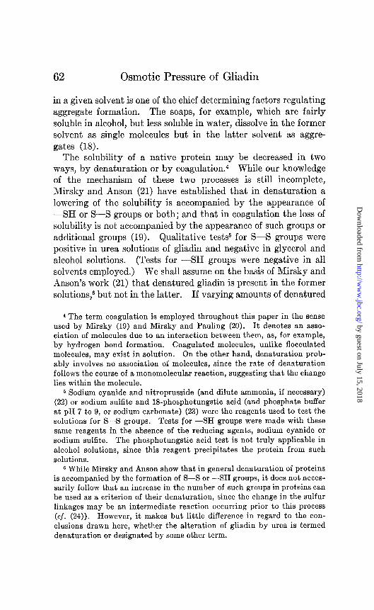
remain for several days in contact with 85 per cent alcohol. Table VI shows that gliadin, after partial coagulation, has a lower osmotic pressure and higher precipitation temperature than has similar gliadin not subjected to partial coagulation. It is there- fore to be concluded that the variations in osmotic pressure of the gliadin preparations of different precipitation temperatures previously studied (Table III) are due to the presence of varying amounts of coagulated gliadin.
This result is in harmony with the observations of Dill and Alsberg. In preparing gliadin, they noticed that if an insufficient
TABLE VI
Comparison of Osmotic Pressure of Partially Coagulated Gliadin with That of Gliadin Not Partially Coagulated
Solvent, 6.66 M urea, 0.05 M phosphate buffer, pFI 7.2. 1 gm. of gliadin Preparation 3,B was placed in a beaker and covered
with 25 cc. of 85 per cent alcohol and allowed to remain quietly for 6 days at room temperature. After the alcohol was decanted off, the gliadin was completely dissolved in 6.66 M urea and immediately precipitated by means of absolute alcohol. The precipitate was washed in the usual man- ner and dried at room temperature in a vacuum desiccator.
Exp%?t Preparation Precipitation Q~p~rn$ P, osmotic P temperature cc. solvent pl%*S3llX F
“C. gm. cm.HzO
156 3,J3 16 1.05 5.96 5.68 157 3, B partially <22* 1.11 4.69 4.23
coagulated
* The preparation was only partially soluble at room temperature.
amount of absolute alcohol was used in precipitating the prot.ein from solution, a part underwent alteration; and some of their earlier preparations contained as much as 50 per cent of a less soluble gliadin. It appears that the use of too small a quantity of absolute alcohol brings the protein, for a short time while on the way to precipitation, into a highly concentrated state. Thus this type of coagulation, brought more clearly to light by Mirsky in studying myosin (19), may be operative when we least expect it. Krejci and Svedberg, in their preparation of gliadin, found 37 per cent of heavy molecules. It is doubtful whether a con-
by guest on July 15, 2018http://w
ww
.jbc.org/D
ownloaded from

N. F. Burk
version of the light normal gliadin molecules into the heavy units takes place to any large extent in ordinary solutions. A precipi- tation process or the formation of a relatively concentrated solu- tion under conditions which are not as yet very clearly defined appears necessary. Gottenberg and Alsberg (27) have suggested the possibility of an alteration of gliadin taking place in its orig- inal source in the wheat berry.
The high molecular weight of gliadin in buffered glycerol solu- tion over that in urea solution can be explained on the basis of an aggregation of the coagulated gliadin molecules present in gliadin samples. Urea solutions are well known for their dis- persive action and it is justifiable to assume that the coagulated
TABLE VII
Osmotic Pressure of Gliadin in Urea Solutions of Varying Salt Content
Solvent, 6.66 M urea containing phosphate buffer; pH 7.2; gliadin Prepa-
Y-
ration C.
Expe%tYt
143 141 142 144
Concentration of P~~a&at
mole
0.05 0.10 0.20 0.40
-
(
-
7, concentration per 100 cc.
solvent
gm. cm.HzO
1.08 6.52 1.06 6.45 1.09 6.27 1.06 5.18
P -c
6.04 6.08 5.75 4.89
molecules are present in the urea solutions studied in an unag- gregated state.
It is well known that the formation of aggregates in solution depends to a large extent upon the presence of electrolytes. A few osmotic pressure experiments were carried out in urea solu- tions in which the electrolyte content was altered by variation in the phosphate buffer concentration. The results are presented in Table VII, which shows that in urea solutions having a buffer concentration greater than 0.1 M, there is a progressive lowering of the osmotic pressure as the salt concentration increases. Again, the results are best interpreted by assuming an aggrega- tion of the coagulated gliadin molecules present. The majority of the molecules present in gliadin are native protein mole- cules (2). If an aggregation of the normal molecules were taking
by guest on July 15, 2018http://w
ww
.jbc.org/D
ownloaded from
66 Osmotic Pressure of Gliadin
place, a much larger effect on the osmotic pressure might be ex- pected.
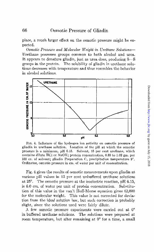
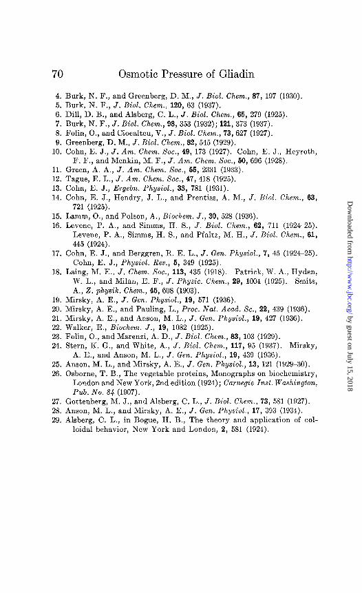
Osmotic Pressure and Molecular Weight in Urethane Solutions- Urethane possesses groups common to both alcohol and urea. It appears to denature gliadin, just as urea does, producing S-S groups in the protein. The solubility of gliadin in urethane solu- tions decreases with temperature and thus resembles the behavior in alcohol solutions.
FIG. 4. Influence of the hydrogen ion activity on osmotic pressure of gliadin in urethane solution. Location of the pH at which the osmotic pressure is a minimum, pH 6.15. Solvent, 15 per cent urethane, which contains dilute HCl or NaOH; protein concentration, 0.89 to 1.02 gm. per 100 cc. of solvent; gliadin Preparation C, precipitation temperature 9”. Ordinates, osmotic pressure in cm. of water per unit of concentration.
Fig. 4 gives the results of osmotic measurements upon gliadin at various pH values in 15 per cent unbuffered urethane solutions at 25”. The osmotic pressure at the isoelectric reaction, pH 6.15, is 6.0 cm. of water per unit of protein concentration. Substitu- tion of this value in the van% Hoff-Morse equation gives 42,000 for the molecular weight. This value is not corrected for devia- tion from the ideal solution law, but such correction is probably slight, since the solutions used were fairly dilute.
A few osmotic pressure experiments were carried out at 0” in buffered urethane solutions. The solutions were prepared at room temperature, but after remaining at 0” for a time, a small
by guest on July 15, 2018http://w
ww
.jbc.org/D
ownloaded from
N. F. Burk 67
amount of a gummy and gelatinous precipitate formed. Hence part of the molecules in the solutions at 0’ were on the way to precipitation, and some had reached that state. The results of the measurements are given in Table V. The osmotic pressure, about 3.1 cm. of water per unit of concentration, is considerably lower than that at 25”. Since the presence of buffer salts in the solution and the lowness of the temperature are conditions favoring insolubility, the results are best interpreted on the basis of an aggregation of the more insoluble gliadin molecules; i.e., those which are present in the coagulated condition. A denatu- ration of the coagulated gliadin molecules, which is presumedly brought about by urethane, may favor the extent of their aggre- gation, since the mean molecular weight of the protein in the ure- thane solutions studied, about 75,000, is slightly higher than that for glycerol solutions (67,000) in which denatured protein is not detectable. On the other hand, as the precipitation tem- perature of the gliadin in the glycerol solutions was 9”, while that in the urethane solutions was 15”, the higher molecular weight can also be explained by the presence of a greater amount of coagulated protein in the urethane solutions studied.
Fractionation of Gliadin-An alcohol solution of gliadin was fractionated by allowing it to remain at 0” for 12 hours. The protein which precipitated was dissolved in warm alcohol, repre- cipitated by means of absolute alcohol, and washed and dried in the usual manner. This fraction, dissolved in 6.66 M urea at pH 7.2, gave an osmotic pressure of 4.46 cm. of water per unit of concentration at C = 2.23, as compared to 7.14 cm. for unfrac- tionated gliadin at the same protein concentration. Thus the heavy units in an alcohol solution of gliadin tend to precipitate out first as the temperature is lowered, a conclusion in agreement with the fractionation experiments of Hauggaard and Johnson (1) and of Krej ci and Svedberg (2).
Reversibility of Altered Gliadin-In urea solutions of gliadin, as previously mentioned, S-S groups appear in the protein, indicat- ing denaturation, or loss of solubility in solvents in which it was originally soluble.
A 6.66 M urea solution of gliadin was dialyzed in a collodion bag against 60 per cent alcohol, until free from urea. No precipita- tion of the protein took place, and a sample of the solution from
by guest on July 15, 2018http://w
ww
.jbc.org/D
ownloaded from
68 Osmotic Pressure of Gliadin
the bag gave no test for S-S groups. Conversely, dialysis of the alcohol solution in the bag against 6.66 M urea brought back the appearance of S-S groups in the protein. Another urea solu- tion of gliadin was dialyzed against water, until free from urea. The gliadin precipitated during dialysis. It was found to be completely soluble in alcohol-water at room temperature and no S-S groups were detectable in the solution. The chemical change produced in gliadin by urea, or the denaturation itself, may therefore be regarded as reversible. Hence we may write,
Gliadin * gliadin S-S
(alcohol solution) (urea solution)
Complete reversibility of the denaturation of other proteins under certain conditions is well known (28). It follows from the above equilibrium, that if altered gliadin, or a sample of gliadin containing protein altered with respect to its disulfide groups, is dissolved in 60 per cent alcohol, such a dissolution should prac- tically free the sample or the solution of this type of altered protein.
The power of 60 or 70 per cent alcohol under certain conditions to reverse the denaturation of gliadin offers an explanation of why gliadin can be boiled for an indefinite time in 70 per cent alcohol without becoming insoluble (cj. (29)). At higher tem- peratures, alcohol solutions lose this power of reversion, for if 70 per cent alcohol solutions of gliadin are heated to 130” in the autoclave, the protein is rendered insoluble (cf. (29)). While most water-soluble proteins are denatured at temperatures rang- ing from 60-loo’, gliadin appears to present an example of a protein of which the so called critical temperature of denaturation, for alcohol solutions, lies in a much higher temperature region.
The molecular weight data show that this reversible reaction which gliadin may undergo does not involve a dissociation.
The writer wishes to express his thanks to Professor E. J. Cohn, Dr. J. T. Edsall, and Dr. J. P. Greenstein for reading the manu- script of this paper and making helpful suggestions.
SUMMARY
1. Five different preparations of gliadin were found to have slightly different osmotic pressures in urea solution. The osmotic
by guest on July 15, 2018http://w
ww
.jbc.org/D
ownloaded from

N. F. Burk 69
pressures of the preparations varied in a more or less regular manner with their precipitation temperatures. The results con- firm the findings of Hauggaard and Johnson and those of Krejci and Svedberg that gliadin is an inhomogeneous protein.
2. From osmotic pressure measurements in alcohol solutions near the isoelectric point the mean molecular weight of well puri- fied gliadin was found to be 41,000; and in urea solutions, 44,000. This comparison therefore shows that gliadin is not dissociated into units of lower molecular weight by urea; it also shows that gliadin possesses a stability relatively greater than certain other proteins, such as hemoglobin, myogen, amandin, and excelsin, whose molecular weights are reduced by urea. Gliadin is prob- ably denatured by urea, and, although S-S groups appear in the protein in urea solution, -SH groups are not present.
3. The osmotic pressure of gliadin, partially coagulated by means of 85 per cent alcohol, was found to be lower in urea solu- tion than that of gliadin not partially coagulated. This suggests that the observed variations in osmotic pressure of different gli- adin preparations are due to varying amounts of coagulated pro- tein. A gliadin fraction, namely, the precipitate obtained by cooling an alcohol solution to O’, also showed an abnormally low osmotic pressure in urea solution.
4. In buffered 75 per cent glycerol solution at 30”, the mean molecular weight of gliadin was found to be 67,000; in buffered 15 per cent urethane solution at O”, 75,000. In these solvents under the conditions described, gliadin is less soluble than in alcohol or urea solutions. The higher mean molecular weights in these solvents are attributed to the existence of aggregates, presumably formed from the coagulated gliadin molecules present in the gliadin preparations.
5. In salt-free urethane solutions at 25”, the mean molecular weight of gliadin was found to be 42,000, the same as its weight in alcohol or urea solutions.
6. The formation in gliadin of S-S groups, associated with the denaturation of proteins, is reversible.
BIBLIOGRAPHY
1. Hauggaard, O., and Johnson, A. H., Compt.-rend. trav. Lab. Carlsberg, 18, No. 2 (1930).
2. Krejci, L., and Svedberg, T., J. Am. Chem. Sot., 67, 946 (1935). 3. S@rensen, S. P. L., Compt.-rend. trav. Lab. Carlsberg, 18, No. 5 (1930).
by guest on July 15, 2018http://w
ww
.jbc.org/D
ownloaded from
Osmotic Pressure of Gliadin
4. Burk, N. F., and Greenberg, D. M., J. Bid. Chem., 87, 197 (1930). 5. Burk, N. F., J. Biol. Chem., 120, 63 (1937). 6. Dill, D. B., and Alsberg, C. L., J. Biol. Chem., 66, 279 (1925). 7. Burk, N. F., J. Biol. Chem., 98, 353 (1932); 121, 373 (1937). 8. Folin, O., and Ciocalteu, V., J. Biol. Chem., 73, 627 (1927). 9. Greenberg, D. M., J. BioZ. Chem., 82, 545 (1929).
10. Cohn, E. J., J. Am. Chem. Xoc., 49, 173 (1927). Cohn, E. J., Heyroth, F. F., and Menkin, M. F., J. Am. Chem. Sot., 60,696 (1928).
11. Green, A. A., J. Am. Chem. Xoc., 66, 2331 (1933). 12. Tague, E. L., J. Am. Chem. Sot., 47, 418 (1925). 13. Cohn, E. J., Ergebn. Physiol., 33, 781 (1931). 14. Cohn, E. J., Hendry, J. L., and Prentiss, A. M., J. BioZ. Chem., 63,
721 (1925). 15. Lamm, O., and Polson, A., Biochem. J., 30, 528 (1936). 16. Levene, P. A., and Simms, H. S., J. BioZ. Chem., 62, 711 (1924-25).
Levene, P. A., Simms, H. S., and Pfaltz, M. H., J. BioZ. Chem., 61, 445 (1924).
17. Cohn, E. J., and Berggren, R. E. L., J. Gen. Physiol., ‘7, 45 (1924-25). Cohn, E. J., Physiol. Rev., 6, 349 (1925).
18. Laing, M. E., J. Chem. Sot., 113, 435 (1918). Patrick, W. A., Hyden, W. L., and Milan, E. F., J. Physic. Chem., 29, 1004 (1925). Smits, A., 2. physik. Chem., 46,608 (1903).
19. Mirsky, A. E., J. Gen. Physiol., 19, 571 (1936). 20. Mirsky, A. E., and Pauling, L., Proc. Nat. Acad. SC., 22, 439 (1936). 21. Mirsky, A. E., and Anson, M. L., J. Gen. Physiol., 19, 427 (1936). 22. Walker, E., Biochem. J., 19, 1082 (1925). 23. Folin, O., and Marenzi, A. D., J. BioZ. Chem., 83, 103 (1929). 24. Stern, K. G., and White, A., J. BioZ. Chem., 117, 95 (1937). Mirsky,
A. E., and Anson, M. L., J. Gen. Physiol., 19, 439 (1936). 25. Anson, M. L., and Mirsky, A. E., J. Gen. Physiol., 13, 121 (1929-30). 26. Osborne, T. B., The vegetable proteins, Monographs on biochemistry,
London and New York, 2nd edition (1924); Carnegie Insl. Washington, Pub. No. 8.4 (1907).
27. Gottenberg, M. J., and Alsberg, C. L., J. BioZ. Chem., 73, 581 (1927). 28. Anson, M. L., and Mirsky, A. E., J. Gen. Physiol., 17, 393 (1934). 29. Alsberg, C. L., in Bogue, H. B., The theory and application of col-
loidal behavior, New York and London, 2, 581 (1924).
by guest on July 15, 2018http://w
ww
.jbc.org/D
ownloaded from
Norval F. BurkGLIADINWEIGHT, AND STABILITY OF
OSMOTIC PRESSURE, MOLECULAR
1938, 124:49-70.J. Biol. Chem.
http://www.jbc.org/content/124/1/49.citation
Access the most updated version of this article at
Alerts:
When a correction for this article is posted•
When this article is cited•
alerts to choose from all of JBC's e-mailClick here
ml#ref-list-1
http://www.jbc.org/content/124/1/49.citation.full.htaccessed free atThis article cites 0 references, 0 of which can be
by guest on July 15, 2018http://w
ww
.jbc.org/D
ownloaded from


![Effect of Molecular Weight and Molecular Distribution on Skin … · 2016-01-07 · based materials . Nevertheless, molecular weight and molecular weight distribution effects on stru[10]](https://static.fdocuments.net/doc/165x107/5e750b4f6204df40457a83af/effect-of-molecular-weight-and-molecular-distribution-on-skin-2016-01-07-based.jpg)


