Osmotic Pressure Escherichia RenderedDetectable Lysozyme · Osmotic Pressure in Escherichia coli as...
7
JouRNAL or BAcmOLoGY, May 1973, p. 549-555 Copyright 0 1973 American Society for Microbiology Vol. 114, No. 2 Printed in U.S.A. Osmotic Pressure in Escherichia coli as Rendered Detectable by Lysozyme Attack PAUL SCHEIE Department of Biophysics, The Pennsylvania State University, University Park, Pennsylvania 16802 Received for publication 8 January 1973 The enhanced susceptibility of plasmolyzed Escherichia coli to lysozyme attack was used to estimate the internal osmotic pressure of these cells under various conditions. Differences were detected between strains, culture media, stages in the growth cycle, and the osmotically active material used to produce plasmolysis. Lysozyme also was found to attack unplasmolyzed cells at 0 C and between 50 and 70 C. The osmotic pressure of bacteria is believed to afford a measure of the concentration of small, free solute molecules contained within the semipermeable plasma membrane. This concentration helps establish the internal envi- ronment in which the chemistry of the cell takes place. Moreover, the differential osmotic pressure, that is, the difference between the osmotic pressure in the cell and that of the medium, will determine how firmly the plasma membrane is pressed against the rigid pep- tidoglycan layer. This differential osmotic pressure thereby may affect one or more of the several membrane-related activities. Studies on the osmotic properties of Esche- richia coli generally have involved varying the osmotic pressure of the medium and observing the response of the cells. Envelopes of E. coli cells are subject to alteration by osmotic stress. Hypotonicity leads to the release of small metabolites (11) and several envelope-bound enzymes (8) as well as to the formation of finger-like protrusions (2) and, perhaps, lysis (12). Hypertonicity leads to plasmolysis during which the plasma membrane shrinks away from the rigid peptidoglycan layer (5, 13). The osmotic pressure of the medium at which plasmolysis first becomes microscopically evi- dent generally is cited as the internal osmotic pressure of the cell. It is tacitly assumed that the plasma membrane is free to shrink away from the cell envelope, at least in some regions. Values so obtained range from 2 atm for resting cells to 15 atm for rapidly growing cells (10). It is, of course, difficult and tedious to quantitate the prevalence of plasmolysis for large numbers of cells by microscope examination. Further- more, several of the studies on plasmolysis have involved washed cells from which many osmotically active particles probably had been removed. Hence, the osmotic pressure of E. coli should not be considered a well-determined parameter. Osmotic injury in the form of plasmolysis renders E. coli susceptible to lysozyme attack (3). Subsequent dilution of such cells in a hypotonic medium can result in the formation of spheroplasts or cell lysis. Lysis, in turn, is easily monitored by turbidity measurements. While this method is commonly utilized in the preparation of protoplasts or gently lysed cells, little attention has been given to the possibility of using the same technique to measure the internal osmotic pressure of E. coli or as an assay for certain injuries, osmotic or other, to the cell envelope. Thermal damage, for in- stance, would seem to be a likely candidate for detection. Thermal inactivation of cells is be- lieved by some to involve a leaky plasma membrane (12), and microscopic observations have shown heat-induced alterations in the appearance of the outer membrane (6). More- over, one might anticipate a role for internal osmotic pressure in thermal inactivation to the extent that outward pressure on a thermally weakened envelope effects irreversible changes Osmotic pressure in E. coli cells subjected to various conditions and assayed by susceptibil- ity to lysozyme attack are reported here. Differ- ences were found between strains, culture me- dia, stages in the growth cycle, and osmotically active materials used in the assay. Investiga- tion of the effect of lysozyme on heated cells indicated an increased susceptibility to attack 549 on June 26, 2020 by guest http://jb.asm.org/ Downloaded from
Transcript of Osmotic Pressure Escherichia RenderedDetectable Lysozyme · Osmotic Pressure in Escherichia coli as...
JouRNAL or BAcmOLoGY, May 1973, p. 549-555Copyright 0 1973 American Society for Microbiology
Vol. 114, No. 2Printed in U.S.A.
Osmotic Pressure in Escherichia coli asRendered Detectable by Lysozyme Attack
PAUL SCHEIEDepartment of Biophysics, The Pennsylvania State University, University Park, Pennsylvania 16802
Received for publication 8 January 1973
The enhanced susceptibility of plasmolyzed Escherichia coli to lysozymeattack was used to estimate the internal osmotic pressure of these cells undervarious conditions. Differences were detected between strains, culture media,stages in the growth cycle, and the osmotically active material used to produceplasmolysis. Lysozyme also was found to attack unplasmolyzed cells at 0 C andbetween 50 and 70 C.
The osmotic pressure of bacteria is believedto afford a measure of the concentration ofsmall, free solute molecules contained withinthe semipermeable plasma membrane. Thisconcentration helps establish the internal envi-ronment in which the chemistry of the celltakes place. Moreover, the differential osmoticpressure, that is, the difference between theosmotic pressure in the cell and that of themedium, will determine how firmly the plasmamembrane is pressed against the rigid pep-tidoglycan layer. This differential osmoticpressure thereby may affect one or more of theseveral membrane-related activities.
Studies on the osmotic properties of Esche-richia coli generally have involved varying theosmotic pressure of the medium and observingthe response of the cells. Envelopes of E. colicells are subject to alteration by osmotic stress.Hypotonicity leads to the release of smallmetabolites (11) and several envelope-boundenzymes (8) as well as to the formation offinger-like protrusions (2) and, perhaps, lysis(12). Hypertonicity leads to plasmolysis duringwhich the plasma membrane shrinks awayfrom the rigid peptidoglycan layer (5, 13). Theosmotic pressure of the medium at whichplasmolysis first becomes microscopically evi-dent generally is cited as the internal osmoticpressure of the cell. It is tacitly assumed thatthe plasma membrane is free to shrink awayfrom the cell envelope, at least in some regions.Values so obtained range from 2 atm for restingcells to 15 atm for rapidly growing cells (10). Itis, of course, difficult and tedious to quantitatethe prevalence of plasmolysis for large numbersof cells by microscope examination. Further-
more, several of the studies on plasmolysishave involved washed cells from which manyosmotically active particles probably had beenremoved. Hence, the osmotic pressure of E. colishould not be considered a well-determinedparameter.Osmotic injury in the form of plasmolysis
renders E. coli susceptible to lysozyme attack(3). Subsequent dilution of such cells in ahypotonic medium can result in the formationof spheroplasts or cell lysis. Lysis, in turn, iseasily monitored by turbidity measurements.While this method is commonly utilized in thepreparation of protoplasts or gently lysed cells,little attention has been given to the possibilityof using the same technique to measure theinternal osmotic pressure of E. coli or as anassay for certain injuries, osmotic or other, tothe cell envelope. Thermal damage, for in-stance, would seem to be a likely candidate fordetection. Thermal inactivation of cells is be-lieved by some to involve a leaky plasmamembrane (12), and microscopic observationshave shown heat-induced alterations in theappearance of the outer membrane (6). More-over, one might anticipate a role for internalosmotic pressure in thermal inactivation to theextent that outward pressure on a thermallyweakened envelope effects irreversible changesOsmotic pressure in E. coli cells subjected to
various conditions and assayed by susceptibil-ity to lysozyme attack are reported here. Differ-ences were found between strains, culture me-dia, stages in the growth cycle, and osmoticallyactive materials used in the assay. Investiga-tion of the effect of lysozyme on heated cellsindicated an increased susceptibility to attack
549
on June 26, 2020 by guesthttp://jb.asm
.org/D
ownloaded from
J. BACrERIOL.
between 50 and 70 C, in addition to whichevidence was found for damage to the en-velopes of some cells when placed at 0 C.
MATERIALS AND METHODSE. coli strains B/r, B.-,, and W3110 obtained from
Stanley Person were grown at 37 C in 10, 15, or 25 mlof medium bubbled with air. Growth media usedwere nutrient broth (Difco) at 8 g/liter and RobertsC-minimal medium with 5% glucose (11).
Microscope observations were carried out with aBausch and Lomb bright-field phase-contrast micro-scope at a magnification of x900.Osmotic effects. Samples (5 ml) of cultures in
exponential phase were placed in 12-ml heavy-wallcentrifuge tubes (Sorvall). The initial absorbance(-0.2) of the suspensions in these tubes was deter-mined at 425 nm with a Bausch and Lomb spectro-photometer, model 20, after which the cells werecentrifuged at 8,000 x g at room temperature for 3min. The supernatant fluid was decanted and theinside walls of the tubes were blotted to remove mostof the remaining liquid. The tubes were than placedin a bath at the appropriate temperature. Roomtemperature was approximately 25 C; a stable 11 Cbath was obtained with running cold tap water; andthe 0 C bath consisted of an ice slurry.Two to three minutes were allowed for the pellet to
reach the desired temperature. The desired sucroseor salt solution (0.1 ml), maintained at the sametemperature as the pellet, was added to the tubes,and the pellets were suspended by a few seconds ofshaking on a Vortex mixer. A 0.1-ml sample of eggwhite lysozyme [56 Ag/ml in 0.0005 M tris (hydroxy-methyl)aminomethane (Tris) at pH 7.2] was addedand the tube was reshaken. After an additional 30 to60 s in the bath the tubes were removed and the cellswere diluted. This dilution was accomplished with 5ml of nutrient broth at 25 C for cells grown innutrient broth and with 5 ml of double-distilledwater for cells grown in the C-minimal medium. Theabsorbance was measured immediately followingdilution and again after 5 to 10 min. It was found thatlittle change occurred after 5 min. Results wereexpressed in terms of the ratio of final absorbance/ini-tial absorbance which, multiplied by 100, is per centinitial absorbance. The mean values of 3 to 8 separatemeasurements were plotted as a function of soluteconcentration along with error bars indicating esti-mates of the standard deviation of the mean (16).One departure from this procedure began with
cells grown in nutrient broth to an initial absorbanceof -0.65 when the osmotic pressure of cells from cul-tures after the exponential phase was measured. Foranother set of experiments cells grown in C-minimalmedium were washed twice with double-distilledwater before being subjected to sucrose and lyso-zyme. The initial absorbance was measured prior topelleting the cells after the second suspension inwater. The lysozyme applied to these washed cellswas dissolved in 0.01 M Tris at pH 7.2.High-temperature effects. Cells to be subjected
to high temperature were grown in C-minimal salts to
a concentration of 1.5 to 2 x 10⁸ cells/ml (absorbance-0.4). A 2-ml amount of this suspension was addedto a preincubated test tube containing 8 ml of C-mini-mal medium lacking the MgCl2 and Na2SO4 but withlysozyme and ethylenediaminetetracetic acid(EDTA) added to give final concentrations of 56ug/ml and 4%, respectively. The tubes were removedat intervals from the bath and the absorbance wasmeasured at 425 nm. The first readings were made 20s after adding the cells to the hot medium and theseserved as the values for initial absorbance. Themeasurement process required removing the tubesfrom the bath for about 15 s per reading.
RESULTSOsmotic pressure and differential pres-
sure. The decrease in absorbance of lysozyme-treated E. coli B/r after subjection to variousconcentrations of sucrose at room temperature(-25 C) is indicated in Fig. 1. Figure 2 showsthe two positions in the growth cycle fromwhich samples were taken. Plasmolysis andlysis first became evident in exponential phasecells at about 0.2 M sucrose and by 0.5 M su-crose the maximum amount of lysis had beenattained. These results agreed with those re-ported by Birdsell and Cota-Robles (3), whoused a similar technique, as well as with thoseof Scheie (13) who noted the percentage ofcells plasmolyzed by counting under a micro-scope. The residual absorbance, even at highconcentrations of sucrose, might be accountedfor in one or more ways. Ghosts of either cellsor spheroplasts contributed a small amount tothe absorbance. On the other hand, up to 20%of the cells may have been resistant to plasmol-ysis; or, it is possible that some of the cellswere permeable to the sucrose after lysozymeattack and did not lyse; or perhaps the outerportions of the cell envelope on some of thecells, even if plasmolyzed, were impermeableto lysozyme and, consequently, the lysozymestill could not reach its substrate. Microscopeobservation of exponential-phase cells treatedwith 0.8 M sucrose and lysozyme indicated thepresence of both rods and spheres after dilu-tion into nutrient broth; hence, more than oneof the above must have been involved.E. coli B/r cells from cultures after exponen-
tial phase were less susceptible to plasmolysisand lysozyme attack for a given concentrationof sucrose. Fifty percent lysis was not exhibiteduntil 1.0 M sucrose was used, at which pointthe maximum amount of lysis probably hadnot been reached.
Results such as those in Fig. 1 make it clearthat all cells in a culture did not have the sameosmotic pressure. Nevertheless, there is some
550 SCHEIE
on June 26, 2020 by guesthttp://jb.asm
.org/D
ownloaded from
OSMOTIC PRESSURE IN E. COLI
W 100z
O 80(ao04
< 60F
, 402
w 20
0.2 04 0.6SUCROSE CONCENTRATION (M)
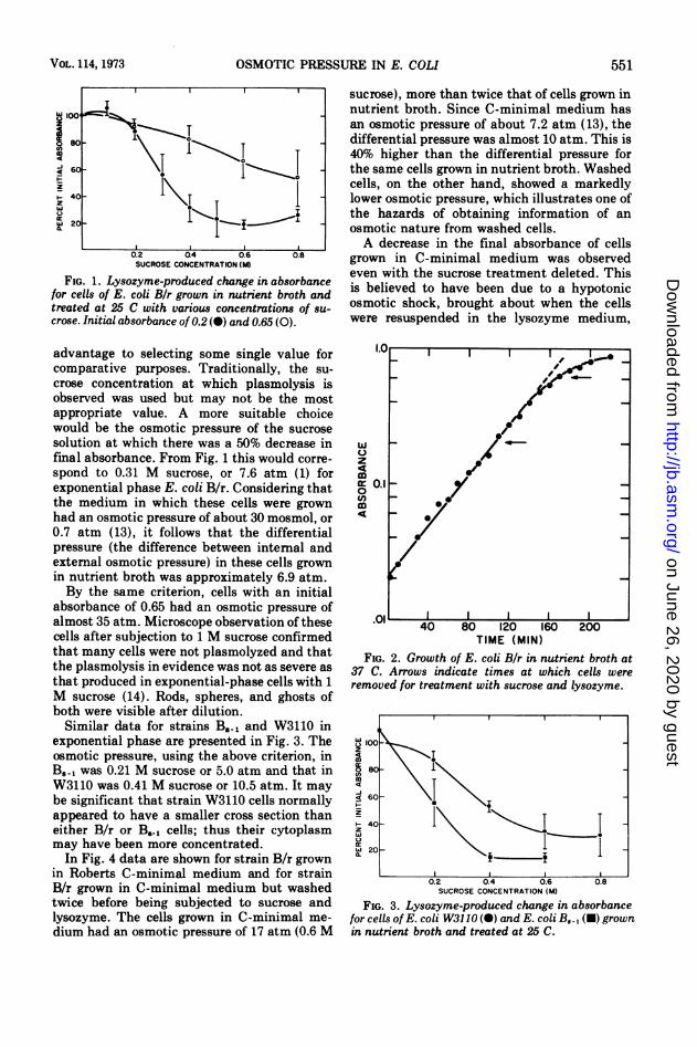
FIG. 1. Lysozyme-produced change in absorbancefor cells of E. coli Bir grown in nutrient broth andtreated at 25 C with various concentrations of su-crose. Initial absorbance of 0.2 (0) and 0.65 (0).
advantage to selecting some single value forcomparative purposes. Traditionally, the su-
crose concentration at which plasmolysis isobserved was used but may not be the mostappropriate value. A more suitable choicewould be the osmotic pressure of the sucrose
solution at which there was a 50% decrease infinal absorbance. From Fig. 1 this would corre-
spond to 0.31 M sucrose, or 7.6 atm (1) forexponential phase E. coli B/r. Considering thatthe medium in which these cells were grownhad an osmotic pressure of about 30 mosmol, or
0.7 atm (13), it follows that the differentialpressure (the difference between intemal andexternal osmotic pressure) in these cells grownin nutrient broth was approximately 6.9 atm.By the same criterion, cells with an initial
absorbance of 0.65 had an osmotic pressure ofalmost 35 atm. Microscope observation of thesecells after subjection to 1 M sucrose confirmedthat many cells were not plasmolyzed and thatthe plasmolysis in evidence was not as severe asthat produced in exponential-phase cells with 1M sucrose (14). Rods, spheres, and ghosts ofboth were visible after dilution.Similar data for strains B5., and W3110 in
exponential phase are presented in Fig. 3. Theosmotic pressure, using the above criterion, inB5.1 was 0.21 M sucrose or 5.0 atm and that inW3110 was 0.41 M sucrose or 10.5 atm. It maybe significant that strain W3110 cells normallyappeared to have a smaller cross section thaneither B/r or B., cells; thus their cytoplasmmay have been more concentrated.In Fig. 4 data are shown for strain B/r grown
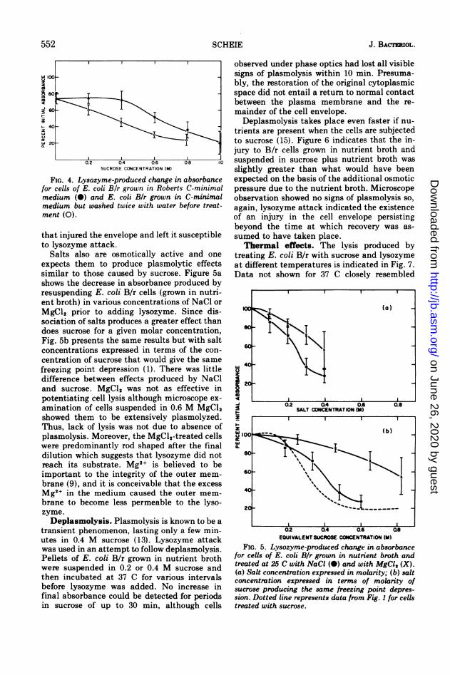
in Roberts C-minimal medium and for strainB/r grown in C-minimal medium but washedtwice before being subjected to sucrose andlysozyme. The cells grown in C-minimal me-
dium had an osmotic pressure of 17 atm (0.6 M
sucrose), more than twice that of cells grown innutrient broth. Since C-minimal medium hasan osmotic pressure of about 7.2 atm (13), thedifferential pressure was almost 10 atm. This is40% higher than the differential pressure forthe same cells grown in nutrient broth. Washedcells, on the other hand, showed a markedlylower osmotic pressure, which illustrates one ofthe hazards of obtaining information of anosmotic nature from washed cells.A decrease in the final absorbance of cells
grown in C-minimal medium was observedeven with the sucrose treatment deleted. Thisis believed to have been due to a hypotonicosmotic shock, brought about when the cellswere resuspended in the lysozyme medium,
I. I
w
z
4
ir 0.1
0/U)mc,
TIME (MIN)FIG. 2. Growth of E. coli Bir in nutrient broth at
37 C. Arrows indicate times at which cells wereremoved for treatment with sucrose and lysozyme.
z4
m
4
z
tL
SUCROSE CONCENTRATION (M)
FIG. 3. Lysozyme-produced change in absorbancefor cells of E. coli W3110 (0) and E. coli BJ., (U) grownin nutrient broth and treated at 25 C.
- 1*I lI
~~~~~
I
_0
0.8
VOL. 114, 1973 551
on June 26, 2020 by guesthttp://jb.asm
.org/D
ownloaded from
J. BACTERIOL.
i 100-
so
ml
z
20-
SUCROSE CONCENTRATION (M)
FIG. 4. Lysozyme-produced change in absorbancefor cells of E. coli B/r grown in Roberts C-minimalmedium (0) and E. coli Bir grown in C-minimalmedium but washed twice with water before treat-ment (0).
that injured the envelope and left it susceptibleto lysozyme attack.
Salts also are osmotically active and oneexpects them to produce plasmolytic effectssimilar to those caused by sucrose. Figure 5ashows the decrease in absorbance produced byresuspending E. coli B/r cells (grown in nutri-ent broth) in various concentrations of NaCl orMgCl2 prior to adding lysozyme. Since dis-sociation of salts produces a greater effect thandoes sucrose for a given molar concentration,Fig. 5b presents the same results but with saltconcentrations expressed in terms of the con-centration of sucrose that would give the samefreezing point depression (1). There was littledifference between effects produced by NaCland sucrose. MgCl2 was not as effective inpotentiating cell lysis although microscope ex-amination of cells suspended in 0.6 M MgCl2showed them to be extensively plasmolyzed.Thus, lack of lysis was not due to absence ofplasmolysis. Moreover, the MgCl2-treated cellswere predominantly rod shaped after the finaldilution which suggests that lysozyme did notreach its substrate. Mg2+ is believed to beimportant to the integrity of the outer mem-brane (9), and it is conceivable that the excessMg2+ in the medium caused the outer mem-brane to become less permeable to the lyso-zyme.Deplasmolysis. Plasmolysis is known to be a
transient phenomenon, lasting only a few min-utes in 0.4 M sucrose (13). Lysozyme attackwas used in an attempt to follow deplasmolysis.Pellets of E. coli B/r grown in nutrient brothwere suspended in 0.2 or 0.4 M sucrose andthen incubated at 37 C for various intervalsbefore lysozyme was added. No increase infinal absorbance could be detected for periodsin sucrose of up to 30 min, although cells
observed under phase optics had lost all visiblesigns of plasmolysis within 10 min. Presuma-bly, the restoration of the original cytoplasmicspace did not entail a return to normal contactbetween the plasma membrane and the re-mainder of the cell envelope.Deplasmolysis takes place even faster if nu-
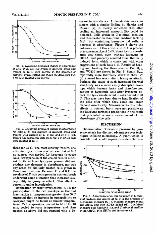
trients are present when the cells are subjectedto sucrose (15). Figure 6 indicates that the in-jury to B/r cells grown in nutrient broth andsuspended in sucrose plus nutrient broth wasslightly greater than what would have beenexpected on the basis of the additional osmoticpressure due to the nutrient broth. Microscopeobservation showed no signs of plasmolysis so,again, lysozyme attack indicated the existenceof an injury in the cell envelope persistingbeyond the time at which recovery was as-sumed to have taken place.Thermal effects. The lysis produced by
treating E. coli B/r with sucrose and lysozymeat different temperatures is indicated in Fig. 7.Data not shown for 37 C closely resembled
wU
z
49-iSI1-Z
EQUIVALENT SUCROSE CONCENTRATION (M)
FIG. 5. Lysozyme-produced change in absorbancefor cells of E. coli B/r grown in nutrient broth andtreated at 25 C with NaCI (0) and with MgCI, (X).(a) Salt concentration expressed in molarity; (b) saltconcentration expressed in terms of molarity ofsucrose producing the same freezing point depres-sion. Dotted line represents data from Fig. 1 for cellstreated with sucrose.
552 SCHEIE
on June 26, 2020 by guesthttp://jb.asm
.org/D
ownloaded from
OSMOTIC PRESSURE IN E. COLI
SUCROSE CONCENTRATION (M)
FIG. 6. Lysozyme-produced change in absorbanceof cells of E. coli Bir grown in nutrient broth andtreated at 25 C with sucrose in the presence ofnutrient broth. Dotted line shows the data from Fig.1 for cells treated with sucrose.
w100§ 80 -
IT~~~~
IzIUw 20-
( L0.2 0.4 0.6 0.8
SUCROSE CONCENTRATION (M)
FIG. 7. Lysozyme-produced change in absorbancefor cells of E. coli B/grown in nutrient broth andtreated with sucrose at 11 C (0) and at 0 C (x).Dotted line represents data from Fig. 1 in which cellswere treated at 25 C.
those for 25 C. The most striking feature, oneexhibited by all three strains, was that at 0 Cno sucrose was needed for lysozyme to causelysis. Resuspension of the cooled cells in nutri-ent broth with no lysozyme present did notproduce any decrease in absorbance, nor wasany noted for similarly treated cells grown inC-minimal medium. Between 11 and 0 C theenvelope of E. coli cells grown in nutrient brothunderwent some alteration that increased sus-ceptibility to lysozyme attack. This effect iscurrently under investigation.
Implication by other investigators (6, 12) forparticipation of the cell envelope in thermalinactivation at temperatures greater than 50 Csuggested that an increase in susceptibility tolysozyme might be found at similar tempera-tures. Cell suspensions heated to 55 C for 10min, cooled to room temperature, and thentreated as above did not respond with a de-
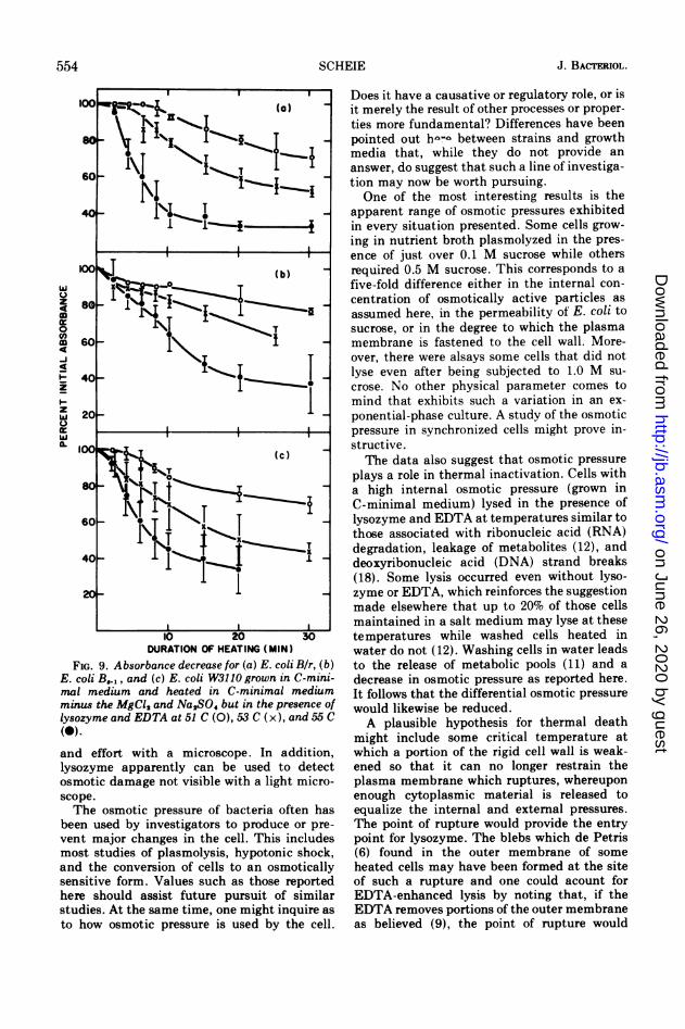
crease in absorbance. Although this was con-sistent with a similar finding by Harries andRussell (7), it merely indicated that aftercooling no increased susceptibility could bedetected. Cells grown in C-minimal mediumand then heated in C-minimal medium lackingMg2+ but containing lysozyme did suffer adecrease in absorbance. Figure 8 shows theenhancement of this effect with EDTA presentat a concentration of 0.4%. Some loss in absorb-ance was noted, even without lysozyme orEDTA, and probably resulted from thermallyinduced lysis, which is consistent with othersuggestions of such lysis (12). Results of heat-ing and treating the three strains, B/r, B..1,and W3110 are shown in Fig. 9. Strain B8l,reportedly more thermally sensitive than B/r(4), showed less sensitivity to lysozyme attack.Perhaps the cause of such increased thermalsensitivity was a more easily disrupted enve-lope which became leaky and therefore notsubject to hypotonic lysis after lysozyme at-tack. No lysis was detected in cells heated to 70C. This may have been due to heat fixation ofthe cells after which they could no longerrespond osmotically. Measurements of similareffects in nutrient broth were not possible asthe lysozyme formed a precipitate in the broththat prevented accurate measurement of theabsorbance of the cells.
DISCUSSIONDetermination of osmotic pressure by lyso-
zyme attack has distinct advantages over tech-niques utilizing microscopy. A quantitation ispossible that would require considerable time
.08
wU
z4 .06 *
04
DURATION OF HEATING (MIN)
FIG. 8. Absorbance of E. coli B/r grown in C-mini-mal medium and heated at 55 C in the presence ofC-minimal medium (0), C-minimal medium minusthe MgCl, and with EDTA added (0), C-minimalmedium plus lysozyme (x), and C-minimal mediumminus MgCI2 plus EDTA and lysozyme (A).
553VOL. 114, 1973
on June 26, 2020 by guesthttp://jb.asm
.org/D
ownloaded from
J. BACTERIOL.
w
z49
It0C,)
o
-J
49
z
z
w
I.
DURATION OF HEATING (MIN)
FIG. 9. Absorbance decrease for (a) E. coli Bir, (b)E. coli B.,, and (c) E. coli W3110 grown in C-mini-mal medium and heated in C-minimal mediumminus the MgCl2 and Na,SO4 but in the presence oflysozyme and EDTA at 51 C (O), 53 C (x), and 55 C(0)-
and effort with a microscope. In addition,lysozyme apparently can be used to detectosmotic damage not visible with a light micro-scope.The osmotic pressure of bacteria often has
been used by investigators to produce or pre-
vent major changes in the cell. This includesmost studies of plasmolysis, hypotonic shock,and the conversion of cells to an osmoticallysensitive form. Values such as those reportedhere should assist future pursuit of similarstudies. At the same time, one might inquire as
to how osmotic pressure is used by the cell.
Does it have a causative or regulatory role, or isit merely the result of other processes or proper-ties more fundamental? Differences have beenpointed out h--- between strains and growthmedia that, while they do not provide ananswer, do suggest that such a line of investiga-tion may now be worth pursuing.One of the most interesting results is the
apparent range of osmotic pressures exhibitedin every situation presented. Some cells grow-ing in nutrient broth plasmolyzed in the pres-ence of just over 0.1 M sucrose while othersrequired 0.5 M sucrose. This corresponds to afive-fold difference either in the internal con-centration of osmotically active particles asassumed here, in the permeability of E. coli tosucrose, or in the degree to which the plasmamembrane is fastened to the cell wall. More-over, there were alsays some cells that did notlyse even after being subjected to 1.0 M su-crose. No other physical parameter comes tomind that exhibits such a variation in an ex-ponential-phase culture. A study of the osmoticpressure in synchronized cells might prove in-structive.The data also suggest that osmotic pressure
plays a role in thermal inactivation. Cells witha high internal osmotic pressure (grown inC-minimal medium) lysed in the presence oflysozyme and EDTA at temperatures similar tothose associated with ribonucleic acid (RNA)degradation, leakage of metabolites (12), anddeoxyribonucleic acid (DNA) strand breaks(18). Some lysis occurred even without lyso-zyme or EDTA, which reinforces the suggestionmade elsewhere that up to 20% of those cellsmaintained in a salt medium may lyse at thesetemperatures while washed cells heated inwater do not (12). Washing cells in water leadsto the release of metabolic pools (11) and adecrease in osmotic pressure as reported here.It follows that the differential osmotic pressurewould likewise be reduced.A plausible hypothesis for thermal death
might include some critical temperature atwhich a portion of the rigid cell wall is weak-ened so that it can no longer restrain theplasma membrane which ruptures, whereuponenough cytoplasmic material is released toequalize the internal and external pressures.The point of rupture would provide the entrypoint for lysozyme. The blebs which de Petris(6) found in the outer membrane of someheated cells may have been formed at the siteof such a rupture and one could acount forEDTA-enhanced lysis by noting that, if theEDTA removes portions of the outer membraneas believed (9), the point of rupture would
554 SCHEIE
on June 26, 2020 by guesthttp://jb.asm
.org/D
ownloaded from
OSMOTIC PRESSURE IN E. COLI
become more rapidly accessible to attack bylysozyme. Furthermore, it also has been re-ported that Aerobacter aerogenes cells heatedin water are more resistant to inactivation thanare those heated in a growth-supporting me-dium (17).One might reasonably expect a lesion in the
membrane to heal once the pressure differen-tial is removed. However, subsequent degrada-tion of RNA may again increase the intemalosmotic pressure, to the point ,of producinglysis, providing the integrity of the peptidogly-can layer had been destroyed. According tosuch an hypothesis one would expect lysozymeto be ineffective when added after cells hadbeen cooled. What remains, of course, is thequestion as to which of the various heat-induced phenomena is the primary cause ofthermal inactivation.
ITrERATURE CITED1. American Optical Co. 1964. Tables of properties of
aqueous solutions related to index of refraction. Amer-ican Optical Co., Buffalo, N.Y.
2. Bayer, M. E. 1967. Response of cell walls of Escherichiacoli to a sudden reduction of the environmentalosmotic pressure. J. Bacteriol. 93:1104-1112.
3. Birdsell, D. C., and E. H. Cota-Robles. 1967. Productionand ultrastructure of lysozyme and ethyleneamine-tetraacetate-lysozyme apheroplasts of EscherichiacpU. J. Bacteriol. 93:427-437.
4. Bridges, B. A., M. J. Ashwood-Smith, and R. J. Munson.1969. Correlation of bacterial sensitivities to ionizingradiation and mild heating. J. Gen. Microbiol.58:115-124.
5. Cota-Robles, E. H. 1963. Electron microscopy of plas-molysis in Escherichia coli. J. Bacteriol. 85:499-503.
6. de Petris, S. 1967. Ultrastructure of the cell wall ofEscherichia coli and chemical nature of its constituentlayers. J. Ultrastruc. Res. 19:45-83.
7. Harries, D., and A. D. Russell. 1967. A note on somechanges in the physical properties of heated suspen-sions of Escherichia coli J. Pharm. Pharmacol.19:740-743.
8. Heppel, L. A. 1967. Selective release of enzymes frombacteria. Science 156:1451-1455.
9. Leive, L., U. K. Shoulin, and S. E. Mergenhagen. 1968.Physical, chemical, and immunological properties oflipopolysaccharide released from Escherichia coli byethylenediaminetetraacetate. J. Biol. Chem.243:6384-391.
10. Mitchell, P., and J. Moyle. 1956. Osmotic function andstructure in bacteria. Symp. Soc. Gen Microbiol.6:150-180.
11. Roberts, R. B., P. H. Abelson, D. B. Cowie, E. T. Bolton,and R. J. Britten. 1957. Studies of biosynthesis inEscherichia coli. Carnegie Inst. Wash. Publ. 607.
12. Russell, A. D., and D. Harries. 1968. Damage to Esche-richia coli on exposure to moist heat. Appl. Microbiol.16:1394-1399.
13. Scheie, P. 0. 1969. Plasmolysis of Escherichia coli B/rwith sucrose. J. Bacteriol. 98:335-340.
14. Scheie, P., and H. Dalen. 1968. Spatial anisotropy inEacherichia cok. J. Bacteriol. 96:1413-1414.
15. Scheie, P., and R. Rehberg. 1972. Response of Esche-richia coli B/r to high concentrations of sucrose in anutrient medium. J. Bacteriol. 109:229-235.
16. Snedecor, G. W. 1956. Statistical methods, p. 38. TheIowa State University Press, Ames, Iowa.
17. Strange, R. E., and M. Shon. 1964. Effects of thermalstress on viability and RNA ofAerobacter aerogenes inaqueous suspension. J. Gen. Microbiol. 34:99-114.
18. Woodcock, E., and G. W. Grigg. 1972. Repair of ther-mally induced DNA breakage in Escherichia coli.Nature N. Biol. 237:76-79.
555VOL. 114, 1973
on June 26, 2020 by guesthttp://jb.asm
.org/D
ownloaded from