Oseltamivir (Tamiflu TM) is a substrate of...
30
Title page Oseltamivir (Tamiflu TM ) is a substrate of PEPT1 Takuo Ogihara, Takashi Kano, Tamae Wagatsuma, Sho Wada, Hikaru Yabuuchi, Shigeki Enomoto, Kaori Morimoto, Yoshiyuki Shirasaka, Shoko Kobayashi and Ikumi Tamai Faculty of Pharmacy, Takasaki University of Health and Welfare, 60, Nakaorui, Takasaki, Gunma 370-0033, Japan (T.O., T.K., S.W., K.M.), Department of Food and Life Science, Takasaki University of Health and Welfare, 37-1, Nakaorui, Takasaki, Gumma 370-1295, Japan (T.W., S.K.) Division of Pharmaceutical Sciences, Graduate School of Natural Science and Technology, Kanazawa University, Kakuma-machi, Kanazawa 920-1192 (Y.S., I.T.) GenoMembrane, Inc., Tsurumi Leading Venture Plaza, 75-1, Ono, Tsurumi, Yokohama, Kanagawa 230-0046 (H.Y., S.E.) DMD Fast Forward. Published on May 13, 2009 as doi:10.1124/dmd.109.026922 Copyright 2009 by the American Society for Pharmacology and Experimental Therapeutics. This article has not been copyedited and formatted. The final version may differ from this version. DMD Fast Forward. Published on May 13, 2009 as DOI: 10.1124/dmd.109.026922 at ASPET Journals on February 14, 2019 dmd.aspetjournals.org Downloaded from
Transcript of Oseltamivir (Tamiflu TM) is a substrate of...

DMD#26922
1
Title page
Oseltamivir (TamifluTM) is a substrate of PEPT1
Takuo Ogihara, Takashi Kano, Tamae Wagatsuma, Sho Wada, Hikaru Yabuuchi, Shigeki Enomoto,
Kaori Morimoto, Yoshiyuki Shirasaka, Shoko Kobayashi and Ikumi Tamai
Faculty of Pharmacy, Takasaki University of Health and Welfare, 60, Nakaorui, Takasaki, Gunma
370-0033, Japan (T.O., T.K., S.W., K.M.),
Department of Food and Life Science, Takasaki University of Health and Welfare, 37-1, Nakaorui,
Takasaki, Gumma 370-1295, Japan (T.W., S.K.)
Division of Pharmaceutical Sciences, Graduate School of Natural Science and Technology,
Kanazawa University, Kakuma-machi, Kanazawa 920-1192 (Y.S., I.T.)
GenoMembrane, Inc., Tsurumi Leading Venture Plaza, 75-1, Ono, Tsurumi, Yokohama, Kanagawa
230-0046 (H.Y., S.E.)
DMD Fast Forward. Published on May 13, 2009 as doi:10.1124/dmd.109.026922
Copyright 2009 by the American Society for Pharmacology and Experimental Therapeutics.
This article has not been copyedited and formatted. The final version may differ from this version.DMD Fast Forward. Published on May 13, 2009 as DOI: 10.1124/dmd.109.026922
at ASPE
T Journals on February 14, 2019
dmd.aspetjournals.org
Dow
nloaded from

DMD#26922
2
Running title page.
Running title:
Oseltamivir is a substrate of PEPT1
Corresponding author:
Prof. Takuo Ogihara, Ph.D.
Laboratory of Biopharmaceutics, Department of Pharmacology, Faculty of Pharmacy, Takasaki
University of Health and Welfare, 60, Nakaorui, Takasaki, Gunma 370-0033, Japan,
Phone; +81-27-352-1180, Fax: ;+81-27-352-1118; E-mail: [email protected]
Document statistics:
The number of text pages: 22
The number of tables: 1
The number of figures: 7
The number of references: 16
Number of words:
Abstract; 148 words
Introduction; 490 words
Discussion; 615 words
PEPT1, peptide transporter1; P-gp, P-glycoprotein; HBSS, Hanks’ balanced salt solution;
DMEM, Dulbecco's modified Eagle's medium; CLtot, total clearance; HPLC, high-performance
liquid chromatography; TEER, transepithelial electrical resistance; FBS, fetal bovine serum; Gly-Sar,
glycyl-sarcosine; BNPP, bis(4-nitrophenyl) phosphate; BSA, bovine serum albumin; Cmax, The
maximum plasma concentration; Tmax, time to Cmax; AUC0-6hr, area under the plasma
concentration-time curve from time 0 to 6 hr; CES, human carboxylesterase
This article has not been copyedited and formatted. The final version may differ from this version.DMD Fast Forward. Published on May 13, 2009 as DOI: 10.1124/dmd.109.026922
at ASPE
T Journals on February 14, 2019
dmd.aspetjournals.org
Dow
nloaded from

DMD#26922
3
ABSTRACT
Oseltamivir, an ester-type prodrug of the neuraminidase inhibitor Ro 64-0802, has been
developed for the treatment of A and B strains of the influenza virus, but has neuropsychiatric and
other side effects. In this study, we characterized the transport across intestinal epithelial cells and
the absorption of oseltamivir in rats. Uptake by Caco-2 cells (human carcinoma cell line) and HeLa
cells transfected with peptide transporter1 (HeLa/PEPT1) was time- and temperature-dependent, and
was inhibited by typical PEPT1 inhibitors such as glycyl-sarcosine (Gly-Sar). The uptake by Caco-2
cells and HeLa/PEPT1 was saturable, with similar Km values. Oseltamivir absorption in adult rats
was greatly reduced by simultaneous administration of milk, casein or Gly-Sar. Further, the plasma
and brain concentrations of oseltamivir were higher in fasting than in non-fasting rats after oral
administration. These results suggest that oseltamivir is a substrate of PEPT1, and that PEPT1 is
involved in its intestinal absorption.
This article has not been copyedited and formatted. The final version may differ from this version.DMD Fast Forward. Published on May 13, 2009 as DOI: 10.1124/dmd.109.026922
at ASPE
T Journals on February 14, 2019
dmd.aspetjournals.org
Dow
nloaded from

DMD#26922
4
INTRODUCTION
Various transporters are expressed on apical and basolateral membranes of intestinal
epithelial cells, serving to take up nutrients and to excrete xenobiotics into the lumen. Influx
transporters are able to accept nutrients and also various drugs as substrates. In particular, peptide
transporter1 (PEPT1, SLC15A1), localized at brush-border membranes of human small intestine
(Saito et al., 1995), plays important roles in the absorption of not only di/tri-peptides (Tamai et al.,
1994), but also peptide-mimetic compounds, such as orally administered beta-lactam antibiotics
(Ganapathy et al., 1995; Sai et al., 1996) and the anticancer agent bestatin (Tomita et al., 1990; Inui
et al., 1992). Recently, several researchers have found that intestinal PEPT1 can transport L-valine
ester prodrugs, such as valacyclovir and valgancyclovir (Balimane et al., 1998; Han et al., 1998;
Sugawara et al., 2000). Therefore, such structural modification of drugs may result in increased
intestinal absorption, mediated by PEPT1.
Oseltamivir phosphate (oseltamivir), manufactured under the trade name TamifluTM as an
ester-type prodrug of the neuraminidase inhibitor Ro 64-0802, has been developed for the treatment
of A and B strains of the influenza virus. This drug has been reported to be associated with
neuropsychiatric side effects (http://www.fda.gov/cder/drug/infopage/tamiflu/QA20051117.htm and
http://www.mhlw.go.jp/ english/index.html), which are likely to be caused by distribution of
oseltamivir and/or its metabolite(s) to the central nervous system. Recently, we examined the
possible role of P-glycoprotein (P-gp) as the determinant of brain distribution of oseltamivir and Ro
64-0802 both in vitro using LLC-GA5-COL150 cells, which over-express human MDR1 P-gp on the
apical membrane, and in vivo using mdr1a/1b knockout mice (Morimoto et al., 2008). The
permeability of oseltamivir in the basolateral-to-apical direction was significantly greater than that in
the opposite direction. The brain distribution of oseltamivir was increased in mdr1a/1b knockout
mice compared with wild-type mice. In contrast, negligible transport of Ro 64-0802 by P-gp was
This article has not been copyedited and formatted. The final version may differ from this version.DMD Fast Forward. Published on May 13, 2009 as DOI: 10.1124/dmd.109.026922
at ASPE
T Journals on February 14, 2019
dmd.aspetjournals.org
Dow
nloaded from
DMD#26922
5
observed in both in vitro and in vivo studies. These results demonstrated that oseltamivir, but not Ro
64-0802, was a substrate of P-gp. Accordingly, low levels of P-gp activity or drug-drug interactions
at P-gp may lead to enhanced brain accumulation of oseltamivir, and this in turn may account for the
central nervous system effects of oseltamivir observed in some patients (Morimoto et al., 2008).
During that research, we noticed that increased plasma concentration and toxicity were observed in
fasted infant rats compared with non-fasted ones. Therefore, we speculated that transporters involved
in the uptake of food components also take part in the absorption of oseltamivir.
The purpose of the present study was to characterize the transport of oseltamivir across
intestinal epithelial cells. We first examined whether oseltamivir is a substrate of PEPT1 by using a
human carcinoma cell line, Caco-2 cells, and HeLa cells stably expressing human PEPT1
(HeLa/PEPT1). A rat in vivo study was then conducted to confirm involvement of PEPT1 in
oseltamivir absorption in the small intestine. Our findings indicate that a drug-food interaction with
potential clinical significance is likely to occur between oseltamivir and milk.
METHODS
Chemicals and animals
Oseltamivir phosphate was purchased from Sequoia Research Products (Pangbourne, UK).
Ro 64-0802 was biologically synthesized from oseltamivir using porcine liver esterase (Sigma, St
Louis, MO) as described previously (Morimoto et al., 2008). The human colon adenocarcinoma cell
line, Caco-2, was obtained from the American Type Culture Collection (Rockville, MD, USA).
HeLa/PEPT1 and HeLa transfected with vector alone (mock) were established as described
previously (Nakanishi et al., 2000). Dulbecco's modified Eagle's medium (DMEM), nonessential
amino acids, penicillin, streptomycin, gentamycin, and Hanks’ balanced salt solution (HBSS) were
all from Invitrogen Corp. (Carlsbad, CA, USA). Fetal bovine serum (FBS) was obtained from ICN
This article has not been copyedited and formatted. The final version may differ from this version.DMD Fast Forward. Published on May 13, 2009 as DOI: 10.1124/dmd.109.026922
at ASPE
T Journals on February 14, 2019
dmd.aspetjournals.org
Dow
nloaded from

DMD#26922
6
Biomedicals, Inc. (Osaka, Japan), and type-I collagen solution was from Nitta Gelatin (Osaka,
Japan). Glycyl-sarcosine (Gly-Sar), bis(4-nitrophenyl) phosphate (BNPP) and bovine serum albumin
(BSA) were from SIGMA (St. Louis, MO). The protein assay kit was purchased from Bio-Rad
Laboratories Inc. (Hercules, CA). All other chemicals and solvents were commercial products of
analytical, HPLC or LC/MS grade as appropriate.
The animal study was performed according to the Guidelines for the Care and Use of
Laboratory Animals at the Takasaki University of Health and Welfare and approved by the
Committee of Ethics of Animal Experimentation of the university. One- and eight- week-old Wistar
rats were purchased from SLC Japan (Hamamatsu, Japan)
Cell culture and cellular quality assessment
Caco-2 cells were cultured in DMEM containing 10% FBS, 1% nonessential amino acids,
100 units/mL penicillin, 0.1 mg/mL streptomycin, and 25 μg/mL gentamycin in a humidified
atmosphere of 5% CO2 at 37°C. Cells at passage number from 60 to 80 were used. For the transport
study, Caco-2 cells were routinely grown to confluence in 75-cm2 tissue culture dishes and seeded
into Transwell inserts coated with type-I collagen (pore size: 0.4 μm; diameter: 12 mm, Costar,
Cambridge, MA, USA). The cells were seeded at a density of 2 x 10⁵ cells/cm2 and monolayers were
formed after culturing for 2 weeks. The integrity of the cell layer was evaluated by measurement of
transepithelial electrical resistance (TEER) with Millicell-ERS equipment (Nihon Millipore, Tokyo,
Japan). Monolayers with a TEER of more than 300 Ω•cm2 were used for the transepithelial
transport experiments. These monolayers were also used for permeation studies, as previously
reported (Kobayashi et al., 2008). TEER of the monolayers was measured before and after each
transport experiment. For the uptake study, Caco-2 cells were seeded at a density of 2 x 10⁵ cells/cm2
on multiwell dishes (Nunc, Naperville, IL) coated with collagen. Cells were grown for 7 days for the
This article has not been copyedited and formatted. The final version may differ from this version.DMD Fast Forward. Published on May 13, 2009 as DOI: 10.1124/dmd.109.026922
at ASPE
T Journals on February 14, 2019
dmd.aspetjournals.org
Dow
nloaded from

DMD#26922
7
uptake experiment (Kimoto et al., 2007).
HeLa/PEPT1 and mock cells were grown in DMEM supplemented with 10% FBS, 100
units/mL penicillin, 0.1 mg/mL streptomycin, 2 mM glutamine, and 1 mg/mL geneticin (G418) as
described previously (Nakanishi et al., 2000). For the uptake assay, each cell line was seeded on
eight-well plates (Nunc, Naperville, IL) and cultured for 3 days.
Transcellular transport and uptake experiments
In the transcellular transport study, monolayers of Caco-2 cells were gently rinsed twice
with HBSS [136.9 mM NaCl, 5.37 mM KCl, 5.55 mM D-glucose, 1.258 mM CaCl2, 0.441 mM
KH2PO4, 0.811 mM MgSO4, 0.337 mM Na2HPO4, 4.047 mM NaHCO3, 10 mM
2-[4-(2-hydroxyethyl)-1-piperazinyl]ethanesulfonic acid (HEPES) for pH 7.4 or 10 mM
2-(N-morpholino)ethanesulfonic acid (MES) for pH 6.0] and left to equilibrate in the same
solution for 20 min at 37°C. To measure the apical-to-basolateral permeability, 1.5 mL of HBSS (pH
7.4, 37°C) was added to the basolateral chamber of the Transwell insert and then 0.5 mL of the test
solution (pH 6.0, 37°C) containing oseltamivir was added to the apical side. After the desired
incubation time at 37°C, the basolateral solution was collected, and replaced with an equal volume
of HBSS. To investigate the effect of esterase inhibitor treatment of Caco-2 cells on the permeability
of oseltamivir in the apical-to-basolateral direction, Caco-2 cells were preincubated with 200 µM
BNPP, a specific carboxylesterase inhibitor (Block et al., 1978; Mentlein et al., 1988) at 37°C for 45
min. We also examined the influence of permeability of oseltamivir across a Caco-2 cell monolayer
in the presence of 10 μM verapamil, an inhibitor of P-gp (Ogihara et al., 2004).
In the case of the uptake study, Caco-2 cells grown on the multidishes were washed with 2
mL of HBSS and preincubated for 20 min. After preincubation, HBSS (300 μL) containing
oseltamivir was added to initiate uptake. The cells were incubated at 37°C for a designated time, and
This article has not been copyedited and formatted. The final version may differ from this version.DMD Fast Forward. Published on May 13, 2009 as DOI: 10.1124/dmd.109.026922
at ASPE
T Journals on February 14, 2019
dmd.aspetjournals.org
Dow
nloaded from

DMD#26922
8
then washed five times with 2 mL of ice-cold HBSS to terminate the uptake.
Uptake study with HeLa cells was also performed at 37°C in HBSS adjusted to pH 6.0.
Cultured cells were washed and preincubated in the buffer without oseltamivir or Ro 64-0802 for 10
min at 37°C. The uptake was initiated by adding HBSS (300 μL) containing oseltamivir or Ro
64-0802. After incubation for designated times at 37°C, the experiment was terminated by removing
the medium, followed by washing five times with 2 mL of ice-cold HBSS. For quantitation of the
drug taken up by the cells, the cells were suspended in acetonitlile/methanol (25/75; vol/vol),
collected with a cell scraper (Asahi Techno Glass Corporation Co. Ltd., Chiba, Japan), and then
sonicated for 30 min, followed by centrifugation at 12000 rpm and 4˚C for 10 min. The supernatant
was evaporated under centrifugal evaporator at 30˚C. The residue was dissolved in 0.1 mL of 10 mM
ammonium acetate buffer (pH7.0) and filtered by passing through a 0.45 µm pore size membrane
filter (Millipore Corporation, Billerica MA, USA). The supernatants were then subjected to LC-MS
analysis.
Cellular protein was determined using a protein assay kit with bovine serum albumin as a
standard.
In vitro data analysis
The permeability (µL/mg protein) was expressed as the apical-to-basolateral side obtained
by dividing the transported amount (µmol/mg protein) at the basolateral side by the initial
concentration (µM) in the apical side. The permeability coefficient (Papp, cm/s) was calculated from
the linear portion of an uptake vs time plot using the follow equation:
Papp = dQ/dt/A/C0
where dQ/dt is the initial permeation rate across the Caco-2 cell monolayer (µmol/s), A is the surface
area of the filter (cm2), and C0 is the initial concentration of the solution in the apical side (µM).
This article has not been copyedited and formatted. The final version may differ from this version.DMD Fast Forward. Published on May 13, 2009 as DOI: 10.1124/dmd.109.026922
at ASPE
T Journals on February 14, 2019
dmd.aspetjournals.org
Dow
nloaded from

DMD#26922
9
Cell-to-medium ratio was obtained by dividing the cellular uptake amount by the
concentration of test compound in the medium. Kinetic parameters for transport activity were
estimated by nonlinear least-squares fitting of the data to the following equation using the MULTI
program:
V = Vmax • S / (Km + S) + Kd • S
where V, S, Km, Vmax, and Kd represent the initial uptake rate, substrate concentration, Michaelis
constant, maximum uptake rate, and first-order rate constant, respectively.
Pharmacokinetic study
The eight-week-old male rats (weights: 185-206g) were deprived of food for 12 hr before
experiments. Oseltamivir was dissolved in distilled water, milk (commercially available cows’ milk),
Gly-Sar solution (20 mM or 125 mM), or casein solution (300 mg/10 mL), and was orally
administered to rats at a single dose of 30 mg/kg (the dosing volume: 10 mL/kg). For the intravenous
injection study, eight-week-old male rats (weights: 177-186g) were administered with 30 mg/kg of
oseltamivir via the jugular vein (the dosing volume: 1 mL/kg). Blood samples were withdrawn from
the jugular vein of rats with a heparinized syringe at designated times under anesthesia induced with
diethyl ether. In the case of infant rats, one-week-old infant rats (weights: 16.5-20.6g) from the same
parental female rat were used. Three of them (2 male, 1 female) were separated from the parental
female rat and starved overnight, and the others (3 male, 2 female) remained with the parental rat
and were fed milk. Oseltamivir dissolved in distilled water was orally administered to infant rats at a
single dose of 30 mg/kg (the dosing volume: 10 mL/kg). Blood samples were collected from the
jugular vein at 30 min after the start of administration under anesthesia induced with diethyl ether,
and then the infant rats were decapitated. The brain was quickly excised, rinsed with ice-cold saline,
blotted dry and weighed. Samples were stored at -30°C until analysis. Blood samples were
This article has not been copyedited and formatted. The final version may differ from this version.DMD Fast Forward. Published on May 13, 2009 as DOI: 10.1124/dmd.109.026922
at ASPE
T Journals on February 14, 2019
dmd.aspetjournals.org
Dow
nloaded from

DMD#26922
10
centrifuged (1700 x g) for 15 min at 4°C to obtain plasma. As it has been reported that rat intestinal
PEPT1 expression shows a diurnal rhythm, all in vivo studies have conducted at the same time of the
day (Pan et al., 2004). Quantification of oseltamivir and Ro 64-0802 in plasma and brain tissues was
performed using reported methods (Wiltshire et al., 2000) with some modification. Briefly, aliquots
of brain tissues (100 mg) were homogenized with 1 mL of 5 mM ammonium acetate buffer, followed
by centrifugation at 1700 x g, and 0.9 mL of the supernatant was subjected to solid-phase extraction
(Empore Mixed Phase Cation, 7 mm/3 mL, 3M Bioanalytical Technologies, St. Paul, MN). The
methods used for the extraction of plasma and brain homogenate were the same. The maximum
plasma concentration (Cmax) and time to Cmax (Tmax) were determined directly from the observed data.
The area under the plasma concentration-time curve from time 0 to 6 hr (AUC0-6hr) was estimated by
the linear trapezoidal method.
Analytical methods
Aliquots (5 µL) of samples containing oseltamivir and Ro 64-0802 were injected into an
HPLC system (LC-20A system, Shimadzu, Kyoto, Japan) equipped with a CapcellpakTM OD column
(150 x 2.0 mm i.d., Shiseido, Tokyo, Japan) using isocratic elution at 0.1 mL/min with 0.05% formic
acid. Analytes were detected using a quadrupole mass spectrometer (LCMS-2010EV; Shimadzu,
Kyoto, Japan) fitted with an electrospray ionization source. Analytes were detected in the positive
mode, and protonated molecular ions at m/z=313 for oseltamivir and m/z=285 for Ro 64-0802 were
monitored. Each value presented is the mean ± S.E.M of three samples. Statistical analysis was
performed by means of Student’s t test. A difference between means was considered to be significant
when the P-value was less than 0.05.
RESULTS
This article has not been copyedited and formatted. The final version may differ from this version.DMD Fast Forward. Published on May 13, 2009 as DOI: 10.1124/dmd.109.026922
at ASPE
T Journals on February 14, 2019
dmd.aspetjournals.org
Dow
nloaded from

DMD#26922
11
Characterization of oseltamivir transport across Caco-2 cell monolayers
We have shown that oseltamivir is a substrate of P-gp (Morimoto et al., 2008), and it was
speculated that it might be a substrate of human carboxylesterase 1 (hCE-1, CES1A1, HU1) and/or
carboxylesterase 2 (hCE-2, hiCE, HU3), which are present in several organs, including small
intestine (Imai, 2006). Therefore, we first examined the effects of CES and P-gp on the permeability
of oseltamivir across Caco-2 cell monolayers. Figure 1A shows the permeability coefficient of
oseltamivir without BNPP pretreatment and absence of verapamil () was 0.45±0.02 x 10⁻⁶ cm/s
(control value). The permeability coefficient of oseltamivir after pretreatment BNPP () or in the
presence of verapamil () was not showed significantly change (0.61±0.07 and 0.60±0.06 x 10⁻⁶
cm/s, respectively). The permeability of oseltamivir after pretreatment with BNPP and in the
presence of verapamil (♦, 1.07±0.10 x 10⁻⁶ cm/s) showed the significantly higher than control value
(p<0.05). Consequently, we decided that subsequent influx studies using Caco-2 cells should be done
after preincubation with BNPP and after adding verapamil to the apical chamber. Figure 1B shows
the inhibitory effects of dipeptide and temperature on oseltamivir permeability across Caco-2 cell
monolayers. Gly-Sar and Trp-Gly, which are substrates of PEPT1, significantly decreased the
permeability coefficient of oseltamivir to 0.39±0.07 and 0.49±0.04 x 10⁻⁶ cm/s, respectively from
0.61±0.04 x 10⁻⁶ cm/s (Fig. 1B). The permeability coefficient was greatly decreased to 0.12±0.01 x
10-6 cm/s at 4°C. Although we examined the concentration dependence of oseltamivir transport
across Caco-2 cell monolayers, kinetic parameters could not be determined (data not shown).
Characterization of oseltamivir uptake by Caco-2 cells
The time course, concentration dependence, effect of PEPT1 substrates and temperature
dependence of oseltamivir uptake by Caco-2 cells were studied and the results are shown in Figure 2
and Figure 3. The uptake of oseltamivir increased linearly up to 1 min (Fig. 2A). Thus, the initial
This article has not been copyedited and formatted. The final version may differ from this version.DMD Fast Forward. Published on May 13, 2009 as DOI: 10.1124/dmd.109.026922
at ASPE
T Journals on February 14, 2019
dmd.aspetjournals.org
Dow
nloaded from

DMD#26922
12
uptake rate was obtained as the slope of the uptake over 1 min, and the incubation time of 1 min was
used in subsequent studies. The concentration dependence of the initial uptake of oseltamivir was
studied over the range from 30 μM to 10 mM (Fig. 2B). The uptake was saturable, and the Km, Vmax,
and Kd values were 6.54 ± 2.03 mM, 45.6 ± 12.0 nmol/min/mg protein, and 0.470 ±0.517
µL/min/mg protein, respectively. The uptake was greatly reduced at 4°C. Both Gly-Sar and Trp-Gly
significantly and concentration-dependently inhibited the uptake of oseltamivir (Fig. 3).
Characterization of oseltamivir uptake in HeLa/PEPT1
The uptake of oseltamivir by HeLa/PEPT1 increased in a time-dependent manner and was
higher than that by HeLa/mock (Fig. 4A). Kinetic parameters of oseltamivir transport via PEPT1
were evaluated. The uptake of oseltamivir by PEPT1 was estimated after subtracting the uptake by
HeLa/mock from those by HeLa/PEPT1. The uptake was saturable, and the Km and the Vmax values
were estimated to be 8.59 ± 1.98 mM and 11.4 ± 1.68 nmol/10 min/mg protein, respectively (Fig.
4B). The uptake was markedly reduced when the temperature was lowered to 4°C (Fig. 5). In
addition, the uptake of oseltamivir was decreased in the presence of PEPT1 substrates, 20 mM
Gly-Sar or Trp-Gly (Fig. 5). On the other hand, the cell-to-medium ratio of Ro 64-0802, an active
metabolite of oseltamivir, was 0.034±0.002 µL/10 min/mg protein, which was not significantly
different from that of mock cells. This result suggested that the active metabolite is not a substrate of
PEPT1.
Plasma concentration of oseltamivir in rats
Figure 6 shows the plasma concentration of unchanged drug after a single oral
administration of oseltamivir (30 mg/kg) in rats (eight-week-old). The pharmacokinetic parameters
of oseltamivir are summarized in Table 1. Coadministration of 20 mM Gly-Sar did not affect
This article has not been copyedited and formatted. The final version may differ from this version.DMD Fast Forward. Published on May 13, 2009 as DOI: 10.1124/dmd.109.026922
at ASPE
T Journals on February 14, 2019
dmd.aspetjournals.org
Dow
nloaded from

DMD#26922
13
oseltamivir pharmacokinetics. However, when 125 mM Gly-Sar was administered concurrently, the
plasma concentration of oseltamivir was dramatically reduced (Fig. 6), and the BA was decreased
from 31.5% to 5.5%. When oseltamivir dissolved in milk was orally administered to rats, the plasma
concentration of oseltamivir was decreased, and the BA was decreased to 11.7%. Concurrent
administration of casein (300 mg/kg) also significantly decreased the plasma concentration of
oseltamivir, and the BA was decreased to 5.5% (Table 1).
Plasma and brain concentrations of oseltamivir in pups
Oseltamivir were administered to fasting (non-breast-fed) rats, which were separated from
the parental female rats overnight, and to non-fasting (breast-fed) rats born from the same mother.
The plasma and brain concentrations of oseltamivir in fasting rats (13.4-15.3 µg/mL and 83.0-134
ng/g brain, respectively) were higher than those in non-fasting ones (0.344~0.884 µg/mL and
4.83~17.2 ng/g brain, respectively) after oral administration of the drug (Fig. 7). The plasma
concentration was well correlated with the brain concentration.
DISCUSSION
We have already demonstrated that oseltamivir is transported by P-gp, and that the brain
distribution is significantly affected by P-gp (Morimoto et al.,2008). During our study we noticed
that the plasma concentration of oseltamivir, as well as its toxicity, was enhanced in fasted baby rats
compared with non-fasted ones. Since baby rats are always fed milk, their gastrointestinal tract
contains large amounts of di- and tripeptides, and we speculated that a peptide transporter might take
part in the absorption of oseltamivir. Accordingly, we examined the intestinal transport of
oseltamivir using Caco-2 cells and PEPT1-expressing HeLa cells. Initial uptake of oseltamivir by
Caco-2 cells was saturable and temperature-dependent, and was inhibited by typical PEPT1
This article has not been copyedited and formatted. The final version may differ from this version.DMD Fast Forward. Published on May 13, 2009 as DOI: 10.1124/dmd.109.026922
at ASPE
T Journals on February 14, 2019
dmd.aspetjournals.org
Dow
nloaded from

DMD#26922
14
substrates (Fig. 2, Fig. 3). The transport properties of oseltamivir across a Caco-2 cell monolayer
(Fig. 1) were consistent with the results of the uptake studies. These findings strongly suggested that
oseltamivir is a substrate of PEPT1 and that its absorption is mediated, at least in part, by PEPT1.
We directly confirmed that PEPT1 transports oseltamivir using PEPT1-expressing HeLa cell (Fig. 4,
Fig. 5). The Km value obtained in HeLa/PEPT1 cells was similar to that obtained in Caco-2 cells (Fig.
4). All these results suggested that the transport of oseltamivir across a Caco-2 cell monolayer was
mediated by PEPT1. Interestingly, Ro 64-0802, which is an active metabolite of oseltamivir, was not
a substrate of PEPT1, although the metabolite with a carboxylic acid moiety superficially seems to
be more similar than oseltamivir to a dipeptide.
To estimate the effects of milk, and protein and peptides derived from milk, on absorption of
oseltamivir, we conducted an in vivo study using eight-week-old and infant rats. Casein is a major
protein in milk, and 300 mg/kg of casein is equivalent of 10 mL/kg of milk as protein content.
Moreover, 1.25 mmol/kg (125 mM) of dipeptide is equivalent to 10 mL/kg of milk, if milk protein is
completely digested to dipeptides. Therefore, we concurrently administered oseltamivir with 10
mL/kg of milk, 300 mg/kg of casein or 125 mM Gly-Sar to eight-week-old rats. Oseltamivir
absorption was greatly reduced by these treatments (Fig. 6), suggesting that PEPT1 is indeed
involved in gastric absorption of oseltamivir in rats. Such a peptide-drug interaction can directly
affect the therapeutic efficacy and safety of substrate drugs, especially in infants, which are routinely
fed milk. We then examined PEPT1-mediated peptide-drug interaction in one-week-old infant rats.
The plasma and brain concentrations of oseltamivir were both higher in fasting rats than in
non-fasting rats after oral administration of the drug (Fig. 7). These results suggest that milk peptides
interacted with oseltamivir on PEPT1 and thereby inhibited absorption of oseltamivir in infant rats.
In this study, we demonstrated that oseltamivir is a substrate of PEPT1, and that this drug
was absorbed at least in part via PEPT1 in small intestine. This result has two important implications.
This article has not been copyedited and formatted. The final version may differ from this version.DMD Fast Forward. Published on May 13, 2009 as DOI: 10.1124/dmd.109.026922
at ASPE
T Journals on February 14, 2019
dmd.aspetjournals.org
Dow
nloaded from

DMD#26922
15
In general, it is thought that intestinal absorption of ester-type prodrugs of carboxylic acids occurs
via simple diffusion, and hence can be improved by increasing the lipophilicity. However, it now
appears that PEPT1 may also play a role. This is consistent with previous reports on
PEPT1-mediated transport of the L-valine ester prodrug valacyclovir (Balimane et al., 1998; Han et
al., 1998; Sugawara et al., 2000). Absorption of various ester-type prodrugs might also be mediated
by the influx transporter PEPT1. Secondly, the absorption of PEPT1 substrates might be influenced
by eating, although so far there are few examples concerning the influence of food components on
the absorption of medicines which are substrates of PEPT1. If other such peptide-drug interactions
occur, they could directly affect the therapeutic efficacy and safety of substrate drugs, especially in
infants. Further studies, including clinical studies, are needed.
This article has not been copyedited and formatted. The final version may differ from this version.DMD Fast Forward. Published on May 13, 2009 as DOI: 10.1124/dmd.109.026922
at ASPE
T Journals on February 14, 2019
dmd.aspetjournals.org
Dow
nloaded from
DMD#26922
16
REFERENCES
Balimane PV, Tamai I, Guo A, Nakanishi T, Kitada H, Leibach FH, Tsuji A and Sinko PJ (1998)
Direct evidence for peptide transporter (PepT1)-mediated uptake of a nonpeptide prodrug,
valacyclovir. Biochem Biophys Res Commun 250: 246-251.
Block W and Arndt R (1978) Chromatographic study on the specificity of
bis-p-nitrophenylphosphate in vivo. Identification of labelled proteins of rat liver after intravenous
injection of bis-p-nitro[14C]phenylphosphate as carboxylesterases and amidases. Biochim Biophys
Acta 524:85-93.
Ganapathy ME, Brandsch M, Prasad PD, Ganapathy V and Leibach FH (1995) Differential
recognition of β-lactam antibiotics by intestinal and renal peptide transporters, PEPT1 and PEPT2. J
Biol Chem 270: 25672-25677.
Han HK, Oh DM and Amidon GL (1998) Cellular uptake mechanism of amino acid ester prodrugs in
Caco-2/hPEPT1 cells overexpressing a human peptide transporter. Pharm Res 15: 1154-1159.
Imai T (2006) Human carboxylesterase isozymes: catalytic properties and rational drug design. Drug
Metab Pharmacokinet 21: 173-185.
Inui K, Tomita Y, Katsura T, Okano T, Takano M and Hori R (1992) H+ coupled active transport of
bestatin via the dipeptide transport system in rabbit intestinal brush-border membranes. J Pharmacol
Exp Ther 260: 482-486.
This article has not been copyedited and formatted. The final version may differ from this version.DMD Fast Forward. Published on May 13, 2009 as DOI: 10.1124/dmd.109.026922
at ASPE
T Journals on February 14, 2019
dmd.aspetjournals.org
Dow
nloaded from

DMD#26922
17
Kimoto E, Seki S, Itagaki S, Matsuura M, Kobayashi M, Hirano T, Goto Y, Tadano K and Iseki K
(2007) Efflux transport of N-monodesethylamiodarone by the human intestinal cell-line caco-2 cells.
Drug Metab Pharmacokinet 22: 307-312.
Kobayashi S, Tanabe S, Sugiyama M and Konishi Y (2008) Transepithelial transport of hesperetin
and hesperidin in intestinal Caco-2 cell monolayers. Biochim Biophys Acta 1778:33-41.
Mentlein R, Rix-Matzen H and Heymann E. (1988) Subcellular localization of non-specific
carboxylesterases, acylcarnitine hydrolase, monoacylglycerol lipase and palmitoyl-CoA hydrolase in
rat liver. Biochim Biophys Acta 964:319-328.
Morimoto K, Nakakariya M, Shirasaka Y, Kakinuma C, Fujita T, Tamai I and Ogihara T (2008)
Oseltamivir (Tamiflu) efflux transport at the blood-brain barrier via P-glycoprotein. Drug Metab
Dispos 36: 6-9.
Nakanishi T, Tamai I, Takaki A and Tsuji A (2000) Cancer cell-targeted drug delivery utilizing
oligopeptide transport activity. Int J Cancer 88: 274-280.
Ogihara T, Matsumoto S and Ohnishi S (2004) Functional characterization of active transport of
progesterone to adrenal cells. J Pharm Pharmacol 56: 79-84.
Pan X, Terada T, Okuda M, and Inui K (2004) The diurnal rhythm of the intestinal transporters
SGLT1 and PEPT1 is regulated by the feeding conditions in rats. J Nutr 134: 2211-22115
This article has not been copyedited and formatted. The final version may differ from this version.DMD Fast Forward. Published on May 13, 2009 as DOI: 10.1124/dmd.109.026922
at ASPE
T Journals on February 14, 2019
dmd.aspetjournals.org
Dow
nloaded from
DMD#26922
18
Sai Y, Tamai I, Sumikawa H, Hayashi K, Nakanishi T, Amano O, Numata M, Iseki S and Tsuji A
(1996) Immunolocalization and pharmacological relevance of oligopeptide transporter Pept1 in
intestinal absorption of β-lactam antibiotics. FEBS Lett 392: 25-29.
Saito H, Okuda M, Terada T, Sasaki S and Inui K (1995) Cloning and characterization of a rat
H+/peptide cotransporter mediating absorption of β-lactam antibiotics in the intestine and kidney. J
Pharmacol Exp Ther 275: 1631-1637.
Sugawara M, Huang W, Fei YJ, Leibach FH, Ganapathy V and Ganapathy ME (2000) Transport of
valganciclovir, a ganciclovir prodrug, via peptide transporters PEPT1 and PEPT2. J Pharm Sci 89:
781-789.
Tamai I, Tomizawa N, Kadowaki A., Terasaki T., Nakayama K., Higashida H and Tsuji A (1994)
Functional expression of intestinal dipeptide/ beta-lactam antibiotic transporter in Xenopus laevis
oocytes. Biochem Pharmacol Exp 48:881-888.
Tomita Y, Katsura T, Okano T, Inui K and Hori R (1990) Transport mechanisms of bestatin in rabbit
intestinal brush-border membranes: Role of H+/dipeptide cotransport system. J Pharmacol Exp Ther
252: 859-862.
Wiltshire H, Wiltshire B, Citron A, Clarke T, Serpe C, Gray D and Herron W (2000) Development of
a high-performance liquid chromatographic-mass spectrometric assay for the specific and sensitive
quantification of Ro 64-0802, an anti-influenza drug, and its pro-drug, oseltamivir, in human and
animal plasma and urine. J Chromatogr B Biomed Sci Appl 745: 373-388.
This article has not been copyedited and formatted. The final version may differ from this version.DMD Fast Forward. Published on May 13, 2009 as DOI: 10.1124/dmd.109.026922
at ASPE
T Journals on February 14, 2019
dmd.aspetjournals.org
Dow
nloaded from

DMD#26922
19
Footnotes
This work was supported in part by a grant-in-aid for scientific research from the Ministry
of Education, Science and Culture, Japan.
This article has not been copyedited and formatted. The final version may differ from this version.DMD Fast Forward. Published on May 13, 2009 as DOI: 10.1124/dmd.109.026922
at ASPE
T Journals on February 14, 2019
dmd.aspetjournals.org
Dow
nloaded from

DMD#26922
20
FIGURE LEGENDS
Figure 1. Effects of inhibitors of carboxylesterase (CES), P-glycoprotein (P-gp) and PEPT1,
and temperature on oseltamivir permeability across a Caco-2 cell monolayer
(A) Caco-2 cells were preincubated with 200 μM BNPP (♦,) or without BNPP (, ) for 40 min.
Permeability of oseltamivir (100 μM) across a Caco-2 cell monolayer was measured in the absence
(,) and presence (♦,) of verapamil (10 μM) at 37°C in HBSS at an apical-side pH of 6.0 and at a
basolateral-side pH of 7.4.
Each point is the mean ± S.E.M. of three experiments. * p<0.05, significantly different from the
control ().
(B) Permeability coefficient of oseltamivir (100 μM) across a Caco-2 cell monolayer was measured
at 37°C (control) or 4°C and in the presence of 10 mM Gly-Sar or 10 mM Trp-Gly at 37°C in HBSS
at an apical-side pH of 6.0 and at a basolateral-side pH of 7.4.
Each column is the mean ±S.E.M. of three experiments. * p<0.05, significantly different from the
control.
Figure 2. Time course (A) and concentration dependence (B) of oseltamivir uptake by Caco-2
cells
(A) Uptake of oseltamivir (100 μM) by Caco-2 cells was measured at 37°C in HBSS (pH 6.0).
Each point is the mean ±S.E.M. of three experiments.
(B) Uptake of oseltamivir by Caco-2 cells was measured for 1 min at 37°C and pH 6.0. The
concentration of oseltamivir was 30 or 300 μM, or 1, 3, or 10 mM.
Each point is the mean ±S.E.M. of three experiments.
Figure 3. Effect of PEPT1 substrates and temperature on oseltamivir uptake by a Caco-2 cell
This article has not been copyedited and formatted. The final version may differ from this version.DMD Fast Forward. Published on May 13, 2009 as DOI: 10.1124/dmd.109.026922
at ASPE
T Journals on February 14, 2019
dmd.aspetjournals.org
Dow
nloaded from

DMD#26922
21
monolayer
Uptake of oseltamivir (100 μM) was measured for 1 min at 37°C or 4°C and pH 6.0. The effect of
PEPT1 inhibitor Gly-Sar or Trp-Gly was examined at 37°C and pH 6.0.
Each column is the mean ±S.E.M. of three experiments. * p<0.05, significantly different from the
control.
Figure 4. Time profile (A) and concentration dependence (B) of oseltamivir uptake by HeLa
cells stably expressing PEPT1
(A) HeLa/PEPT1 () or mock () cells were incubated for the indicated periods at 37°C in HBSS
(pH 6.0) containing 100 μM oseltamivir.
Each point is the mean ± S.E.M. of three experiments. * p<0.05, significantly different from the
control.
(B) The uptake of oseltamivir by HeLa/PEPT1 () or mock () cells was determined at 10 min
during incubation at 37°C and pH 6.0. The concentration of oseltamivir used was 30, 100 or 300 μM,
or 1, 3, or 10 mM.
Each point is the mean ±S.E.M. of three experiments. * p<0.05, significantly different from the
control.
Figure 5. Effect of PEPT1 substrate and temperature on oseltamivir uptake by HeLa cells
stably expressing PEPT1
Uptake of oseltamivir (100 μM) by HeLa/PEPT1 cells was measured at 37°C or 4°C and in the
presence of 20 mM Gly-Sar or 20 mM Tpr-Gly for 10 min at 37°C and pH 6.0. Each column is the
mean±S.E.M. of three experiments. * p<0.05, significantly different from the control.
This article has not been copyedited and formatted. The final version may differ from this version.DMD Fast Forward. Published on May 13, 2009 as DOI: 10.1124/dmd.109.026922
at ASPE
T Journals on February 14, 2019
dmd.aspetjournals.org
Dow
nloaded from

DMD#26922
22
Figure 6. Plasma concentration of oseltamivir after oral administration to rats and inhibitory
effect of peptides on oseltamivir absorption
Oseltamivir dissolved in distilled water (), milk (), 20 mM Gly-Sar solution (◊), 125 mM Gly-Sar
solution () or 300 mg/10 mL casein solution (Δ) was orally administered (30 mg/kg).
Figure 7. Influence of peptides derived from milk on oseltamivir absorption in infant rats
Oseltamivir (30 mg/kg) was orally administered to one-week-old infant rats. Breast-fed male rats ()
and female rats () stayed with the parental female rat until shortly before the test. Nonbreast-fed
male rats () and female rats () were separated from the parental female rat overnight. The line
represents the correlation between plasma and brain concentrations: Brain concentration (ng/g brain)
= 6.84 x Plasma concentration (μg/mL) + 6.26 (R=0.975, p<0.05).
This article has not been copyedited and formatted. The final version may differ from this version.DMD Fast Forward. Published on May 13, 2009 as DOI: 10.1124/dmd.109.026922
at ASPE
T Journals on February 14, 2019
dmd.aspetjournals.org
Dow
nloaded from
DMD#26922
23
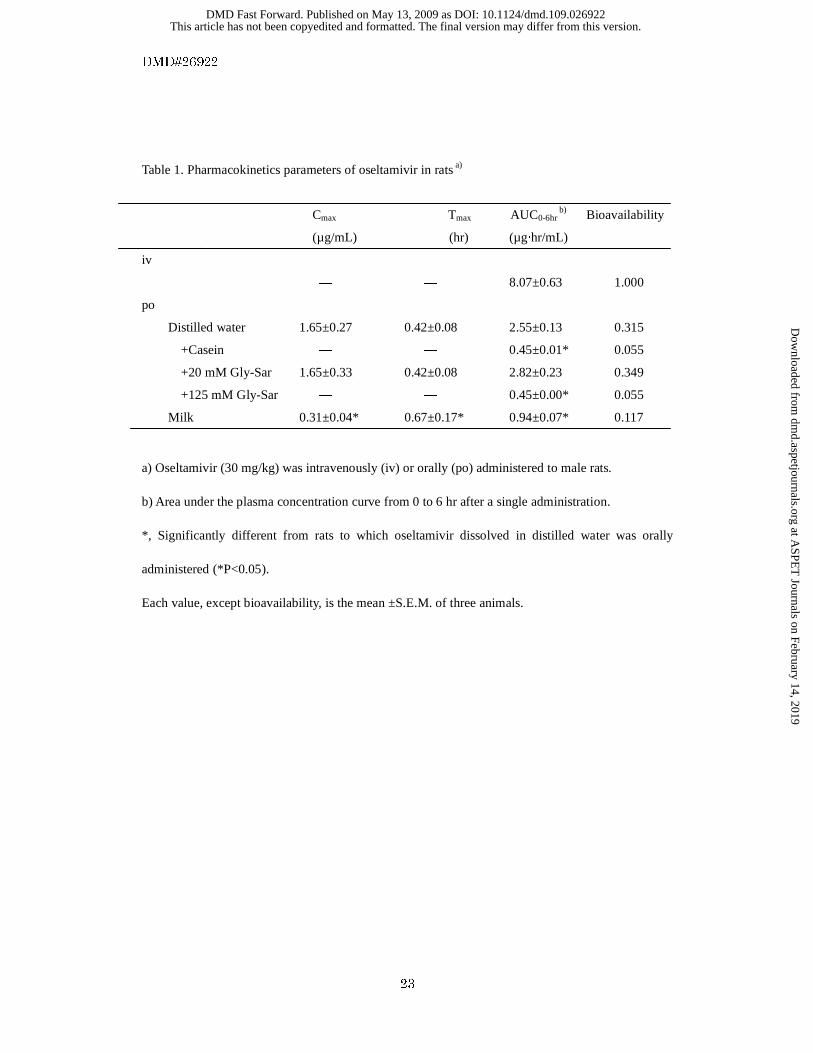
Table 1. Pharmacokinetics parameters of oseltamivir in rats a)
Cmax Tmax AUC0-6hr b) Bioavailability
(µg/mL) (hr) (µg·hr/mL)
iv
― ― 8.07±0.63 1.000
po
Distilled water 1.65±0.27 0.42±0.08 2.55±0.13 0.315
+Casein ― ― 0.45±0.01* 0.055
+20 mM Gly-Sar 1.65±0.33 0.42±0.08 2.82±0.23 0.349
+125 mM Gly-Sar ― ― 0.45±0.00* 0.055
Milk 0.31±0.04* 0.67±0.17* 0.94±0.07* 0.117
a) Oseltamivir (30 mg/kg) was intravenously (iv) or orally (po) administered to male rats.
b) Area under the plasma concentration curve from 0 to 6 hr after a single administration.
*, Significantly different from rats to which oseltamivir dissolved in distilled water was orally
administered (*P<0.05).
Each value, except bioavailability, is the mean ±S.E.M. of three animals.
This article has not been copyedited and formatted. The final version may differ from this version.DMD Fast Forward. Published on May 13, 2009 as DOI: 10.1124/dmd.109.026922
at ASPE
T Journals on February 14, 2019
dmd.aspetjournals.org
Dow
nloaded from

This article has not been copyedited and formatted. The final version may differ from this version.DMD Fast Forward. Published on May 13, 2009 as DOI: 10.1124/dmd.109.026922
at ASPE
T Journals on February 14, 2019
dmd.aspetjournals.org
Dow
nloaded from

This article has not been copyedited and formatted. The final version may differ from this version.DMD Fast Forward. Published on May 13, 2009 as DOI: 10.1124/dmd.109.026922
at ASPE
T Journals on February 14, 2019
dmd.aspetjournals.org
Dow
nloaded from

This article has not been copyedited and formatted. The final version may differ from this version.DMD Fast Forward. Published on May 13, 2009 as DOI: 10.1124/dmd.109.026922
at ASPE
T Journals on February 14, 2019
dmd.aspetjournals.org
Dow
nloaded from

This article has not been copyedited and formatted. The final version may differ from this version.DMD Fast Forward. Published on May 13, 2009 as DOI: 10.1124/dmd.109.026922
at ASPE
T Journals on February 14, 2019
dmd.aspetjournals.org
Dow
nloaded from

This article has not been copyedited and formatted. The final version may differ from this version.DMD Fast Forward. Published on May 13, 2009 as DOI: 10.1124/dmd.109.026922
at ASPE
T Journals on February 14, 2019
dmd.aspetjournals.org
Dow
nloaded from

This article has not been copyedited and formatted. The final version may differ from this version.DMD Fast Forward. Published on May 13, 2009 as DOI: 10.1124/dmd.109.026922
at ASPE
T Journals on February 14, 2019
dmd.aspetjournals.org
Dow
nloaded from

This article has not been copyedited and formatted. The final version may differ from this version.DMD Fast Forward. Published on May 13, 2009 as DOI: 10.1124/dmd.109.026922
at ASPE
T Journals on February 14, 2019
dmd.aspetjournals.org
Dow
nloaded from


![Pharmacokinetics and Disposition of the Thiouracil ...dmd.aspetjournals.org/content/dmd/44/2/209.full.pdf · acetamide] (Fig. 1) (Ruggeri et al., 2015). PF-06282999 is a suicide substrate](https://static.fdocuments.net/doc/165x107/5c65789e09d3f2a36e8cde93/pharmacokinetics-and-disposition-of-the-thiouracil-dmd-acetamide-fig.jpg)