OSCE Stations for Medical Finals Book 2 - Medical Revision · PDF fileOSCE Stations for...
24
OSCE Stations for Medical Finals Book 2 Adam Feather FRCP Senior Lecturer in Medical Education, St. Bartholomew’s and the Royal London School of Medicine and Dentistry, Consultant Geriatrician Newham University Hospital NHS Trust Ashling Lillis BA (Cantab) MB BS MRCP(UK) Acute Medicine Registrar, King George Hospital, Essex Tony Joy MBChB MRCS(Eng) DCH ‘Darzi’ Fellow in Clinical Leadership Registrar in Emergency Medicine North East Thames Rotation London John S P Lumley MS FRCS Emeritus Professor of Vascular Surgery, St Bartholomew’s and the Royal London School of Medicine and Dentistry
Transcript of OSCE Stations for Medical Finals Book 2 - Medical Revision · PDF fileOSCE Stations for...

OSCE Stations for Medical Finals
Book 2
Adam Feather FRCP
Senior Lecturer in Medical Education,
St. Bartholomew’s and the Royal London School of Medicine and Dentistry,
Consultant Geriatrician
Newham University Hospital NHS Trust
Ashling Lillis BA (Cantab) MB BS MRCP(UK)
Acute Medicine Registrar,
King George Hospital,
Essex
Tony Joy MBChB MRCS(Eng) DCH
‘Darzi’ Fellow in Clinical Leadership
Registrar in Emergency Medicine
North East Thames Rotation
London
John S P Lumley MS FRCS
Emeritus Professor of Vascular Surgery,
St Bartholomew’s and the Royal London
School of Medicine and Dentistry

v
Contents
About the authors vi
Preface vii
Acknowledgements viii
How to use this book ix
Introductory chapter xi
Glossary xxiv
SCENARIOS
1. No post today 1
2. Collapse 23
3. One too many 36
4. Lost for words 53
5. Breast lump 75
6. Terrible legs 88
7. Heart broken 110
8. Running on empty 125
9. Frequent and profuse 140
10. All very confusing 166
11. Hyper, Hyper 190
12. All fall down 212
13. Lose my breath 234
14. Abdominal agony 248
15. Weaker and weaker 264
16. The pit of despair 278
17. A little rash 311
18. Drunk and disorderly 336
19. Blood in the pan 361
Clinical !ndings 379
Blank charts 391
Station index 397
Subject index 399

SC
EN
AR
IO 1
1
Scenario 1: ‘No post today’
Station 1
History 10-minute stationYou are the FY1 doctor on call with the Acute Medical Team. !e next patient is
Mr Mohammed Iqbal, who has been brought into the Emergency Department by
ambulance after suffering an episode of severe chest pain while at work.
Please take a focussed history from the patient with a view to presenting the
history and likely diagnosis to the Medical Registrar.
You will be assessed on the following areas, as well as the content and diagnostic
reasoning of your history – take them into account in your presentation.
Professionalism
Professional appearance (NHS dress code) – including general appearance, hair and jewellery
Maintains patient and personal safety
Polite introduction; identi!es patient or interviewee correctly; con!rms patient’s date of birth from
name band or other source
Obtains informal consent; maintains patient’s privacy
Displays empathetic and caring attitudes and behaviours throughout.
Process
Good organisation and structure; appropriate use of open and closed questions
Appropriate "uency/rhythm/pace to the interview – this may change depending on environment
and acute nature of the problem
Appropriate time for the patient to respond/reply to questions
Appropriate acknowledgement of di#cult or emotional areas of the patient’s history.
Communication skills
Demonstrates caring and sympathetic attitude
Asks open questions
Invites patient to ask questions and answers them appropriately
Addresses patient’s ideas, concerns and expectations.

‘ N O P O S T T O D A Y ’
2
SC
EN
AR
IO 1
Station 2
Examination 10-minute stationAfter completing and presenting the history, the Medical Registrar asks you to
perform a focussed examination of the patient. Mr Iqbal has been seen by the nurse in
the resus area, and is attached to a cardiac monitor. !e nurse has recorded Mr Iqbal’s
observations on the chart. You may ask for these during your assessment.
Please present the relevant findings to your colleague in an appropriate manner
for a busy medical on call (if you do not have a model, please read and present the
information on page 379).
You will be assessed on the following areas, as well as the content and skills of your
examination – take them into account in your presentation.
Professionalism
Professional appearance; maintains infection control standards, including hand cleaning and
appropriate use of gloves and aprons
Maintains patient and personal safety
Polite introduction; identi!es patient and con!rms date of birth from name band or other source
Obtains informal consent; maintains patient privacy and dignity
Displays empathetic and caring attitudes and behaviours throughout.
Process
Appropriate "uency/rhythm/pace to the examination – this may change depending on environment
and acute nature of the problem
Organisation and structure of examination; sensitive and empathetic approach
Uses appropriate clinical techniques throughout
Maintains privacy and dignity throughout.
Clinical communication
Explains proposed examination/procedure: explains examination/procedure as it proceeds
O#ers information in a clear, structured and "uent manner, avoiding jargon
Listens to patient and responds appropriately
Demonstrates appropriate body language.
‘ N O P O S T T O D A Y ’
SC
EN
AR
IO 1
3
Station 3
Data interpretation 10-minute stationMr Iqbal’s ECG shows global T-wave inversion, and he is transferred immediately
to the CCU for ongoing management. ! e Medical Registrar on call is one of the
cardiology trainees and hands you fi ve ECGs belonging to patients on the CCU.
Please indicate whether each of the statements are TRUE (T) or FALSE (F)
regarding each of the ECGs shown below. You may assume all ECGs are running at
25 mm/s.
1
A ! e rhythm shown is sinus arrhythmia.
B ! ere is evidence of fi rst-degree heart block.
C ! ere is anterior ST depression.
D ! ere are inverted T waves in the lateral and high lateral leads.
E ! ere are features to suggest an acute inferior STEMI.

‘ N O P O S T T O D A Y ’
4
SC
EN
AR
IO 1
2
A ! ere is left axis deviation.
B ! ere is inferior ST segment depression.
C ! ere is ST segment depression in the septo-lateral leads.
D ! ere is evidence of poor anterior R-wave progression.
E ! e rhythm shown is atrial fi brillation.
3
A ! ere is both a nodal and sinus rhythm demonstrated.
B ! e axis is deviated to the right.
C ! ere is ST depression in the infero-lateral leads.
D ! ere is anterior ST elevation.
E ! is ECG pattern represents left main stem obstruction.

‘ N O P O S T T O D A Y ’
SC
EN
AR
IO 1
5
4
A ! e rate is approximately 150 bpm.
B ! ere is left axis deviation.
C ! ere is T-wave inversion in the high-lateral leads.
D ! ere is normal anterior R-wave progression.
E ! is patient is likely to have a normal LV ejection fraction.

‘ N O P O S T T O D A Y ’
6
SC
EN
AR
IO 1
5
A ! is is Mobitz type I second-degree heart block.
B ! e axis is normal.
C ! ere is anterior ST elevation.
D ! ere is ST elevation in the infero-lateral leads.
E ! ere is ST depression in the high lateral leads.
Station 4
Procedural skills 10-minute stationMr Iqbal requires a second intravenous cannula inserted for his intravenous insulin
infusion.
Please write up the fi xed-rate intravenous insulin infusion on the chart provided.
Mr Mohammed Iqbal; DOB 23/08/57; Hospital No. 5463721; Consultant
Dr Westmore; Ward CCU; Bleep 332
Procedure A
Please make up the insulin infusion using the equipment provided, explaining the
process to the examiner as you proceed.
Equipment provided
50 ml syringe, insulin (100 unit vial), 0.9% saline – 250 ml bag
Needles to draw up medications, extension tubing
‘ N O P O S T T O D A Y ’
SC
EN
AR
IO 1
7
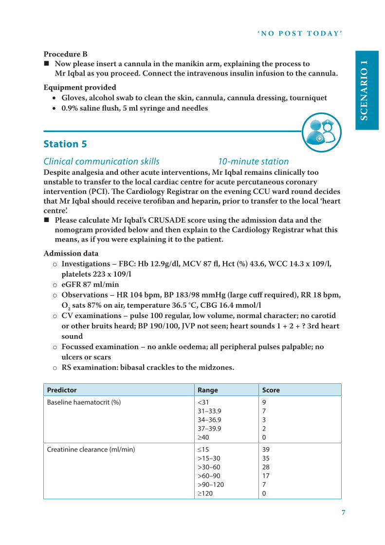
Procedure B
Now please insert a cannula in the manikin arm, explaining the process to
Mr Iqbal as you proceed. Connect the intravenous insulin infusion to the cannula.
Equipment provided
Gloves, alcohol swab to clean the skin, cannula, cannula dressing, tourniquet
0.9% saline flush, 5 ml syringe and needles
Station 5
Clinical communication skills 10-minute stationDespite analgesia and other acute interventions, Mr Iqbal remains clinically too
unstable to transfer to the local cardiac centre for acute percutaneous coronary
intervention (PCI). !e Cardiology Registrar on the evening CCU ward round decides
that Mr Iqbal should receive terofiban and heparin, prior to transfer to the local ‘heart
centre’.
Please calculate Mr Iqbal’s CRUSADE score using the admission data and the
nomogram provided below and then explain to the Cardiology Registrar what this
means, as if you were explaining it to the patient.
Admission data
o Investigations – FBC: Hb 12.9g/dl, MCV 87 fl, Hct (%) 43.6, WCC 14.3 x 10⁹/l,
platelets 223 x 109/l
o eGFR 87 ml/min
o Observations – HR 104 bpm, BP 183/98 mmHg (large cuff required), RR 18 bpm,
O2 sats 87% on air, temperature 36.5 °C, CBG 16.4 mmol/l
o CV examinations – pulse 100 regular, low volume, normal character; no carotid
or other bruits heard; BP 190/100, JVP not seen; heart sounds 1 + 2 + ? 3rd heart
sound
o Focussed examination – no ankle oedema; all peripheral pulses palpable; no
ulcers or scars
o RS examination: bibasal crackles to the midzones.
Predictor Range Score
Baseline haematocrit (%) <31
31–33.9
34–36.9
37–39.9
40
9
7
3
2
0
Creatinine clearance (ml/min) 15
>15–30
>30–60
>60–90
>90–120
120
39
35
28
17
7
0

‘ N O P O S T T O D A Y ’
8
SC
EN
AR
IO 1
Predictor Range Score
Heart rate (bpm) 70
71–80
81–90
91–100
101–110
111–120
121
0
1
3
6
8
10
11
Sex Male
Female
0
8
Signs of CHF at presentation No
Yes
0
7
Prior vascular disease No
Yes
0
6
Diabetes mellitus No
Yes
0
6
Systolic blood pressure (mmHg) 90
91–100
101–120
121–180
181–200
201
10
8
5
1
3
5
Note: heart rate is truncated at <70 bpm
CrCI: Cockcroft-Gault is truncated at >90 ml/min; prior vascular disease is de!ned as prior PAD or
stroke
The CRUSADE bleeding score to assess baseline risk of major bleeding in non-ST-segment
elevation myocardial infarction
Risk group CRUSADE score Risk of major bleed
Very low <21 3.1%
Low 21–30 5.5%
Moderate 31–40 8.6%
High 41–50 11.9%
Very high >50 19.5%

‘ N O P O S T T O D A Y ’
SC
EN
AR
IO 1
9
Answers
Station 1 – History
Patient script
You are Mr Iqbal Mohammed (DOB 23 August 1957), a 54-year-old postal worker
in the local post office depot. You were brought into the Emergency Department
this morning at about 09:30 after having suffered a severe episode of chest pain.
Over the past 4–6 weeks you have been suffering with occasional episodes of chest
pain that you initially thought was ‘bad indigestion’. �e first episode occurred
while you were at work pushing some of the heavy trolleys around the depot. �e
pain was very tight and ‘squeezing’, ‘like a band around my chest’. It only lasted
about a minute but was really bad and took your breath away. You had to sit down
for about 5 minutes before you felt OK again. Since then you’ve had two to three
similar episodes, the last one while playing football with your grandson. �is
was a more severe and prolonged episode, lasting about 5 minutes or so, but you
had to laugh it off as your grandson became frightened. �e pain was severe, and
central, radiating to your jaw and left hand. You were sweaty and felt sick but it
went off by itself after you lay down on the ground after about 5–10 minutes.
Today’s episode came on at the depot, just after you had finished your morning tea
break. You were walking up a flight of stairs when the pain suddenly came on. �is
was different to all the other episodes.
�e pain was severe (the worst you’ve experienced), ‘squeezing and crushing the
life out of me’. �e pain was central, radiating to your jaw, left and right arms, but
not through to your back. You collapsed down the stairs and your work colleague
pulled you to safety and made a pillow for you with his jacket. Your colleague said
‘it looked like all the life had drained out of me’ and you were dizzy and faint but
don’t think you passed out. �e pain continued for at least 10–15 minutes before
the ambulance men arrived and gave you oxygen and some tablets up under your
upper gums. You still feel as if you have a little chest discomfort but nothing like
the pain you had earlier.
If asked you deny any symptoms of heart failure, eg peripheral oedema,
orthopnoea, PND, shortness of breath, wheeze, dry cough or frothy white sputum.
Risk factors: your father and his parents all died of heart and stroke problems
before the age of 70. You have two close cousins who have had heart attacks in the
last few years. You smoked 20+ cigarettes/day until 4 or 5 years ago but stopped
when your grandson was born. You do not drink any alcohol.

‘ N O P O S T T O D A Y ’
10
SC
EN
AR
IO 1
Your doctor told you recently that you were very overweight and this was putting
you at risk for high blood pressure and diabetes. You are 5 feet 8 inches (1 m 72
cm) and 15 and a half stone. You don’t know about your blood pressure. Your
doctor told you to lose weight or he would have to start you on diabetes tablets.
You do not know about your cholesterol and have never had symptoms suggestive
of a stroke, peripheral vascular disease or IHD.
You have no significant previous medical history; you had an appendicetomy at 11
years old.
You are not on any regular medications, but do take the occasional ‘zantac’, Rennie
tablets and paracetamol. You have no known allergies.
You are married with five children aged 26, 24, 21, 20 and 17 years. !e youngest
three still live at home, and two of them are unemployed. You work in the local
post office depot and are the shop steward of the local postal union.
You think you may have had a heart attack this morning as the pain was so bad
and you felt so unwell. You are hoping that it’s nothing too serious, as you need to
get back to work soon. You are the main breadwinner in the house, and everyone
is reliant on you.

‘ N O P O S T T O D A Y ’
SC
EN
AR
IO 1
11
CONTENT
Identi es key information
Pain: chronological progression; onset, frequency, duration, character, radiation, relieving
and exacerbating factors
Associated features: shortness of breath, nausea and vomiting, dizziness, presyncope and
syncope, feeling unwell, washed out, palpitations.
Includes important negatives, including systemic enquiry
No radiation to the back (thoracic aortic aneurysm)
No features of heart failure: peripheral oedema, orthopnoea, PND, shortness of breath,
wheeze, dry cough, frothy white sputum
No features suggestive of gastrointestinal or hepatobiliary disease
Excludes other systemic symptoms.
Identi es key information from rest of history
Cardiovascular risk: family history, known IHD, stroke or PVD, diabetes mellitus,
hypertension, smoking, alcohol, others
Relevant facts about employment, housing, social support, life stressors.
Completing the patient history
Drug and allergy history: Zantac and indigestion tablets; occasional paracetamol; no
known drug allergies
Previous medical history: nil known
Social and occupational history: as above.
Summarises important areas of the history back to the patient
Invites patient to ask questions and deals with them appropriately
Establishes patient’s ideas, concerns and expectations
A B C D E
PROFESSIONALISM
A B C D E
A B C D E
PROCESS
A B C D E
COMMUNICATION

‘ N O P O S T T O D A Y ’
12
SC
EN
AR
IO 1
CLINICAL DIAGNOSTIC REASONING
Please present your history
Candidate o#ers a logical, well-structured account of the history.
What is your diagnosis?
Candidate o#ers the correct diagnosis and appropriate di#erentials
The patient has presented with ACS but the history suggests a severe episode: MI?
Can you describe how you would manage this patient? What three essential bedside
(clinical) investigations would you perform?
Nurse in high-dependency area, cardiac monitor, IV access, aspirin and clopidogrel,
oxygen and some further pain relief, eg IV morphine and anti-emetic; full examination
and further investigations
BP, CBG, ECG, oxygen sats ABGs.
Demonstrates safe, sensible and appropriate management plan
Demonstrates clear and logical diagnostic reasoning
GLOBAL HISTORY MARK
A B C D E
Station 2 – Examination
Patient script (see also page 379)
If you are an actor/patient, read the patient history and physical signs fully – when
the candidate comes to an abnormal site in their examination, act out tenderness
and/or volunteer the relevant physical sign.
A B C D E

‘ N O P O S T T O D A Y ’
SC
EN
AR
IO 1
13
CONTENT
Exposes and positions patient correctly and maintains comfort
Comments on wellbeing of patient, ie well or unwell
‘Feet to face’
Observes, and comments on patient and surroundings from foot of bed
Evidence of previous cardiac surgery, eg sternotomy, JVP, anaemia, colour/perfusion.
Asks for appropriate/relevant clinical observations
HR 104 bpm, BP 183/98 mmHg (large cu! required), RR 18 bpm, O2 sats 87% on air,
temperature 36.5 °C, CBG 16.4 mmol/l
Urinalysis: WCC nil, protein 1+, blood negative, nitrites negative, glucose 3+.
General/systemic examination
Hands and upper limbs: tar staining, perfusion of hands, anaemia, stigmata of
hyperlipidaemia; comments on general signs, eg clubbing, leuconychia
Face and neck: signs of anaemia, peri-orbital xanthelasma, central cyanosis.
Focussed examination
Inspection: sternotomy scar, JVP – makes appropriate assessment, including correct
positioning of patient, correct technique; comments correctly on JVP
Palpation: carotid pulse – comments on character and presence of bruits
Apex beat: position and character
Assesses and comments on heaves and thrills
Auscultation: listens in correct areas, assesses for radiation, manoeuvres patient correctly,
appropriate use of stethoscope – bell and diaphragm.
Completes examination by identifying relevant additional clinical signs
Signs of left and right heart failure: bibasal crackles, pleural e!usions, peripheral oedema;
hepatomegaly/ascites
Signs of PVD and generalised atherosclerosis: AAA, peripheral pulses, abdominal bruits.
Thanks patient, o!ers assistance, maintains patient’s dignity and privacy until they are
dressed
A B C D E
PROFESSIONALISM
A B C D E
A B C D E
PROCESS
A B C D E
COMMUNICATION

‘ N O P O S T T O D A Y ’
14
SC
EN
AR
IO 1
CLINICAL DIAGNOSTIC REASONING
Correctly identi!es the relevant physical signs, including important negative !ndings
What does a third heart sound represent?
Fluid overload in keeping with his heart failure
Given these clinical !ndings, can you name three acute therapeutic interventions you
would arrange for this gentleman in the Emergency Department (he has already had
oxygen, GTN spray and anti-platelet treatment)?
Ongoing pain: further GTN or IV morphine and anti-emetics
Pulmonary oedema (bibasal crackles and hypoxia): oxygen to maintain sats between 94
and 98%, IV GTN infusion and IV furosemide; may require IV diamorphine (venodilator
and analgesic, as above)
Hypertension: this would also be accounted for with the GTN infusion
CBG >11.0 mmol/l: IV insulin infusion (either sliding scale or FRIVII).
Demonstrates safe, sensible and appropriate management plan
Demonstrates clear and logical diagnostic reasoning
GLOBAL EXAMINATION MARK
A B C D E
Station 3 – Data interpretation
1 A True
B True
C False
D False
E True
This ECG demonstrates a large inferior STEMI with high lateral (I and aVL) ST depression.
The antero-lateral leads are normal. There is sinus arrhythmia with !rst-degree heart block.
2 A False
B True
C True
D True
E False
A B C D E

‘ N O P O S T T O D A Y ’
SC
EN
AR
IO 1
15
This ECG demonstrates atrial �utter with 2 : 1 block. This is commonly seen in older patients
who are acutely unwell with chest pathology, eg chest infection or pulmonary embolism,
especially if there is underlying ischaemic heart disease. The axis is moving towards the left
but because leads I and II are both very positive, this means the axis will remain within normal
limits. There is septo-lateral (V3–V6) ST depression extending into the high lateral (I and aVL)
leads. There is relatively absent R-wave progression in leads V1–V3.
3 A True
B False
C True
D True
E True
This ECG demonstrates a large anterior STEMI with deep ST depression in the infero-lateral
leads. The rhythm is unstable owing to the acute ischaemia and starts as a nodal rhythm but
progresses into sinus arrhythmia. The axis is neutral as all three leads are almost isoelectric.
As all three territories of the heart demonstrate acute ischaemia, this would suggest there is
left main stem disease with a dominant left circum�ex artery.
4 A True
B True
C True
D False
E False
This ECG demonstrates atrial �utter with 2 : 1 block, giving the classical regular appearance at
a rate of 150 bpm. There are several features of underlying ischaemic heart disease including
left axis deviation, partial left bundle branch block and poor anterior R-wave progression.
There is T-wave inversion in the high lateral leads (I and aVL). Given the rate and the features
of the IHD, this patient will rapidly develop acute heart failure. They are unlikely to have a
normal LV ejection fraction.
5 A False
B True
C True
D True
E True
This ECG demonstrates a massive STEMI involving the antero-lateral and inferior territories.
There is also ST depression in lead aVL. There is third-degree (complete) heart block with a
normal axis. This again may well represent left main stem disease with a dominant circum�ex
artery. This patient needs urgent coronary intervention.
GLOBAL DATA INTERPRETATION MARK
A B C D E

‘ N O P O S T T O D A Y ’
16
SC
EN
AR
IO 1
Station 4 – Procedural skills
Infusion prescriptions continued SC = subcutane-
ous
IVC = intravenous central
IVP = intravenous peripheral
Date
&
time
Route Infusion Fluid Medication Duration Rate Prescriber’s
signature &
bleep no.
Date
given
Given by
/ Added
by
Check
by
Start
time
Finish
time
Pharmacy
Name &
strength
Volume Approved name with
expiry / unit number
Dose
07/
05IV 0.9 % saline
Exp:
Batch/unit no:
50 ml ACTRAPID INSULIN
50
UNITS
AF 622
07/05 IV Exp:
Batch/unit no:
Exp:
Batch/unit no:
Exp:
Batch/unit no:
Exp:
Batch/unit no:
Exp:
Batch/unit no:
Exp:
Batch/unit no:
AF 622
Exp:
Batch/unit no:
Exp:
Batch/unit no:
Allergies, sensitivities and adverse drug reactions Patient details/addressograph
No known allergies Initials Gender M / F NHS/ Hospital No: 5463721
Not possible to ascertain Date Weight (kg) Date
Medicine/substance Reaction & Severity Initials & Date 100 kg Surname: IQBAL
HeightFirst name: Mohammed
Alerts Surface area (m2)Date of birth: 23.08.57
TO RUN IV AT 0.1 UNITS/=10u/kg/hrkg/hr
50 UNITS OF ACTRAPID INSULIN IN 50 mls OF 0.9% SALINE
TO RUN IV ACCORDING TO SLIDING SCALE CBG UNITS/HOUR 0�4.0 0 4.1�7.0 1.0
7.1�11.0 2.0 11.1�15.0 3.0 15.1�21.0 4.0 21.1�28.0 5.0 >28.0 6.0 Please call DR if CBG >28.0

‘ N O P O S T T O D A Y ’
SC
EN
AR
IO 1
17
Procedure A: Intravenous insulin infusion
CONTENT
Identi es and sets out equipment correctly; maintains aseptic technique throughout
Insulin vial (100 units)
250 ml bag of 0.9% saline
Syringes
Needles
Medication label (sticker) to be completed by candidate.
Correctly performs the procedure
Identi!es patient from hospital bracelet (one cannot verbally con!rm patient’s name and
DOB as he is confused)
Puts on gloves
For both drugs (0.9% saline and insulin): checks vial/bag for correct name of drug and
expiry date
Checks correct patient identity against prescription on the chart
Breaks open 0.9% saline and draws up 50 ml into 50 ml syringe using ‘green’ needle
Draws up 50 units of insulin using the appropriate needle and syringe
Using a second needle adds the insulin to the normal saline
Ensures insulin is adequately mixed with saline
Correctly completes medication label and applies it to the syringe.
Obtains an acceptable/appropriate result
Disposes of all sharps and other items correctly
Ensures patient receives correct advice about what to do next and follow-up
Ensures nursing sta" or other healthcare professionals receive correct information about
the consequences/outcome of the procedure/task
A B C D E
PROFESSIONALISM
A B C D E
A B C D E
PROCESS

‘ N O P O S T T O D A Y ’
18
SC
EN
AR
IO 1
Procedure B: Cannulation
CONTENT
Exposes and positions patient correctly and maintains comfort
Exposes forearm and assesses for appropriate vein
Identi!es and sets out equipment correctly; maintains aseptic technique throughout
Gloves
Alcohol swab to clean the skin
Appropriate cannula (G14 or G16)
Cannula dressing
Tourniquet
Extension tubing for cannula
0.9% saline !ush
5 ml syringe and needles
Labelled insulin infusion
Sharps bin.
Correctly performs the procedure
Puts on gloves
Identi"es patient’s ID from patient, against hospital bracelet and the prescription chart
Checks correct details of the infusion against prescription chart
Primes extension tubing using 0.9% saline !ush
Connects tubing to insulin infusion syringe
Applies tourniquet to manikin arm
Identi"es appropriate vein to site cannula
Cleans skin with alcohol swab
Inserts cannula using appropriate technique: needle held at 45° to skin, smooth insertion
under skin into vein
Acknowledges !ashback of blood
Withdraws introducer to a degree, then inserts cannula to hub
Releases tourniquet, presses on proximal vein while withdrawing introducer, connects cap
Applies dressing
Flushes the cannula using prepared 5 ml, 0.9% saline !ush
Connects primed extension tubing and insulin infusion.
Obtains an acceptable/appropriate result
Disposes of all sharps and other items correctly
Ensures patient receives correct advice about what to do next and follow-up
Ensures nursing sta" or other healthcare professionals receive correct information about
the consequences/outcome of the procedure/task
A B C D E
PROFESSIONALISM
A B C D E
A B C D E
PROCESS

‘ N O P O S T T O D A Y ’
SC
EN
AR
IO 1
19
GLOBAL PROCEDURE MARK
A B C D E
Station 5 – Clinical communication skills
Patient script
You have been told that you are very unwell and need to be transferred to the
‘heart attack centre’ as soon as possible. However you need to be ‘a bit better’ and
as such need to first go to the heart ward to make sure you are safe to travel.
You have also been told that you will need to be on lots of medicines to keep your
heart safe and that these have all been shown to help people like you when they
are having a heart attack.
However, one of the doctors has told you that many of the medicines may make
you bleed and that someone will be coming to explain this to you.
You have been told that aspirin stops the blood forming clots that in turn cause
heart attacks, but you don’t really know about the other medicines.
When told you have a 1 : 8–1 : 9 risk of having a major bleed, you tell the ‘doctor’
that this is TOO HIGH a RISK and that you don’t want the medicines. However, if
the candidate is sympathetic, calm and reassuring, you will be convinced that this
is a worthwhile risk. If the candidate is unsympathetic, bullying or aggressive, you
should not change your mind.
If the candidate uses medical jargon or terms that you don’t think an intelligent
lay person would know or understand you should challenge them to explain these
to you.
You expect the doctors ‘know best’ but are very worried about the risk of the
bleeding.

‘ N O P O S T T O D A Y ’
20
SC
EN
AR
IO 1
CONTENT
Con rms reason for discussion – to talk to patient about the new medications and their
risks and bene ts
Establishes what patient wishes to know; gains agreement/informal consent to participate
in the discussion
Reviews patient’s current understanding of clinical situation and summarises what has
happened so far
Establishes patient’s ideas, concerns and expectations
Explains the key, important information; invites patient to ask questions and is able to
deal with them appropriately
Explains the aims of treatment and their bene�ts: to stop further clot progression and
possible myocardial damage
Introduces the idea of possible risks: may cause signi�cant bleeding, eg from the bowel
or possibly inside the brain
Introduces idea of objective scoring system and how it can be used to calculate risk
Shows patient that calculated CRUSADE score is 41
Correctly explains the relative risk (11.9%, ie approx 1 : 8–1 : 9 risk) using clear, jargon-free
and understandable language
Deals sympathetically and appropriately with patient’s initial response (‘If it’s that high
doctor I don’t want it’)
Revisits ideas, concerns and expectations and deals with them appropriately
Avoids giving wrong or disinformation
Avoids aggressive or judgemental language.
Summarises important areas of the consultation back to the patient
Clari es patient’s nal position
If patient is willing to take treatment, records this in the medical notes and prescribes the
medications
If patient is NOT willing to take the treatment, records this in the notes BUT may ask one
of his/her seniors to come and revisit the issues once the patient has had time to think
about them.
Formally ends the consultation and ensures appropriate follow-up has been discussed
A B C D E
PROFESSIONALISM
A B C D E
A B C D E
PROCESS

‘ N O P O S T T O D A Y ’
SC
EN
AR
IO 1
21
CLINICAL DIAGNOSTIC REASONING
Correctly calculates CRUSADE score (41)
Correctly identi!es and explains what a relative risk of 11.9% means
Successfully negotiates a satisfactory, safe management plan
Demonstrates clear and logical diagnostic reasoning
GLOBAL CLINICAL COMMUNICATION MARK
A B C D E
Scenario 1: Reflection and consolidation
History
Mr Iqbal is a 54-year-old British-born Asian man. He was brought into the Emergency Department at about
09.30 after having su!ered a severe episode of chest pain. Over the past 4–6 weeks he has been su!ering
with occasional episodes of chest pain that he initially thought was ‘bad indigestion’. The "rst episode
occurred while he was at work pushing some of the heavy trolleys around the postal depot. The pain was
very tight and ‘squeezing’, ‘like a band around his chest’. It only lasted about a minute but was really bad and
took his breath away. He had to sit down for about 5 minutes before he felt OK again. Since then he’s had two
to three similar episodes, the last one while playing football with his grandson. This was a more severe and
prolonged episode, lasting about 5–10 minutes. The pain was severe, and central, radiating to his jaw and left
hand. He was sweaty and felt sick but it went o! by itself after he lay down on the ground.
Today’s episode came on at the postal depot just after his morning tea. He was walking up a #ight of stairs when
the pain came on suddenly. This was the most severe pain he’d ever experienced and he described it as ‘squeezing
and crushing the life out of me’. The pain was central, radiating to his jaw, left and right arms. He collapsed down
the stairs but a work colleague pulled him to safety and made a pillow for him with a jacket. He was dizzy and faint
but didn’t pass out. The pain continued for at least 10–15 minutes before the ambulance men arrived and gave him
oxygen and some buccal nitrate. On arrival he still had a little chest discomfort. He had no symptoms of heart failure.
Risk factors:
Strong family history of IHD and CV disease. including his father and grandparents dying of heart
and stroke problems before the age of 70; Mr Iqbal also has two close cousins who have had MI in
the last few years
Probable pre-diabetes
Ex-smoker: 20+ cigarettes/day until 4 or 5 years ago
No known hypertension, stroke or PVD
Alcohol: nil
Weight: grossly overweight, being 5’8’’ and 15 and a half stone
No regular medications; no known allergies
PMH – no signi"cant history
Married with "ve children aged 26–17 years; the youngest three still live at home, two of them are
unemployed
Works in the local post o$ce depot; shop steward of the local postal union and is the main bread-winner.
A B C D E

‘ N O P O S T T O D A Y ’
22
SC
EN
AR
IO 1
Examination
On examination, Mr Iqbal is an obese, middle-aged Asian man. On arrival in the Emergency Department he
looked unwell, pale and clammy and still had slight chest discomfort.
Vital observations were: HR 104 bpm, BP 183/98 mmHg (large cu! required), RR 18 bpm, O2 sats 87%
on air, temperature 36.5 °C, CBG 16.4 mmol/l
Feet to face: very overweight but nil else of note
General examination: "ngers of right hand heavily tar stained; no anaemia, no stigmata of
hyperlipidaemia
No carotid or other bruits heard
CV examination: pulse 100 regular, low volume, normal character; BP 190/100 mmHg, JVP – not seen;
HS 1 + 2 + ? 3rd heart sound
RS examination: bibasal crackles to the midzones
No ankle oedema
All peripheral pulses palpable.
Abdomen: obese, no organomegaly or masses
Neurology: not formally assessed.
In summary, this is a 64-year-old Asian man with features suggestive of obesity, T2DM, ACS, LVF and poorly
controlled hypertension.
Investigations
Blood tests including FBC, U&Es, RBG, lipids, troponin (at 12 hours)
CXR: to con"rm the features of heart failure; other features to look for include calci"ed valves, cardiomegaly
and signs of respiratory disease eg features of COPD (chronic smoker)
ECG: to con"rm or exclude signs of:
Acute or chronic IHD, eg ST segment changes, T-wave changes, left bundle branch block
Arrhythmia and signs of heart block
Hypertensive changes, eg left axis deviation, voltage criteria of LVH.
Management
Acute ACS protocol, including LMWH or fondaparinux, aspirin and clopidogrel, anti-anginals – beta blockers,
nitrates
Mr Iqbal has ongoing pain and therefore requires further anti-platelet and anticoagulants (see NICE
guidelines below)
Treatment of LVF: IV nitrates and furosemide
IV insulin infusion
Secondary prophylaxis: statin, anti-platelets, anti-hypertensives
Further investigation according to GRACE or similar score – once stabilised Mr Iqbal will need PCI and
possible stenting
Will need dietitian and diabetes specialist nurse review and will require a sensible but e!ective weight-loss
programme.
Further reading and web links
http://www.crusadebleedingscore.org/index.html
http://www.nice.org.uk/nicemedia/live/11552/33013/33013.pdf
NICE guidelines of ACS and NSTEMI
http://www.nice.org.uk/nicemedia/live/12947/47918/47918.pdf
NICE guidelines on the management of cardiac chest pain of recent onset